Embed Size (px)
Citation preview

Because a number of cells in the autonomic nervous system act in conjunction, they have relinquished their independence to function as a coherent whole.
—Otto Appenzeller and Emilio Oribe, in The Autonomic Nervous System, 1997
Background Basics
Membrane receptors
Neurotransmitters
Second messenger systems
Catecholamines
Up- and down-regulation
Tonic and antagonistic control
Organization of the nervous system
Neuron structure
Synapses
Nerves
Action potentials
Slow synaptic potentials
The Autonomic Division Autonomic Refl exes Are Important for Homeostasis
Antagonistic Control Is a Hallmark of the Autonomic Division
Autonomic Pathways Have Two Eff erent Neurons in Series
Sympathetic and Parasympathetic Branches Originate in Diff erent Regions
The Autonomic Nervous System Uses a Variety of Chemical Signals
Autonomic Pathways Control Smooth and Cardiac Muscle and Glands
Autonomic Neurotransmitters Are Synthesized in the Axon
Autonomic Receptors Have Multiple Subtypes
The Adrenal Medulla Secretes Catecholamines
Autonomic Agonists and Antagonists Are Important Tools in Research
and Medicine
Primary Disorders of the Autonomic Nervous System Are Relatively
Uncommon
Summary of Sympathetic and Parasympathetic Branches
The Somatic Motor Division A Somatic Motor Pathway Consists of One Neuron
The Neuromuscular Junction Contains Nicotinic Receptors
Eff erent Division: Autonomic and Somatic Motor Control 11
Muscle fi bers with motor neurons terminating at neuromuscular junctions.
From Chapter 11 of Human Physiology: An Integrated Approach, Sixth Edition. Dee Unglaub Silverthorn. Copyright © 2013 by Pearson Education, Inc. All rights reserved.
397
Eff erent Division: Autonomic and Somatic Motor Control
The picnic lunch was wonderful. You are now dozing on the grass in the warm spring sunlight as you let the meal digest. Suddenly you feel something moving across your
lower leg. You open your eyes, and as they adjust to the bright light, you see a four-foot-long snake slithering over your foot. More by instinct than reason, you fl ing the snake into the grass while scrambling to a safe perch on top of the nearby picnic ta-ble. You are breathing heavily, and your heart is pounding.
In less than a second, your body has gone from a state of quiet rest and digestion to a state of panic and frantic activity. Th is refl ex reaction is integrated and coordinated through the central nervous system (CNS), then carried out by the eff erent division of the peripheral nervous system (PNS). Th e fi bers of efferent neurons are bundled together into nerves that carry commands from the CNS to the muscles and glands of the body. Some nerves, called mixed nerves, also carry sensory informa-tion through aff erent fi bers.
Th e eff erent division of the peripheral nervous system can be subdivided into somatic motor neurons , which control skel-etal muscles, and autonomic neurons , which control smooth muscle, cardiac muscle, many glands, and some adipose tis-sue. Th e somatic and autonomic divisions are sometimes called the voluntary and involuntary divisions of the nervous system, respectively. However, this distinction does not always hold true. Although most movement controlled by somatic pathways requires conscious thought, some skeletal muscle refl exes, such as swallowing and the knee jerk refl ex, are involuntary. And al-though autonomic refl exes are mainly involuntary, a person can use biofeedback training to learn to modulate some autonomic functions, such as heart rate and blood pressure.
We begin our study of the eff erent division of the PNS by looking at the autonomic division. Th en we consider the somatic motor division.
The Autonomic Division Th e autonomic division of the eff erent nervous system (or auto-nomic nervous system for short) is also known in older writings as the vegetative nervous system , refl ecting the observation that its functions are not under voluntary control. Th e word auto-nomic comes from the same roots as autonomous , meaning self-governing . Another name for the autonomic division is visceral nervous system because of its control over internal organs.
The autonomic division is subdivided into sympathetic and parasympathetic branches (often called the sympathetic and parasympathetic nervous systems ). Some parts of the sym-pathetic branch were first described by the Greek physician Claudius Galen (ca. c.e. 130–200), who is famous for his compi-lation of anatomy, physiology, and medicine as they were known during his time. As a result of his dissections, Galen proposed that “animal spirits” fl owed from the brain to the tissues through hollow nerves, creating “sympathy” between the diff erent parts of the body. Galen’s “sympathy” later gave rise to the name for the sympathetic branch. Th e prefi x para -, for the parasympa-thetic branch, means beside or alongside .
The sympathetic and parasympathetic branches can be distinguished anatomically, but there is no simple way to sepa-rate the actions of the two branches on their targets. They are distinguished best by the type of situation in which they are most active. Th e picnic scene that began the chapter illustrates the two extremes at which the sympathetic and parasympathetic branches function. If you are resting quietly aft er a meal, the parasympa-thetic branch is dominant, taking command of the routine, quiet activities of day-to-day living, such as dig estion. Consequently, parasympathetic neurons are sometimes said to control “rest and digest” functions.
In contrast, the sympathetic branch is dominant in stress-ful situations, such as the potential threat from the snake. One of the most dramatic examples of sympathetic action is the fight-or-flight response, in which the brain triggers massive simultaneous sympathetic discharge throughout the body. As the body prepares to fi ght or fl ee, the heart speeds up; blood vessels to muscles of the arms, legs, and heart dilate; and the liver starts to produce glucose to provide energy for muscle contraction. Diges-tion becomes a low priority when life and limb are threatened, and so blood is diverted from the gastrointestinal tract to skeletal muscles.
The massive sympathetic discharge that occurs in fi ght-or-fl ight situations is mediated through the hypothalamus and is a total-body response to a crisis. If you have ever been scared by the squealing of brakes or a sudden sound in the
R U N N I N G P R O B L E M
A Powerful Addiction
Every day, more than 1.3 billion people around the world intentionally absorb a chemical that kills about 5 million people a year. Why would people knowingly poison themselves? If you’ve guessed that the chemical is nicotine, you already know part of the answer. One of more than 4000 chemicals found in tobacco, nicotine is highly addictive. So powerful is this addiction that fewer than 20% of tobacco users are able to quit smoking the fi rst time they try. Shanika, a smoker for six years, is attempting for the second time to stop smoking. The odds are in her favor this time, however, because she has made an appointment with her physician to discuss all the options available to help her break her addiction to nicotine and smoking.
398
Eff erent Division: Autonomic and Somatic Motor Control
11
Autonomic Refl exes Are Important for Homeostasis
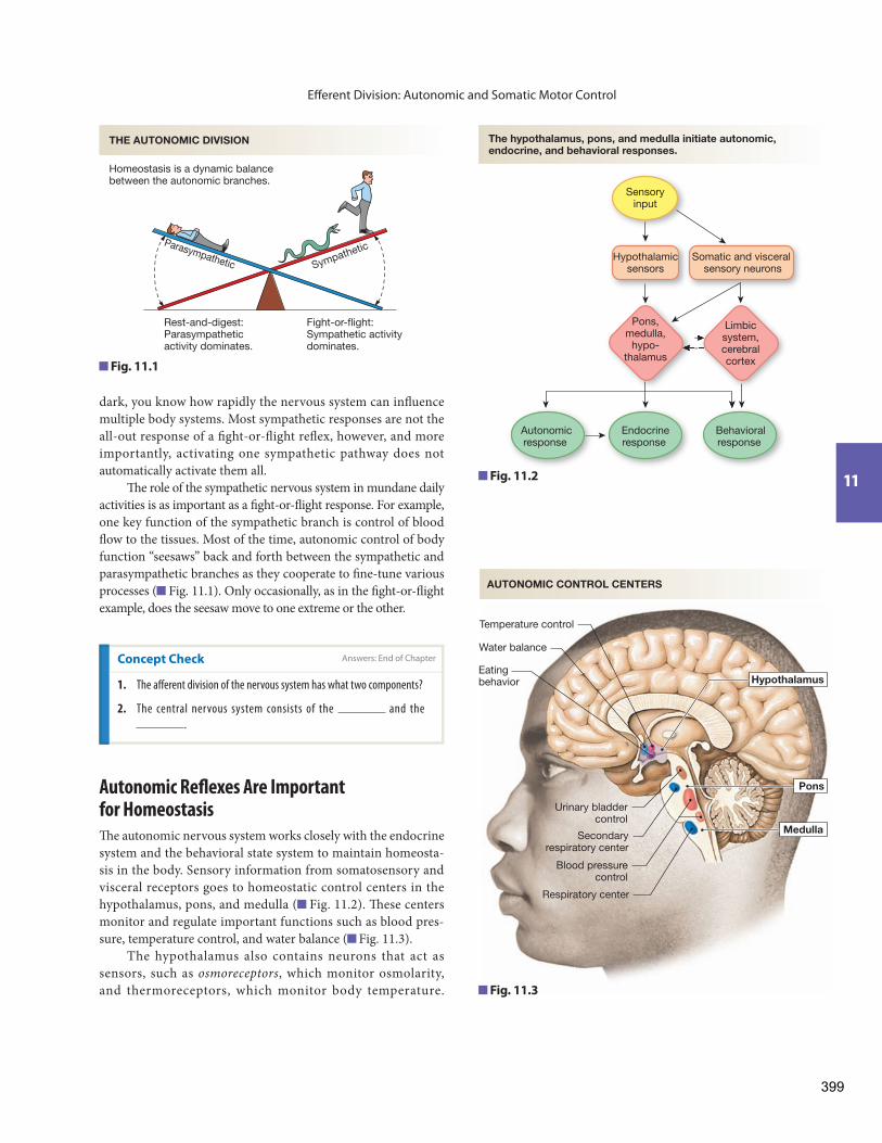
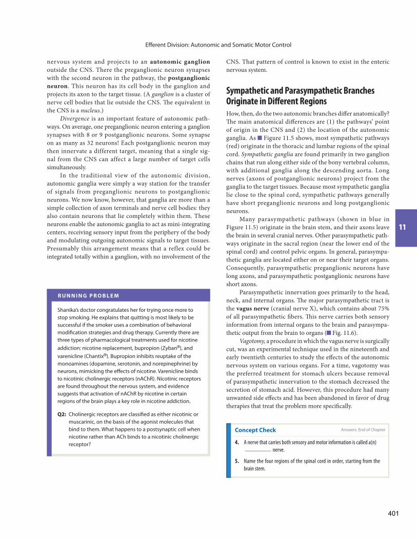
Th e autonomic nervous system works closely with the endocrine system and the behavioral state system to maintain homeosta-sis in the body. Sensory information from somatosensory and visceral receptors goes to homeostatic control centers in the hypothalamus, pons, and medulla ( Fig. 11.2 ). Th ese centers monitor and regulate important functions such as blood pres-sure, temperature control, and water balance ( Fig. 11.3 ).
The hypothalamus also contains neurons that act as sensors, such as osmoreceptors , which monitor osmolarity, and thermoreceptors, which monitor body temperature.
dark, you know how rapidly the nervous system can infl uence multiple body systems. Most sympathetic responses are not the all-out response of a fi ght-or-fl ight refl ex, however, and more importantly, activating one sympathetic pathway does not automatically activate them all.
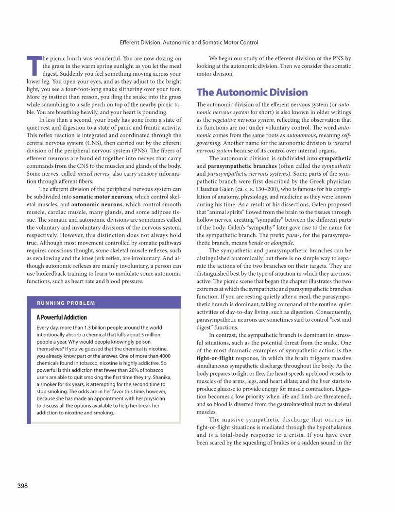
Th e role of the sympathetic nervous system in mundane daily activities is as important as a fi ght-or-fl ight response. For example, one key function of the sympathetic branch is control of blood fl ow to the tissues. Most of the time, autonomic control of body function “seesaws” back and forth between the sympathetic and parasympathetic branches as they cooperate to fi ne-tune various processes ( Fig. 11.1 ). Only occasionally, as in the fi ght-or-fl ight example, does the seesaw move to one extreme or the other.
Concept Check Answers: End of Chapter
1. The aff erent division of the nervous system has what two components?
2. The central nervous system consists of the and the
.
Rest-and-digest:Parasympatheticactivity dominates.
Fight-or-flight:Sympathetic activitydominates.
Homeostasis is a dynamic balancebetween the autonomic branches.
Parasympathetic Sympathetic
THE AUTONOMIC DIVISION
Fig. 11.1
Eating behavior
Secondaryrespiratory center
Respiratory center
Hypothalamus
Medulla
Pons
Urinary bladdercontrol
Water balance
Temperature control
Blood pressurecontrol
AUTONOMIC CONTROL CENTERS
Fig. 11.3
Pons,medulla,
hypo-thalamus
Limbicsystem,cerebralcortex
Sensoryinput
Endocrineresponse
Behavioralresponse
Autonomicresponse
Hypothalamicsensors
Somatic and visceral sensory neurons
The hypothalamus, pons, and medulla initiate autonomic, endocrine, and behavioral responses.
Fig. 11.2
399

Eff erent Division: Autonomic and Somatic Motor Control
internal environment, (2) up-down regulation by tonic control, (3) antagonistic control, and (4) chemical signals with diff erent eff ects in diff erent tissues.
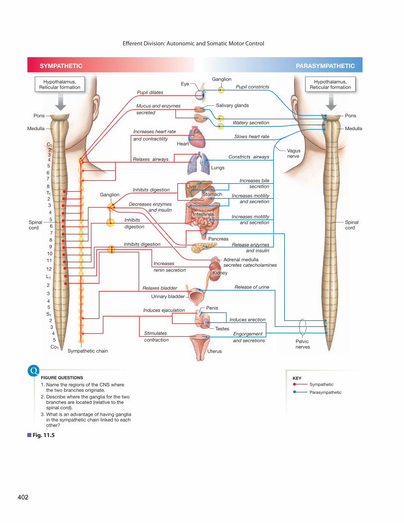
Most internal organs are under antagonistic control, in which one autonomic branch is excitatory and the other branch is inhibitory (see the table in Figure 11.5 ). For example, sym-pathetic innervation increases heart rate, while parasympa-thetic stimulation decreases it. Consequently, heart rate can be regulated by altering the relative proportions of sympathetic and parasympathetic control.
Exceptions to dual antagonistic innervation include the sweat glands and the smooth muscle in most blood vessels. Th ese tissues are innervated only by the sympathetic branch and rely strictly on tonic (up-down) control.
Although the two autonomic branches are usually antago-nistic in their control of a given target tissue, they sometimes work cooperatively on different tissues to achieve a common goal. For example, blood fl ow for penile erection is under con-trol of the parasympathetic branch, and muscle contraction for sperm ejaculation is directed by the sympathetic branch.
In some autonomic pathways, the neurotransmitter receptor determines the response of the target tissue. For instance, most blood vessels contain one type of adrenergic receptor that causes smooth muscle contraction (vasoconstriction). However, some blood vessels also contain a second type of adrenergic receptor that causes smooth muscle relaxation (vasodilation). Both receptors are activated by catecholamines. In this example the receptor, not the chemical signal, determines the response.
Motor output from the hypothalamus and brain stem creates autonomic responses, endocrine responses, and behavioral responses such as drinking, food-seeking, and temperature regulation (getting out of the heat, putting on a sweater). Th ese behavioral responses are integrated in brain centers responsible for motivated behaviors and control of movement.
In addition, sensory information integrated in the cerebral cortex and limbic system can create emotions that influence autonomic output, as Figure 11.2 illustrates. Blushing, faint-ing at the sight of a hypodermic needle, and “butterfl ies in the stomach” are all examples of emotional infl uences on autonomic functions. Understanding the autonomic and hormonal control of organ systems is the key to understanding the maintenance of homeostasis in virtually every system of the body.
Some autonomic refl exes are capable of taking place with-out input from the brain. Th ese spinal refl exes include urination, defecation, and penile erection—body functions that can be infl uenced by descending pathways from the brain but do not require this input. For example, people with spinal cord injuries that disrupt communication between the brain and spinal cord may retain some spinal refl exes but lose the ability to sense or control them.
Antagonistic Control Is a Hallmark of the Autonomic Division
The sympathetic and parasympathetic branches of the au-tonomic nervous system display all four of Walter Cannon’s properties of homeostasis: (1) preservation of the fi tness of the
Concept Check
3. Defi ne homeostasis.
R U N N I N G P R O B L E M
Neuroscientists have learned that addictive behaviors develop because certain chemicals act as positive reinforcers in the brain, creating physical and psychological dependence. Nicotine is an addictive drug that enhances dopamine release in the brain’s reward centers and creates pleasurable sensations. Over time, the brain also begins to associate the social aspects of cigarette smoking with pleasure, a conditioned response that makes quitting diffi cult. If smokers do stop smoking, they may suff er from unpleasant physical withdrawal symptoms, including lethargy, hunger, and irritability.
Q1: To avoid withdrawal symptoms, people continue to smoke, resulting in chronically elevated nicotine levels in their blood. Nicotine binds to nicotinic acetylcholine receptors (nAChR). What is the usual response of cells that are chronically exposed to elevated concentrations of a signal molecule?
Autonomic pathways consist of two neuronsthat synapse in an autonomic ganglion.
CNS Autonomicganglion
Preganglionicneuron
Targettissue
Postganglionicneuron
Fig. 11.4
Autonomic Pathways Have Two Eff erent Neurons in Series
All autonomic pathways (sympathetic and parasympathetic) consist of two neurons in series ( Fig. 11.4 ). Th e fi rst neuron, called the preganglionic neuron , originates in the central
Answers: End of Chapter
400
Eff erent Division: Autonomic and Somatic Motor Control
11
CNS. That pattern of control is known to exist in the enteric nervous system.
Sympathetic and Parasympathetic Branches Originate in Diff erent Regions
How, then, do the two autonomic branches diff er anatomically? Th e main anatomical diff erences are (1) the pathways’ point of origin in the CNS and (2) the location of the autonomic ganglia. As Figure 11.5 shows, most sympathetic pathways (red) originate in the thoracic and lumbar regions of the spinal cord. Sympathetic ganglia are found primarily in two ganglion chains that run along either side of the bony vertebral column, with additional ganglia along the descending aorta. Long nerves (axons of postganglionic neurons) project from the ganglia to the target tissues. Because most sympathetic ganglia lie close to the spinal cord, sympathetic pathways generally have short preganglionic neurons and long postganglionic neurons.
Many parasympathetic pathways (shown in blue in Figure 11.5 ) originate in the brain stem, and their axons leave the brain in several cranial nerves. Other parasympathetic path-ways originate in the sacral region (near the lower end of the spinal cord) and control pelvic organs. In general, parasympa-thetic ganglia are located either on or near their target organs. Consequently, parasympathetic preganglionic neurons have long axons, and parasympathetic postganglionic neurons have short axons.
Parasympathetic innervation goes primarily to the head, neck, and internal organs. Th e major parasympathetic tract is the vagus nerve (cranial nerve X), which contains about 75% of all parasympathetic fi bers. Th is nerve carries both sensory information from internal organs to the brain and parasympa-thetic output from the brain to organs ( Fig. 11.6 ).
Vagotomy, a procedure in which the vagus nerve is surgically cut, was an experimental technique used in the nineteenth and early twentieth centuries to study the eff ects of the autonomic nervous system on various organs. For a time, vagotomy was the preferred treatment for stomach ulcers because removal of parasympathetic innervation to the stomach decreased the secretion of stomach acid. However, this procedure had many unwanted side eff ects and has been abandoned in favor of drug therapies that treat the problem more specifi cally.
nervous system and projects to an autonomic ganglionoutside the CNS. There the preganglionic neuron synapses with the second neuron in the pathway, the postganglionic neuron . This neuron has its cell body in the ganglion and projects its axon to the target tissue. (A ganglion is a cluster of nerve cell bodies that lie outside the CNS. Th e equivalent in the CNS is a nucleus .)
Divergence is an important feature of autonomic path-ways. On average, one preganglionic neuron entering a ganglion synapses with 8 or 9 postganglionic neurons. Some synapse on as many as 32 neurons! Each postganglionic neuron may then innervate a different target, meaning that a single sig-nal from the CNS can affect a large number of target cells simultaneously.
In the traditional view of the autonomic division, autonomic ganglia were simply a way station for the transfer of signals from preganglionic neurons to postganglionic neurons. We now know, however, that ganglia are more than a simple collection of axon terminals and nerve cell bodies: they also contain neurons that lie completely within them. These neurons enable the autonomic ganglia to act as mini-integrating centers, receiving sensory input from the periphery of the body and modulating outgoing autonomic signals to target tissues. Presumably this arrangement means that a reflex could be integrated totally within a ganglion, with no involvement of the
R U N N I N G P R O B L E M
Shanika’s doctor congratulates her for trying once more to stop smoking. He explains that quitting is most likely to be successful if the smoker uses a combination of behavioral modifi cation strategies and drug therapy. Currently there are three types of pharmacological treatments used for nicotine addiction: nicotine replacement, bupropion (Zyban ® ), and varenicline (Chantix ® ). Bupropion inhibits reuptake of the monoamines (dopamine, serotonin, and norepinephrine) by neurons, mimicking the eff ects of nicotine. Varenicline binds to nicotinic cholinergic receptors (nAChR). Nicotinic receptors are found throughout the nervous system, and evidence suggests that activation of nAChR by nicotine in certain regions of the brain plays a key role in nicotine addiction.
Q2: Cholinergic receptors are classifi ed as either nicotinic or muscarinic, on the basis of the agonist molecules that bind to them. What happens to a postsynaptic cell when nicotine rather than ACh binds to a nicotinic cholinergic receptor?
Concept Check Answers: End of Chapter
4. A nerve that carries both sensory and motor information is called a(n)
nerve.
5. Name the four regions of the spinal cord in order, starting from the
brain stem.
401
Eff erent Division: Autonomic and Somatic Motor Control
Ganglion
Heart
Pupil dilates
Mucus and enzymessecreted
Increases heart rate
and contractility Slows heart rate
Relaxes airways Constricts airways
Watery secretion
Pupil constricts
Inhibits digestion
Inhibits digestion
Inhibits digestion
Increasesrenin secretion
Relaxes bladder
Induces ejaculation
Induces erection
Release of urine
Release enzymesand insulin
Decreases enzymesand insulin
Increases motilityand secretion
Increases motility and secretion
Increases bilesecretion
Stimulatescontraction
Engorgementand secretions
Lungs
Liver
Stomach
Pancreas
Adrenal medullasecretes catecholamines
Kidney
Penis
Testes
Uterus
Urinary bladder
Intestines
Salivary glands
Eye
Ganglion
Pons
Medulla
Spinalcord
Pons
Medulla
Spinalcord
Vagusnerve
Sympathetic chain
Pelvicnerves
C1234567
8T123
4
567
89
1011
12
L1
2
3
45S1
2345
Co1
Hypothalamus,Reticular formation
Hypothalamus,Reticular formation
SYMPATHETIC PARASYMPATHETIC
Parasympathetic
Sympathetic1. Name the regions of the CNS where the two branches originate.2. Describe where the ganglia for the two branches are located (relative to the spinal cord).3. What is an advantage of having ganglia in the sympathetic chain linked to each other?
FIGURE QUESTIONS KEY
Fig. 11.5
402

Eff erent Division: Autonomic and Somatic Motor Control
11
The Autonomic Nervous System Uses a Variety of Chemical Signals
Chemically, the sympathetic and parasympathetic branches can be distinguished by their neurotransmitters and receptors, using the following rules and Figure 11.6 :
1 Both sympathetic and parasympathetic preganglionic neurons release acetylcholine (ACh) onto nicotinic cholinergic receptors (AChR) on the postganglionic cell.
2 Most postganglionic sympathetic neurons secrete nor-epinephrine (NE) onto adrenergic receptors on the target cell.
3 Most postganglionic parasympathetic neurons secrete acetyl-choline onto muscarinic cholinergic receptors on the target cell.
However, there are some exceptions to these rules. A few sympathetic postganglionic neurons, such as those that terminate on sweat glands, secrete ACh rather than norepi-nephrine. These neurons are therefore called sympathetic cholinergic neurons .
A small number of autonomic neurons secrete neither norepinephrine nor acetylcholine and are known as nonadren-ergic, noncholinergic neurons . Some of the chemicals they use as neurotransmitters include substance P, somatostatin, vasoac-tive intestinal peptide (VIP), adenosine, nitric oxide, and ATP. Th e nonadrenergic, noncholinergic neurons are assigned to ei-ther the sympathetic or parasympathetic branch according to where their preganglionic fi bers leave the nerve cord.
Pupil of eye Dilates
Dilates
Bronchiolesdilate
Constricts
Mucus, enzymes
Increases rateand force ofcontraction
Decreasesmotility andsecretion
Decreasesenzymesecretion
Inhibits insulin secretion
Secretescatecholamines
Increases reninsecretion
Urinary retention
Fat breakdown
Ejaculation(male)
Depends onstage of cycle
Generallyinhibitory
Salivary glands
Digestive tract
Heart
Lungs
Kidney
Uterus
Urinary bladder
Arterioles and veins
Exocrinepancreas
Endocrinepancreas
Adrenalmedulla
Adipose tissue
Male and femalesex organs
Lymphoid tissue
EFFECTORORGAN
SYMPATHETICRESPONSE
ADRENERGICRECEPTOR
α
α and β2
β1
αβ2
β2*
α, β2
α
α
β1
α, β2
β
α
α, β2
α, β2
**All parasympathetic responses are mediated by muscarinic receptors.
*Hormonal epinephrine only
—
Constricts
Watery secretion
Slows rate
Release of urine
Erection
Bronchiolesconstrict
Increasesmotility andsecretion
Increasesenzymesecretion
Stimulatesinsulin secretion
Depends onstage of cycle
PARASYMPATHETICRESPONSE **
——
——
——
——
——
CNS CNS
Autonomicganglion
Norepinephrine
ACh
Adrenergicreceptor
Sympathetic pathways use acetylcholine andnorepinephrine.
Parasympathetic pathwaysuse acetylcholine.
Nicotinicreceptor
Muscarinic receptor
ACh
Target tissueT T
FIGURE QUESTIONS
1. Identify all: - cholinergic neurons - adrenergic neurons - preganglionic neurons - postganglionic neurons2. Which pathway will have longer preganglionic neurons? (Hint: See Fig. 11.5.)
Fig. 11.6 Sympathetic and parasympathetic neurotransmitters and receptors
403

Eff erent Division: Autonomic and Somatic Motor Control
Autonomic Pathways Control Smooth and Cardiac Muscle and Glands
The targets of autonomic neurons are smooth muscle, cardiac muscle, many exocrine glands, a few endocrine glands, lymphoid tissues, and some adipose tissue. The synapse between a postganglionic autonomic neuron and its target cell is called the neuroeff ector junction (recall that targets are also called eff ectors).
The structure of an autonomic synapse differs from the model synapse. Autonomic postganglionic axons end with a se-ries of swollen areas at their distal ends, like beads spaced out along a string ( Fig. 11.7 a). Each of these swellings, known as a varicosity { varicosus, abnormally enlarged or swollen}, contains vesicles fi lled with neurotransmitter.
Th e branched ends of the axon lie across the surface of the target tissue, but the underlying target cell membrane does not
NE
Axon varicosity
Adrenergicreceptor
Tyrosine
Exocytosis
Ca2+
Voltage-gated Ca2+ channel
Action potential
Axon
Active transport
Response
MAO
Bloodvessel
G
Diffuses away
Action potential arrives at the varicosity.
Depolarization opens voltage-gated Ca2+ channels.
Ca2+ entry triggers exocytosis of synaptic vesicles.
NE binds to adrenergicreceptor on target.
NE is metabolized by monoamine oxidase (MAO).
Receptor activation ceases when NE diffuses away from the synapse.
NE can be taken back intosynaptic vesicles for re-release.
NE is removed from the synapse.
NE
1
1
2
2
4
4
5
5
3
3
66
7
7
8
8
Target cell
Mitochondrion
VaricositiesSmooth muscle cells
Vesicle containingneurotransmitter
Axon ofpostganglionic
autonomicneuron
Varicosity
AUTONOMIC SYNAPSES
(a) Autonomic varicosities release neurotransmitter over the surface of target cells.
(b) Norepinephrine (NE) release and removal at a sympathetic neuroeffector junction
Fig. 11.7
404
Eff erent Division: Autonomic and Somatic Motor Control
11
possess clusters of neurotransmitter receptors in specific sites. Instead, the neurotransmitter is simply released into the interstitial fl uid to diff use to wherever the receptors are located. Th e result is a less-directed form of communication than that which occurs between a somatic motor neuron and a skeletal muscle. The diff use release of autonomic neurotransmitter means that a single postganglionic neuron can aff ect a large area of target tissue.
Th e release of autonomic neurotransmitters is subject to modulation from a variety of sources. For example, sympathetic varicosities contain receptors for hormones and for paracrines such as histamine. These modulators may either facilitate or inhibit neurotransmitter release. Some preganglionic neurons co-secrete neuropeptides along with acetylcholine. Th e peptides act as neuromodulators, producing slow synaptic potentials that modify the activity of postganglionic neurons.
Autonomic Neurotransmitters Are Synthesized in the Axon
In the autonomic division, neurotransmitters are synthesized in the axon varicosities ( Fig. 11.7 b). The primary autonomic neurotransmitters are acetylcholine (ACh) and norepineph-rine, both small molecules easily synthesized by cytoplasmic enzymes. Neurotransmitter made in the varicosities is packaged into synaptic vesicles for storage.
Neurotransmitter release follows the pattern found in other cells: depolarization—calcium signal—exocytosis. When an action potential arrives at the varicosity, voltage-gated Ca 2+ channels open, Ca 2+ enters the neuron, and the synaptic vesicle contents are released by exocytosis. Once neurotransmitters are released into the synapse, they either diff use through the inter-stitial fl uid until they encounter a receptor on the target cell or drift away from the synapse.
The concentration of neurotransmitter in the synapse is a major factor in the control that an autonomic neuron exerts
on its target: more neurotransmitter means a longer or stronger response. Th e concentration of neurotransmitter in a synapse is infl uenced by its rate of breakdown or removal ( Fig. 11.7 b). Neurotransmitter activation of its receptor terminates when the neurotransmitter (1) diffuses away, (2) is metabolized by enzymes in the extracellular fl uid, or (3) is actively transported into cells around the synapse. Th e uptake of neurotransmitter by varicosities allows neurons to reuse the chemicals.
Th ese steps are shown for norepinephrine in Figure 11.7 b. Norepinephrine is synthesized in the varicosity from the amino acid tyrosine. Once released into the synapse, norepinephrine may combine with an adrenergic receptor on the target cell, dif-fuse away, or be transported back into the varicosity. Inside the neuron, recycled norepinephrine is either repackaged into vesi-cles or broken down by monoamine oxidase (MAO), the main enzyme responsible for degradation of catecholamines.
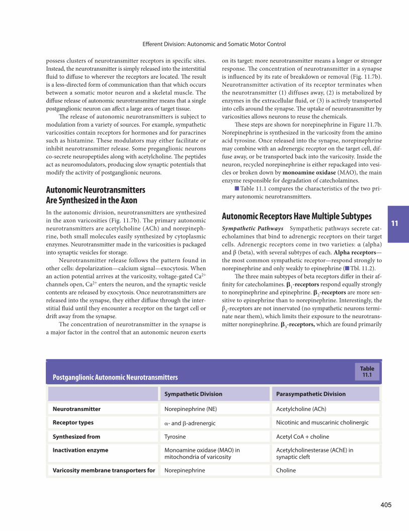
Table 11.1 compares the characteristics of the two pri-mary autonomic neurotransmitters.
Autonomic Receptors Have Multiple Subtypes
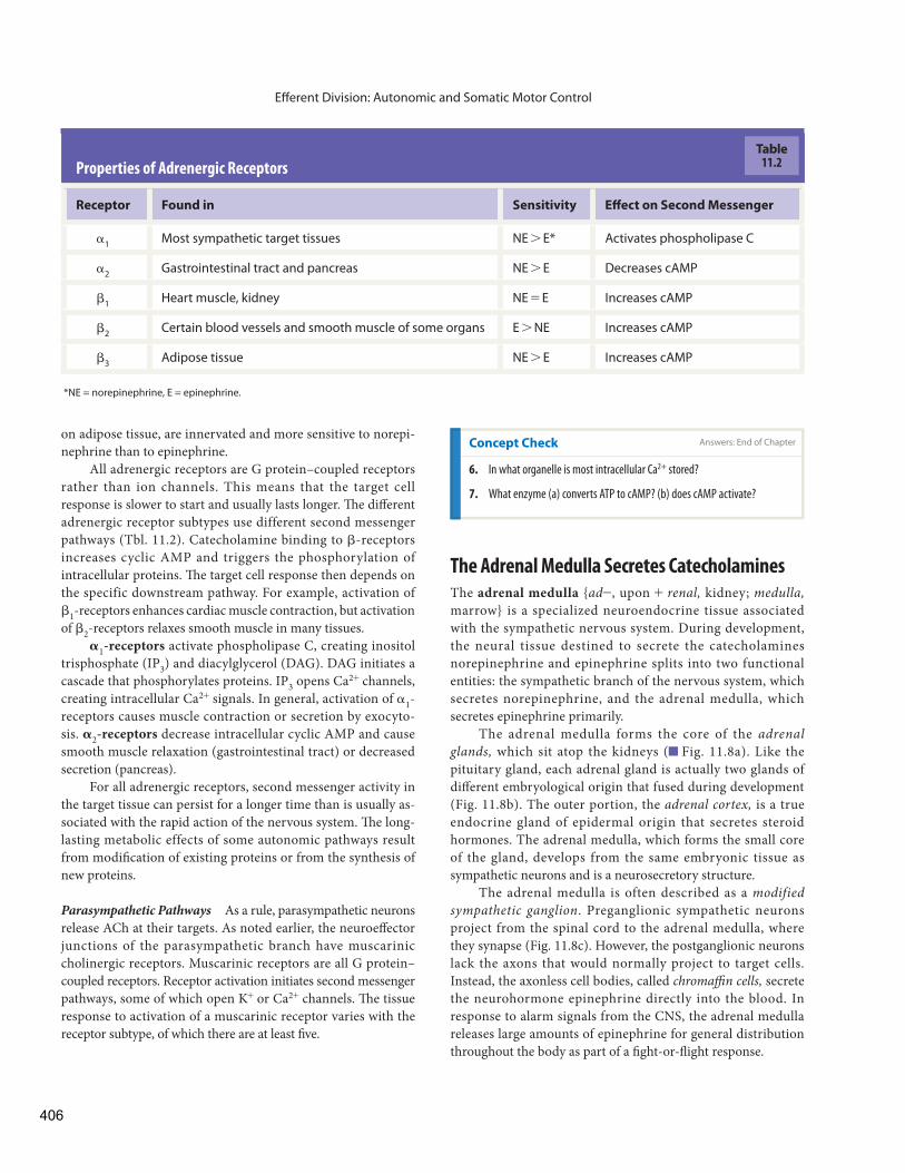
Sympathetic Pathways Sympathetic pathways secrete cat-echolamines that bind to adrenergic receptors on their target cells. Adrenergic receptors come in two varieties: α (alpha) and β (beta), with several subtypes of each. Alpha receptors —the most common sympathetic receptor— respond strongly to norepinephrine and only weakly to epinephrine ( Tbl. 11.2 ).
Th e three main subtypes of beta receptors diff er in their af-fi nity for catecholamines. 6 1 -receptors respond equally strongly to norepinephrine and epinephrine. 6 2 -receptors are more sen-sitive to epinephrine than to norepinephrine. Interestingly, the β 2 -receptors are not innervated (no sympathetic neurons termi-nate near them), which limits their exposure to the neurotrans-mitter norepinephrine. 6 3 -receptors, which are found primarily
Postganglionic Autonomic Neurotransmitters
Sympathetic Division Parasympathetic Division
Neurotransmitter Norepinephrine (NE) Acetylcholine (ACh)
Receptor types c- and d-adrenergic Nicotinic and muscarinic cholinergic
Synthesized from Tyrosine Acetyl CoA + choline
Inactivation enzyme Monoamine oxidase (MAO) in mitochondria of varicosity
Acetylcholinesterase (AChE) in synaptic cleft
Varicosity membrane transporters for Norepinephrine Choline
Table11.1
405
Eff erent Division: Autonomic and Somatic Motor Control
The Adrenal Medulla Secretes Catecholamines
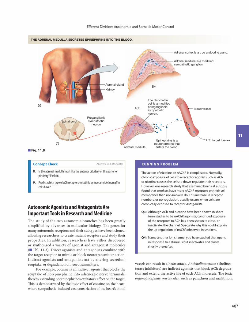
The adrenal medulla { ad -, upon + renal, kidney; medulla,marrow} is a specialized neuroendocrine tissue associated with the sympathetic nervous system. During development, the neural tissue destined to secrete the catecholamines norepinephrine and epinephrine splits into two functional entities: the sympathetic branch of the nervous system, which secretes norepinephrine, and the adrenal medulla, which secretes epinephrine primarily.
The adrenal medulla forms the core of the adrenal glands, which sit atop the kidneys ( Fig. 11.8 a). Like the pituitary gland, each adrenal gland is actually two glands of diff erent embryological origin that fused during development ( Fig. 11.8 b). The outer portion, the adrenal cortex, is a true endocrine gland of epidermal origin that secretes steroid hormones. The adrenal medulla, which forms the small core of the gland, develops from the same embryonic tissue as sympathetic neurons and is a neurosecretory structure.
The adrenal medulla is often described as a modified sympathetic ganglion . Preganglionic sympathetic neurons project from the spinal cord to the adrenal medulla, where they synapse ( Fig. 11.8 c). However, the postganglionic neurons lack the axons that would normally project to target cells. Instead, the axonless cell bodies, called chromaffi n cells, secrete the neurohormone epinephrine directly into the blood. In response to alarm signals from the CNS, the adrenal medulla releases large amounts of epinephrine for general distribution throughout the body as part of a fi ght-or-fl ight response.
on adipose tissue, are innervated and more sensitive to norepi-nephrine than to epinephrine.
All adrenergic receptors are G protein–coupled receptors rather than ion channels. This means that the target cell response is slower to start and usually lasts longer. Th e diff erent adrenergic receptor subtypes use different second messenger pathways ( Tbl. 11.2 ). Catecholamine binding to d-receptors increases cyclic AMP and triggers the phosphorylation of intracellular proteins. Th e target cell response then depends on the specific downstream pathway. For example, activation of d 1 -receptors enhances cardiac muscle contraction, but activation of d 2 -receptors relaxes smooth muscle in many tissues.
5 1 -receptors activate phospholipase C, creating inositol trisphosphate (IP 3 ) and diacylglycerol (DAG). DAG initiates a cascade that phosphorylates proteins. IP 3 opens Ca 2+ channels, creating intracellular Ca 2+ signals. In general, activation of c 1 -receptors causes muscle contraction or secretion by exocyto-sis. 5 2 -receptors decrease intracellular cyclic AMP and cause smooth muscle relaxation (gastrointestinal tract) or decreased secretion (pancreas).
For all adrenergic receptors, second messenger activity in the target tissue can persist for a longer time than is usually as-sociated with the rapid action of the nervous system. Th e long-lasting metabolic effects of some autonomic pathways result from modifi cation of existing proteins or from the synthesis of new proteins.
Parasympathetic Pathways As a rule, parasympathetic neurons release ACh at their targets. As noted earlier, the neuroeff ector junctions of the parasympathetic branch have muscarinic cholinergic receptors. Muscarinic receptors are all G protein–coupled receptors. Receptor activation initiates second messenger pathways, some of which open K + or Ca 2+ channels. Th e tissue response to activation of a muscarinic receptor varies with the receptor subtype, of which there are at least fi ve.
Properties of Adrenergic Receptors
Receptor Found in Sensitivity Eff ect on Second Messenger
c 1 Most sympathetic target tissues NE 7 E* Activates phospholipase C
c 2 Gastrointestinal tract and pancreas NE 7 E Decreases cAMP
d 1 Heart muscle, kidney NE = E Increases cAMP
d 2 Certain blood vessels and smooth muscle of some organs E 7 NE Increases cAMP
d 3 Adipose tissue NE 7 E Increases cAMP
*NE = norepinephrine, E = epinephrine.
Table11.2
Concept Check Answers: End of Chapter
6. In what organelle is most intracellular Ca 2+ stored?
7. What enzyme (a) converts ATP to cAMP? (b) does cAMP activate?
406
Eff erent Division: Autonomic and Somatic Motor Control
11
Autonomic Agonists and Antagonists Are Important Tools in Research and Medicine
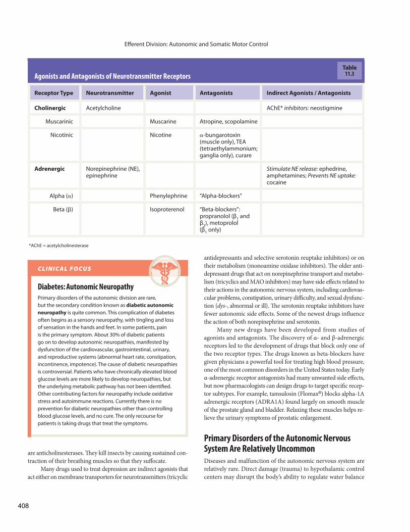
The study of the two autonomic branches has been greatly simplified by advances in molecular biology. The genes for many autonomic receptors and their subtypes have been cloned, allowing researchers to create mutant receptors and study their properties. In addition, researchers have either discovered or synthesized a variety of agonist and antagonist molecules ( Tbl. 11.3 ). Direct agonists and antagonists combine with the target receptor to mimic or block neurotransmitter action. Indirect agonists and antagonists act by altering secretion, reuptake, or degradation of neurotransmitters.
For example, cocaine is an indirect agonist that blocks the reuptake of norepinephrine into adrenergic nerve terminals, thereby extending norepinephrine’s excitatory eff ect on the target. Th is is demonstrated by the toxic eff ect of cocaine on the heart, where sympathetic-induced vasoconstriction of the heart’s blood
Concept Check Answers: End of Chapter
8. Is the adrenal medulla most like the anterior pituitary or the posterior
pituitary? Explain.
9. Predict which type of ACh receptors (nicotinic or muscarinic) chromaffi n
cells have?
R U N N I N G P R O B L E M
The action of nicotine on nAChR is complicated. Normally, chronic exposure of cells to a receptor agonist such as ACh or nicotine causes the cells to down-regulate their receptors. However, one research study that examined brains at autopsy found that smokers have more nAChR receptors on their cell membranes than nonsmokers do. This increase in receptor numbers, or up-regulation, usually occurs when cells are chronically exposed to receptor antagonists .
Q3: Although ACh and nicotine have been shown in short-term studies to be nAChR agonists, continued exposure of the receptors to ACh has been shown to close, or inactivate, the channel. Speculate why this could explain the up-regulation of nAChR observed in smokers.
Q4: Name another ion channel you have studied that opens in response to a stimulus but inactivates and closes shortly thereafter.
vessels can result in a heart attack. Anticholinesterases (cholines-terase inhibitors) are indirect agonists that block ACh degrada-tion and extend the active life of each ACh molecule. Th e toxic organophosphate insecticides, such as parathion and malathion,
ACh
Adrenal medulla is a modifiedsympathetic ganglion.
Adrenal medulla
Adrenal cortex is a true endocrine gland.
Adrenal gland(b)
Kidney
(a)
The chromaffincell is a modifiedpostganglionicsympatheticneuron.
Preganglionicsympathetic
neuronSpinal cord
(c)
Blood vessel
Epinephrine is aneurohormone thatenters the blood.
To target tissues
THE ADRENAL MEDULLA SECRETES EPINEPHRINE INTO THE BLOOD.
Fig. 11.8
407
Eff erent Division: Autonomic and Somatic Motor Control
are anticholinesterases. Th ey kill insects by causing sustained con-traction of their breathing muscles so that they suff ocate.
Many drugs used to treat depression are indirect agonists that act either on membrane transporters for neurotransmitters (tricyclic
Diabetes: Autonomic Neuropathy
Primary disorders of the autonomic division are rare, but the secondary condition known as diabetic autonomic neuropathy is quite common. This complication of diabetes often begins as a sensory neuropathy, with tingling and loss of sensation in the hands and feet. In some patients, pain is the primary symptom. About 30% of diabetic patients go on to develop autonomic neuropathies, manifested by dysfunction of the cardiovascular, gastrointestinal, urinary, and reproductive systems (abnormal heart rate, constipation, incontinence, impotence). The cause of diabetic neuropathies is controversial. Patients who have chronically elevated blood glucose levels are more likely to develop neuropathies, but the underlying metabolic pathway has not been identifi ed. Other contributing factors for neuropathy include oxidative stress and autoimmune reactions. Currently there is no prevention for diabetic neuropathies other than controlling blood glucose levels, and no cure. The only recourse for patients is taking drugs that treat the symptoms.
C L I N I C A L F O C U S
Agonists and Antagonists of Neurotransmitter Receptors
Receptor Type Neurotransmitter Agonist Antagonists Indirect Agonists / Antagonists
Cholinergic Acetylcholine AChE* inhibitors: neostigmine
Muscarinic Muscarine Atropine, scopolamine
Nicotinic Nicotine c-bungarotoxin (muscle only), TEA (tetraethylammonium; ganglia only), curare
Adrenergic Norepinephrine (NE), epinephrine
Stimulate NE release: ephedrine, amphetamines; Prevents NE uptake: cocaine
Alpha (c) Phenylephrine “Alpha-blockers”
Beta (d) Isoproterenol “Beta-blockers”: propranolol (β 1 and β 2 ), metoprolol (β 1 only)
*AChE = acetylcholinesterase
Table11.3
antidepressants and selective serotonin reuptake inhibitors) or on their metabolism (monoamine oxidase inhibitors). Th e older anti-depressant drugs that act on norepinephrine transport and metabo-lism (tricyclics and MAO inhibitors) may have side eff ects related to their actions in the autonomic nervous system, including cardiovas-cular problems, constipation, urinary diffi culty, and sexual dysfunc-tion { dys -, abnormal or ill}. Th e serotonin reuptake inhibitors have fewer autonomic side eff ects. Some of the newest drugs infl uence the action of both norepinephrine and serotonin.
Many new drugs have been developed from studies of agonists and antagonists. The discovery of α- and β-adrenergic receptors led to the development of drugs that block only one of the two receptor types. The drugs known as beta-blockers have given physicians a powerful tool for treating high blood pressure, one of the most common disorders in the United States today. Early α-adrenergic receptor antagonists had many unwanted side eff ects, but now pharmacologists can design drugs to target specifi c recep-tor subtypes. For example, tamsulosin (Flomax ® ) blocks alpha-1A adrenergic receptors (ADRA1A) found largely on smooth muscle of the prostate gland and bladder. Relaxing these muscles helps re-lieve the urinary symptoms of prostatic enlargement.
Primary Disorders of the Autonomic Nervous System Are Relatively Uncommon
Diseases and malfunction of the autonomic nervous system are relatively rare. Direct damage (trauma) to hypothalamic control centers may disrupt the body’s ability to regulate water balance
408
Eff erent Division: Autonomic and Somatic Motor Control
11
After discussing her options with her doctor, Shanika decides to try the nicotine patch, one form of nicotine replacement therapy. These adhesive patches allow the former smoker to gradually decrease nicotine levels in the body, preventing withdrawal symptoms during the time the cells are down-regulating their receptors back to the normal number. When Shanika reads the package insert prior to applying her fi rst nicotine patch, she notices a warning to keep the patches away from children. An overdose of nicotine (highly unlikely when the patch is used as directed) could result in complete paralysis of the respiratory muscles (the diaphragm and the skeletal muscles of the chest wall).
Q5: Why might excessive levels of nicotine cause respiratory paralysis?
R U N N I N G P R O B L E M
or temperature. Generalized sympathetic dysfunction may result from systemic diseases such as cancer and diabetes mellitus. Th ere are also some conditions, such as multiple system atrophy, in which the CNS control centers for autonomic functions degenerate.
In many cases of sympathetic dysfunction, the symptoms are manifested most strongly in the cardiovascular system, when
diminished sympathetic input to blood vessels results in abnor-mally low blood pressure. Other prominent symptoms of sym-pathetic pathology include urinary incontinence { in -, unable + continere, to contain}, which is the loss of bladder control, and impotence, which is the inability to achieve or sustain a penile erection.
Occasionally, patients suffer from primary autonomic failure when sympathetic neurons degenerate. In the face of continuing diminished sympathetic input, target tissues up-regulate, putting more receptors into the cell membrane to maximize the cell’s response to available norepinephrine. This increase in receptor abundance leads to denervation hypersensitivity, a state in which the administration of exogenous adrenergic agonists causes a greater-than-expected response.
Summary of Sympathetic and Parasympathetic Branches
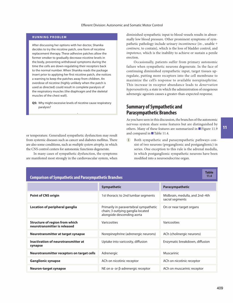
As you have seen in this discussion, the branches of the autonomic nervous system share some features but are distinguished by others. Many of these features are summarized in Figure 11.9 and compared in Table 11.4 .
1 Both sympathetic and parasympathetic pathways con-sist of two neurons (preganglionic and postganglionic) in series. One exception to this rule is the adrenal medulla, in which postganglionic sympathetic neurons have been modifi ed into a neuroendocrine organ.
Comparison of Sympathetic and Parasympathetic Branches
Sympathetic Parasympathetic
Point of CNS origin 1st thoracic to 2nd lumbar segments Midbrain, medulla, and 2nd–4th sacral segments
Location of peripheral ganglia Primarily in paravertebral sympathetic chain; 3 outlying ganglia located alongside descending aorta
On or near target organs
Structure of region from which neurotransmitter is released
Varicosities Varicosities
Neurotransmitter at target synapse Norepinephrine (adrenergic neurons) ACh (cholinergic neurons)
Inactivation of neurotransmitter at synapse
Uptake into varicosity, diffusion Enzymatic breakdown, diffusion
Neurotransmitter receptors on target cells Adrenergic Muscarinic
Ganglionic synapse ACh on nicotinic receptor ACh on nicotinic receptor
Neuron-target synapse NE on α- or β-adrenergic receptor ACh on muscarinic receptor
Table11.4
409

Efferent Divisions of the Nervous System
SOMATIC MOTOR PATHWAY
AUTONOMIC PATHWAYS
Comparison of Somatic Motor and Autonomic Divisions
Number of neurons in efferent path
Neurotransmitter/receptor at neuron-target synapse
Target tissue
1
ACh/nicotinic
2
Skeletal muscle
Axon terminals
Excitatory only: muscle contracts
Axons only
Posture and movement
Smooth and cardiac muscle; some endocrineand exocrine glands; some adipose tissue
ACh/muscarinic or NE/α- or β-adrenergic
Varicosities and axon terminals
Excitatory or inhibitory
Preganglionic axons, ganglia, postganglionic neurons
Visceral function, including movement in internal organs and secretion; control of metabolism
Neurotransmitter released from
Effects on target tissue
Peripheral components found outside the CNS
Summary of function
SOMATIC MOTOR AUTONOMIC
Using the figure, compare: (a) number of neurons in somatic motor and autonomic pathways (b) receptors on target cells of somatic motor, sympathetic, and parasympathetic pathways (c) neurotransmitters used on target cells of somatic motor, sympathetic, and parasympathetic pathways (d) receptor subtypes for epinephrine with subtypes for norepinephrine
Muscarinicreceptor
KEY
FIGURE QUESTIONS
ACh = acetylcholine
E = epinephrine
NE = norepinephrine
Ganglion
Nicotinic receptor
Nicotinic receptor
Autonomic targets:
• Smooth and cardiac muscles
• Some endocrine and exocrine glands
• Some adipose tissueAChACh
β1 receptor
β2 receptor
α receptor
Adrenal medulla
Adrenal cortexBlood vessel
E
E
NE
AChCNS
CNS
CNS
CNS
Target:skeletal muscle
Nicotinic receptor
(a) Parasympathetic Pathway
(b) Sympathetic Pathway
(c) Adrenal Sympathetic Pathway
ACh
Fig. 11.9 E S S E N T I A L S
410
Eff erent Division: Autonomic and Somatic Motor Control
11
signal molecules. Th ese signal molecules play a critical role in the formation and maintenance of neuromuscular junctions.
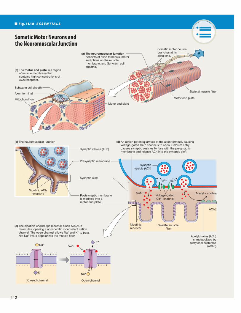
On the postsynaptic side of the neuromuscular junction, the muscle cell membrane that lies opposite the axon terminal is modifi ed into a motor end plate , a series of folds that look like shallow gutters ( Fig. 11.10 c). Along the upper edge of each gut-ter, nicotinic ACh receptor (nAChR) channels cluster together in an active zone. Between the axon and the muscle, the synaptic cleft is fi lled with a fi brous matrix whose collagen fi bers hold the axon terminal and the motor end plate in the proper alignment. Th e matrix also contains acetylcholinesterase (AChE), the en-zyme that rapidly deactivates ACh by degrading it into acetyl and choline.
2 All preganglionic autonomic neurons secrete acetylcho-line onto nicotinic receptors. Most sympathetic neurons secrete norepinephrine onto adrenergic receptors. Most parasympathetic neurons secrete acetylcholine onto mus-carinic receptors.
3 Sympathetic pathways originate in the thoracic and lum-bar regions of the spinal cord. Parasympathetic pathways leave the CNS at the brain stem and in the sacral region of the spinal cord.
4 Most sympathetic ganglia are located close to the spinal cord (are paravertebral ). Parasympathetic ganglia are located close to or in the target tissue.
5 Th e sympathetic branch controls functions that are useful in stress or emergencies (fi ght-or-fl ight). Th e parasympa-thetic branch is dominant during rest-and-digest activities.
The Somatic Motor Division Somatic motor pathways, which control skeletal muscles, diff er from autonomic pathways both anatomically and functionally (see the table in Figure 11.9 ). Somatic motor pathways have a single neuron that originates in the CNS and projects its axon to the target tissue, which is always a skeletal muscle. Somatic pathways are always excitatory, unlike autonomic pathways, which may be either excitatory or inhibitory.
A Somatic Motor Pathway Consists of One Neuron
Th e cell bodies of somatic motor neurons are located either in the ventral horn of the spinal cord or in the brain, with a long single axon projecting to the skeletal muscle target ( Fig. 11.9 ). Th ese myelinated axons may be a meter or more in length, such as the somatic motor neurons that innervate the muscles of the foot and hand.
Somatic motor neurons branch close to their targets. Each branch divides into a cluster of enlarged axon terminals that lie on the surface of the skeletal muscle fi ber ( Fig. 11.10 a). Th is branching structure allows a single motor neuron to control many muscle fi bers at one time.
Th e synapse of a somatic motor neuron on a muscle fi ber is called the neuromuscular junction , or NMJ ( Fig. 11.10 b). Like all other synapses, the NMJ has three components: (1) the motor neuron’s presynaptic axon terminal fi lled with synaptic vesicles and mitochondria, (2) the synaptic cleft, and (3) the postsynaptic membrane of the skeletal muscle fi ber.
In addition, the neuromuscular junction includes extensions of Schwann cells that form a thin layer covering the top of the axon terminals. For years it was thought that this cell layer simply pro-vided insulation to speed up the conduction of the action potential, but we now know that Schwann cells secrete a variety of chemical
Concept Check
10. Is the ventral horn of the spinal cord, which contains the cell bodies of
somatic motor neurons, gray matter or white matter?
The Neuromuscular Junction Contains Nicotinic Receptors
As in all neurons, action potentials arriving at the axon terminal open voltage-gated Ca 2+ channels in the membrane. Calcium diffuses into the cell down its electrochemical gradient, triggering the release of ACh-containing synaptic vesicles. Acetylcholine diffuses across the synaptic cleft and combines with nicotinic receptor channels (nAChR) on the skeletal muscle membrane ( Fig. 11.10 d).
The nAChR channels of skeletal muscle are similar but not identical to the nicotinic ACh receptors found on neurons. This difference is illustrated by the fact that the snake toxin a- bungarotoxin binds to nicotinic skeletal muscle receptors but not to those in autonomic ganglia. Both muscle and neuronal nAChR proteins have fi ve subunits encircling the central pore. However, skeletal muscle has c, d, f, and 2 subunit isoforms, while neuronal nAchR has only the c and d isoforms. Th ese iso-forms of nAChR are inactivated with extended exposure to ACh or other agonists.
Nicotinic cholinergic receptors are chemically gated ion channels with two binding sites for ACh ( Fig. 11.10 d). When ACh binds to the receptor, the channel gate opens and allows monovalent cations to fl ow through. In skeletal muscle, net Na +entry into the muscle fi ber depolarizes it, triggering an action potential that causes contraction of the skeletal muscle cell.
Acetylcholine acting on a skeletal muscle’s motor end plate is always excitatory and creates muscle contraction. Th ere is no antagonistic innervation to relax skeletal muscles. Instead, relaxation occurs when the somatic motor neurons are
Answers: End of Chapter
411
+ + + + + + + + ++
- - - - - - - - --+ + + + + + + + + +
- - - - - - - - - -
Fig. 11.10 E S S E N T I A L S
Somatic Motor Neurons and the Neuromuscular Junction
(c) The neuromuscular junction
Somatic motor neuron branches at its distal end.
Skeletal muscle fiber
Motor end plate
Motor end plateMitochondrion
Synaptic vesicle (ACh)
Synapticvesicle (ACh)
Presynaptic membrane
Synaptic cleft
Postsynaptic membrane is modified into a motor end plate.
Schwann cell sheath
Axon terminal
Nicotinic AChreceptors
Skeletal muscle fiber
AChE
K+
Na+
Na+
K+
Voltage-gatedCa2+ channel
ACh
ACh
Ca2+ Ca2+
Acetyl + choline
Nicotinicreceptor
Closed channel Open channel
Acetylcholine (ACh) is metabolized by
acetylcholinesterase (AChE).
(e) The nicotinic cholinergic receptor binds two ACh molecules, opening a nonspecific monovalent cation channel. The open channel allows Na+ and K+ to pass. Net Na+ influx depolarizes the muscle fiber.
(a) The neuromuscular junction consists of axon terminals, motor end plates on the muscle membrane, and Schwann cell sheaths.
(b) The motor end plate is a region of muscle membrane that contains high concentrations of ACh receptors.
(d) An action potential arrives at the axon terminal, causing voltage-gated Ca2+ channels to open. Calcium entry causes synaptic vesicles to fuse with the presynaptic membrane and release ACh into the synaptic cleft.
412

Eff erent Division: Autonomic and Somatic Motor Control
11
inhibited in the CNS, preventing ACh release. You will learn later about how inhibition of somatic motor pathways controls body movement.
Somatic motor neurons do more than simply create contrac-tions: they are necessary for muscle health. “Use it or lose it” is a cli-ché that is very appropriate to the dynamics of muscle mass because disrupting synaptic transmission at the neuromuscular junction has devastating eff ects on the entire body. Without communication between the motor neuron and the muscle, the skeletal muscles for movement and posture weaken, as do the skeletal muscles for breathing. In the severest cases, loss of respiratory function can be fatal unless the patient is placed on artifi cial ventilation. Myasthenia gravis, a disease characterized by loss of ACh receptors, is the most common disorder of the neuromuscular junction.
A Powerful Addiction
R U N N I N G P R O B L E M C O N C L U S I O N
Shanika is determined to stop smoking this time because her grandfather, a smoker for many years, was just diagnosed with lung cancer. Finding that the patch alone does not stop her craving for a cigarette, she attends behavioral modifi cation classes. In these classes, she learns to avoid situations that make her likely to smoke and to substitute other activities, such as chewing gum, for smoking. After six months, Shanika proudly informs her family that she thinks she has kicked the habit.
Nicotine replacement may not be the ideal treatment for smoking cessation because although the former smoker is no longer exposed to cigarette smoke, the nicotine addiction may remain. Varenicline (Chantix ® ) acts as a partial nAChR agonist and may help
break the addiction. However, unwanted side eff ects, such as nightmares and psychological disturbances, have been reported with its use. Some smokers have quit with the help of bupropion (Zyban ® ), a drug that is also used as an antidepressant. Two drugs that act on cannabinoid receptors were eff ective in clinical trials but were withdrawn from the market after people taking them exhibited serious psychological side eff ects. A vaccine against nicotine is currently in the last stages of clinical trials in the United States. To learn more about nicotine addiction and smoking cessation programs, see Medline Plus ( www.nlm.nih.gov/medlineplus ). Check your understanding of this running problem by comparing your answers to the information in the following summary table.
Concept Check Answers: End of Chapter
11. Compare gating and ion selectivity of acetylcholine receptor-channels
in the motor end plate with that of ion channels along the axon of a
somatic motor neuron.
12. A nonsmoker who chews nicotine-containing gum might notice an
increase in heart rate, a function controlled by sympathetic neurons.
Postganglionic sympathetic neurons secrete norepinephrine, not ACh,
so how could nicotine aff ect heart rate?
13. Patients with myasthenia gravis have a defi ciency of ACh receptors on
their skeletal muscles and have weak muscle function as a result. Why
would administration of an anticholinesterase drug (one that inhibits
acetylcholinesterase) improve muscle function in these patients?
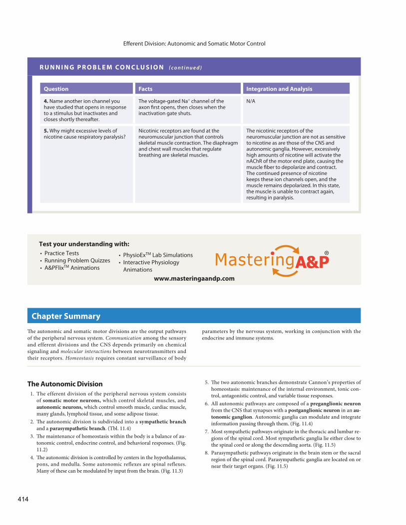
Question Facts Integration and Analysis
1. What is the usual response of cells that are chronically exposed to elevated concentrations of a signal molecule?
A cell exposed to elevated concentrations of a signal molecule will decrease (down-regulate) its receptors for that molecule.
Down-regulation of receptors allows a cell to respond normally even if the concen-tration of ligand is elevated.
2. What happens to a postsynaptic cell when nicotine rather than ACh binds to a nicotinic cholinergic receptor?
Nicotine is an agonist of ACh. Agonists mimic the activity of a ligand.
Nicotine binding to a nAChR will open ion channels in the postsynaptic cell, and the cell will depolarize. This is the same eff ect that ACh binding creates.
3. Although ACh and nicotine have been shown in short-term studies to be nAChR agonists, continued exposure of the receptors to ACh has been shown to close, or inactivate, the channel. Speculate why this could explain the up-regulation of nAChR observed in smokers.
Chronic exposure to an agonist usually causes down-regulation. Chronic exposure to an antagonist usually causes up-regulation. nAChR channels open with initial exposure to agonists but close with continued exposure.
Although nicotine is a short-term agonist, it appears to be having the same eff ect as an antagonist during long-term exposure. With both antagonism and the inactivation described here, the cell’s activity decreases. The cell subsequently up-regulates the number of receptors in an attempt to restore activity.
413
Eff erent Division: Autonomic and Somatic Motor Control
R U N N I N G P R O B L E M C O N C L U S I O N ( c o n t i n u e d )
• PhysioExTM Lab Simulations • Interactive Physiology
Animations
Test your understanding with:
www.masteringaandp.com
• Practice Tests • Running Problem Quizzes • A&PFlixTM Animations
Th e autonomic and somatic motor divisions are the output pathways of the peripheral nervous system. Communication among the sensory and efferent divisions and the CNS depends primarily on chemical signaling and molecular interactions between neurotransmitters and their receptors. Homeostasis requires constant surveillance of body
Chapter Summary
parameters by the nervous system, working in conjunction with the endocrine and immune systems.
The Autonomic Division 1. The efferent division of the peripheral nervous system consists
of somatic motor neurons, which control skeletal muscles, and autonomic neurons, which control smooth muscle, cardiac muscle, many glands, lymphoid tissue, and some adipose tissue.
2. Th e autonomic division is subdivided into a sympathetic branch and a parasympathetic branch . ( Tbl. 11.4 )
3. Th e maintenance of homeostasis within the body is a balance of au-tonomic control, endocrine control, and behavioral responses. ( Fig. 11.2 )
4. Th e autonomic division is controlled by centers in the hypothalamus, pons, and medulla. Some autonomic reflexes are spinal reflexes. Many of these can be modulated by input from the brain. ( Fig. 11.3 )
5. Th e two autonomic branches demonstrate Cannon’s properties of homeostasis: maintenance of the internal environment, tonic con-trol, antagonistic control, and variable tissue responses.
6. All autonomic pathways are composed of a preganglionic neuron from the CNS that synapses with a postganglionic neuron in an au-tonomic ganglion . Autonomic ganglia can modulate and integrate information passing through them. ( Fig. 11.4 )
7. Most sympathetic pathways originate in the thoracic and lumbar re-gions of the spinal cord. Most sympathetic ganglia lie either close to the spinal cord or along the descending aorta. ( Fig. 11.5 )
8. Parasympathetic pathways originate in the brain stem or the sacral region of the spinal cord. Parasympathetic ganglia are located on or near their target organs. ( Fig. 11.5 )
Question Facts Integration and Analysis
4. Name another ion channel you have studied that opens in response to a stimulus but inactivates and closes shortly thereafter.
The voltage-gated Na + channel of the axon fi rst opens, then closes when the inactivation gate shuts.
N/A
5. Why might excessive levels of nicotine cause respiratory paralysis?
Nicotinic receptors are found at the neuromuscular junction that controls skeletal muscle contraction. The diaphragm and chest wall muscles that regulate breathing are skeletal muscles.
The nicotinic receptors of the neuromuscular junction are not as sensitive to nicotine as are those of the CNS and autonomic ganglia. However, excessively high amounts of nicotine will activate the nAChR of the motor end plate, causing the muscle fi ber to depolarize and contract. The continued presence of nicotine keeps these ion channels open, and the muscle remains depolarized. In this state, the muscle is unable to contract again, resulting in paralysis.
414
Eff erent Division: Autonomic and Somatic Motor Control
11
9. The primary autonomic neurotransmitters are acetylcholine and norepinephrine . All preganglionic neurons secrete ACh onto nicotinic cholinergic receptors . As a rule, postganglionic sym-pathetic neurons secrete norepinephrine onto adrenergic recep-tors, and postganglionic parasympathetic neurons secrete ACh onto muscarinic cholinergic receptors . ( Fig. 11.6 , Tbl. 11.1 )
10. The synapse between an autonomic neuron and its target cells is called the neuroeff ector junction .
11. Postganglionic autonomic axons end with varicosities from which neurotransmitter is released. (Figs. 11.7, 11.8)
12. Th e adrenal medulla secretes epinephrine and is controlled by sym-pathetic preganglionic neurons. ( Fig. 11.8 )
13. Adrenergic receptors are G protein–coupled receptors. Alpha receptors respond most strongly to norepinephrine. 6 1 -receptors respond equally to norepinephrine and epinephrine. 6 2 -receptors are not associated with sympathetic neurons and respond most strongly to epinephrine. 6 3 -receptors respond most strongly to norepinephrine. ( Fig. 11.9 , Tbl. 11.2 )
14. Cholinergic muscarinic receptors are also G protein–coupled receptors.
The Somatic Motor Division 15. Somatic motor pathways, which control skeletal muscles, have a
single neuron that originates in the CNS and terminates on a skel-etal muscle. Somatic motor neurons are always excitatory and cause muscle contraction. ( Fig. 11.9 )
16. A single somatic motor neuron controls many muscle fi bers at one time.
17. Th e synapse of a somatic motor neuron on a muscle fi ber is called the neuromuscular junction. Th e muscle cell membrane is modi-fi ed into a motor end plate that contains a high concentration of nicotinic ACh receptors. ( Fig. 11.10 )
18. ACh binding to nicotinic receptor opens cation channels. Net Na + entry into the muscle fi ber depolarizes the fi ber. Acetylcholine in the synapse is broken down by the enzyme acetylcholinesterase. ( Fig. 11.10 )
Questions
Level One Reviewing Facts and Terms 1. Name the two eff erent divisions of the peripheral nervous system.
What type of eff ectors does each control? 2. Th e autonomic nervous system is sometimes called the
nervous system. Why is this an appropriate name? List some func-tions controlled by the autonomic nervous system.
3. What are the two branches of the autonomic nervous system? How are these branches distinguished from each other anatomically and physiologically?
4. Which neurosecretory endocrine gland is closely allied to the sym-pathetic branch?
5. Neurons that secrete acetylcholine are described as neurons, whereas those that secrete norepinephrine are called either or neurons.
6. List four things that can happen to autonomic neurotransmitters aft er they are released into a synapse.
7. The main enzyme responsible for catecholamine degradation is , abbreviated as .
8. Somatic motor pathways (a) are excitatory or inhibitory? (b) are composed of a single neuron or a preganglionic and a post-
ganglionic neuron? (c) synapse with glands or with smooth, cardiac, or skeletal muscle?
9. What is acetylcholinesterase? Describe its action. 10. What kind of receptor is found on the postsynaptic cell in a neuro-
muscular junction?
Level Two Reviewing Concepts 11. What is the advantage of divergence of neural pathways in the auto-
nomic nervous system? 12. Compare and contrast
(a) neuroeff ector junctions and neuromuscular junctions. (b) alpha, beta, muscarinic, and nicotinic receptors. Describe where
each is found and the ligands that bind to them.
13. Concept map: Use the following terms to make a map comparing the somatic motor division and the sympathetic and parasympa-thetic branches of the autonomic division.
You may add terms.
• acetylcholine • adipose tissue • alpha receptor • autonomic division • beta receptor • cardiac muscle • cholinergic receptor • eff erent division • endocrine gland • exocrine gland • ganglion
• muscarinic receptor • nicotinic receptor • norepinephrine • one-neuron pathway • parasympathetic branch • skeletal muscle • smooth muscle • somatic motor division • sympathetic branch • two-neuron pathway
14. Compare and contrast (a) autonomic ganglia and CNS nuclei. (b) the adrenal medulla and the posterior pituitary gland. (c) axon terminals and varicosities.
15. If a target cell’s receptor is (use items in left column), the neuron(s) releasing neurotransmitter onto the receptor must be (use all appropriate items from the right column).
(a) nicotinic cholinergic (b) adrenergic α (c) muscarinic cholinergic (d) adrenergic β
1. somatic motor neuron 2. autonomic preganglionic
neuron 3. sympathetic
postganglionic neuron 4. parasympathetic
postganglionic neuron
16. Ganglia contain the cell bodies of (choose all that apply) (a) somatic motor neurons (b) preganglionic autonomic neurons (c) interneurons
415
Eff erent Division: Autonomic and Somatic Motor Control
(d) postganglionic autonomic neurons (e) sensory neurons
Level Three Problem Solving 17. If nicotinic receptor channels allow both Na + and K + to flow
through, why does Na + infl ux exceed K + effl ux? 18. You have discovered a neuron that innervates an endocrine cell in
the intestine. To learn more about this neuron, you place a marker substance at the endocrine cell synapse. Th e marker is taken into the neuron and transported in a vesicle by retrograde axonal transport to the nerve cell body. (a) By what process is the marker probably taken into the axon
terminal? (b) Th e nerve cell body is found in a ganglion very close to the en-
docrine cell. To which branch of the peripheral nervous system does the neuron probably belong? (Be as specifi c as you can.)
(c) Which neurotransmitter do you predict will be secreted by the neuron onto the endocrine cell?
19. The Huaorani Indians of South America use blowguns to shoot darts poisoned with curare at monkeys. Curare is a plant toxin that binds to and inactivates nicotinic ACh receptors. What happens to a monkey struck by one of these darts?
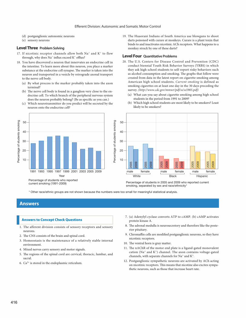
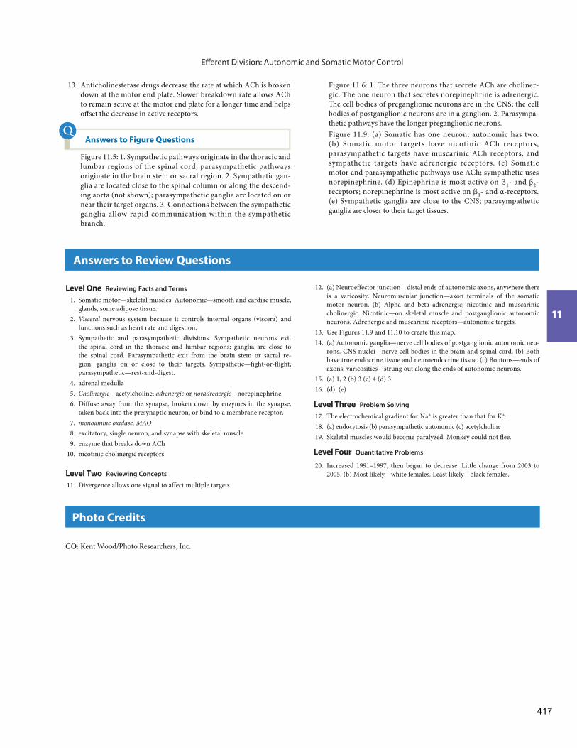
Level Four Quantitative Problems 20. The U.S. Centers for Disease Control and Prevention (CDC)
conduct biennial Youth Risk Behavior Surveys (YRBS) in which they ask high school students to self-report risky behaviors such as alcohol consumption and smoking. Th e graphs that follow were created from data in the latest report on cigarette smoking among American high school students. Current smoking is defined as smoking cigarettes on at least one day in the 30 days preceding the survey. ( http://www.cdc.gov/mmwr/pdf/ss/ss5905.pdf ) (a) What can you say about cigarette smoking among high school
students in the period from 1991 to 2009? (b) Which high school students are most likely to be smokers? Least
likely to be smokers?
7. (a) Adenylyl cyclase converts ATP to cAMP. (b) cAMP activates protein kinase A.
8. Th e adrenal medulla is neurosecretory and therefore like the poste-rior pituitary.
9. Chromaffi n cells are modifi ed postganglionic neurons, so they have nicotinic receptors.
10. Th e ventral horn is gray matter. 11. Th e nAChR of the motor end plate is a ligand-gated monovalent
cation (Na + and K + ) channel. The axon contains voltage-gated channels, with separate channels for Na + and K + .
12. Postganglionic sympathetic neurons are activated by ACh acting on nicotinic receptors. Th is means that nicotine also excites sympa-thetic neurons, such as those that increase heart rate.
1. The afferent division consists of sensory receptors and sensory neurons.
2. Th e CNS consists of the brain and spinal cord. 3. Homeostasis is the maintenance of a relatively stable internal
environment. 4. Mixed nerves carry sensory and motor signals. 5. Th e regions of the spinal cord are cervical, thoracic, lumbar, and
sacral. 6. Ca 2+ is stored in the endoplasmic reticulum.
Answers to Concept Check Questions
Answers
1991 1993 1995 1997 1999 2001
Year
10
20
30
40
50
Per
cent
age
of s
tud
ents
sm
okin
g
2003 2005 2009
2009
2005
2009
2005
2009
2005
2009
2005
2009
2005
2009
2005
Percentage of students who reported current smoking (1991-2009)
* Other race/ethnic groups are not shown because the numbers were too small for meaningful statistical analysis.
male female male female male female
10
20
30
40
50
Per
cent
age
of s
tud
ents
sm
okin
g
Percentage of students in 2005 and 2009 who reported current smoking, separated by sex and race/ethnicity*
White Black Hispanic
416
Eff erent Division: Autonomic and Somatic Motor Control
11
Figure 11.6 : 1. Th e three neurons that secrete ACh are choliner-gic. The one neuron that secretes norepinephrine is adrenergic. Th e cell bodies of preganglionic neurons are in the CNS; the cell bodies of postganglionic neurons are in a ganglion. 2. Parasympa-thetic pathways have the longer preganglionic neurons.
Figure 11.9 : (a) Somatic has one neuron, autonomic has two. (b) Somatic motor targets have nicotinic ACh receptors, parasympathetic targets have muscarinic ACh receptors, and sympathetic targets have adrenergic receptors. (c) Somatic motor and parasympathetic pathways use ACh; sympathetic uses norepinephrine. (d) Epinephrine is most active on d 1 - and β 2 -receptors; norepinephrine is most active on d 1 - and α-receptors. (e) Sympathetic ganglia are close to the CNS; parasympathetic ganglia are closer to their target tissues. Answers: p. A1
13. Anticholinesterase drugs decrease the rate at which ACh is broken down at the motor end plate. Slower breakdown rate allows ACh to remain active at the motor end plate for a longer time and helps off set the decrease in active receptors.
Answers to Figure Questions
Figure 11.5 : 1. Sympathetic pathways originate in the thoracic and lumbar regions of the spinal cord; parasympathetic pathways originate in the brain stem or sacral region. 2. Sympathetic gan-glia are located close to the spinal column or along the descend-ing aorta (not shown); parasympathetic ganglia are located on or near their target organs. 3. Connections between the sympathetic ganglia allow rapid communication within the sympathetic branch.
Answers to Figure Questions
Level One Reviewing Facts and Terms
1. Somatic motor—skeletal muscles. Autonomic—smooth and cardiac muscle, glands, some adipose tissue.
2. Visceral nervous system because it controls internal organs (viscera) and functions such as heart rate and digestion.
3. Sympathetic and parasympathetic divisions. Sympathetic neurons exit the spinal cord in the thoracic and lumbar regions; ganglia are close to the spinal cord. Parasympathetic exit from the brain stem or sacral re-gion; ganglia on or close to their targets. Sympathetic—fight-or-flight; parasympathetic—rest-and-digest.
4. adrenal medulla 5. Cholinergic—acetylcholine; adrenergic or noradrenergic —norepinephrine. 6. Diffuse away from the synapse, broken down by enzymes in the synapse,
taken back into the presynaptic neuron, or bind to a membrane receptor. 7. monoamine oxidase, MAO 8. excitatory, single neuron, and synapse with skeletal muscle 9. enzyme that breaks down ACh 10. nicotinic cholinergic receptors
Level Two Reviewing Concepts
11. Divergence allows one signal to affect multiple targets.
12. (a) Neuroeffector junction—distal ends of autonomic axons, anywhere there is a varicosity. Neuromuscular junction—axon terminals of the somatic motor neuron. (b) Alpha and beta adrenergic; nicotinic and muscarinic cholinergic. Nicotinic—on skeletal muscle and postganglionic autonomic neurons. Adrenergic and muscarinic receptors—autonomic targets.
13. Use Figures 11.9 and 11.10 to create this map. 14. (a) Autonomic ganglia—nerve cell bodies of postganglionic autonomic neu-
rons. CNS nuclei—nerve cell bodies in the brain and spinal cord. (b) Both have true endocrine tissue and neuroendocrine tissue. (c) Boutons—ends of axons; varicosities—strung out along the ends of autonomic neurons.
15. (a) 1, 2 (b) 3 (c) 4 (d) 3 16. (d), (e)
Level Three Problem Solving
17. The electrochemical gradient for Na+ is greater than that for K+ . 18. (a) endocytosis (b) parasympathetic autonomic (c) acetylcholine 19. Skeletal muscles would become paralyzed. Monkey could not flee.
Level Four Quantitative Problems
20. Increased 1991–1997, then began to decrease. Little change from 2003 to 2005. (b) Most likely—white females. Least likely—black females.
Answers to Review Questions
Photo Credits
CO: Kent Wood/Photo Researchers, Inc.
417

418

Muscles
From Chapter 12 of Human Physiology: An Integrated Approach, Sixth Edition. Dee Unglaub Silverthorn. Copyright © 2013 by Pearson Education, Inc. All rights reserved.
419