Embed Size (px)
Citation preview
Gastric secreting cells of a mouse
Organisms could not have evolved without relatively impermeable membranes to surround the cell constituents.
— E. N. Harvey, in H. Davson and J. F. Danielli’s The Permeability of Natural Membranes (1952).
5 Membrane Dynamics Homeostasis Does Not Mean Equilibrium
Osmosis and Tonicity The Body Is Mostly Water
The Body Is in Osmotic Equilibrium
Osmolarity Describes the Number of Particles in Solution
Tonicity Describes the Volume Change of a Cell
Transport Processes Cell Membranes Are Selectively Permeable
Diff usion Lipophilic Molecules Cross Membranes by Simple Diff usion
Protein-Mediated Transport Membrane Proteins Have Four Major Functions
Channel Proteins Form Open, Water-Filled Passageways
Carrier Proteins Change Conformation to Move Molecules
Facilitated Diff usion Uses Carrier Proteins
Active Transport Moves Substances Against Their Concentration Gradients
Carrier-Mediated Transport Exhibits Specifi city, Competition, and Saturation
Vesicular Transport Phagocytosis Creates Vesicles Using the Cytoskeleton
Endocytosis Creates Smaller Vesicles
Exocytosis Releases Molecules Too Large for Transport Proteins
Epithelial Transport Epithelial Transport May Be Paracellular or Transcellular
Transcellular Transport of Glucose Uses Membrane Proteins
Transcytosis Uses Vesicles to Cross an Epithelium
The Resting Membrane Potential Electricity Review
The Cell Membrane Enables Separation of Electrical Charge in the Body
The Resting Membrane Potential Is Due Mostly to Potassium
Changes in Ion Permeability Change the Membrane Potential
Integrated Membrane Processes: Insulin Secretion Background Basics
Polar and nonpolar molecules
Protein and lipid structure
Cell junctions
Molarity and solutions
Membrane structure
Cytoskeleton
Types of epithelia
Enzymes
From Chapter 5 of Human Physiology: An Integrated Approach, Sixth Edition. Dee Unglaub Silverthorn. Copyright © 2013 by Pearson Education, Inc. All rights reserved.
137
I n 1992 the medical personnel at isolated Atoifi Hospital in the Solomon Islands of the South Pacific were faced with a di-lemma. A patient was vomiting and needed intravenous (IV)
fl uids, but the hospital’s supply had run out, and it would be sev-eral days before a plane could bring more. Th eir solution was to try something they had only heard about—make an IV of coconut water, the sterile solution that forms in the hollow center of devel-oping coconuts. For two days the patient received a slow drip of fl uid into his veins directly from young coconuts suspended next to his bed. He soon recovered and was well enough to go home. *
No one knows who fi rst tried coconut water as an IV solu-tion, although stories have been passed down that both the Japa-nese and the British used it in the Pacifi c Th eater of Operations during World War II. Choosing the appropriate IV solution is more than a matter of luck, however. It requires a solid under-standing of the body’s compartments and of the ways diff erent solutes pass between them.
Homeostasis Does Not Mean Equilibrium
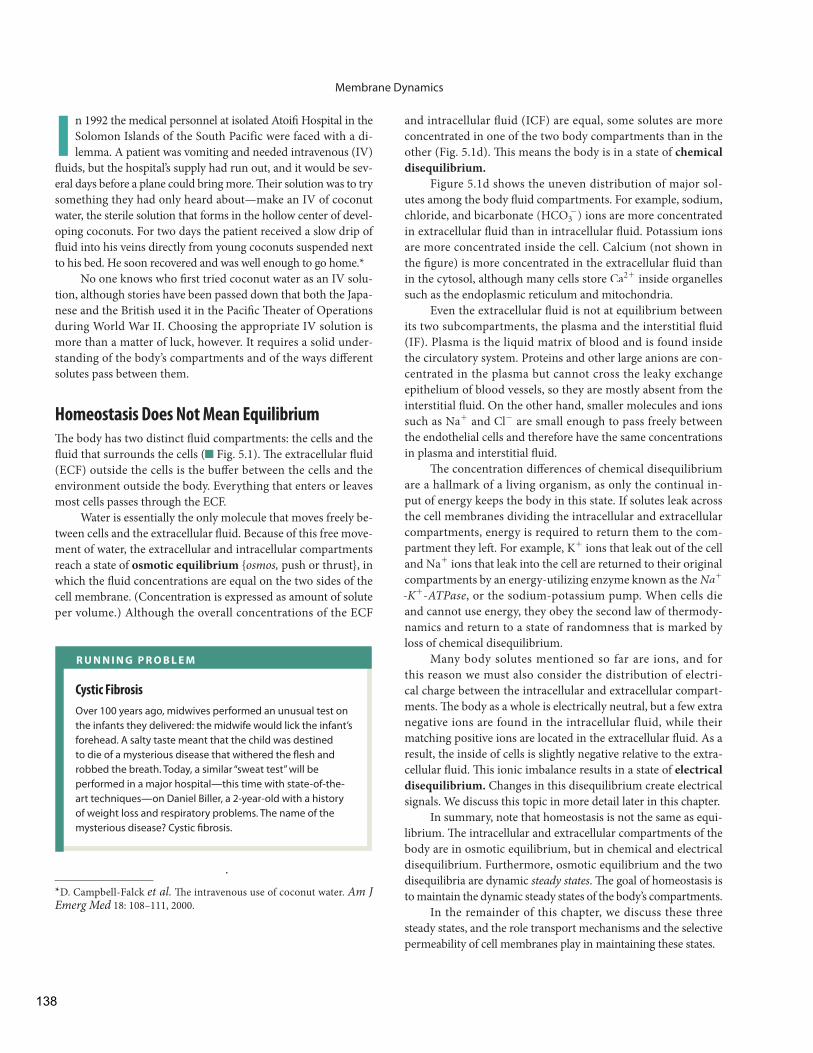
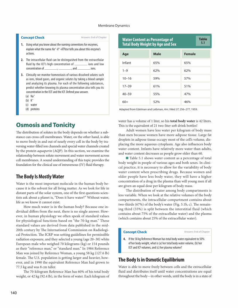
Th e body has two distinct fl uid compartments: the cells and the fl uid that surrounds the cells ( Fig. 5.1 ). Th e extracellular fl uid (ECF) outside the cells is the buff er between the cells and the environment outside the body. Everything that enters or leaves most cells passes through the ECF.
Water is essentially the only molecule that moves freely be-tween cells and the extracellular fl uid. Because of this free move-ment of water, the extracellular and intracellular compartments reach a state of osmotic equilibrium { osmos, push or thrust}, in which the fl uid concentrations are equal on the two sides of the cell membrane. (Concentration is expressed as amount of solute per volume.) Although the overall concentrations of the ECF
* D. Campbell-Falck et al. Th e intravenous use of coconut water. Am J Emerg Med 18: 108–111, 2000.
and intracellular fl uid (ICF) are equal, some solutes are more concentrated in one of the two body compartments than in the other ( Fig. 5.1 d). Th is means the body is in a state of chemical disequilibrium.
Figure 5.1 d shows the uneven distribution of major sol-utes among the body fl uid compartments. For example, sodium,chloride, and bicarbonate (HCO3
-) ions are more concentratedin extracellular fl uid than in intracellular fl uid. Potassium ions are more concentrated inside the cell. Calcium (not shown in the fi gure) is more concentrated in the extracellular fl uid thanin the cytosol, although many cells store Ca2 + inside organellessuch as the endoplasmic reticulum and mitochondria.
Even the extracellular fl uid is not at equilibrium between its two subcompartments, the plasma and the interstitial fl uid (IF) . Plasma is the liquid matrix of blood and is found inside the circulatory system. Proteins and other large anions are con-centrated in the plasma but cannot cross the leaky exchange epithelium of blood vessels, so they are mostly absent from the interstitial fl uid. On the other hand, smaller molecules and ions such as Na+ and Cl- are small enough to pass freely between the endothelial cells and therefore have the same concentrations in plasma and interstitial fl uid.
Th e concentration diff erences of chemical disequilibrium are a hallmark of a living organism, as only the continual in-put of energy keeps the body in this state. If solutes leak across the cell membranes dividing the intracellular and extracellular compartments, energy is required to return them to the com-partment they left . For example, K+ ions that leak out of the cell and Na+ ions that leak into the cell are returned to their original compartments by an energy-utilizing enzyme known as the Na+
- K+ - ATPase , or the sodium-potassium pump. When cells die and cannot use energy, they obey the second law of thermody-namics and return to a state of randomness that is marked by loss of chemical disequilibrium.
Many body solutes mentioned so far are ions, and for this reason we must also consider the distribution of electri-cal charge between the intracellular and extracellular compart-ments. Th e body as a whole is electrically neutral, but a few extra negative ions are found in the intracellular fluid, while their matching positive ions are located in the extracellular fl uid. As a result, the inside of cells is slightly negative relative to the extra-cellular fl uid. Th is ionic imbalance results in a state of electrical disequilibrium. Changes in this disequilibrium create electrical signals. We discuss this topic in more detail later in this chapter.
In summary, note that homeostasis is not the same as equi-librium. Th e intracellular and extracellular compartments of the body are in osmotic equilibrium, but in chemical and electrical disequilibrium. Furthermore, osmotic equilibrium and the two disequilibria are dynamic steady states . Th e goal of homeostasis is to maintain the dynamic steady states of the body’s compartments.
In the remainder of this chapter, we discuss these three steady states, and the role transport mechanisms and the selective permeability of cell membranes play in maintaining these states.
R U N N I N G P R O B L E M
Cystic Fibrosis
Over 100 years ago, midwives performed an unusual test on the infants they delivered: the midwife would lick the infant’s forehead. A salty taste meant that the child was destined to die of a mysterious disease that withered the fl esh and robbed the breath. Today, a similar “sweat test” will be performed in a major hospital—this time with state-of-the-art techniques—on Daniel Biller, a 2-year-old with a history of weight loss and respiratory problems. The name of the mysterious disease? Cystic fi brosis.
Membrane Dynamics
138
Fig. 5.1 E S S E N T I A L S
Interstitial fluid
Plasma
Cellmembrane
Intracellular fluid
KEY
Na
K
Cl
HCO3
Proteins
KEY
1. Using the ECF volume shown in (b), calculate the volumes of the plasma and interstitial fluid.2. What is this person's total body water volume?3. Use your answers from the two questions above to calculate the percentage of total body water in the plasma and interstitial fluid. 4. A woman weighs 121 pounds. Using the standard proportions for the fluid compartments, calculate her ECF, ICF, and plasma volumes. (2.2 lb = 1 kg. 1 kg water = 1 L)
GRAPH QUESTIONS
5. How does the ion composition of plasma differ from that of the IF?6. What ions are concentrated in the ECF? In the ICF?
GRAPH QUESTIONS
(a) The body fluids are in two compartments: the extracellular fluid (ECF) and intracellular fluid (ICF). The ECF and ICF are in osmotic equilibrium but have very different chemical composition.
(b) This figure shows the compartment volumes for the “standard” 70-kg man.
(c) Fluid compartments are often illustrated with box diagrams like this one.
Body Fluid Compartments
20%
40%
60%
80%
100%
Per
cent
of t
otal
bod
y w
ater
Intracellularfluid (ICF)
Intracellularfluid
Interstitialfluid
Pla
sma
ECF1/3
ICF2/3
Extracellularfluid (ECF)
Plasma (25% of ECF)
Interstitial Fluid (75% of ECF)
28 L
14 L
(d) The body compartments are in a state of chemical disequilibrium. The cell membrane is a selectively permeable barrier between the ECF and ICF.
20
40
60
80
100
120
140
160
Ion
conc
entr
atio
n (m
mol
/L)
Intracellular fluid Interstitial fluid Plasma
Intracellular fluid is 2/3 of the total body water volume. Material moving into and out of the ICF must cross the cell membrane.
Extracellular fluid includes all fluid outside the cells. The ECF is 1/3 of the body fluid volume.The ECF consists of:
• Interstitial fluid (IF), which lies between the circulatory system and the cells, is 75% of the ECF volume.
• Plasma, the liquid matrix of blood, is 25% of the ECF volume. Substances moving between the plasma and interstitiial fluid must cross the leaky exchange epithelium of the capillary wall.
139
Membrane Dynamics
water has a volume of 1 liter, so his total body water is 42 liters. Th is is the equivalent of 21 two-liter soft drink bottles!
Adult women have less water per kilogram of body mass than men because women have more adipose tissue. Large fat droplets in adipose tissue occupy most of the cell’s volume, dis-placing the more aqueous cytoplasm . Age also infl uences body water content. Infants have relatively more water than adults, and water content decreases as people grow older than 60.
Table 5.1 shows water content as a percentage of total body weight in people of various ages and both sexes. In clini-cal practice, it is necessary to allow for the variability of body water content when prescribing drugs. Because women and older people have less body water, they will have a higher concentration of a drug in the plasma than will young men if all are given an equal dose per kilogram of body mass.
The distribution of water among body compartments is less variable. When we look at the relative volumes of the body compartments, the intracellular compartment contains about two-thirds (67%) of the body’s water ( Fig. 5.1 b, c). Th e remain-ing third (33%) is split between the interstitial fluid (which contains about 75% of the extracellular water) and the plasma (which contains about 25% of the extracellular water).
Water Content as Percentage of Total Body Weight by Age and Sex
Age Male Female
Infant 65% 65%
1–9 62% 62%
10–16 59% 57%
17–39 61% 51%
40–59 55% 47%
60+ 52% 46%
Adapted from Edelman and Leibman, Am J Med 27; 256–277, 1959.
Table5.1
Concept Check Answers: End of Chapter
1. Using what you know about the naming conventions for enzymes ,
explain what the name Na+ - K+ -ATPase tells you about this enzyme’s
actions.
2. The intracellular fluid can be distinguished from the extracellular
fluid by the ICF’s high concentration of ions and low
concentration of , , and ions.
3. Clinically we monitor homeostasis of various dissolved solutes such
as ions, blood gases, and organic solutes by taking a blood sample
and analyzing its plasma. For each of the following substances,
predict whether knowing its plasma concentration also tells you its
concentration in the ECF and the ICF. Defend your answer.
(a) Na+
(b) K +
(c) water
(d) proteins
Osmosis and Tonicity Th e distribution of solutes in the body depends on whether a sub-stance can cross cell membranes. Water, on the other hand, is able to move freely in and out of nearly every cell in the body by tra-versing water-fi lled ion channels and special water channels created by the protein aquaporin (AQP). In this section, we examine the relationship between solute movement and water movement across cell membranes. A sound understanding of this topic provides the foundation for the clinical use of intravenous (IV) fl uid therapy.
The Body Is Mostly Water
Water is the most important molecule in the human body be-cause it is the solvent for all living matter. As we look for life in distant parts of the solar system, one of the fi rst questions scien-tists ask about a planet is, “Does it have water?” Without water, life as we know it cannot exist.
How much water is in the human body? Because one in-dividual diff ers from the next, there is no single answer. How-ever, in human physiology we often speak of standard values for physiological functions based on “the 70-kg man.” These standard values are derived from data published in the mid-20 th century by Th e International Commission on Radiologi-cal Protection. Th e ICRP was setting guidelines for permissible radiation exposure, and they selected a young (age 20–30) white European male who weighed 70 kilograms (kg) or 154 pounds as their “reference man,” or “standard man.” In 1984 Reference Man was joined by Reference Woman, a young 58 kg (127.6 lb) female. Th e U.S. population is getting larger and heavier, how-ever, and in 1990 the equivalent Reference Man had grown to 77.5 kg and was 8 cm taller.
Th e 70-kilogram Reference Man has 60% of his total body weight, or 42 kg (92.4 lb), in the form of water. Each kilogram of
Concept Check
4. If the 58 kg Reference Woman has total body water equivalent to 50%
of her body weight, what is (a) her total body water volume, (b) her
ECF and ICF volumes, and (c) her plasma volume?
The Body Is in Osmotic Equilibrium
Water is able to move freely between cells and the extracellular fluid and distributes itself until water concentrations are equal throughout the body—in other words, until the body is in a state of
Answers: End of Chapter
140
Membrane Dynamics
5
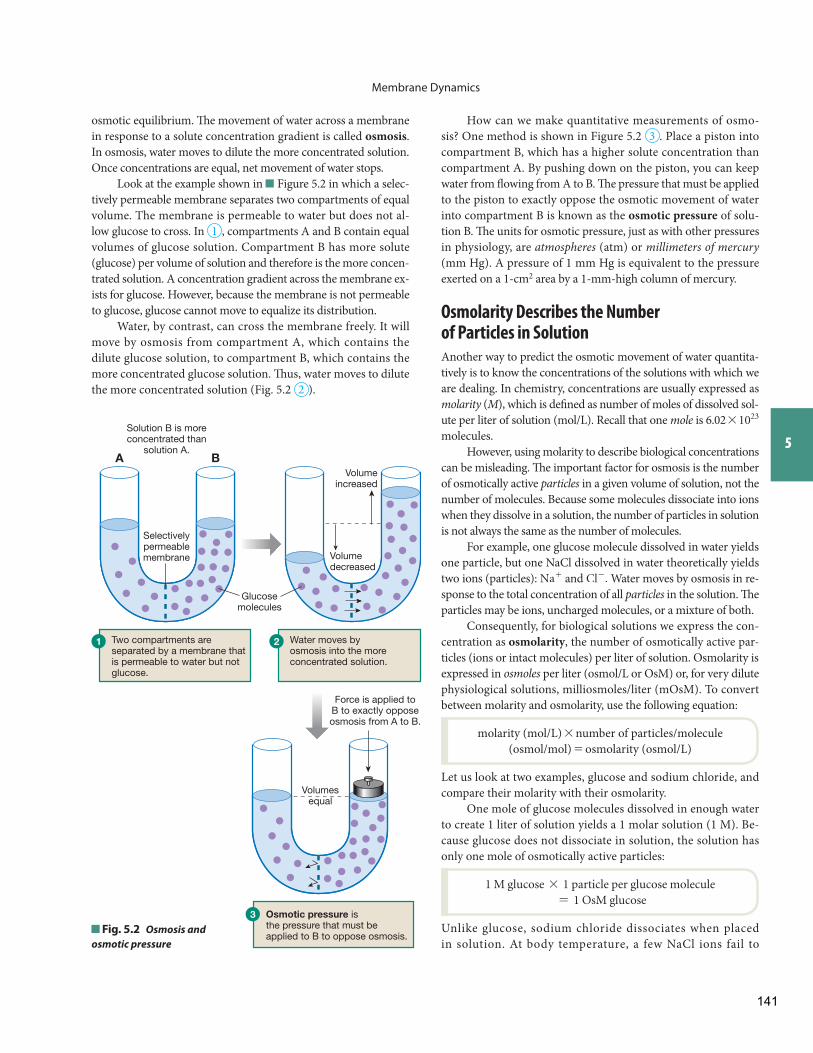
How can we make quantitative measurements of osmo-sis? One method is shown in Figure 5.2 3 . Place a piston into compartment B, which has a higher solute concentration than compartment A. By pushing down on the piston, you can keep water from fl owing from A to B. Th e pressure that must be applied to the piston to exactly oppose the osmotic movement of water into compartment B is known as the osmotic pressure of solu-tion B. Th e units for osmotic pressure, just as with other pressures in physiology, are atmospheres (atm) or millimeters of mercury (mm Hg). A pressure of 1 mm Hg is equivalent to the pressure exerted on a 1-cm 2 area by a 1-mm-high column of mercury.
Osmolarity Describes the Number of Particles in Solution
Another way to predict the osmotic movement of water quantita-tively is to know the concentrations of the solutions with which weare dealing. In chemistry, concentrations are usually expressed as molarity ( M ), which is defi ned as number of moles of dissolved sol-ute per liter of solution (mol/L). Recall that one mole is 6.02 * 1023 molecules.
However, using molarity to describe biological concentrations can be misleading. Th e important factor for osmosis is the number of osmotically active particles in a given volume of solution, not the number of molecules. Because some molecules dissociate into ions when they dissolve in a solution, the number of particles in solution is not always the same as the number of molecules.
For example, one glucose molecule dissolved in water yields one particle, but one NaCl dissolved in water theoretically yields two ions (particles): Na+ and Cl- . Water moves by osmosis in re-sponse to the total concentration of all particles in the solution. Th e particles may be ions, uncharged molecules, or a mixture of both.
Consequently, for biological solutions we express the con-centration as osmolarity , the number of osmotically active par-ticles (ions or intact molecules) per liter of solution. Osmolarity is expressed in osmoles per liter (osmol/L or OsM) or, for very dilute physiological solutions, milliosmoles/liter (mOsM). To convert between molarity and osmolarity, use the following equation:
molarity (mol/L) * number of particles/molecule (osmol/mol) = osmolarity (osmol/L)
Let us look at two examples, glucose and sodium chloride, and compare their molarity with their osmolarity.
One mole of glucose molecules dissolved in enough water to create 1 liter of solution yields a 1 molar solution (1 M). Be-cause glucose does not dissociate in solution, the solution has only one mole of osmotically active particles:
1 M glucose * 1 particle per glucose molecule= 1 OsM glucose
Unlike glucose, sodium chloride dissociates when placed in solution. At body temperature, a few NaCl ions fail to
osmotic equilibrium. Th e movement of water across a membrane in response to a solute concentration gradient is called osmosis . In osmosis, water moves to dilute the more concentrated solution. Once concentrations are equal, net movement of water stops.
Look at the example shown in Figure 5.2 in which a selec-tively permeable membrane separates two compartments of equal volume. The membrane is permeable to water but does not al-low glucose to cross. In 1 , compartments A and B contain equal volumes of glucose solution. Compartment B has more solute (glucose) per volume of solution and therefore is the more concen-trated solution. A concentration gradient across the membrane ex-ists for glucose. However, because the membrane is not permeable to glucose, glucose cannot move to equalize its distribution.
Water, by contrast, can cross the membrane freely. It will move by osmosis from compartment A, which contains the dilute glucose solution, to compartment B, which contains the more concentrated glucose solution. Th us, water moves to dilute the more concentrated solution ( Fig. 5.2 2 ).
Force is applied toB to exactly opposeosmosis from A to B.
Volumesequal
Osmotic pressure isthe pressure that must beapplied to B to oppose osmosis.
Volumeincreased
Volumedecreased
Two compartments are separated by a membrane that is permeable to water but not glucose.
Water moves byosmosis into the moreconcentrated solution.
Glucosemolecules
Selectivelypermeablemembrane
Solution B is moreconcentrated than
solution A.
3
1 2
A B
Fig. 5.2 Osmosis and osmotic pressure
141
Membrane Dynamics
Osmolarity says nothing about what the particles are or how they behave. Before we can predict whether osmosis will take place be-tween any two solutions divided by a membrane, we must know the properties of the membrane and of the solutes on each side of it.
If the membrane is permeable only to water and not to any solutes, water will move by osmosis from a less concentrated (hyposmotic) solution into a more concentrated (hyperosmotic) solution, as illustrated in Figure 5.2 . Most biological systems are not this simple, however. Biological membranes are selectively permeable and allow some solutes to cross in addition to water. To predict the movement of water into and out of cells, you must know the tonicity of the solution, explained in the next section.
Tonicity Describes the Volume Change of a Cell
Tonicity { tonikos, pertaining to stretching} is a physiological term used to describe a solution and how that solution would affect cell volume if the cell were placed in the solution and allowed to come to equilibrium ( Tbl. 5.3 ).
If a cell placed in the solution gains water at equilibrium and swells, we say that the solution is hypotonic to the cell.
If the cell loses water and shrinks at equilibrium, the solu-tion is said to be hypertonic .
If the cell in the solution does not change size at equilib-rium, the solution is isotonic .
separate, so instead of 2 ions per NaCl, the dissociation factoris about 1.8.
Thus, one mole of NaCl dissociates in solution to yield1.8 moles of particles ( Na+ , Cl- , and NaCl). The result is a1.8 OsM solution:
1 mole NaCl/L * 1.8 osmoles/mole NaCl= 1.8 osmol NaCl/L
Osmolarity describes only the number of particles in the so-lution. It says nothing about the composition of the particles. A 1 OsM solution could be composed of pure glucose or pure Na+ and Cl- or a mixture of all three solutes.
Th e normal osmolarity of the human body ranges from 280 to 296 milliosmoles per liter (mOsM). In this course, to simplify calculations we will round that number up slightly to 300 mOsM.
A term related to osmolarity is osmolality. Osmolality is concentration expressed as osmoles of solute per kilogram of water. Because biological solutions are dilute and little of their weight comes from solute, physiologists oft en use the terms os-molarity and osmolality interchangeably. Osmolality is usually used in clinical situations because it is easy to estimate people’s body water content by weighing them.
Clinicians estimate a person’s fl uid loss in dehydration by equating weight loss to fl uid loss. Because 1 liter of pure water weighs 1 kilogram, a decrease in body weight of 1 kilogram (or 2.2 lb) is considered equivalent to the loss of 1 liter of body fl uid. A baby with diarrhea can easily be weighed to estimate its fl uid loss. A decrease of 1.1 pounds (0.5 kg) of body weight is as-sumed to mean the loss of 500 mL of fl uid. Th is calculation pro-vides a quick estimate of how much fl uid needs to be replaced.
Concept Check
5. A mother brings her baby to the emergency room because he has lost
fl uid through diarrhea and vomiting for two days. The staff weighs
the baby and fi nds that he has lost 2 pounds. If you assume that the
reduction in weight is due to water loss, what volume of water has the
baby lost (2.2 pounds = 1 kilogram) ?
Comparing Osmolarities of Two Solutions Osmolarity is a prop-erty of every solution. You can compare the osmolarities of diff erent solutions as long as the concentrations are expressed in the same units—for example, as milliosmoles per liter. If two solutions con-tain the same number of solute particles per unit volume, we say that the solutions are isosmotic { iso -, equal}. If solution A has a higher osmolarity (contains more particles per unit volume, is more con-centrated) than solution B, we say that solution A is hyperosmoticto solution B. In the same example, solution B, with fewer osmoles per unit volume, is hyposmotic to solution A. Table 5.2 shows some examples of comparative osmolarities.
Osmolarity is a colligative property of solutions, meaning it depends strictly on the number of particles per liter of solution.
Comparing Osmolarities
Solution A = 1 OsM Glucose
Solution B =
2 OsM Glucose Solution C = 1 OsM NaCl
A is hyposmotic to B
B is hyperosmotic to A
C is isosmotic to A
A is isosmotic to C
B is hyperosmotic to C
C is hyposmotic to B
Table5.2
Tonicity of Solutions
Solution
Cell Behavior When Placed in the Solution
Description of the Solution Relative to the Cell
A Cell swells Solution A is hypotonic.
B Cell doesn’t change size
Solution B is isotonic.
C Cell shrinks Solution C is hypertonic.
Table5.3
Answers: End of Chapter
142
Membrane Dynamics
5
2 If the cell has a lower concentration of nonpenetrating solutes than the solution, there will be net movement of water out of the cell. Th e cell shrinks, and the solution is hypertonic .
3 If the concentrations of nonpenetrating solutes are the same in the cell and the solution, there will be no net movement of water at equilibrium. Th e solution is isotonic to the cell.
How does tonicity relate osmolarity? Figure 5.3 shows the possible combinations of osmolarity and tonicity. A hyposmotic so-lution is always hypotonic, no matter what its composition. Th e cell will always have a higher concentration of nonpenetrating solutes than the solution, and water will move into the cell (rule 1 above).
An isosmotic solution may be isotonic or hypotonic, but can never be hypertonic because it can never have a higher con-centration of nonpenetrating solutes than the cell. If all solutes in the isosmotic solution are nonpenetrating, then the solution is also isotonic. If there are any penetrating solutes in the isos-motic solution, the solution will be hypotonic.
Hyperosmotic solutions may be hypertonic, isotonic, or hypotonic. Th eir tonicity depends on the relative concentration of nonpenetrating solutes in the solution compared to the cell, as described in the list above.
Normally tonicity is explained using a single cell that is placed into a solution, but here we will use a more physi-ologically appropriate system: a two-compartment box model that represents the total body divided into ECF and ICF (see Fig. 5.1 c). To simplify the calculations, we will use a 3- liter body, with 2 liters in the ICF and 1 liter in the ECF. We assume that the starting osmolarity is 300 mOsM (0.3 OsM) and that solutes in each compartment are nonpenetrating (NP) and cannot move into the other compartment. By defi ning volumes and concentrations, we can use the equation solute/volume = concentration (S>V = C) to mathematically determine changes to volumes and osmolarity. Concentration is osmolarity.
Always begin by defi ning the starting conditions. Th is may be the person’s normal state or it may be the altered state that you are trying to return to normal. An example of this would be trying to restore normal volume and osmolarity in a person who has become dehydrated through sweat loss.
Figure 5.4 shows the starting conditions for the 3-liter body both as a box diagram and in a table. Th e table format al-lows you to deal with an example mathematically if you know the volumes of the body and of the solution added or lost.
By convention, we always describe the tonicity of the solu-tion relative to the cell. How, then, does tonicity differ from osmolarity?
1 Osmolarity describes the number of solute particles dis-solved in a volume of solution. It has units, such as os-moles/liter. Th e osmolarity of a solution can be measured by a machine called an osmometer . Tonicity has no units; it is only a comparative term.
2 Osmolarity can be used to compare any two solutions, and the relationship is reciprocal (solution A is hyperosmotic to solution B; therefore, solution B is hyposmotic to solution A). Tonicity always compares a solution and a cell, and by convention, tonicity is used to describe only the solution—for example, “Solution A is hypotonic to red blood cells.”
3 Osmolarity alone does not tell you what happens to a cell placed in a solution. Tonicity by defi nition tells you what happens to cell volume at equilibrium when the cell is placed in the solution.
Th is third point is the one that is most confusing to stu-dents. Why can’t osmolarity be used to predict tonicity? The reason is that the tonicity of a solution depends not only on its concentration (osmolarity) but also on the nature of the solutes in the solution.
By nature of the solutes, we mean whether the solute par-ticles can cross the cell membrane. If the solute particles (ions or molecules) can enter the cell, we call them penetrating solutes . We call particles that cannot cross the cell membrane nonpen-etrating solutes . Tonicity depends on the concentration of non-penetrating solutes only. Let’s see why this is true.
First, some preliminary information. Th e most important nonpenetrating solute in physiology is NaCl. If a cell is placed in a solution of NaCl, the Na+ and Cl- ions do not enter the cell. Th is makes NaCl a nonpenetrating solute. (In reality, a few Na+ ions may leak across, but they are immediately transported back to the extracellular fl uid by the Na+ - K+ -ATPase. For this reason NaCl is considered a functionally nonpenetrating solute.)
By convention, we assume that cells are fi lled with other types of nonpenetrating solutes. In other words, the solutes in-side the cell are unable to leave so long as the cell membrane remains intact. Now we are ready to see why osmolarity alone cannot be used to predict tonicity.
Suppose you know the composition and osmolarity of a solution. How can you fi gure out the tonicity of the solution without actually putting a cell in it? Th e key lies in knowing the relative concentrations of nonpenetrating solutes in the cell and in the solution . Water will always move until the concentrations of nonpenetrating solutes in the cell and the solution are equal.
Here are the rules for predicting tonicity:
1 If the cell has a higher concentration of nonpenetrating solutes than the solution, there will be net movement of water into the cell. Th e cell swells, and the solution is hypotonic . Fig. 5.3 The relationship between osmolarity and tonicity
TONICITY
Hypotonic
Isotonic
Hypertonic
Hyposmotic
√ √
√
√
√
√
Isosmotic Hyperosmotic
OSMOLARITY
143
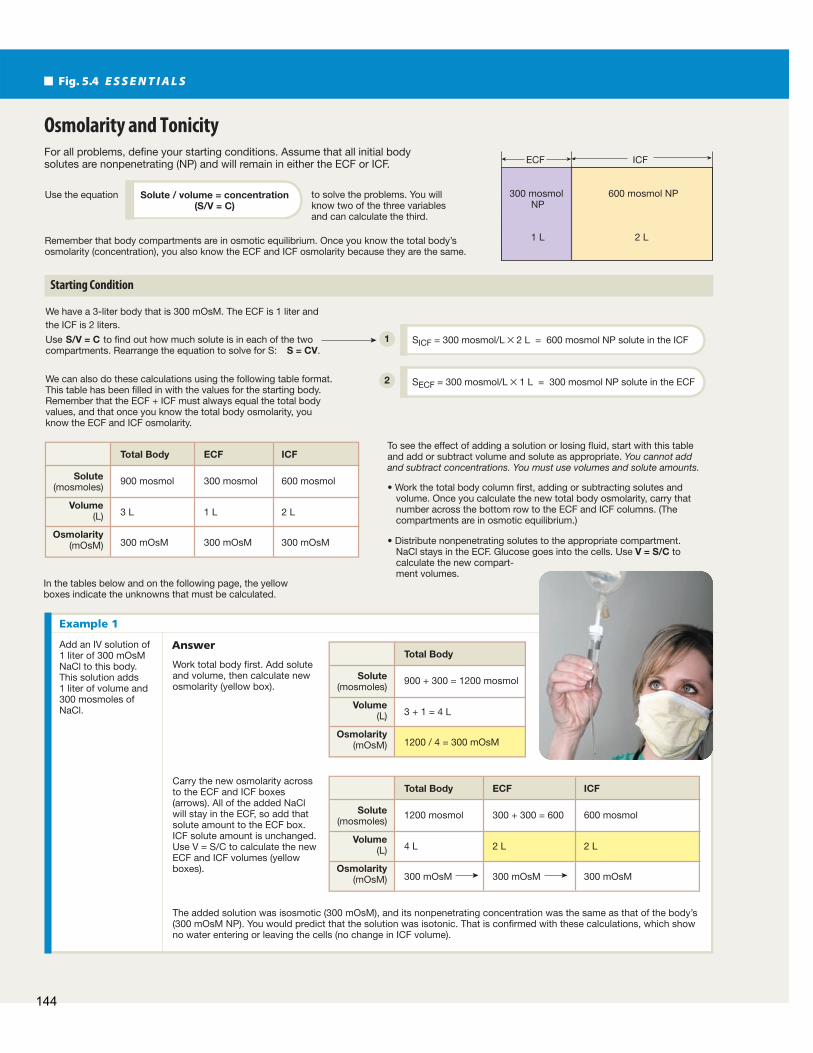
For all problems, define your starting conditions. Assume that all initial body solutes are nonpenetrating (NP) and will remain in either the ECF or ICF.
to solve the problems. You will know two of the three variables and can calculate the third.
Remember that body compartments are in osmotic equilibrium. Once you know the total body’s osmolarity (concentration), you also know the ECF and ICF osmolarity because they are the same.
In the tables below and on the following page, the yellow boxes indicate the unknowns that must be calculated.
We have a 3-liter body that is 300 mOsM. The ECF is 1 liter and the ICF is 2 liters.
Use S/V = C to find out how much solute is in each of the two compartments. Rearrange the equation to solve for S: S = CV.
We can also do these calculations using the following table format. This table has been filled in with the values for the starting body. Remember that the ECF + ICF must always equal the total body values, and that once you know the total body osmolarity, you know the ECF and ICF osmolarity.
To see the effect of adding a solution or losing fluid, start with this table and add or subtract volume and solute as appropriate. You cannot add and subtract concentrations. You must use volumes and solute amounts.
volume. Once you calculate the new total body osmolarity, carry that number across the bottom row to the ECF and ICF columns. (The compartments are in osmotic equilibrium.)
NaCl stays in the ECF. Glucose goes into the cells. Use V = S/C to calculate the new compart-ment volumes.
Osmolarity and Tonicity
Starting Condition
Solute / volume = concentration(S/V = C)
Use the equation
SICF = 300 mosmol/L × 2 L = 600 mosmol NP solute in the ICF
SECF = 300 mosmol/L × 1 L = 300 mosmol NP solute in the ECF
Solute(mosmoles)
900 mosmol 300 mosmol 600 mosmol
300 mOsM 300 mOsM 300 mOsM
3 L 1 L 2 LVolume
(L)
Osmolarity(mOsM)
Total Body ECF ICF
Solute(mosmoles)
900 + 300 = 1200 mosmol
1200 / 4 = 300 mOsM
3 + 1 = 4 LVolume
(L)
Osmolarity(mOsM)
Total Body
Solute(mosmoles)
1200 mosmol 300 + 300 = 600 600 mosmol
300 mOsM 300 mOsM 300 mOsM
4 L 2 L 2 LVolume
(L)
Osmolarity(mOsM)
Total Body ECF ICF
AnswerAdd an IV solution of 1 liter of 300 mOsM NaCl to this body. This solution adds 1 liter of volume and 300 mosmoles of NaCl.
Work total body first. Add solute and volume, then calculate new osmolarity (yellow box).
Carry the new osmolarity across to the ECF and ICF boxes (arrows). All of the added NaCl will stay in the ECF, so add that solute amount to the ECF box. ICF solute amount is unchanged. Use V = S/C to calculate the new ECF and ICF volumes (yellow boxes).
The added solution was isosmotic (300 mOsM), and its nonpenetrating concentration was the same as that of the body’s (300 mOsM NP). You would predict that the solution was isotonic. That is confirmed with these calculations, which show no water entering or leaving the cells (no change in ICF volume).
600 mosmol NP300 mosmol NP
1 L 2 L
ECF ICF
Fig. 5.4 E S S E N T I A L S
Example 1
1
2
144
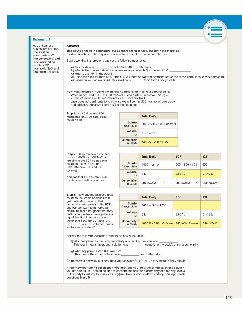
Solute(mosmoles)
900 + 500 = 1400 mosmol
1400/5 = 280 mOsM
3 + 2 = 5 LVolume
(L)
Osmolarity(mOsM)
Total Body
Solute(mosmoles)
1400 mosmol 300 + 500 = 800 600
280 mOsM 280 mOsM 280 mOsM
5 L 2.857 L 2.143 LVolume
(L)
Osmolarity(mOsM)
Total Body ECF ICF
AnswerAdd 2 liters of a 500 mOsM solution. The solution is equal parts NaCl (nonpenetrating) and urea (penetrating), so it has 250 mosmol/L NaCl and 250 mosmol/L urea.
Step 1: Add 2 liters and 500
column first.
Step 2: Carry the new osmolarity across to ECF and ICF. NaCl all remains in the ECF, so add that solute to the ECF column. Calculate new ECF and ICF volumes.
volume = total body volume.
This solution has both penetrating and nonpenetrating solutes, but only nonpenetrating solutes contribute to tonicity and cause water to shift between compartments.
Before working this problem, answer the following questions:
Now work the problem using the starting conditions table as your starting point.What did you add? 2 L of (250 mosmol/L urea and 250 mosmol/L NaCl) =2 liters of volume + 500 mosmol urea + 500 mosmol NaCl.Urea does not contribute to tonicity, so we will set the 500 mosmol of urea aside and add only the volume and NaCl in the first step:
(a) This solution is __________ osmotic to the 300 mOsM body. (b) What is the concentration of nonpenetrating solutes [NP] in the solution? _______________ (c) What is the [NP] in the body? ________ (d) Using the rules for tonicity in Table 5.4, will there be water movement into or out of the cells? If so, in what direction? (e) Based on your answer in (d), this solution is ________ tonic to this body’s cells.
Answer the following questions from the values in the table:
If you know the starting conditions of the body and you know the composition of a solution you are adding, you should be able to describe the solution’s osmolarity and tonicity relative to the body by asking the questions in (a)–(e). Now test yourself by working Concept Check questions 8 and 9.
(f) What happened to the body osmolarity after adding the solution? _____________ This result means the added solution was ___________osmotic to the body’s starting osmolarity. (g) What happened to the ICF volume? ________________________ This means the added solution was ____________tonic to the cells.
Solute(mosmoles) 1400 + 500 = 1900
1900/5 = 380 mOsM 380 mOsM 380 mOsM
5 L 2.857 L 2.143 LVolume
(L)
Osmolarity(mOsM)
Total Body ECF ICFStep 3: Now add the reserved urea solute to the whole body solute to get the final osmolarity. That osmolarity carries over to the ECF and ICF compartments. Urea will distribute itself throughout the body until its concentration everywhere is equal, but it will not cause any water shift between ECF and ICF. So the ECF and ICF volumes remain as they were in step 2.
Example 2
145
Membrane Dynamics
nonpenetrating solutes. When the 300 mOsM urea solution mixes with the ECF, the added volume of the urea solution dilutes the nonpenetrating solutes of the ECF. (S>V = C. Th e same amount of NP solute in a larger volume means a lower NP concentration.)
Now the nonpenetrating concentration of the ECF is less than 300 mOsM. The cells still have a nonpenetrating solute concentration of 300 mOsM, so water moves into the cells to equalize the nonpenetrating concentrations. (Rule: water moves into the compartment with the higher concentration of NP solutes.) Th e cells gain water and volume. Th is means the urea solution is hypotonic, even though it is isosmotic.
Example 2 in Figure 5.4 shows how combining penetrat-ing and nonpenetrating solutes can complicate the situation. Th is example asks you to describe the solution’s osmolarity and tonicity based on its composition before you do the mathemati-cal calculations. Th is skill is important for clinical situations, when you will not know exact body fl uid volumes for the person needing an IV. Table 5.4 lists some rules to help you distin-guish between osmolarity and tonicity.
Understanding the diff erence between osmolarity and tonicity is critical to making good clinical decisions about intravenous (IV) fl uid therapy. Th e choice of IV fl uid depends on how the clinician wants the solutes and water to distribute between the extracellular and intracellular fl uid compartments. If the problem is dehydrated cells, the appropriate IV solution is hypotonic because the cells need fl uid. If the situation requires fl uid that remains in the extracellular fl uid to replace blood loss, an isotonic IV solution is used. In medi-cine, the tonicity of a solution is usually the important consideration.
Table 5.5 lists some common IV solutions and their approximate osmolarity and tonicity relative to the normal hu-man cell. What about the coconut water described at the start of the chapter? Chemical analysis shows that it is not an ideal IV solution, although it is useful for emergencies. It is isosmotic to human plasma but is hypotonic, with Na+ concentrations much lower than normal ECF and high concentrations of glucose and fructose, along with amino acids.
Th e body’s volumes and concentration will change as the result of adding or losing solutes, water, or both—the law of mass balance . Additions to the body normally come through the ingestion of food and drink, but in medical situations solutions can be added directly to the ECF through intravenous (IV) infu-sions. Signifi cant solute and water loss may occur with sweating, vomiting and diarrhea, or blood loss.
Once you have defi ned the starting conditions, you add or subtract volume and solutes to fi nd the body’s new osmolarity. Th e fi nal step is to determine whether the ECF and ICF volumes will change as a result of the water and solute gain or loss. In this last step, you must separate the added solutes into penetrating solutes and nonpenetrating solutes.
In our examples, we use three solutes: NaCl, urea, and glucose. NaCl is considered nonpenetrating. Any NaCl added to the body remains in the ECF. Urea is freely penetrating and behaves as if the cell membranes dividing the ECF and ICF do not exist. An added load of urea distributes itself until the urea concentration is the same throughout the body.
Glucose (also called dextrose ) is an unusual solute. Like all solutes, it fi rst goes into the ECF. Over time, however, 100% of added glucose will enter the cells. When glucose enters the cells, it is phosphorylated to glucose 6-phosphate (G-6-P) and cannot leave the cell again. So although glucose enters cells, it is not freely penetrating because it stays in the cell and adds to the cell’s nonpenetrating solutes.
Giving someone a glucose solution is the same as giving them a slow infusion of pure water because glucose 6-phosphate is the first step in the aerobic metabolism of glucose. The end products of aerobic glucose metabolism are CO2 and water.
Th e examples shown in Figure 5.4 walk you through the process of adding and subtracting solutions to the body. Ask the following questions when you are evaluating the eff ects of a solution on the body:
1 What is the osmolarity of this solution relative to the body? ( Tbl. 5.2 )
2 What is the tonicity of this solution? (Use Fig. 5.3 to help eliminate possibilities.) To determine tonicity, compare the concentration of the nonpenetrating solutes in the solution to the body concentration. (All body solutes are consid-ered to be nonpenetrating.)
For example, consider a solution that is 300 mOsM— isosmotic to a body that is 300 mOsM. Th e solution’s tonicity depends on the concentration of nonpenetrating solutes in the solution. If the solu-tion is 300 mOsM NaCl, the solution’s nonpenetrating solute con-centration is equal to that of the body. When the solution mixes with the ECF, the ECF nonpenetrating concentration and osmolar-ity do not change. No water will enter or leave the cells (the ICF compartment), and the solution is isotonic. You can calculate this for yourself by working through Example 1 in Figure 5.4 .
Now suppose the 300 mOsM solution has urea as its only solute. Urea is a penetrating solute, so this solution has no
Rules for Osmolarity and Tonicity
1. Assume that all intracellular solutes are nonpenetrating.
2. Compare osmolarities before the cell is exposed to the solution. (At equilibrium, the cell and solution are always isosmotic.)
3. Tonicity of a solution describes the volume change of a cell at equilibrium ( Tbl. 5.6 ).
4. Determine tonicity by comparing nonpenetrating solute concentrations in the cell and the solution. Net water movement is into the compartment with the higher concentration of nonpenetrating solutes.
5. Hyposmotic solutions are always hypotonic.
Table5.4
146
Membrane Dynamics
5
Transport Processes Humans are large complex organisms, and the movement of ma-terial within and between body compartments is necessary for communication. Th is movement requires a variety of transport mechanisms. Some require an outside source of energy, such as that stored in the high-energy bond of ATP , while other trans-port processes use only the kinetic or potential energy already in the system . Movement between compartments usually means a molecule must cross one or more cell membranes. Movement within a compartment is less restricted. For this reason, biologi-cal transport is another theme that you will encounter repeat-edly as you study the organ systems.
Th e most general form of biological transport is the bulk fl ow of fl uids within a compartment. Although many people equate fl uids with liquids, in physics both gases and liquids are considered fl uids because they fl ow. Th e main diff erence between the two fl uids is that gases are compressible because their molecules are so far apart in space. Liquids, especially water, are not compressible. (Think of squeezing on a water balloon.)
In bulk fl ow, a pressure gradient causes fl uid to fl ow from regions of higher pressure to regions of lower pressure. As the fl uid fl ows, it carries with it all of its component parts, including substances dissolved or suspended in it. Blood moving through the circulatory system is an excellent example of bulk fl ow. Th e heart acts as a pump that creates a region of high pressure, push-ing plasma with its dissolved solutes and the suspended blood cells through the blood vessels. Air fl ow in the lungs is another example of bulk flow that you will encounter as you study physiology.
Other forms of transport are more specifi c than bulk fl ow. When we discuss them, we must name the molecule or mol-ecules that are moving. Transport mechanisms you will learn about in the following sections include diffusion, protein- mediated transport, and vesicular transport.
Intravenous Solutions
Solution Also Known As Osmolarity Tonicity
0.9% saline* Normal saline Isosmotic Isotonic
5% dextrose † in 0.9% saline D5–normal saline Hyperosmotic Isotonic
5% dextrose in water D5W Isosmotic Hypotonic
0.45% saline Half-normal saline Hyposmotic Hypotonic
5% dextrose in 0.45% saline D5–half-normal saline Hyperosmotic Hypotonic
*Saline = NaCl. † Dextrose = glucose.
Table5.5
Concept Check Answers: End of Chapter
6. Which of the following solutions has/have the most water per unit
volume: 1 M glucose, 1 M NaCl, or 1 OsM NaCl?
7. Two compartments are separated by a membrane that is permeable to water
and urea but not to NaCl. Which way will water move when the following
solutions are placed in the two compartments? (Hint: Watch the units!)
Compartment A Membrane Compartment B
(a) 1 M NaCl I 1 OsM NaCl
(b) 1 M urea I 2 M urea
(c) 1 OsM NaCl I 1 OsM urea
8. Use the same 3-liter, 300 mOsM body as in Figure 5.4 for this problem.
Add 1 liter of 260 mOsM glucose to the body and calculate the new body
volumes and osmolarity once all the glucose has entered the cells and
been phosphorylated. Before you do the calculations, make the following
predictions: This solution is osmotic to the body and is
tonic to the body’s cells.
9. Use the same 3-liter, 300 mOsM body as in Figure 5.4 for this problem. A
3-liter person working in the hot sun loses 500 mL of sweat that is equivalent
to a 130 mOsM NaCl solution. Assume all NaCl loss comes from the ECF.
(a) The sweat lost is osmotic to the body. This means
that the osmolarity of the body after the sweat loss will ( increase/
decrease/not change? ).
(b) As a result of this sweat loss, the body’s cell volume will ( increase/
decrease/not change? ).
(c) Using the table, calculate what happens to volume and osmolarity
as a result of this sweat loss. Do the results of your calculations
match your answers in (a) and (b)?
10. You have a patient who lost 1 liter of blood, and you need to restore volume
quickly while waiting for a blood transfusion to arrive from the blood bank.
(a) Which would be better to administer: 5% dextrose in water or
0.9% NaCl in water? ( Hint : Think about how these solutes distribute
in the body.) Defend your choice.
(b) How much of your solution of choice would you have to administer
to return blood volume to normal?
147
Membrane Dynamics
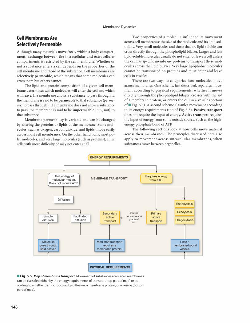
Two properties of a molecule influence its movement across cell membranes: the size of the molecule and its lipid sol-ubility. Very small molecules and those that are lipid soluble can cross directly through the phospholipid bilayer. Larger and less lipid-soluble molecules usually do not enter or leave a cell unless the cell has specifi c membrane proteins to transport these mol-ecules across the lipid bilayer. Very large lipophobic molecules cannot be transported on proteins and must enter and leave cells in vesicles.
There are two ways to categorize how molecules move across membranes. One scheme, just described, separates move-ment according to physical requirements: whether it moves directly through the phospholipid bilayer, crosses with the aid of a membrane protein, or enters the cell in a vesicle (bottom of Fig. 5.5 ). A second scheme classifi es movement according to its energy requirements (top of Fig. 5.5 ). Passive transport does not require the input of energy. Active transport requires the input of energy from some outside source, such as the high- energy phosphate bond of ATP.
The following sections look at how cells move material across their membranes. The principles discussed here also apply to movement across intracellular membranes, when substances move between organelles.
Cell Membranes Are Selectively Permeable
Although many materials move freely within a body compart-ment, exchange between the intracellular and extracellular compartments is restricted by the cell membrane. Whether or not a substance enters a cell depends on the properties of the cell membrane and those of the substance. Cell membranes are selectively permeable , which means that some molecules can cross them but others cannot.
The lipid and protein composition of a given cell mem-brane determines which molecules will enter the cell and which will leave . If a membrane allows a substance to pass through it, the membrane is said to be permeable to that substance { perme-are, to pass through}. If a membrane does not allow a substance to pass, the membrane is said to be impermeable { im -, not} to that substance.
Membrane permeability is variable and can be changed by altering the proteins or lipids of the membrane. Some mol-ecules, such as oxygen, carbon dioxide, and lipids, move easily across most cell membranes. On the other hand, ions, most po-lar molecules, and very large molecules (such as proteins), enter cells with more diffi culty or may not enter at all.
Uses energy ofmolecular motion.
Does not require ATP.
Diffusion
Simplediffusion
Facilitateddiffusion Phagocytosis
Exocytosis
Endocytosis
Secondaryactive
transport
Primaryactive
transport
PHYSICAL REQUIREMENTS
Mediated transportrequires a
membrane protein.
Moleculegoes throughlipid bilayer.
Uses amembrane-bound
vesicle.
Requires energyfrom ATP.
ENERGY REQUIREMENTS
MEMBRANE TRANSPORT
createsconcentration
gradientfor
Fig. 5.5 Map of membrane transport. Movement of substances across cell membranes can be classifi ed either by the energy requirements of transport (top part of map) or ac-cording to whether transport occurs by diff usion, a membrane protein, or a vesicle (bottom part of map).
148
Membrane Dynamics
5
gradient, also known as a chemical gradient . We say that molecules diff use down the gradient, from higher concentra-tion to lower concentration.
Th e rate of diff usion depends on the magnitude of the concentration gradient. Th e larger the concentration gra-dient, the faster diff usion takes place. For example, when you open a bottle of cologne, the rate of diff usion is most rapid as the molecules fi rst escape from the bottle into the air. Later, when the cologne has spread evenly throughout the room, the rate of diff usion has dropped to zero because there is no longer a concentration gradient.
3 Net movement of molecules occurs until the concentration is equal everywhere . Once molecules of a given substance have distributed themselves evenly, the system reaches equilib-rium and diff usion stops. Individual molecules are still mov-ing at equilibrium, but for each molecule that exits an area, another one enters. Th e dynamic equilibrium state in diff u-sion means that the concentration has equalized throughout the system but molecules continue to move.
4 Diff usion is rapid over short distances but much slower over long distances . Albert Einstein studied the diffusion of molecules in solution and found that the time required for a molecule to diff use from point A to point B is propor-tional to the square of the distance from A to B. In other words, if the distance doubles from 1 to 2, the time needed for diff usion increases from 12 to 22 (from 1 to 4).
What does the slow rate of diffusion over long dis-tances mean for biological systems? In humans, nutrients take five seconds to diffuse from the blood to a cell that is 100 μm from the nearest capillary. At that rate, it would take years for nutrients to diff use from the small intestine to cells in the big toe, and the cells would starve to death.
To overcome the limitations of diff usion over distance, organisms use various transport mechanisms that speed up the movement of molecules. Most multicellular organ-isms have some form of circulatory system to bring oxygen and nutrients rapidly from the point at which they enter the body to the cells.
5 Diff usion is directly related to temperature . At higher tem-peratures, molecules move faster. Because diff usion results from molecular movement, the rate of diff usion increases as temperature increases. Generally, changes in temperature do not signifi cantly aff ect diff usion rates in humans because we maintain a relatively constant body temperature.
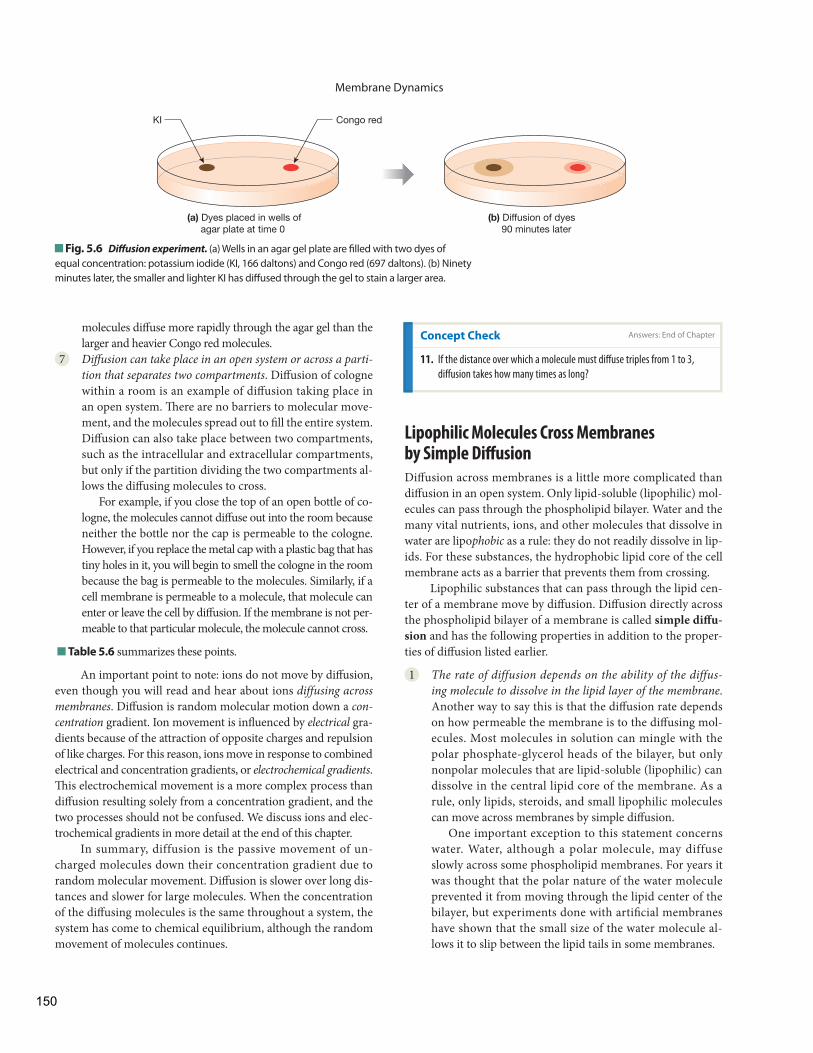
6 Diff usion rate is inversely related to molecular weight and size . Smaller molecules require less energy to move over a distance and therefore diffuse faster. Einstein showed that friction between the surface of a particle and the medium through which it diffuses is a source of resistance to movement. He calculated that diff usion is inversely proportional to the radius of the molecule: the larger the molecule, the slower its diff u-sion through a given medium. Th e experiment in Figure 5.6 shows that the smaller and lighter potassium iodide (KI)
Diff usion Passive transport across membranes uses the kinetic energy in-herent in molecules. Gas molecules and molecules in solution constantly move from one place to another, bouncing off other molecules or off the sides of any container holding them. When molecules start out concentrated in one area of an enclosed space, their motion causes them to spread out gradually until they distribute evenly throughout the available space. Th is pro-cess is known as diff usion.
Diffusion { diffundere, to pour out} may be defined as the movement of molecules from an area of higher concentration of the molecules to an area of lower concentration of the molecules.* If you leave a bottle of cologne open and later notice its fragrance across the room, it is because the aromatic molecules in the cologne have diff used from where they are more concentrated (in the bottle) to where they are less concentrated (across the room).
Diff usion has the following seven properties:
1 Diff usion is a passive process . By passive, we mean that dif-fusion does not require the input of energy from some outside source. Diff usion uses only the kinetic energy pos-sessed by all molecules.
2 Molecules move from an area of higher concentration to an area of lower concentration . A diff erence in the concentration of a substance between two places is called a concentration
R U N N I N G P R O B L E M
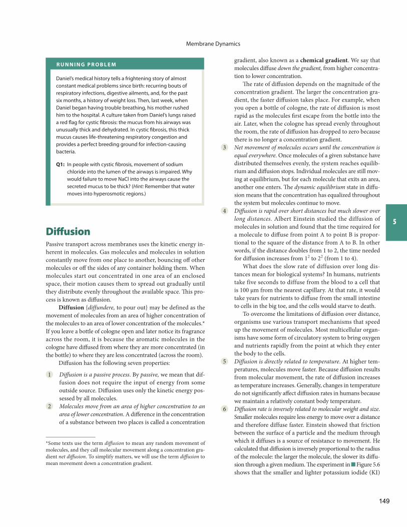
Daniel’s medical history tells a frightening story of almost constant medical problems since birth: recurring bouts of respiratory infections, digestive ailments, and, for the past six months, a history of weight loss. Then, last week, when Daniel began having trouble breathing, his mother rushed him to the hospital. A culture taken from Daniel’s lungs raised a red fl ag for cystic fi brosis: the mucus from his airways was unusually thick and dehydrated. In cystic fi brosis, this thick mucus causes life-threatening respiratory congestion and provides a perfect breeding ground for infection-causing bacteria.
Q1: In people with cystic fi brosis, movement of sodium chloride into the lumen of the airways is impaired. Why would failure to move NaCl into the airways cause the secreted mucus to be thick? (Hint: Remember that water moves into hyperosmotic regions.)
*Some texts use the term diff usion to mean any random movement of molecules, and they call molecular movement along a concentration gra-dient net diff usion . To simplify matters, we will use the term diff usion to mean movement down a concentration gradient.
149
Membrane Dynamics
Lipophilic Molecules Cross Membranes by Simple Diff usion
Diff usion across membranes is a little more complicated than diff usion in an open system. Only lipid-soluble (lipophilic) mol-ecules can pass through the phospholipid bilayer. Water and the many vital nutrients, ions, and other molecules that dissolve in water are lipo phobic as a rule: they do not readily dissolve in lip-ids. For these substances, the hydrophobic lipid core of the cell membrane acts as a barrier that prevents them from crossing.
Lipophilic substances that can pass through the lipid cen-ter of a membrane move by diff usion. Diff usion directly across the phospholipid bilayer of a membrane is called simple diff u-sion and has the following properties in addition to the proper-ties of diff usion listed earlier.
1 The rate of diffusion depends on the ability of the diffus-ing molecule to dissolve in the lipid layer of the membrane . Another way to say this is that the diff usion rate depends on how permeable the membrane is to the diff using mol-ecules. Most molecules in solution can mingle with the polar phosphate-glycerol heads of the bilayer , but only nonpolar molecules that are lipid-soluble (lipophilic) can dissolve in the central lipid core of the membrane. As a rule, only lipids, steroids, and small lipophilic molecules can move across membranes by simple diff usion.
One important exception to this statement concerns water. Water, although a polar molecule, may diffuse slowly across some phospholipid membranes. For years it was thought that the polar nature of the water molecule prevented it from moving through the lipid center of the bilayer, but experiments done with artifi cial membranes have shown that the small size of the water molecule al-lows it to slip between the lipid tails in some membranes.
molecules diff use more rapidly through the agar gel than the larger and heavier Congo red molecules.
7 Diff usion can take place in an open system or across a parti-tion that separates two compartments . Diff usion of cologne within a room is an example of diff usion taking place in an open system. Th ere are no barriers to molecular move-ment, and the molecules spread out to fi ll the entire system. Diff usion can also take place between two compartments, such as the intracellular and extracellular compartments, but only if the partition dividing the two compartments al-lows the diff using molecules to cross.
For example, if you close the top of an open bottle of co-logne, the molecules cannot diff use out into the room because neither the bottle nor the cap is permeable to the cologne. However, if you replace the metal cap with a plastic bag that has tiny holes in it, you will begin to smell the cologne in the room because the bag is permeable to the molecules. Similarly, if a cell membrane is permeable to a molecule, that molecule can enter or leave the cell by diff usion. If the membrane is not per-meable to that particular molecule, the molecule cannot cross.
Table 5.6 summarizes these points.
An important point to note: ions do not move by diff usion, even though you will read and hear about ions diffusing across membranes . Diff usion is random molecular motion down a con-centration gradient. Ion movement is infl uenced by electrical gra-dients because of the attraction of opposite charges and repulsion of like charges. For this reason, ions move in response to combined electrical and concentration gradients, or electrochemical gradients . Th is electrochemical movement is a more complex process than diff usion resulting solely from a concentration gradient, and the two processes should not be confused. We discuss ions and elec-trochemical gradients in more detail at the end of this chapter.
In summary, diffusion is the passive movement of un-charged molecules down their concentration gradient due to random molecular movement. Diff usion is slower over long dis-tances and slower for large molecules. When the concentration of the diff using molecules is the same throughout a system, the system has come to chemical equilibrium, although the random movement of molecules continues.
Fig. 5.6 Diff usion experiment. (a) Wells in an agar gel plate are fi lled with two dyes of equal concentration: potassium iodide (KI, 166 daltons) and Congo red (697 daltons). (b) Ninety minutes later, the smaller and lighter KI has diff used through the gel to stain a larger area.
KI Congo red
(a) Dyes placed in wells of agar plate at time 0
(b) Diffusion of dyes 90 minutes later
Concept Check
11. If the distance over which a molecule must diff use triples from 1 to 3,
diff usion takes how many times as long?
Answers: End of Chapter
150
Membrane Dynamics
5
reaches the cells is not enough to sustain any muscular ac-tivity and the patient is confi ned to bed.
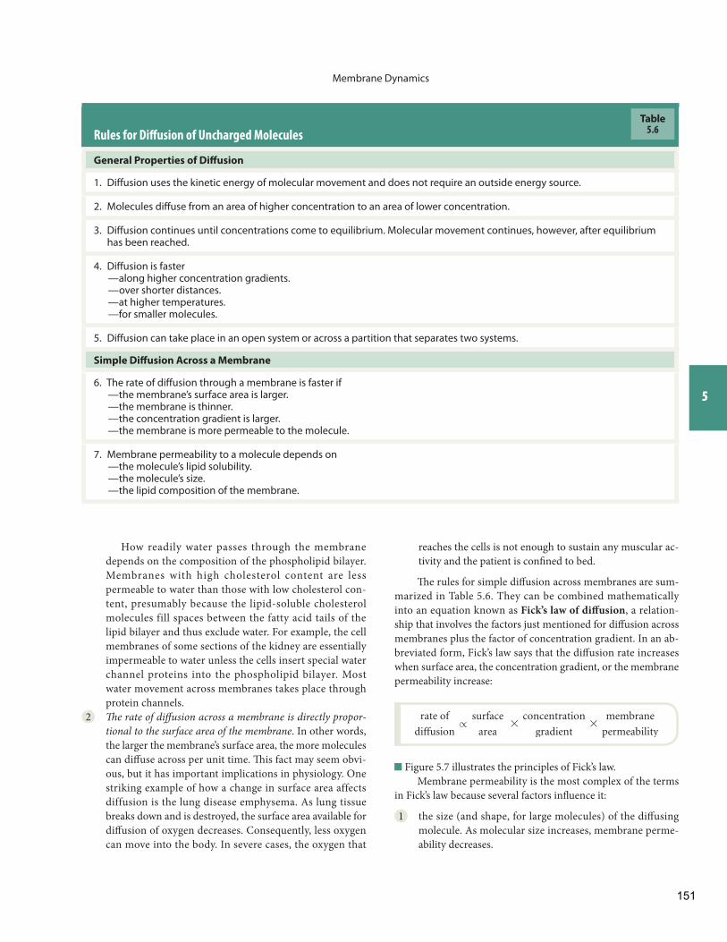
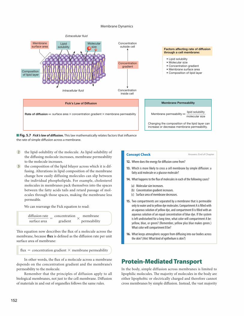
Th e rules for simple diff usion across membranes are sum-marized in Table 5.6 . They can be combined mathematically into an equation known as Fick’s law of diff usion , a relation-ship that involves the factors just mentioned for diff usion across membranes plus the factor of concentration gradient. In an ab-breviated form, Fick’s law says that the diff usion rate increases when surface area, the concentration gradient, or the membrane permeability increase:
rate of
diffusion r
surface
area *
concentration
gradient*
membrane
permeability
Figure 5.7 illustrates the principles of Fick’s law. Membrane permeability is the most complex of the terms
in Fick’s law because several factors infl uence it:
1 the size (and shape, for large molecules) of the diff using molecule. As molecular size increases, membrane perme-ability decreases.
Rules for Diff usion of Uncharged Molecules
General Properties of Diff usion
1. Diffusion uses the kinetic energy of molecular movement and does not require an outside energy source.
2. Molecules diffuse from an area of higher concentration to an area of lower concentration.
3. Diffusion continues until concentrations come to equilibrium. Molecular movement continues, however, after equilibrium has been reached.
4. Diffusion is faster —along higher concentration gradients. —over shorter distances. —at higher temperatures. —for smaller molecules.
5. Diffusion can take place in an open system or across a partition that separates two systems.
Simple Diff usion Across a Membrane
6. The rate of diffusion through a membrane is faster if —the membrane’s surface area is larger. —the membrane is thinner. —the concentration gradient is larger. —the membrane is more permeable to the molecule.
7. Membrane permeability to a molecule depends on —the molecule’s lipid solubility. —the molecule’s size. —the lipid composition of the membrane.
Table5.6
How readily water passes through the membrane depends on the composition of the phospholipid bilayer. Membranes with high cholesterol content are less permeable to water than those with low cholesterol con-tent, presumably because the lipid-soluble cholesterol molecules fill spaces between the fatty acid tails of the lipid bilayer and thus exclude water. For example, the cell membranes of some sections of the kidney are essentially impermeable to water unless the cells insert special water channel proteins into the phospholipid bilayer. Most water movement across membranes takes place through protein channels.
2 Th e rate of diff usion across a membrane is directly propor-tional to the surface area of the membrane . In other words, the larger the membrane’s surface area, the more molecules can diff use across per unit time. Th is fact may seem obvi-ous, but it has important implications in physiology. One striking example of how a change in surface area affects diffusion is the lung disease emphysema. As lung tissue breaks down and is destroyed, the surface area available for diff usion of oxygen decreases. Consequently, less oxygen can move into the body. In severe cases, the oxygen that
151
Membrane Dynamics
Protein-Mediated Transport In the body, simple diffusion across membranes is limited to lipophilic molecules. Th e majority of molecules in the body are either lipophobic or electrically charged and therefore cannot cross membranes by simple diff usion. Instead, the vast majority
2 the lipid-solubility of the molecule. As lipid solubility of the diff using molecule increases, membrane permeability to the molecule increases.
3 the composition of the lipid bilayer across which it is dif-fusing. Alterations in lipid composition of the membrane change how easily diffusing molecules can slip between the individual phospholipids. For example, cholesterol molecules in membranes pack themselves into the spaces between the fatty acids tails and retard passage of mol-ecules through those spaces , making the membrane less permeable.
We can rearrange the Fick equation to read:
diffusion rate
surface area=
concentration
gradient*
membrane
permeability
Th is equation now describes the fl ux of a molecule across the membrane, because fl ux is defi ned as the diff usion rate per unit surface area of membrane:
flux = concentration gradient * membrane permeability
In other words, the fl ux of a molecule across a membrane depends on the concentration gradient and the membrane’s permeability to the molecule.
Remember that the principles of diffusion apply to all biological membranes, not just to the cell membrane. Diff usion of materials in and out of organelles follows the same rules.
Fig. 5.7 Fick’s law of diff usion. This law mathematically relates factors that infl uence the rate of simple diff usion across a membrane.
Rate of diffusion surface area × concentration gradient × membrane permeability
Extracellular fluid
Membranesurface area
Intracellular fluid
Compositionof lipid layer
Lipidsolubility
Molecularsize
Concentrationoutside cell
Concentrationinside cell
Concentrationgradient
Fick's Law of Diffusion
lipid solubility
molecular size Membrane permeability
Changing the composition of the lipid layer can increase or decrease membrane permeability.
Factors affecting rate of diffusionthrough a cell membrane:
• Lipid solubility • Molecular size • Concentration gradient • Membrane surface area • Composition of lipid layer
Membrane Permeability
∝ ∝
Concept Check Answers: End of Chapter
12. Where does the energy for diff usion come from?
13. Which is more likely to cross a cell membrane by simple diffusion: a
fatty acid molecule or a glucose molecule?
14. What happens to the fl ux of molecules in each of the following cases?
(a) Molecular size increases.
(b) Concentration gradient increases.
(c) Surface area of membrane decreases.
15. Two compartments are separated by a membrane that is permeable
only to water and to yellow dye molecules. Compartment A is fi lled with
an aqueous solution of yellow dye, and compartment B is fi lled with an
aqueous solution of an equal concentration of blue dye. If the system
is left undisturbed for a long time, what color will compartment A be:
yellow, blue, or green? (Remember, yellow plus blue makes green.)
What color will compartment B be?
16. What keeps atmospheric oxygen from diff using into our bodies across
the skin? ( Hint: What kind of epithelium is skin?)
152
Membrane Dynamics
5
Structural Proteins Th e structural proteins have three major roles.
1 Th ey connect the membrane to the cytoskeleton to main-tain the shape of the cell . The microvilli of transporting epithelia are one example of membrane shaping by the cytoskeleton .
2 Th ey create cell junctions that hold tissues together, such as tight junctions and gap junctions .
3 They attach cells to the extracellular matrix by linking cytoskeleton fi bers to extracellular collagen and other pro-tein fi bers.
Enzymes Membrane enzymes catalyze chemical reactions that take place either on the cell’s external surface or just inside the cell. For example, enzymes on the external surface of cells lining the small intestine are responsible for digesting peptides and carbohydrates. Enzymes attached to the intracellular surface of many cell membranes play an important role in transferring signals from the extracellular environment to the cytoplasm .
Receptors Membrane receptor proteins are part of the body’s chemical signaling system. The binding of a receptor with its ligand usually triggers another event at the membrane
of solutes cross membranes with the help of membrane proteins, a process we call mediated transport .
If mediated transport is passive and moves molecules down their concentration gradient, and if net transport stops when concentrations are equal on both sides of the membrane, the process is known as facilitated diffusion ( Fig. 5.5 ). If protein-mediated transport requires energy from ATP or another outside source and moves a substance against its concentration gradient, the process is known as active transport .
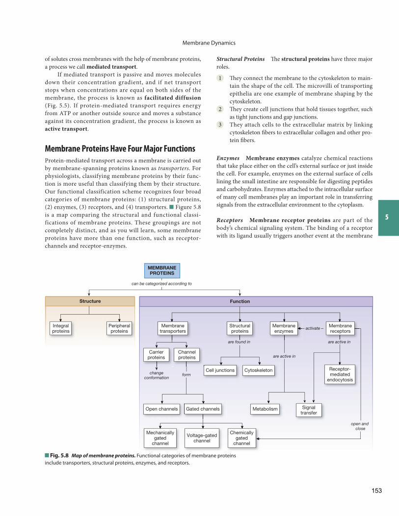
Membrane Proteins Have Four Major Functions
Protein-mediated transport across a membrane is carried out by membrane-spanning proteins known as transporters . For physiologists, classifying membrane proteins by their func-tion is more useful than classifying them by their structure. Our functional classification scheme recognizes four broad categories of membrane proteins: (1) structural proteins, (2) enzymes, (3) receptors, and (4) transporters. Figure 5.8 is a map comparing the structural and functional classi-fications of membrane proteins. These groupings are not completely distinct, and as you will learn, some membrane proteins have more than one function, such as receptor-channels and receptor-enzymes.
MEMBRANEPROTEINS
Integralproteins
Peripheralproteins
Membranetransporters
Structure
are found in
form
are active in
are active in
activate
changeconformation
Carrierproteins
Channelproteins
Cell junctions
Gated channelsOpen channels
Cytoskeleton
Function
Structuralproteins
Membraneenzymes
Signaltransfer
Metabolism
Receptor-mediated
endocytosis
Chemicallygated
channel
Voltage-gatedchannel
Mechanicallygated
channel
can be categorized according to
open and close
Membranereceptors
Fig. 5.8 Map of membrane proteins. Functional categories of membrane proteins include transporters, structural proteins, enzymes, and receptors.
153
Membrane Dynamics
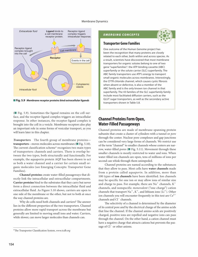
( Fig. 5.9 ). Sometimes the ligand remains on the cell sur-face, and the receptor-ligand complex triggers an intracellular response. In other instances, the receptor-ligand complex is brought into the cell in a vesicle. Membrane receptors also play an important role in some forms of vesicular transport, as you will learn later in this chapter.
Transporters The fourth group of membrane proteins—transporters —moves molecules across membranes ( Fig. 5.10 ). Th e current classification scheme * recognizes two main types of transporters: channels and carriers. There is overlap be-tween the two types, both structurally and functionally. For example, the aquaporin protein AQP has been shown to act as both a water channel and a carrier for certain small or-ganic molecules (see Emerging Concepts: Transporter Gene Families).
Channel proteins create water-fi lled passageways that di-rectly link the intracellular and extracellular compartments. Carrier proteins bind to the substrates that they carry but never form a direct connection between the intracellular fluid and extracellular fluid. As Figure 5.10 shows, carriers are open to one side of the membrane or the other, but not to both at once the way channel proteins are.
Why do cells need both channels and carriers? Th e answer lies in the diff erent properties of the two transporters. Channel proteins allow more rapid transport across the membrane but generally are limited to moving small ions and water. Carriers, while slower, can move larger molecules than channels can.
* Th e Transporter Classifi cation System, www.tcdb.org
Cell membrane
Ligand binds toa cell membrane receptor protein.
Receptor
Receptor-ligand complex triggers intracellular response.
Receptor-ligand complex broughtinto the cell
Cytoplasmicvesicle
Events in the cell
Extracellular fluid
Intracellular fluid
Fig. 5.9 Membrane receptor proteins bind extracellular ligands
E M E R G I N G C O N C E P T S
Transporter Gene Families
One outcome of the Human Genome project has been the recognition that many proteins are closely related to each other, both within and across species. As a result, scientists have discovered that most membrane transporters for organic solutes belong to one of two gene “superfamilies”: the ATP-binding cassette (ABC) superfamily or the solute carrier (SLC) superfamily. The ABC family transporters use ATP’s energy to transport small organic molecules across membranes. Interestingly, the CFTR chloride channel, which causes cystic fibrosis when absent or defective, is also a member of the ABC family and is the only known ion channel in that superfamily. The 43 families of the SLC superfamily family include most facilitated diffusion carriers, such as the GLUT sugar transporters, as well as the secondary active transporters shown in Table 5.8 .
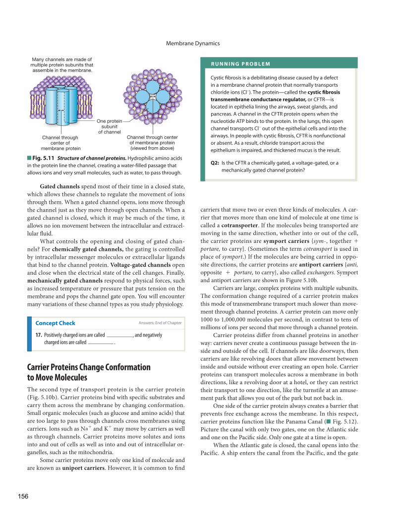
Channel Proteins Form Open, Water-Filled Passageways
Channel proteins are made of membrane-spanning protein subunits that create a cluster of cylinders with a tunnel or porethrough the center. Nuclear pore complexes and gap junctions can be considered very large forms of channels. We restrict use of the term “channel” to smaller channels whose centers are nar-row, water-fi lled pores ( Fig. 5.11 ). Movement through these smaller channels is mostly restricted to water and ions. When water-fi lled ion channels are open, tens of millions of ions per second can whisk through them unimpeded.
Channel proteins are named according to the substances that they allow to pass. Most cells have water channels made from a protein called aquaporin . In addition, more than 100 types of ion channels have been identified. Ion channels may be specific for one ion or may allow ions of similar size and charge to pass. For example, there are Na+ channels, K+
channels, and nonspecific monovalent (“one-charge”) cation channels that transport Na+ , K+ , and lithium ions (Li+). Other ion channels you will encounter frequently in this text are Ca2 +
channels and Cl- channels. Th e selectivity of a channel is determined by the diameter
of its central pore and by the electrical charge of the amino acids that line the channel. If the channel amino acids are positively charged, positive ions are repelled and negative ions can pass through the channel. On the other hand, a cation channel must have a negative charge that attracts cations but prevents the pas-sage of Cl- or other anions.
154
on a chain that swings up and blocks the mouth of the channel ( Fig. 5.10 a). One type of channel in neurons has two diff erent gates.
Channels can be classifi ed according to whether their gates are usually open or usually closed. Open channels spend most of their time with their gate open, allowing ions to move back and forth across the membrane without regulation. Th ese gates may occasionally flicker closed, but for the most part these channels behave as if they have no gates. Open channels are sometimes called either leak channels or pores, as in water pores .
Channel proteins are like narrow doorways into the cell. If the door is closed, nothing can go through. If the door is open, there is a continuous passage between the two rooms con-nected by the doorway. Th e open or closed state of a channel is determined by regions of the protein molecule that act like swinging “gates.”
According to current models, channel “gates” take several forms. Some channel proteins have gates in the middle of the protein’s pore. Other gates are part of the cytoplasmic side of the membrane protein. Such a gate can be envisioned as a ball
Membrane transporters are membrane-spanning proteins that help move lipophobic molecules across membranes.
Membrane Transporters
MEMBRANE TRANSPORTERS
(a) Channel proteins create a water-filled pore. (b) Carrier proteins never form an open channel between the two sides of the membrane.
can be classifiedcan be classified
Gated channelsopen and close in
response to signals.
Close-up views of transporters are shown in the top two rows and distant views in the bottom row. Primary active transport is indicated by ATP on the protein.
Open channelsor pores
are usually open.
Uniport carrierstransport only onekind of substrate.
Antiport carriersmove substrates inopposite directions.
Symport carriers move two ormore substrates in the same
direction across the membrane.
Cotransporters
ECF
ICF
Cellmembrane
Carrier opento ICF
Same carrieropen to ECF
GluGlu
ATP
ATP
Open Closed
Fig. 5.10 E S S E N T I A L S
Na+ Na+
K+
155
Membrane Dynamics
Carrier Proteins Change Conformation to Move Molecules
The second type of transport protein is the carrier protein ( Fig. 5.10 b). Carrier proteins bind with specifi c substrates and carry them across the membrane by changing conformation. Small organic molecules (such as glucose and amino acids) that are too large to pass through channels cross membranes using carriers. Ions such as Na+ and K+ may move by carriers as well as through channels. Carrier proteins move solutes and ions into and out of cells as well as into and out of intracellular or-ganelles, such as the mitochondria.
Some carrier proteins move only one kind of molecule and are known as uniport carriers . However, it is common to fi nd
Gated channels spend most of their time in a closed state, which allows these channels to regulate the movement of ions through them. When a gated channel opens, ions move through the channel just as they move through open channels. When a gated channel is closed, which it may be much of the time, it allows no ion movement between the intracellular and extracel-lular fl uid.
What controls the opening and closing of gated chan-nels? For chemically gated channels, the gating is controlled by intracellular messenger molecules or extracellular ligands that bind to the channel protein. Voltage-gated channels open and close when the electrical state of the cell changes. Finally, mechanically gated channels respond to physical forces, such as increased temperature or pressure that puts tension on the membrane and pops the channel gate open. You will encounter many variations of these channel types as you study physiology.
Fig. 5.11 Structure of channel proteins. Hydrophilic amino acids in the protein line the channel, creating a water-fi lled passage that allows ions and very small molecules, such as water, to pass through.
Channel through center of
membrane protein
Channel through centerof membrane protein(viewed from above)
One proteinsubunit
of channel
Many channels are made of multiple protein subunits that assemble in the membrane.
Concept Check
17. Positively charged ions are called , and negatively
charged ions are called .
carriers that move two or even three kinds of molecules. A car-rier that moves more than one kind of molecule at one time is called a cotransporter . If the molecules being transported are moving in the same direction, whether into or out of the cell, the carrier proteins are symport carriers { sym -, together +
portare, to carry}. (Sometimes the term cotransport is used in place of symport .) If the molecules are being carried in oppo-site directions, the carrier proteins are antiport carriers { anti,opposite + portare, to carry}, also called exchangers . Symport and antiport carriers are shown in Figure 5.10 b.
Carriers are large, complex proteins with multiple subunits. The conformation change required of a carrier protein makes this mode of transmembrane transport much slower than move-ment through channel proteins. A carrier protein can move only 1000 to 1,000,000 molecules per second, in contrast to tens of millions of ions per second that move through a channel protein.
Carrier proteins diff er from channel proteins in another way: carriers never create a continuous passage between the in-side and outside of the cell. If channels are like doorways, then carriers are like revolving doors that allow movement between inside and outside without ever creating an open hole. Carrier proteins can transport molecules across a membrane in both directions, like a revolving door at a hotel, or they can restrict their transport to one direction, like the turnstile at an amuse-ment park that allows you out of the park but not back in.
One side of the carrier protein always creates a barrier that prevents free exchange across the membrane. In this respect, carrier proteins function like the Panama Canal ( Fig. 5.12 ). Picture the canal with only two gates, one on the Atlantic side and one on the Pacifi c side. Only one gate at a time is open.
When the Atlantic gate is closed, the canal opens into the Pacific. A ship enters the canal from the Pacific, and the gate
R U N N I N G P R O B L E M
Cystic fi brosis is a debilitating disease caused by a defect in a membrane channel protein that normally transports chloride ions (Cl-). The protein—called the cystic fi brosis transmembrane conductance regulator , or CFTR—is located in epithelia lining the airways, sweat glands, and pancreas. A channel in the CFTR protein opens when the nucleotide ATP binds to the protein. In the lungs, this open channel transports Cl- out of the epithelial cells and into the airways. In people with cystic fi brosis, CFTR is nonfunctional or absent. As a result, chloride transport across the epithelium is impaired, and thickened mucus is the result.
Q2: Is the CFTR a chemically gated, a voltage-gated, or a mechanically gated channel protein?
Answers: End of Chapter
156
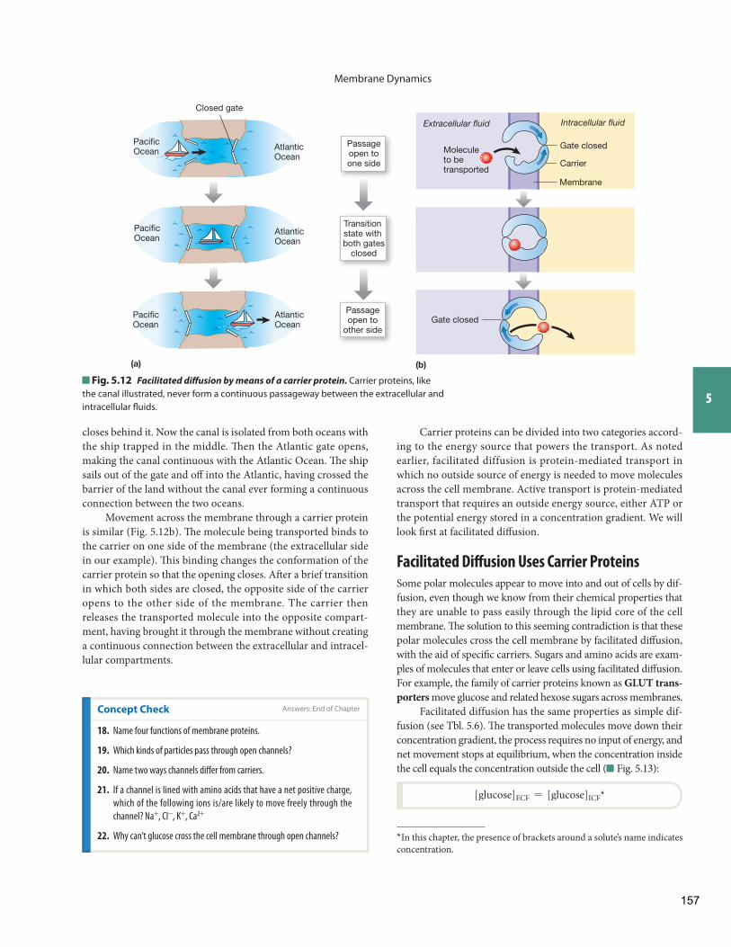
Membrane Dynamics
5
Carrier proteins can be divided into two categories accord-ing to the energy source that powers the transport. As noted earlier, facilitated diffusion is protein-mediated transport in which no outside source of energy is needed to move molecules across the cell membrane. Active transport is protein-mediated transport that requires an outside energy source, either ATP or the potential energy stored in a concentration gradient. We will look fi rst at facilitated diff usion.
Facilitated Diff usion Uses Carrier Proteins
Some polar molecules appear to move into and out of cells by dif-fusion, even though we know from their chemical properties that they are unable to pass easily through the lipid core of the cell membrane. Th e solution to this seeming contradiction is that these polar molecules cross the cell membrane by facilitated diff usion, with the aid of specifi c carriers. Sugars and amino acids are exam-ples of molecules that enter or leave cells using facilitated diff usion. For example, the family of carrier proteins known as GLUT trans-porters move glucose and related hexose sugars across membranes.
Facilitated diffusion has the same properties as simple dif-fusion (see Tbl. 5.6 ). Th e transported molecules move down their concentration gradient, the process requires no input of energy, and net movement stops at equilibrium, when the concentration inside the cell equals the concentration outside the cell ( Fig. 5.13 ):
[glucose]ECF = [glucose]ICF *
* In this chapter, the presence of brackets around a solute’s name indicates concentration.
closes behind it. Now the canal is isolated from both oceans with the ship trapped in the middle. Th en the Atlantic gate opens, making the canal continuous with the Atlantic Ocean. Th e ship sails out of the gate and off into the Atlantic, having crossed the barrier of the land without the canal ever forming a continuous connection between the two oceans.
Movement across the membrane through a carrier protein is similar ( Fig. 5.12 b). Th e molecule being transported binds to the carrier on one side of the membrane (the extracellular side in our example). Th is binding changes the conformation of the carrier protein so that the opening closes. Aft er a brief transition in which both sides are closed, the opposite side of the carrier opens to the other side of the membrane. The carrier then releases the transported molecule into the opposite compart-ment, having brought it through the membrane without creating a continuous connection between the extracellular and intracel-lular compartments.
Fig. 5.12 Facilitated diff usion by means of a carrier protein. Carrier proteins, like the canal illustrated, never form a continuous passageway between the extracellular and intracellular fl uids.
Membrane
PacificOcean
AtlanticOcean
PacificOcean
AtlanticOcean
PacificOcean Atlantic
Ocean
Closed gate
Transition state with both gates
closed
Passageopen toone side
Passageopen to
other side
Intracellular fluid
Molecule to betransported
Gate closed
Gate closed
Carrier
Extracellular fluid
(a) (b)
Concept Check Answers: End of Chapter
18. Name four functions of membrane proteins.
19. Which kinds of particles pass through open channels?
20. Name two ways channels diff er from carriers.
21. If a channel is lined with amino acids that have a net positive charge,
which of the following ions is/are likely to move freely through the
channel? Na+, Cl-, K+, Ca2+
22. Why can’t glucose cross the cell membrane through open channels?
157
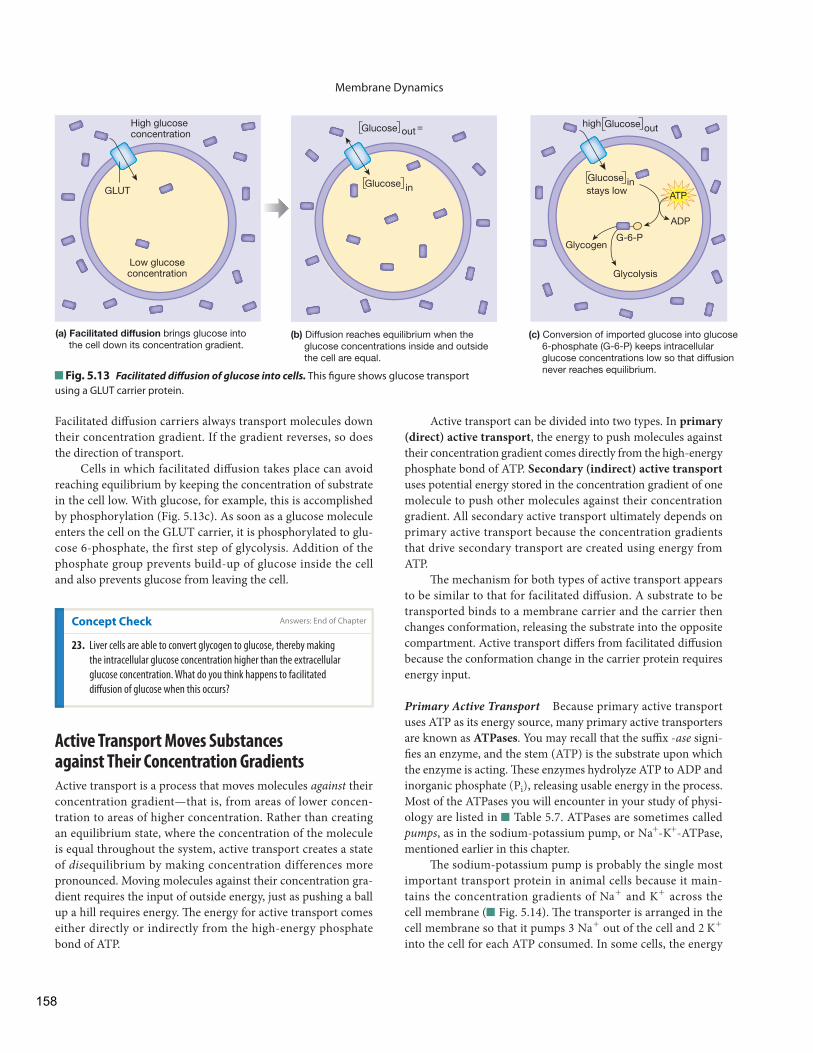
Membrane Dynamics
Active Transport Moves Substances against Their Concentration Gradients
Active transport is a process that moves molecules against their concentration gradient—that is, from areas of lower concen-tration to areas of higher concentration. Rather than creating an equilibrium state, where the concentration of the molecule is equal throughout the system, active transport creates a state of dis equilibrium by making concentration differences more pronounced. Moving molecules against their concentration gra-dient requires the input of outside energy, just as pushing a ball up a hill requires energy . Th e energy for active transport comes either directly or indirectly from the high-energy phosphate bond of ATP.
Facilitated diff usion carriers always transport molecules down their concentration gradient. If the gradient reverses, so does the direction of transport.
Cells in which facilitated diff usion takes place can avoid reaching equilibrium by keeping the concentration of substrate in the cell low. With glucose, for example, this is accomplished by phosphorylation ( Fig. 5.13 c). As soon as a glucose molecule enters the cell on the GLUT carrier, it is phosphorylated to glu-cose 6-phosphate, the first step of glycolysis . Addition of the phosphate group prevents build-up of glucose inside the cell and also prevents glucose from leaving the cell.
Fig. 5.13 Facilitated diff usion of glucose into cells. This fi gure shows glucose transport using a GLUT carrier protein.
High glucose concentration
Glucose
Glucose
in
out =
(a) Facilitated diffusion brings glucose into the cell down its concentration gradient.
(b) Diffusion reaches equilibrium when the glucose concentrations inside and outside the cell are equal.
(c) Conversion of imported glucose into glucose 6-phosphate (G-6-P) keeps intracellular glucose concentrations low so that diffusion never reaches equilibrium.
Glucose out
Glucose in
high
ATP
ADP
G-6-PGlycogen
Glycolysis
stays lowGLUT
Low glucoseconcentration
Concept Check
23. Liver cells are able to convert glycogen to glucose, thereby making
the intracellular glucose concentration higher than the extracellular
glucose concentration. What do you think happens to facilitated
diff usion of glucose when this occurs?
Active transport can be divided into two types. In primary (direct) active transport , the energy to push molecules against their concentration gradient comes directly from the high-energy phosphate bond of ATP. Secondary (indirect) active transportuses potential energy stored in the concentration gradient of one molecule to push other molecules against their concentration gradient. All secondary active transport ultimately depends on primary active transport because the concentration gradients that drive secondary transport are created using energy from ATP.
Th e mechanism for both types of active transport appears to be similar to that for facilitated diff usion. A substrate to be transported binds to a membrane carrier and the carrier then changes conformation, releasing the substrate into the opposite compartment. Active transport diff ers from facilitated diff usion because the conformation change in the carrier protein requires energy input.
Primary Active Transport Because primary active transport uses ATP as its energy source, many primary active transporters are known as ATPases . You may recall that the suffi x - ase signi-fi es an enzyme, and the stem (ATP) is the substrate upon which the enzyme is acting . Th ese enzymes hydrolyze ATP to ADP and inorganic phosphate (Pi), releasing usable energy in the process. Most of the ATPases you will encounter in your study of physi-ology are listed in Table 5.7 . ATPases are sometimes called pumps , as in the sodium-potassium pump, or Na+ - K+ -ATPase, mentioned earlier in this chapter.
Th e sodium-potassium pump is probably the single most important transport protein in animal cells because it main-tains the concentration gradients of Na+ and K+ across the cell membrane ( Fig. 5.14 ). Th e transporter is arranged in the cell membrane so that it pumps 3 Na+ out of the cell and 2 K+ into the cell for each ATP consumed. In some cells, the energy
Answers: End of Chapter
158
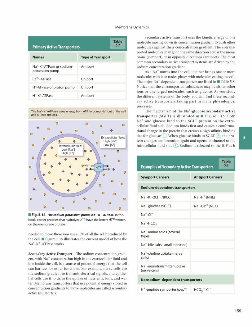
Membrane Dynamics
5
Primary Active Transporters
Names Type of Transport
Na+ - K+ -ATPase or sodium-potassium pump
Antiport
Ca2+ -ATPase Uniport
H+ -ATPase or proton pump Uniport
H+ - K+ -ATPase Antiport
Table5.7
Na+
K+ATP
Extracellular fluid: High [Na+] Low [K+]Intracellular fluid:
Low [Na+] High [K+]
The Na+-K+-ATPase uses energy from ATP to pump Na+ out of the cell and K+ into the cell.
Fig. 5.14 The sodium-potassium pump, Na+ - K + -ATPase. In this book, carrier proteins that hydrolyze ATP have the letters ATP written on the membrane protein.
Examples of Secondary Active Transporters
Symport Carriers Antiport Carriers
Sodium-dependent transporters
Na+ - K+ - 2Cl- (NKCC) Na+ - H+ (NHE)
Na+ -glucose (SGLT) Na+ - Ca2 + (NCX)
Na+ - Cl-
Na+ - HCO3-
Na+ -amino acids (several types)
Na+ -bile salts (small intestine)
Na+ -choline uptake (nerve cells)
Na+ -neurotransmitter uptake (nerve cells)
Nonsodium-dependent transporters
H+ -peptide symporter (pepT) HCO 3- -Cl-
Table5.8
needed to move these ions uses 30% of all the ATP produced by the cell. Figure 5.15 illustrates the current model of how the Na+ - K+ -ATPase works.
Secondary Active Transport Th e sodium concentration gradi-ent, with Na+ concentration high in the extracellular fl uid and low inside the cell, is a source of potential energy that the cell can harness for other functions. For example, nerve cells use the sodium gradient to transmit electrical signals, and epithe-lial cells use it to drive the uptake of nutrients, ions, and wa-ter. Membrane transporters that use potential energy stored in concentration gradients to move molecules are called secondary active transporters .
Secondary active transport uses the kinetic energy of one molecule moving down its concentration gradient to push other molecules against their concentration gradient. The cotrans-ported molecules may go in the same direction across the mem-brane (symport) or in opposite directions (antiport). Th e most common secondary active transport systems are driven by the sodium concentration gradient.
As a Na+ moves into the cell, it either brings one or more molecules with it or trades places with molecules exiting the cell. Th e major Na+ -dependent transporters are listed in Table 5.8 . Notice that the cotransported substances may be either other ions or uncharged molecules, such as glucose. As you study the diff erent systems of the body, you will fi nd these second-ary active transporters taking part in many physiological processes.
The mechanism of the Na+ - glucose secondary active transporter (SGLT) is illustrated in Figure 5.16 . Both Na+ and glucose bind to the SGLT protein on the extra-cellular fl uid side. Sodium binds fi rst and causes a conforma-tional change in the protein that creates a high-affi nity binding site for glucose 1 . When glucose binds to SGLT 2 , the pro-tein changes conformation again and opens its channel to the intracellular fl uid side 3 . Sodium is released to the ICF as it
159
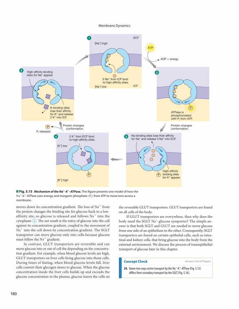
Membrane Dynamics
the reversible GLUT transporters. GLUT transporters are found on all cells of the body.
If GLUT transporters are everywhere, then why does the body need the SGLT Na+ -glucose symporter? The simple an-swer is that both SGLT and GLUT are needed to move glucose from one side of an epithelium to the other. Consequently, SGLT transporters are found on certain epithelial cells, such as intes-tinal and kidney cells, that bring glucose into the body from the external environment. We discuss the process of transepithelial transport of glucose later in this chapter.
moves down its concentration gradient. Th e loss of Na+ from the protein changes the binding site for glucose back to a low-affinity site, so glucose is released and follows Na+ into the cytoplasm 4 . Th e net result is the entry of glucose into the cell against its concentration gradient, coupled to the movement of Na+ into the cell down its concentration gradient. The SGLT transporter can move glucose only into cells because glucose must follow the Na+ gradient.
In contrast, GLUT transporters are reversible and can move glucose into or out of cell the depending on the concentra-tion gradient. For example, when blood glucose levels are high, GLUT transporters on liver cells bring glucose into those cells. During times of fasting, when blood glucose levels fall, liver cells convert their glycogen stores to glucose. When the glucose concentration inside the liver cells builds up and exceeds the glucose concentration in the plasma, glucose leaves the cells on
2 K+ from ECF bind to high-affinity sites.
ADP + energy
ATP
ATPase is phosphorylatedwith Pi from ATP.
Protein changesconformation.
High-affinity binding sites for K+ appear.
34
3 Na+ from ICF bind to high-affinity sites.
P
PP
P
ICF
ECF
Protein changesconformation.
1
25
[Na+] high
[Na+] low
[K+] low
[K+] high
Na-binding sites lose their affinityfor Na+ and release 3 Na+ into ECF.
Pi released.
K-binding sites lose their affinity for K+ and release 2 K+ into ICF.
High-affinity binding sites for Na+ appear.
Fig. 5.15 Mechanism of the Na+ - K + -ATPase. This fi gure presents one model of how the Na+ - K+ -ATPase uses energy and inorganic phosphate (Pi) from ATP to move ions across a membrane.
Concept Check
24. Name two ways active transport by the Na+ - K+ -ATPase ( Fig. 5.15 )
diff ers from secondary transport by the SGLT ( Fig. 5.16 ).
Answers: End of Chapter
160
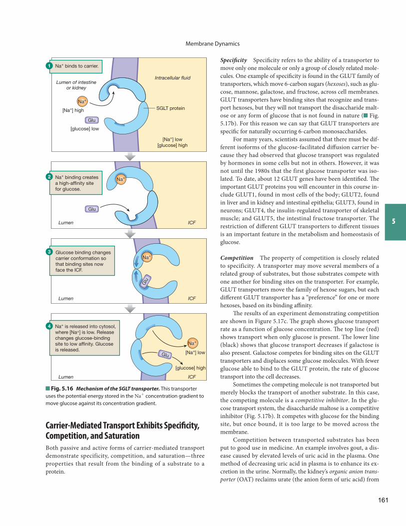
Membrane Dynamics
5
Specifi city Specifi city refers to the ability of a transporter to move only one molecule or only a group of closely related mole-cules. One example of specifi city is found in the GLUT family of transporters, which move 6-carbon sugars ( hexoses ), such as glu-cose, mannose, galactose, and fructose , across cell membranes. GLUT transporters have binding sites that recognize and trans-port hexoses, but they will not transport the disaccharide malt-ose or any form of glucose that is not found in nature ( Fig. 5.17 b). For this reason we can say that GLUT transporters are specifi c for naturally occurring 6-carbon monosaccharides.
For many years, scientists assumed that there must be dif-ferent isoforms of the glucose-facilitated diff usion carrier be-cause they had observed that glucose transport was regulated by hormones in some cells but not in others. However, it was not until the 1980s that the first glucose transporter was iso-lated. To date, about 12 GLUT genes have been identifi ed. Th e important GLUT proteins you will encounter in this course in-clude GLUT1, found in most cells of the body; GLUT2, found in liver and in kidney and intestinal epithelia; GLUT3, found in neurons; GLUT4, the insulin-regulated transporter of skeletal muscle; and GLUT5, the intestinal fructose transporter. The restriction of diff erent GLUT transporters to diff erent tissues is an important feature in the metabolism and homeostasis of glucose.
Competition The property of competition is closely related to specificity. A transporter may move several members of a related group of substrates, but those substrates compete with one another for binding sites on the transporter. For example, GLUT transporters move the family of hexose sugars, but each diff erent GLUT transporter has a “preference” for one or more hexoses, based on its binding affi nity.
Th e results of an experiment demonstrating competition are shown in Figure 5.17 c. Th e graph shows glucose transport rate as a function of glucose concentration. Th e top line (red) shows transport when only glucose is present. The lower line (black) shows that glucose transport decreases if galactose is also present. Galactose competes for binding sites on the GLUT transporters and displaces some glucose molecules. With fewer glucose able to bind to the GLUT protein, the rate of glucose transport into the cell decreases.
Sometimes the competing molecule is not transported but merely blocks the transport of another substrate. In this case, the competing molecule is a competitive inhibitor . In the glu-cose transport system, the disaccharide maltose is a competitive inhibitor ( Fig. 5.17 b). It competes with glucose for the binding site, but once bound, it is too large to be moved across the membrane.
Competition between transported substrates has been put to good use in medicine. An example involves gout, a dis-ease caused by elevated levels of uric acid in the plasma. One method of decreasing uric acid in plasma is to enhance its ex-cretion in the urine. Normally, the kidney’s organic anion trans-porter (OAT) reclaims urate (the anion form of uric acid) from
Na+
[Na+] low[glucose] high
[glucose] low
[Na+] high
[glucose] high
[Na+] low
SGLT protein
Glu
Glu
Glu
Glu
Lumen of intestineor kidney
Intracellular fluid
Na+
Na+
Na+
Glucose binding changes carrier conformation so that binding sites nowface the ICF.
Na+ is released into cytosol, where [Na+] is low. Releasechanges glucose-bindingsite to low affinity. Glucoseis released.
Na+ binding creates a high-affinity site for glucose.
Na+ binds to carrier.1
2
3
4
ICFLumen
ICFLumen
ICFLumen
Fig. 5.16 Mechanism of the SGLT transporter. This transporter uses the potential energy stored in the Na+ concentration gradient to move glucose against its concentration gradient.
Carrier-Mediated Transport Exhibits Specifi city, Competition, and Saturation
Both passive and active forms of carrier-mediated transport demonstrate specificity, competition, and saturation—three properties that result from the binding of a substrate to a protein.
161
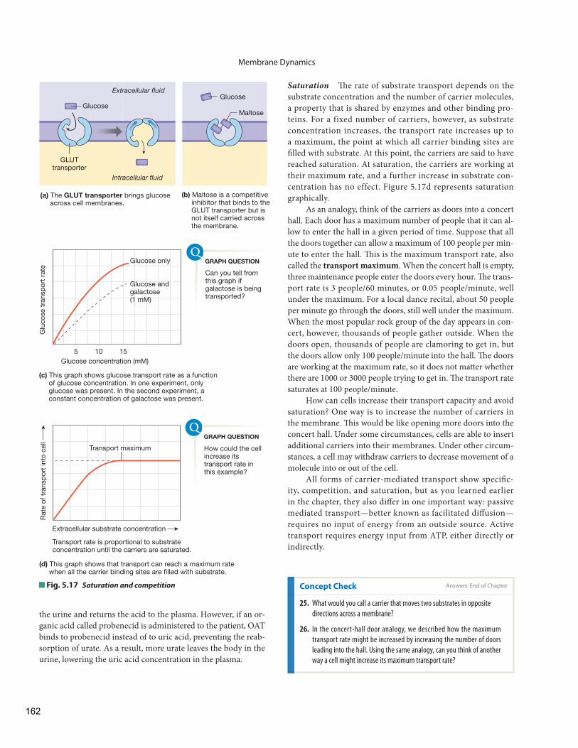
Membrane Dynamics
Saturation Th e rate of substrate transport depends on the substrate concentration and the number of carrier molecules, a property that is shared by enzymes and other binding pro-teins . For a fixed number of carriers, however, as substrate concentration increases, the transport rate increases up to a maximum, the point at which all carrier binding sites are fi lled with substrate. At this point, the carriers are said to have reached saturation. At saturation, the carriers are working at their maximum rate, and a further increase in substrate con-centration has no effect. Figure 5.17 d represents saturation graphically.
As an analogy, think of the carriers as doors into a concert hall. Each door has a maximum number of people that it can al-low to enter the hall in a given period of time. Suppose that all the doors together can allow a maximum of 100 people per min-ute to enter the hall. Th is is the maximum transport rate, also called the transport maximum . When the concert hall is empty, three maintenance people enter the doors every hour. Th e trans-port rate is 3 people/60 minutes, or 0.05 people/ minute, well under the maximum. For a local dance recital, about 50 people per minute go through the doors, still well under the maximum. When the most popular rock group of the day appears in con-cert, however, thousands of people gather outside. When the doors open, thousands of people are clamoring to get in, but the doors allow only 100 people/minute into the hall. Th e doors are working at the maximum rate, so it does not matter whether there are 1000 or 3000 people trying to get in. Th e transport rate saturates at 100 people/minute.
How can cells increase their transport capacity and avoid saturation? One way is to increase the number of carriers in the membrane. Th is would be like opening more doors into the concert hall. Under some circumstances, cells are able to insert additional carriers into their membranes. Under other circum-stances, a cell may withdraw carriers to decrease movement of a molecule into or out of the cell.
All forms of carrier-mediated transport show specifi c-ity, competition, and saturation, but as you learned earlier in the chapter, they also diff er in one important way: passive mediated transport—better known as facilitated diff usion—requires no input of energy from an outside source. Active transport requires energy input from ATP, either directly or indirectly.
(b) Maltose is a competitive inhibitor that binds to the GLUT transporter but is not itself carried across the membrane.
(a) The GLUT transporter brings glucose across cell membranes.
(c) This graph shows glucose transport rate as a function of glucose concentration. In one experiment, only glucose was present. In the second experiment, a constant concentration of galactose was present.
(d) This graph shows that transport can reach a maximum rate when all the carrier binding sites are filled with substrate.
GlucoseGlucose
Maltose
GLUTtransporter
Intracellular fluid
Extracellular fluid
Transport rate is proportional to substrate concentration until the carriers are saturated.
Rat
e of
tra
nsp
ort
into
cel
l
Extracellular substrate concentration
Transport maximum
Glu
cose
tra
nsp
ort
rate
Glucose concentration (mM)
Glucose only
Glucose andgalactose (1 mM)
5 10 15
Can you tell from this graph if galactose is being transported?
How could the cell increase its transport rate in this example?
GRAPH QUESTION
GRAPH QUESTION
Fig. 5.17 Saturation and competition
the urine and returns the acid to the plasma. However, if an or-ganic acid called probenecid is administered to the patient, OAT binds to probenecid instead of to uric acid, preventing the reab-sorption of urate. As a result, more urate leaves the body in the urine, lowering the uric acid concentration in the plasma.
Concept Check Answers: End of Chapter
25. What would you call a carrier that moves two substrates in opposite
directions across a membrane?
26. In the concert-hall door analogy, we described how the maximum
transport rate might be increased by increasing the number of doors
leading into the hall. Using the same analogy, can you think of another
way a cell might increase its maximum transport rate?
162
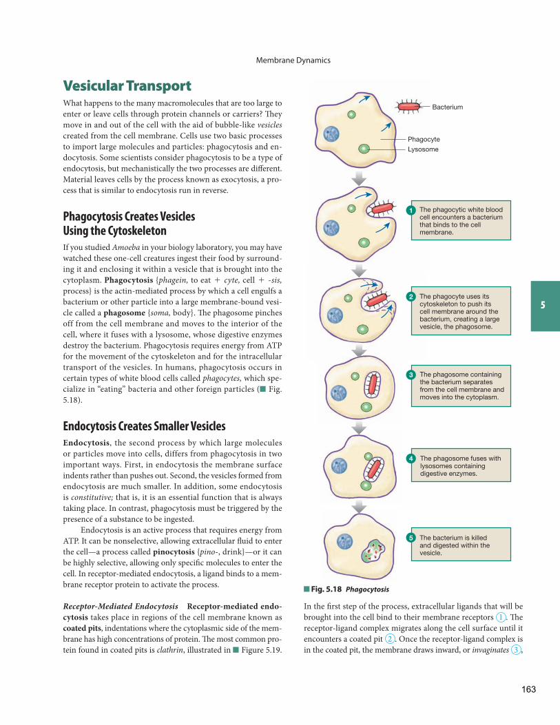
Membrane Dynamics
5
Vesicular Transport What happens to the many macromolecules that are too large to enter or leave cells through protein channels or carriers? Th ey move in and out of the cell with the aid of bubble-like vesicles created from the cell membrane. Cells use two basic processes to import large molecules and particles: phagocytosis and en-docytosis. Some scientists consider phagocytosis to be a type of endocytosis, but mechanistically the two processes are diff erent. Material leaves cells by the process known as exocytosis, a pro-cess that is similar to endocytosis run in reverse.
Phagocytosis Creates Vesicles Using the Cytoskeleton
If you studied Amoeba in your biology laboratory, you may have watched these one-cell creatures ingest their food by surround-ing it and enclosing it within a vesicle that is brought into the cytoplasm. Phagocytosis { phagein, to eat + cyte, cell + - sis, process} is the actin-mediated process by which a cell engulfs a bacterium or other particle into a large membrane-bound vesi-cle called a phagosome { soma, body}. Th e phagosome pinches off from the cell membrane and moves to the interior of the cell, where it fuses with a lysosome , whose digestive enzymes destroy the bacterium. Phagocytosis requires energy from ATP for the movement of the cytoskeleton and for the intracellular transport of the vesicles. In humans, phagocytosis occurs in certain types of white blood cells called phagocytes, which spe-cialize in “eating” bacteria and other foreign particles ( Fig. 5.18 ).
Endocytosis Creates Smaller Vesicles
Endocytosis , the second process by which large molecules or particles move into cells, differs from phagocytosis in two important ways. First, in endocytosis the membrane surface indents rather than pushes out. Second, the vesicles formed from endocytosis are much smaller. In addition, some endocytosis is constitutive; that is, it is an essential function that is always taking place. In contrast, phagocytosis must be triggered by the presence of a substance to be ingested.
Endocytosis is an active process that requires energy from ATP. It can be nonselective, allowing extracellular fl uid to enter the cell—a process called pinocytosis { pino -, drink}—or it can be highly selective, allowing only specifi c molecules to enter the cell. In receptor-mediated endocytosis, a ligand binds to a mem-brane receptor protein to activate the process.
Receptor-Mediated Endocytosis Receptor-mediated endo-cytosis takes place in regions of the cell membrane known as coated pits , indentations where the cytoplasmic side of the mem-brane has high concentrations of protein. Th e most common pro-tein found in coated pits is clathrin , illustrated in Figure 5.19 .
Bacterium
Lysosome
Phagocyte
The phagocytic white bloodcell encounters a bacterium that binds to the cell membrane.
The phagocyte uses itscytoskeleton to push itscell membrane around thebacterium, creating a largevesicle, the phagosome.
The phagosome containing the bacterium separates from the cell membrane andmoves into the cytoplasm.
The phagosome fuses with lysosomes containing digestive enzymes.
The bacterium is killedand digested within thevesicle.
1
2
3
4
5
Fig. 5.18 Phagocytosis
In the fi rst step of the process, extracellular ligands that will be brought into the cell bind to their membrane receptors 1 . Th e receptor-ligand complex migrates along the cell surface until it encounters a coated pit 2 . Once the receptor-ligand complex is in the coated pit, the membrane draws inward, or invaginates 3 ,
163
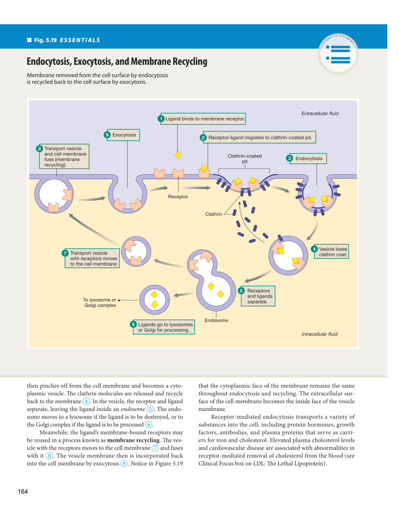
that the cytoplasmic face of the membrane remains the same throughout endocytosis and recycling. The extracellular sur-face of the cell membrane becomes the inside face of the vesicle membrane.
Receptor-mediated endocytosis transports a variety of substances into the cell, including protein hormones, growth factors, antibodies, and plasma proteins that serve as carri-ers for iron and cholesterol. Elevated plasma cholesterol levels and cardiovascular disease are associated with abnormalities in receptor-mediated removal of cholesterol from the blood (see Clinical Focus box on LDL: Th e Lethal Lipoprotein).
then pinches off from the cell membrane and becomes a cyto-plasmic vesicle. Th e clathrin molecules are released and recycle back to the membrane 4 . In the vesicle, the receptor and ligand separate, leaving the ligand inside an endosome 5 . The endo-some moves to a lysosome if the ligand is to be destroyed, or to the Golgi complex if the ligand is to be processed 6 .
Meanwhile, the ligand’s membrane-bound receptors may be reused in a process known as membrane recycling . Th e ves-icle with the receptors moves to the cell membrane 7 and fuses with it 8 . The vesicle membrane then is incorporated back into the cell membrane by exocytosis 9 . Notice in Figure 5.19
Fig. 5.19 E S S E N T I A L S
Membrane removed from the cell surface by endocytosis is recycled back to the cell surface by exocytosis.
Endocytosis, Exocytosis, and Membrane Recycling
Ligand binds to membrane receptor.
Clathrin-coatedpit
Receptor
Extracellular fluid
Intracellular fluid
To lysosome orGolgi complex
Receptor-ligand migrates to clathrin-coated pit.
Endocytosis
Vesicle losesclathrin coat.
Ligands go to lysosomesor Golgi for processing.
Transport vesicle with receptors moves to the cell membrane.
Transport vesicle and cell membranefuse (membranerecycling).
Exocytosis
Clathrin
Endosome
1
2
3
4
5
6
7
8
9
Receptors and ligands separate.
164
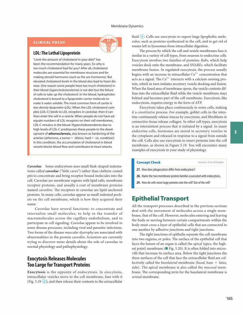
Membrane Dynamics
5
fl uid 9 . Cells use exocytosis to export large lipophobic mole-cules, such as proteins synthesized in the cell, and to get rid of wastes left in lysosomes from intracellular digestion.
Th e process by which the cell and vesicle membranes fuse is similar in a variety of cell types, from neurons to endocrine cells. Exocytosis involves two families of proteins: Rabs, which help vesicles dock onto the membrane, and SNAREs , which facilitate membrane fusion. In regulated exocytosis, the process usually begins with an increase in intracellular Ca2 + concentration that acts as a signal. Th e Ca2 + interacts with a calcium-sensing pro-tein, which in turn initiates secretory vesicle docking and fusion. When the fused area of membrane opens, the vesicle contents dif-fuse into the extracellular fl uid while the vesicle membrane stays behind and becomes part of the cell membrane. Exocytosis, like endocytosis, requires energy in the form of ATP.
Exocytosis takes place continuously in some cells, making it a constitutive process. For example, goblet cells in the intes-tine continuously release mucus by exocytosis, and fi broblasts in connective tissue release collagen. In other cell types, exocytosis is an intermittent process that is initiated by a signal. In many endocrine cells, hormones are stored in secretory vesicles in the cytoplasm and released in response to a signal from outside the cell. Cells also use exocytosis to insert proteins into the cell membrane, as shown in Figure 5.19 . You will encounter many examples of exocytosis in your study of physiology.
Caveolae Some endocytosis uses small fl ask-shaped indenta-tions called caveolae (“little caves”) rather than clathrin-coated pits to concentrate and bring receptor-bound molecules into the cell. Caveolae are membrane regions with lipid raft s , membrane receptor proteins, and usually a coat of membrane proteins named caveolins . Th e receptors in caveolae are lipid- anchored proteins . In many cells, caveolae appear as small indented pock-ets on the cell membrane, which is how they acquired their name.
Caveolae have several functions: to concentrate and internalize small molecules, to help in the transfer of macromolecules across the capillary endothelium, and to participate in cell signaling. Caveolae appear to be involved in some disease processes, including viral and parasitic infections. Two forms of the disease muscular dystrophy are associated with abnormalities in the protein caveolin. Scientists are currently trying to discover more details about the role of caveolae in normal physiology and pathophysiology.
Exocytosis Releases Molecules Too Large for Transport Proteins
Exocytosis is the opposite of endocytosis. In exocytosis, intracellular vesicles move to the cell membrane, fuse with it ( Fig. 5.19 8 ), and then release their contents to the extracellular
LDL: The Lethal Lipoprotein
“Limit the amount of cholesterol in your diet!” has been the recommendation for many years. So why is too much cholesterol bad for you? After all, cholesterol molecules are essential for membrane structure and for making steroid hormones (such as the sex hormones). But elevated cholesterol levels in the blood also lead to heart dis-ease. One reason some people have too much cholesterol in their blood ( hypercholesterolemia ) is not diet but the failure of cells to take up the cholesterol. In the blood, hydrophobic cholesterol is bound to a lipoprotein carrier molecule to make it water soluble. The most common form of carrier is low-density lipoprotein (LDL) . When the LDL-cholesterol com-plex (LDL-C) binds to LDL receptors in caveolae, then it can then enter the cell in a vesicle. When people do not have ad-equate numbers of LDL receptors on their cell membranes, LDL-C remains in the blood. Hypercholesterolemia due to high levels of LDL-C predisposes these people to the devel-opment of atherosclerosis, also known as hardening of the arteries { atheroma, a tumor + skleros, hard + - sis, condition}. In this condition, the accumulation of cholesterol in blood vessels blocks blood fl ow and contributes to heart attacks.
C L I N I C A L F O C U S
Concept Check Answers: End of Chapter
27. How does phagocytosis diff er from endocytosis?
28. Name the two membrane protein families associated with endocytosis.
29. How do cells move large proteins into the cell? Out of the cell?
Epithelial Transport All the transport processes described in the previous sections deal with the movement of molecules across a single mem-brane, that of the cell. However, molecules entering and leaving the body or moving between certain compartments within the body must cross a layer of epithelial cells that are connected to one another by adhesive junctions and tight junctions .
Th e tight junctions of epithelia separate the cell membrane into two regions, or poles. Th e surface of the epithelial cell that faces the lumen of an organ is called the apical { apex, the high-est point} membrane ( Fig. 5.20 ). It is oft en folded into micro-villi that increase its surface area. Below the tight junctions the three surfaces of the cell that face the extracellular fl uid are col-lectively called the basolateral membrane { basal, base + latus, side}. The apical membrane is also called the mucosal mem-brane. Th e corresponding term for the basolateral membrane is serosal membrane.
165
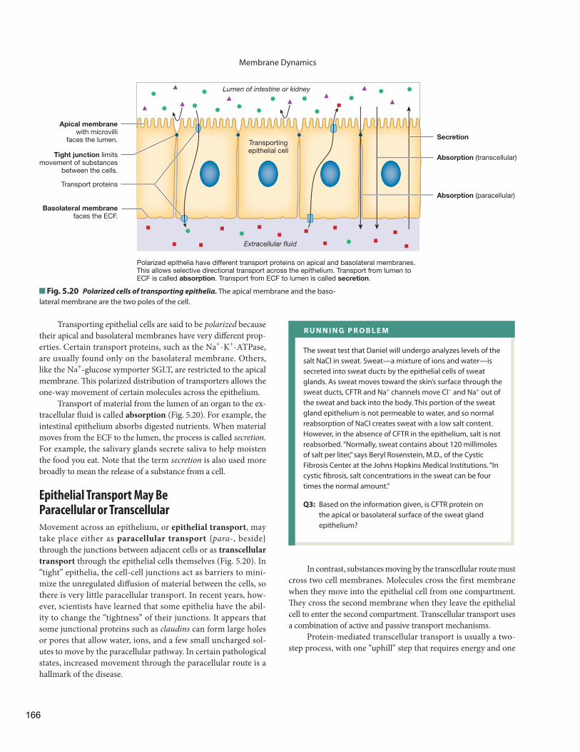
Membrane Dynamics
In contrast, substances moving by the transcellular route must cross two cell membranes. Molecules cross the first membrane when they move into the epithelial cell from one compartment. Th ey cross the second membrane when they leave the epithelial cell to enter the second compartment. Transcellular transport uses a combination of active and passive transport mechanisms.
Protein-mediated transcellular transport is usually a two-step process, with one “uphill” step that requires energy and one
Transporting epithelial cells are said to be polarized because their apical and basolateral membranes have very diff erent prop-erties. Certain transport proteins, such as the Na+ - K+ -ATPase, are usually found only on the basolateral membrane. Others, like the Na+ -glucose symporter SGLT, are restricted to the apical membrane. Th is polarized distribution of transporters allows the one-way movement of certain molecules across the epithelium.
Transport of material from the lumen of an organ to the ex-tracellular fl uid is called absorption ( Fig. 5.20 ). For example, the intestinal epithelium absorbs digested nutrients. When material moves from the ECF to the lumen, the process is called secretion . For example, the salivary glands secrete saliva to help moisten the food you eat. Note that the term secretion is also used more broadly to mean the release of a substance from a cell.
Epithelial Transport May Be Paracellular or Transcellular
Movement across an epithelium, or epithelial transport , may take place either as paracellular transport { para- , beside} through the junctions between adjacent cells or as transcellular transport through the epithelial cells themselves ( Fig. 5.20 ). In “tight” epithelia, the cell-cell junctions act as barriers to mini-mize the unregulated diff usion of material between the cells, so there is very little paracellular transport. In recent years, how-ever, scientists have learned that some epithelia have the abil-ity to change the “tightness” of their junctions. It appears that some junctional proteins such as claudins can form large holes or pores that allow water, ions, and a few small uncharged sol-utes to move by the paracellular pathway. In certain pathological states, increased movement through the paracellular route is a hallmark of the disease.
Apical membranewith microvilli
faces the lumen.
Tight junction limitsmovement of substances
between the cells.
Transportingepithelial cell
Basolateral membranefaces the ECF.
Lumen of intestine or kidney
Extracellular fluid
Transport proteins
Absorption (transcellular)
Secretion
Absorption (paracellular)
Polarized epithelia have different transport proteins on apical and basolateral membranes. This allows selective directional transport across the epithelium. Transport from lumen to ECF is called absorption. Transport from ECF to lumen is called secretion.
Fig. 5.20 Polarized cells of transporting epithelia. The apical membrane and the baso-lateral membrane are the two poles of the cell.
R U N N I N G P R O B L E M
The sweat test that Daniel will undergo analyzes levels of the salt NaCl in sweat. Sweat—a mixture of ions and water—is secreted into sweat ducts by the epithelial cells of sweat glands. As sweat moves toward the skin’s surface through the sweat ducts, CFTR and Na+ channels move Cl- and Na+ out of the sweat and back into the body. This portion of the sweat gland epithelium is not permeable to water, and so normal reabsorption of NaCl creates sweat with a low salt content. However, in the absence of CFTR in the epithelium, salt is not reabsorbed. “Normally, sweat contains about 120 millimoles of salt per liter,” says Beryl Rosenstein, M.D., of the Cystic Fibrosis Center at the Johns Hopkins Medical Institutions. “In cystic fi brosis, salt concentrations in the sweat can be four times the normal amount.”
Q3: Based on the information given, is CFTR protein on the apical or basolateral surface of the sweat gland epithelium?
166
Membrane Dynamics
5
systems: the SGLT-mediated secondary active transport of glucose with Na+ from the lumen into the epithelial cell at the apical mem-brane, followed by the movement of Na+ and glucose out of the cell and into the extracellular fl uid on separate transporters. Sodium moves out by primary active transport via a Na+ - K+ -ATPase, and glucose leaves the cell by facilitated diff usion on GLUT carriers.
Figure 5.21 shows the process in detail. Th e glucose con-centration in the transporting epithelial cell is higher than the glucose concentration in either the extracellular fl uid or the lu-men of the kidney or intestine. For this reason, moving glucose from the lumen into the cell requires the input of energy—in this case, energy stored in the Na+ concentration gradient. So-dium ions in the lumen bind to the SGLT carrier, as previously described (see Fig. 5.16 ), and bring glucose with them into the cell. Th e energy needed to move glucose against its concentra-tion gradient comes from the kinetic energy of Na+ moving down its concentration gradient ( Fig. 5.21 1 ).
Once glucose is in the epithelial cell, it leaves by mov-ing down its concentration gradient on the facilitated diff usion GLUT transporter in the basolateral membrane ( Fig. 5.21 2 ). Na+ is pumped out of the cell on the basolateral side using
“downhill” step in which the molecule moves passively down its gradient. You will see these steps in the example of glucose trans-port that follows. Molecules that are too large to be moved by membrane proteins can be transported across the cell in vesicles.
Th e cells of transporting epithelia can alter their permeabil-ity by selectively inserting or withdrawing membrane proteins. Transporters pulled out of the membrane may be destroyed in lysosomes, or they may be stored in vesicles inside the cell, ready to be reinserted into the membrane in response to a sig-nal (another example of membrane recycling). Most epithelial transport you will study in this course involves the transporting epithelia of intestine and kidney, which are specialized to selec-tively transport molecules into and out of the body.
Transcellular Transport of Glucose Uses Membrane Proteins
Th e absorption of glucose from the lumen of the kidney tubule or intestine to the extracellular fl uid is an important example of directional movement across a transporting epithelium. Transepithelial movement of glucose involves three transport
[Glucose]low
[Glucose]high
[Glucose]low
[Na+]high
[Na+]low
[Na+]high
Apicalmembrane
Glu
Glu
Glu
Na+
Na+
Na+
Na+
K+
K+
Basolateralmembrane
Extracellularfluid
Lumen of kidneyor intestine
Glu
GLUT transporter transfers glucose to ECF by facilitated diffusion.
Na+-K+-ATPase pumps Na+ out of the cell, keepingICF Na+ concentration low.
Na+- glucose symporter brings glucose into cell against its gradient using energy stored in the Na+ concentration gradient.
ATP
Epithelialcell
• Match each transporter to its location. 1. GLUT (a) apical membrane 2. Na+-glucose (b) basolateral membrane symporter 3. Na+-K+-ATPase• Is glucose movement across the basolateral membrane active or passive? Explain.• Why doesn't Na+ movement at the apical membrane require ATP?
1
1
2
2
3
3
FIGURE QUESTIONS
Fig. 5.21 Transepithelial transport of glucose. This process involves indirect (second-ary) active transport of glucose across the apical membrane and glucose diff usion across the basolateral membrane.
167
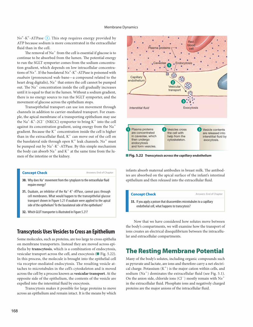
Membrane Dynamics
infants absorb maternal antibodies in breast milk. Th e antibod-ies are absorbed on the apical surface of the infant’s intestinal epithelium and then released into the extracellular fl uid.
Na+ - K+ -ATPase 3 . This step requires energy provided by ATP because sodium is more concentrated in the extracellular fl uid than in the cell.
Th e removal of Na+ from the cell is essential if glucose is to continue to be absorbed from the lumen. Th e potential energy to run the SGLT symporter comes from the sodium concentra-tion gradient, which depends on low intracellular concentra-tions of Na+ . If the basolateral Na+ - K+ -ATPase is poisoned with ouabain (pronounced wah-bane—a compound related to the heart drug digitalis), Na+ that enters the cell cannot be pumped out. Th e Na+ concentration inside the cell gradually increases until it is equal to that in the lumen. Without a sodium gradient, there is no energy source to run the SGLT symporter, and the movement of glucose across the epithelium stops.
Transepithelial transport can use ion movement through channels in addition to carrier-mediated transport. For exam-ple, the apical membrane of a transporting epithelium may use the Na+ - K+ - 2Cl- (NKCC) symporter to bring K+ into the cell against its concentration gradient, using energy from the Na+
gradient. Because the K+ concentration inside the cell is higher than in the extracellular fl uid, K+ can move out of the cell on the basolateral side through open K+ leak channels. Na+ must be pumped out by Na+ - K+ -ATPase. By this simple mechanism the body can absorb Na+ and K+ at the same time from the lu-men of the intestine or the kidney.
Concept Check Answers: End of Chapter
30. Why does Na+ movement from the cytoplasm to the extracellular fl uid
require energy?
31. Ouabain, an inhibitor of the Na+ - K+ -ATPase, cannot pass through
cell membranes. What would happen to the transepithelial glucose
transport shown in Figure 5.21 if ouabain were applied to the apical
side of the epithelium? To the basolateral side of the epithelium?
32. Which GLUT transporter is illustrated in Figure 5.21 ?
Transcytosis Uses Vesicles to Cross an Epithelium
Some molecules, such as proteins, are too large to cross epithelia on membrane transporters. Instead they are moved across epi-thelia by transcytosis , which is a combination of endocytosis, vesicular transport across the cell, and exocytosis ( Fig. 5.22 ). In this process, the molecule is brought into the epithelial cell via receptor-mediated endocytosis. The resulting vesicle at-taches to microtubules in the cell’s cytoskeleton and is moved across the cell by a process known as vesicular transport . At the opposite side of the epithelium, the contents of the vesicle are expelled into the interstitial fl uid by exocytosis.
Transcytosis makes it possible for large proteins to move across an epithelium and remain intact. It is the means by which
Fig. 5.22 Transcytosis across the capillary endothelium
Plasma proteins
Red bloodcell
Endocytosis
Vesiculartransport
Exocytosis
Caveolae
Interstitial fluid
1
2
3
Capillaryendothelium
1 2 3Plasma proteins are concentrated in caveolae, which then undergo endocytosis and form vesicles.
Vesicles cross the cell with help from the cytoskeleton.
Vesicle contents are released intointerstitial fluid by exocytosis.
Concept Check
33. If you apply a poison that disassembles microtubules to a capillary
endothelial cell, what happens to transcytosis?
Now that we have considered how solutes move between the body’s compartments, we will examine how the transport of ions creates an electrical disequilibrium between the intracellu-lar and extracellular compartments.
The Resting Membrane Potential Many of the body’s solutes, including organic compounds such as pyruvate and lactate, are ions and therefore carry a net electri-cal charge. Potassium (K+) is the major cation within cells, and sodium (Na+) dominates the extracellular fl uid (see Fig. 5.1 ). On the anion side, chloride ions (Cl-) mostly remain with Na+ in the extracellular fl uid. Phosphate ions and negatively charged proteins are the major anions of the intracellular fl uid.
Answers: End of Chapter
168
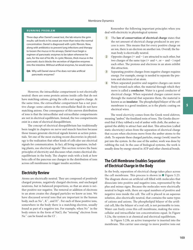
Membrane Dynamics
5
Remember the following important principles when you deal with electricity in physiological systems:
1 The law of conservation of electrical charge states that the net amount of electrical charge produced in any pro-cess is zero. Th is means that for every positive charge on an ion, there is an electron on another ion. Overall, the hu-man body is electrically neutral.
2 Opposite charges (+ and - ) are attracted to each other, but two charges of the same type ( + and + , or - and - ) repel each other. Th e protons and electrons in an atom exhibit this attraction.
3 Separating positive charges from negative charges requires energy. For example, energy is needed to separate the pro-tons and electrons of an atom.
4 When separated positive and negative charges can move freely toward each other, the material through which they move is called a conductor . Water is a good conductor of electrical charge. When separated charges cannot move through the material that separates them, the material is known as an insulator . Th e phospholipid bilayer of the cell membrane is a good insulator, as is the plastic coating on electrical wires.
Th e word electricity comes from the Greek word elektron, meaning “amber,” the fossilized resin of trees. Th e Greeks discov-ered that if they rubbed a rod of amber with cloth, the amber ac-quired the ability to attract hair and dust. Th is attraction (called static electricity) arises from the separation of electrical charge that occurs when electrons move from the amber atoms to the cloth. To separate these charged particles, energy (work) must be put into the system. In the case of the amber, work was done by rubbing the rod. In the case of biological systems, the work is usually done by energy stored in ATP and other chemical bonds.
The Cell Membrane Enables Separation of Electrical Charge in the Body
In the body, separation of electrical charge takes place across the cell membrane. Th is process is shown in Figure 5.23 . Th e diagram shows an artifi cial cell fi lled with molecules that dissociate into positive and negative ions, represented by the plus and minus signs. Because the molecules were electrically neutral to begin with, there are equal numbers of positive and negative ions inside the cell. Th e cell is placed in an aqueous solution, also electrically neutral, that contains the same types of cations and anions. Th e phospholipid bilayer of the artifi -cial cell, like the bilayer of a real cell, is not permeable to ions. Water can freely cross this cell membrane, making the extra-cellular and intracellular ion concentrations equal. In Figure 5.23 a, the system is at chemical and electrical equilibrium.
In Figure 5.23 b, an active transporter is inserted into the membrane. Th is carrier uses energy to move positive ions out
However, the intracellular compartment is not electrically neutral: there are some protein anions inside cells that do not have matching cations, giving the cells a net negative charge. At the same time, the extracellular compartment has a net posi-tive charge: some cations in the extracellular fl uid do not have matching anions. One consequence of this uneven distribution of ions is that the intracellular and extracellular compartments are not in electrical equilibrium. Instead, the two compartments exist in a state of electrical disequilibrium .
Th e concept of electrical disequilibrium has traditionally been taught in chapters on nerve and muscle function because those tissues generate electrical signals known as action poten-tials. Yet one of the most exciting recent discoveries in physiol-ogy is the realization that other kinds of cells also use electrical signals for communication. In fact, all living organisms, includ-ing plants, use electrical signals! Th is section reviews the basic principles of electricity and discusses what creates electrical dis-equilibrium in the body. Th e chapter ends with a look at how beta cells of the pancreas use changes in the distribution of ions across cell membranes to trigger insulin secretion.
Electricity Review
Atoms are electrically neutral . Th ey are composed of positively charged protons, negatively charged electrons, and uncharged neutrons, but in balanced proportions, so that an atom is nei-ther positive nor negative. Th e removal or addition of electrons to an atom creates the charged particles we know as ions. We have discussed several ions that are important in the human body, such as Na+ , K+ , and H+ . For each of these positive ions, somewhere in the body there is a matching electron, usually found as part of a negative ion. For example, when Na+ in the body enters in the form of NaCl, the “missing” electron from Na+ can be found on the Cl- .
R U N N I N G P R O B L E M
Three days after Daniel’s sweat test, the lab returns the grim results: salt levels in his sweat are more than twice the normal concentration. Daniel is diagnosed with cystic fi brosis. Now, along with antibiotics to prevent lung infections and therapy to loosen the mucus in his airways, Daniel must begin a regimen of pancreatic enzymes to be taken whenever he eats, for the rest of his life. In cystic fi brosis, thick mucus in the pancreatic ducts blocks the secretion of digestive enzymes into the intestine. Without artifi cial enzymes, he would starve.
Q4: Why will Daniel starve if he does not take artifi cial pancreatic enzymes?
169
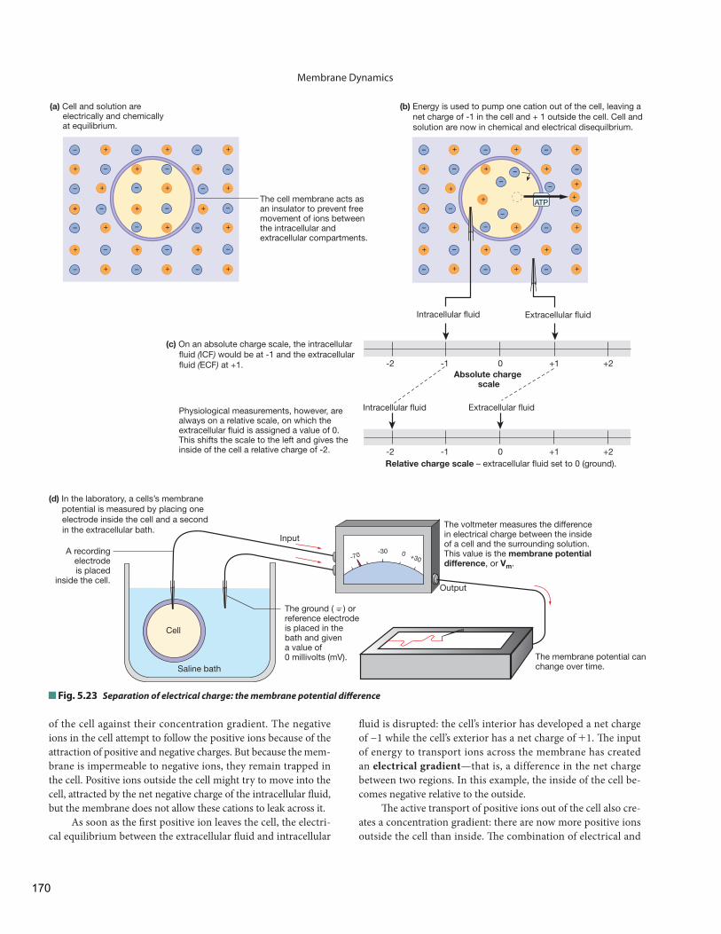
Membrane Dynamics
+–+– +–
+ –+ + –
+–+– +–
+–+– +–
+–+– +–
–
+ –+ + ––
+ –+ + ––
+–+– +–
+ –+ + –
–+–
+
+
+–+
+
– +–
–
–– –
–
+
+
–+ +
+
––
+
– –+ –
-2 -1 0 +1 +2
Intracellular fluid Extracellular fluid
Absolute charge scale
Intracellular fluid Extracellular fluid
-2 -1 0 +1 +2Relative charge scale – extracellular fluid set to 0 (ground).
– –
The voltmeter measures the difference in electrical charge between the inside of a cell and the surrounding solution. This value is the membrane potential difference, or Vm.
Cell
Saline bath
A recordingelectrodeis placed
inside the cell.
Input
The ground ( ) or reference electrode is placed in the bath and given a value of 0 millivolts (mV).
Output
-70 -30 0 +30
ATP
(b) Energy is used to pump one cation out of the cell, leaving a net charge of -1 in the cell and + 1 outside the cell. Cell and solution are now in chemical and electrical disequilbrium.
(c) On an absolute charge scale, the intracellular fluid (ICF) would be at -1 and the extracellular fluid (ECF) at +1.
(d) In the laboratory, a cells’s membrane potential is measured by placing one electrode inside the cell and a second in the extracellular bath.
(a) Cell and solution are electrically and chemically at equilibrium.
The cell membrane acts as an insulator to prevent free movement of ions between the intracellular and extracellular compartments.
Physiological measurements, however, are always on a relative scale, on which the extracellular fluid is assigned a value of 0. This shifts the scale to the left and gives the inside of the cell a relative charge of -2.
The membrane potential can change over time.
Fig. 5.23 Separation of electrical charge: the membrane potential diff erence
of the cell against their concentration gradient. The negative ions in the cell attempt to follow the positive ions because of the attraction of positive and negative charges. But because the mem-brane is impermeable to negative ions, they remain trapped in the cell. Positive ions outside the cell might try to move into the cell, attracted by the net negative charge of the intracellular fl uid, but the membrane does not allow these cations to leak across it.
As soon as the fi rst positive ion leaves the cell, the electri-cal equilibrium between the extracellular fl uid and intracellular
fl uid is disrupted: the cell’s interior has developed a net charge of −1 while the cell’s exterior has a net charge of +1. Th e input of energy to transport ions across the membrane has created an electrical gradient —that is, a difference in the net charge between two regions. In this example, the inside of the cell be-comes negative relative to the outside.
Th e active transport of positive ions out of the cell also cre-ates a concentration gradient: there are now more positive ions outside the cell than inside. Th e combination of electrical and
170
Membrane Dynamics
5
When the recording electrode is placed inside a living cell, the voltmeter measures the membrane potential—in other words, the electrical diff erence between the intracellular fl uid and the extra-cellular fl uid. A recorder connected to the voltmeter can make a recording of the membrane potential versus time.
For nerve and muscle cells, the voltmeter will record a rest-ing membrane potential between −40 and −90 mV, indicating that the intracellular fl uid is negative relative to the extracellular fl uid (0 mV). (Th roughout this discussion, remember that the extracellular fl uid is not really neutral because it has excess posi-tive charges that exactly balance the excess negative charges in-side the cell, as shown in Figure 5.23 c. Th e total body remains electrically neutral at all times.)
The Resting Membrane Potential Is Due Mostly to Potassium
Which ions create the resting membrane potential in animal cells? The artificial cell shown in Figure 5.23 b used an active transport protein to move an unspecifi ed positive ion across a membrane that was otherwise impermeable to ions. But what processes go on in living cells to create an electrical gradient?
Real cells are not completely impermeable to all ions. Th ey have open channels and protein transporters that allow ions to move between the cytoplasm and the extracellular fl uid. We can use a diff erent artifi cial cell to show how the resting membrane potential arises in a typical living cell.
The artificial cell in Figure 5.24 has a membrane that is impermeable to ions. The cell contains K+ and large nega-tively charged proteins, represented by Pr- . Th e cell is placed in a solution of Na+ and Cl- . Both the cell and the solution are electrically neutral, and the system is in electrical equilibrium. However, it is not in chemical equilibrium. Th ere are concentra-tion gradients for all four types of ions in the system, and they would all diff use down their respective concentration gradients if they could cross the cell membrane.
In Figure 5.24 b, a K+ leak channel is inserted into the membrane, making it permeable only to K+ . Because there is no K+ in the extracellular fl uid initially, some K+ ions leak out of the cell, moving down their concentration gradient. As K+ leaves the cell, the negatively charged proteins, Pr- , are unable to follow because the cell membrane is not permeable to them. Th e proteins gradually build up a negative charge inside the cell as more and more K+ diff uses out of the cell.
If the only force acting on K+ were the concentration gra-dient, K+ would leak out of the cell until the K+ concentration inside the cell equaled the K+ concentration outside. Th e loss of positive ions from the cell creates an electrical gradient, however. Because opposite charges attract each other, the negative pro-teins inside the cell try to pull K+ back into the cell. At some point in this process, the electrical force attracting K+ into the cell becomes equal in magnitude to the chemical concentration
concentration gradients is called an electrochemical gradient . Th e cell remains in osmotic equilibrium because water can move freely across the membrane in response to solute movement.
An electrical gradient between the extracellular fl uid and the intracellular fl uid is known as the resting membrane poten-tial diff erence, or membrane potential for short. Although the name sounds intimidating, we can break it apart to see what it means.
1 Th e resting part of the name comes from the fact that this electrical gradient is seen in all living cells, even those that appear to be without electrical activity. In these “resting” cells, the membrane potential has reached a steady state and is not changing.
2 The potential part of the name comes from the fact that the electrical gradient created by active transport of ions across the cell membrane is a form of stored, or potential, energy, just as concentration gradients are a form of po-tential energy. When oppositely charged molecules come back together, they release energy that can be used to do work, in the same way that molecules moving down their concentration gradient can do work. The work done by electrical energy includes opening voltage-gated mem-brane channels and sending electrical signals.
3 Th e diff erence part of the name is to remind you that the membrane potential represents a diff erence in the amount of electrical charge inside and outside the cell. Th e word dif-ference is usually dropped from the name, as noted earlier.
In living systems, we measure electrical gradients on a rela-tive scale rather than an absolute scale. Figure 5.23 c compares the two scales. On the absolute scale, the extracellular fl uid in our simple example has a net charge of +1 from the positive ion it gained, and the intracellular fl uid has a net charge of −1 from the now unbalanced negative ion that was left behind.
However, in real life we cannot measure electrical charge as numbers of electrons gained or lost. Instead we use a device that measures the diff erence in electrical charge between two points. Th is device artifi cially sets the net electrical charge of one side of the membrane to 0 and measures the net charge of the second side relative to the fi rst. In our example, resetting the extracel-lular fl uid net charge to 0 gives the intracellular fl uid a net charge of −2, and we call this value the cell’s resting membrane potential.
Th e equipment for measuring a cell’s membrane potential is depicted in Figure 5.23 . Electrodes are created from hollow glass tubes drawn to very fine points. These micropipettes are fi lled with a liquid that conducts electricity and then connected to a voltmeter, which measures the electrical diff erence between two points in units of either volts (V) or millivolts (mV). A re-cording electrode is inserted through the cell membrane into the cytoplasm of the cell. A reference electrode is placed in the exter-nal bath, which represents the extracellular fl uid.
In living systems, by convention, the extracellular fl uid is des-ignated as the ground and assigned a charge of 0 mV ( Fig. 5.23 c).
171
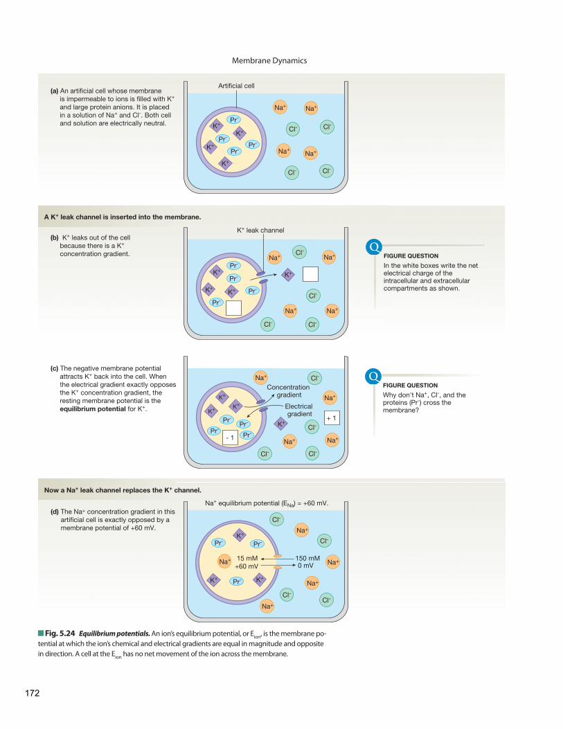
Membrane Dynamics
Cl-Cl-
Artificial cell
Cl-
Cl-
Cl-
Cl-
Cl-
Cl-
Cl-
Cl-
K+
K+
K+
K+
Pr-
Pr-
Pr-
K+
K+K+
K+Pr-
Pr-
Pr-
Pr-
Pr-
Na+Na+
Cl-Cl-
Na+Na+
Na+ Na+
Na+Na+
K+ K+
K+
K+Concentration
gradient
Electrical gradient
Pr-
Pr-Pr-
Pr-
Na+
Na+
Na+
Na+
- 1
+ 1
(a) An artificial cell whose membrane is impermeable to ions is filled with K+
and large protein anions. It is placed in a solution of Na+ and Cl-. Both cell and solution are electrically neutral.
(b) K+ leaks out of the cell because there is a K+ concentration gradient.
(c) The negative membrane potential attracts K+ back into the cell. When the electrical gradient exactly opposes the K+ concentration gradient, the resting membrane potential is the equilibrium potential for K+.
K+ leak channel
150 mM15 mM0 mV+60 mV
Cl-
Cl-
Cl-Cl-
Na+
Na+
Na+
Na+
Na+
K+
Pr-Pr-
Pr-
Na+ equilibrium potential (ENa) = +60 mV.(d) The Na+ concentration gradient in this
artificial cell is exactly opposed by a membrane potential of +60 mV.
A K+ leak channel is inserted into the membrane.
Now a Na+ leak channel replaces the K+ channel.
K+K+
In the white boxes write the net electrical charge of the intracellular and extracellular compartments as shown.
FIGURE QUESTION
Why don't Na+, Cl-, and the proteins (Pr-) cross the membrane?
FIGURE QUESTION
Fig. 5.24 Equilibrium potentials. An ion’s equilibrium potential, or E ion , is the membrane po-tential at which the ion’s chemical and electrical gradients are equal in magnitude and opposite in direction. A cell at the E ion has no net movement of the ion across the membrane.
172
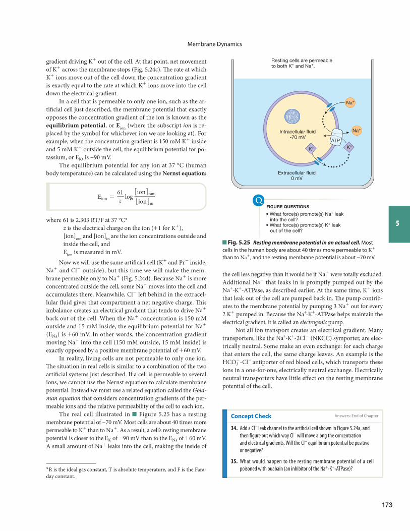
Membrane Dynamics
5
the cell less negative than it would be if Na+ were totally excluded. Additional Na+ that leaks in is promptly pumped out by the Na+ - K+ -ATPase, as described earlier. At the same time, K+ ions that leak out of the cell are pumped back in. Th e pump contrib-utes to the membrane potential by pumping 3 Na+ out for every 2 K+ pumped in. Because the Na+ - K+ -ATPase helps maintain the electrical gradient, it is called an electrogenic pump.
Not all ion transport creates an electrical gradient. Many transporters, like the Na+ - K+ - 2Cl- (NKCC) symporter, are elec-trically neutral. Some make an even exchange: for each charge that enters the cell, the same charge leaves. An example is the HCO3
- - Cl- antiporter of red blood cells, which transports these ions in a one-for-one, electrically neutral exchange. Electrically neutral transporters have little eff ect on the resting membrane potential of the cell.
gradient driving K+ out of the cell. At that point, net movement of K+ across the membrane stops ( Fig. 5.24 c). Th e rate at which K+ ions move out of the cell down the concentration gradient is exactly equal to the rate at which K+ ions move into the cell down the electrical gradient.
In a cell that is permeable to only one ion, such as the ar-tifi cial cell just described, the membrane potential that exactly opposes the concentration gradient of the ion is known as the equilibrium potential , or E ion (where the subscript ion is re-placed by the symbol for whichever ion we are looking at). For example, when the concentration gradient is 150 mM K+ inside and 5 mM K+ outside the cell, the equilibrium potential for po-tassium, or EK, is −90 mV.
The equilibrium potential for any ion at 37 °C (human body temperature) can be calculated using the Nernst equation:
Eion =61z
log 3ion4out
3ion4in
where 61 is 2.303 RT/F at 37 °C * z is the electrical charge on the ion ( +1 for K+ ), [ion] out and [ion] in are the ion concentrations outside and inside the cell, and E ion is measured in mV.
Now we will use the same artifi cial cell ( K+ and Pr- inside, Na+ and Cl- outside), but this time we will make the mem-brane permeable only to Na+ ( Fig. 5.24 d). Because Na+ is more concentrated outside the cell, some Na+ moves into the cell and accumulates there. Meanwhile, Cl- left behind in the extracel-lular fl uid gives that compartment a net negative charge. Th is imbalance creates an electrical gradient that tends to drive Na+
back out of the cell. When the Na+ concentration is 150 mM outside and 15 mM inside, the equilibrium potential for Na+
(ENa) is +60 mV. In other words, the concentration gradient moving Na+ into the cell (150 mM outside, 15 mM inside) is exactly opposed by a positive membrane potential of +60 mV.
In reality, living cells are not permeable to only one ion. Th e situation in real cells is similar to a combination of the two artifi cial systems just described. If a cell is permeable to several ions, we cannot use the Nernst equation to calculate membrane potential. Instead we must use a related equation called the Gold-man equation that considers concentration gradients of the per-meable ions and the relative permeability of the cell to each ion.
The real cell illustrated in Figure 5.25 has a resting membrane potential of −70 mV. Most cells are about 40 times more permeable to K+ than to Na+ . As a result, a cell’s resting membrane potential is closer to the EK of -90 mV than to the ENa of +60 mV. A small amount of Na+ leaks into the cell, making the inside of
* R is the ideal gas constant, T is absolute temperature, and F is the Fara-day constant.
Extracellular fluid0 mV
• What force(s) promote(s) Na+ leak into the cell?• What force(s) promote(s) K+ leak out of the cell?
Intracellular fluid-70 mV
Na+
Na+
K+K+
ATP
FIGURE QUESTIONS
Resting cells are permeableto both K+ and Na+.
Fig. 5.25 Resting membrane potential in an actual cell. Most cells in the human body are about 40 times more permeable to K+ than to Na+ , and the resting membrane potential is about −70 mV.
Concept Check Answers: End of Chapter
34. Add a Cl- leak channel to the artifi cial cell shown in Figure 5.24 a, and
then fi gure out which way Cl- will move along the concentration
and electrical gradients. Will the Cl- equilibrium potential be positive
or negative?
35. What would happen to the resting membrane potential of a cell
poisoned with ouabain (an inhibitor of the Na+ - K+ -ATPase)?
173
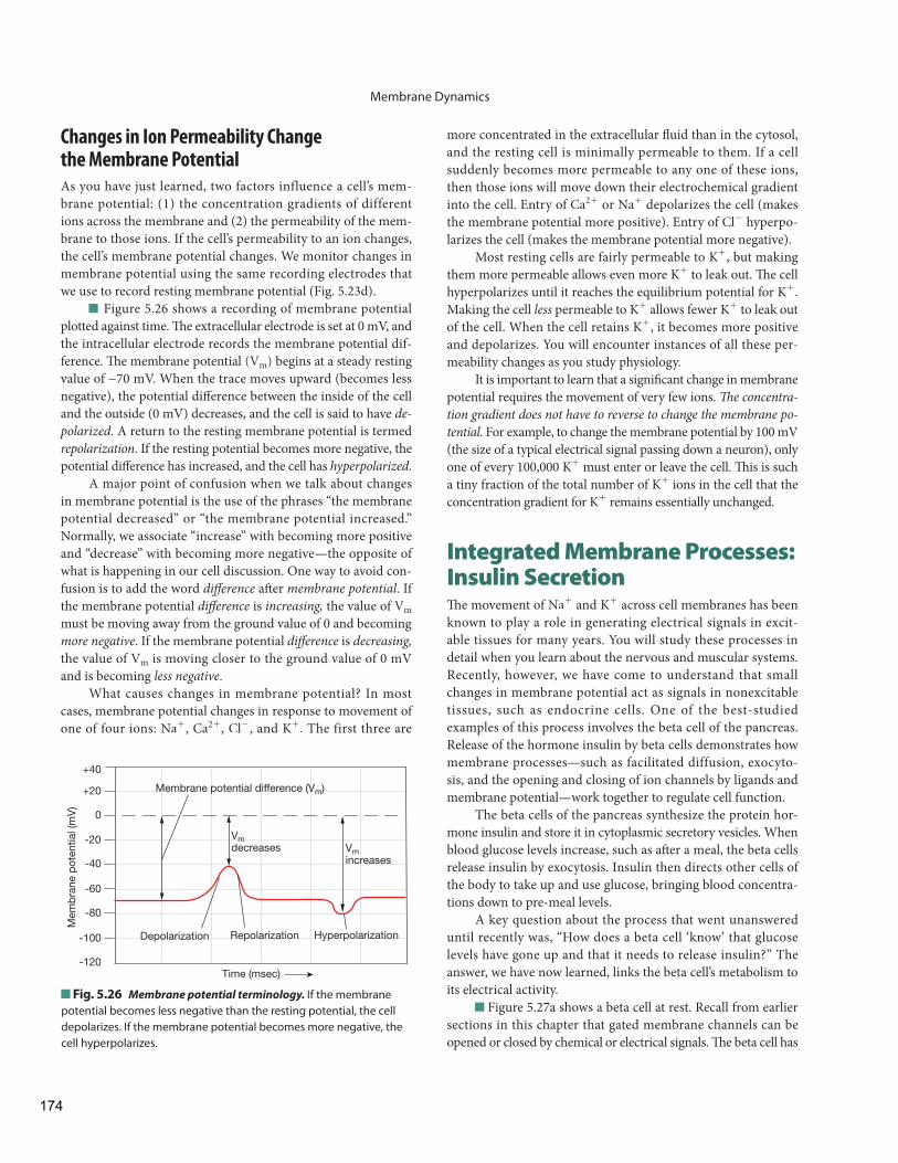
Membrane Dynamics
more concentrated in the extracellular fl uid than in the cytosol, and the resting cell is minimally permeable to them. If a cell suddenly becomes more permeable to any one of these ions, then those ions will move down their electrochemical gradient into the cell. Entry of Ca2 + or Na+ depolarizes the cell (makes the membrane potential more positive). Entry of Cl- hyperpo-larizes the cell (makes the membrane potential more negative).
Most resting cells are fairly permeable to K+ , but making them more permeable allows even more K+ to leak out. Th e cell hyperpolarizes until it reaches the equilibrium potential for K+ . Making the cell less permeable to K+ allows fewer K+ to leak out of the cell. When the cell retains K+ , it becomes more positive and depolarizes. You will encounter instances of all these per-meability changes as you study physiology.
It is important to learn that a signifi cant change in membrane potential requires the movement of very few ions. Th e concentra-tion gradient does not have to reverse to change the membrane po-tential. For example, to change the membrane potential by 100 mV (the size of a typical electrical signal passing down a neuron), only one of every 100,000 K+ must enter or leave the cell. Th is is such a tiny fraction of the total number of K+ ions in the cell that the concentration gradient for K+ remains essentially unchanged.
Integrated Membrane Processes: Insulin Secretion Th e movement of Na+ and K+ across cell membranes has been known to play a role in generating electrical signals in excit-able tissues for many years. You will study these processes in detail when you learn about the nervous and muscular systems. Recently, however, we have come to understand that small changes in membrane potential act as signals in nonexcitable tissues, such as endocrine cells. One of the best-studied examples of this process involves the beta cell of the pancreas. Release of the hormone insulin by beta cells demonstrates how membrane processes—such as facilitated diffusion, exocyto-sis, and the opening and closing of ion channels by ligands and membrane potential—work together to regulate cell function.
The beta cells of the pancreas synthesize the protein hor-mone insulin and store it in cytoplasmic secretory vesicles. When blood glucose levels increase, such as aft er a meal, the beta cells release insulin by exocytosis. Insulin then directs other cells of the body to take up and use glucose, bringing blood concentra-tions down to pre-meal levels.
A key question about the process that went unanswered until recently was, “How does a beta cell ‘know’ that glucose levels have gone up and that it needs to release insulin?” The answer, we have now learned, links the beta cell’s metabolism to its electrical activity.
Figure 5.27 a shows a beta cell at rest. Recall from earlier sections in this chapter that gated membrane channels can be opened or closed by chemical or electrical signals. Th e beta cell has
Changes in Ion Permeability Change the Membrane Potential
As you have just learned, two factors influence a cell’s mem-brane potential: (1) the concentration gradients of different ions across the membrane and (2) the permeability of the mem-brane to those ions. If the cell’s permeability to an ion changes, the cell’s membrane potential changes. We monitor changes in membrane potential using the same recording electrodes that we use to record resting membrane potential ( Fig. 5.23 d).
Figure 5.26 shows a recording of membrane potential plotted against time. Th e extracellular electrode is set at 0 mV, and the intracellular electrode records the membrane potential dif-ference. Th e membrane potential (Vm) begins at a steady resting value of −70 mV. When the trace moves upward (becomes less negative), the potential diff erence between the inside of the cell and the outside (0 mV) decreases, and the cell is said to have de-polarized . A return to the resting membrane potential is termed repolarization . If the resting potential becomes more negative, the potential diff erence has increased, and the cell has hyperpolarized .
A major point of confusion when we talk about changes in membrane potential is the use of the phrases “the membrane potential decreased” or “the membrane potential increased.” Normally, we associate “increase” with becoming more positive and “decrease” with becoming more negative—the opposite of what is happening in our cell discussion. One way to avoid con-fusion is to add the word diff erence aft er membrane potential . If the membrane potential diff erence is increasing, the value of Vm must be moving away from the ground value of 0 and becoming more negative . If the membrane potential diff erence is decreasing, the value of Vm is moving closer to the ground value of 0 mV and is becoming less negative .
What causes changes in membrane potential? In most cases, membrane potential changes in response to movement of one of four ions: Na+ , Ca2 + , Cl- , and K+ . The first three are
Fig. 5.26 Membrane potential terminology. If the membrane potential becomes less negative than the resting potential, the cell depolarizes. If the membrane potential becomes more negative, the cell hyperpolarizes.
HyperpolarizationRepolarizationDepolarization
Time (msec)
Vmdecreases Vm
increases
Membrane potential difference (Vm)
-60
-80
-100
-120
-40
-20
+20
+40
Mem
bra
ne p
oten
tial (
mV
)
0
174
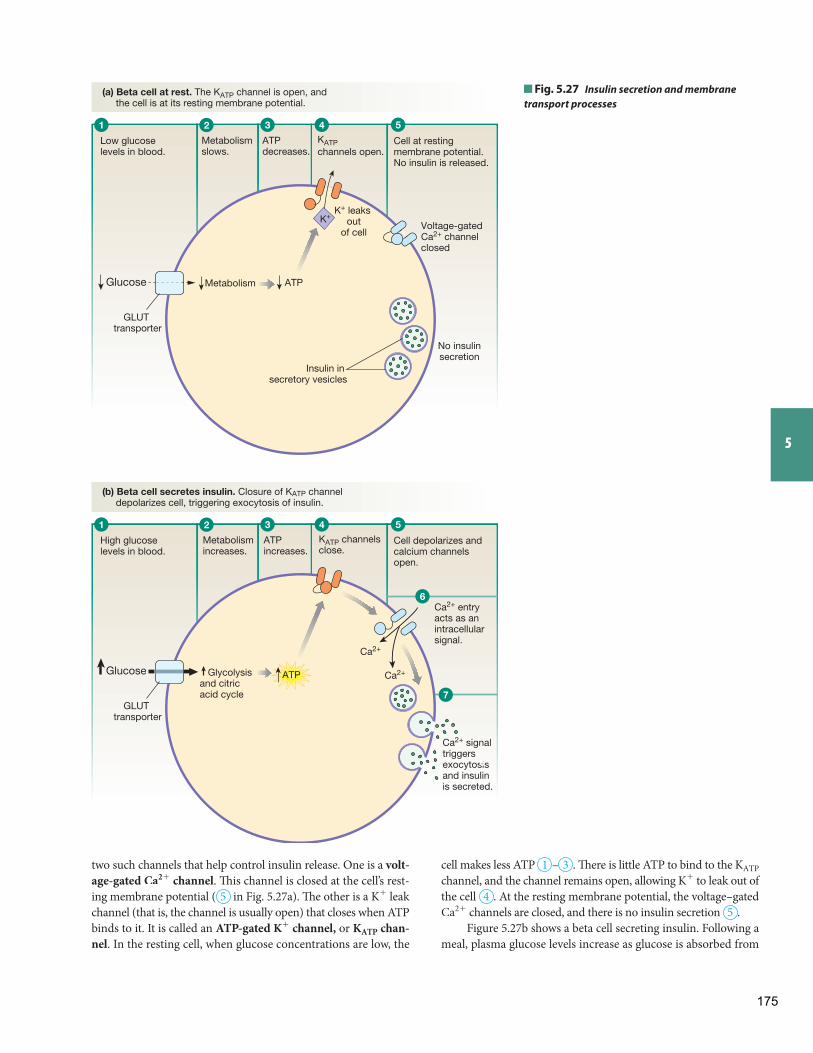
5
K+
Low glucose levels in blood.
No insulinsecretion
Metabolismslows.
ATPdecreases.
ATPMetabolismGlucose
Cell at restingmembrane potential.No insulin is released.
KATP channels open.
Insulin in secretory vesicles
K+ leaks out
of cellVoltage-gated Ca2+ channel closed
Glycolysisand citric acid cycle
ATP
Ca2+ signal triggersexocytosis and insulin is secreted.
Ca2+
Ca2+
GLUT transporter
(a) Beta cell at rest. The KATP channel is open, and the cell is at its resting membrane potential.
(b) Beta cell secretes insulin. Closure of KATP channel depolarizes cell, triggering exocytosis of insulin.
High glucose levels in blood.
Metabolismincreases.
ATPincreases.
Glucose
Cell depolarizes andcalcium channelsopen.
KATP channels close.
Ca2+ entry acts as anintracellularsignal.
GLUT transporter
1 2 3 4 5
1 2 3 4 5
6
7
6
Fig. 5.27 Insulin secretion and membrane transport processes
two such channels that help control insulin release. One is a volt-age-gated Ca2 + channel . Th is channel is closed at the cell’s rest-ing membrane potential ( 5 in Fig. 5.27 a). Th e other is a K+ leak channel (that is, the channel is usually open) that closes when ATP binds to it. It is called an ATP-gated K+ channel , or KATP chan-nel . In the resting cell, when glucose concentrations are low, the
cell makes less ATP 1 – 3 . Th ere is little ATP to bind to the KATP channel, and the channel remains open, allowing K+ to leak out of the cell 4 . At the resting membrane potential, the voltage–gated Ca2 + channels are closed, and there is no insulin secretion 5 .
Figure 5.27 b shows a beta cell secreting insulin. Following a meal, plasma glucose levels increase as glucose is absorbed from
175
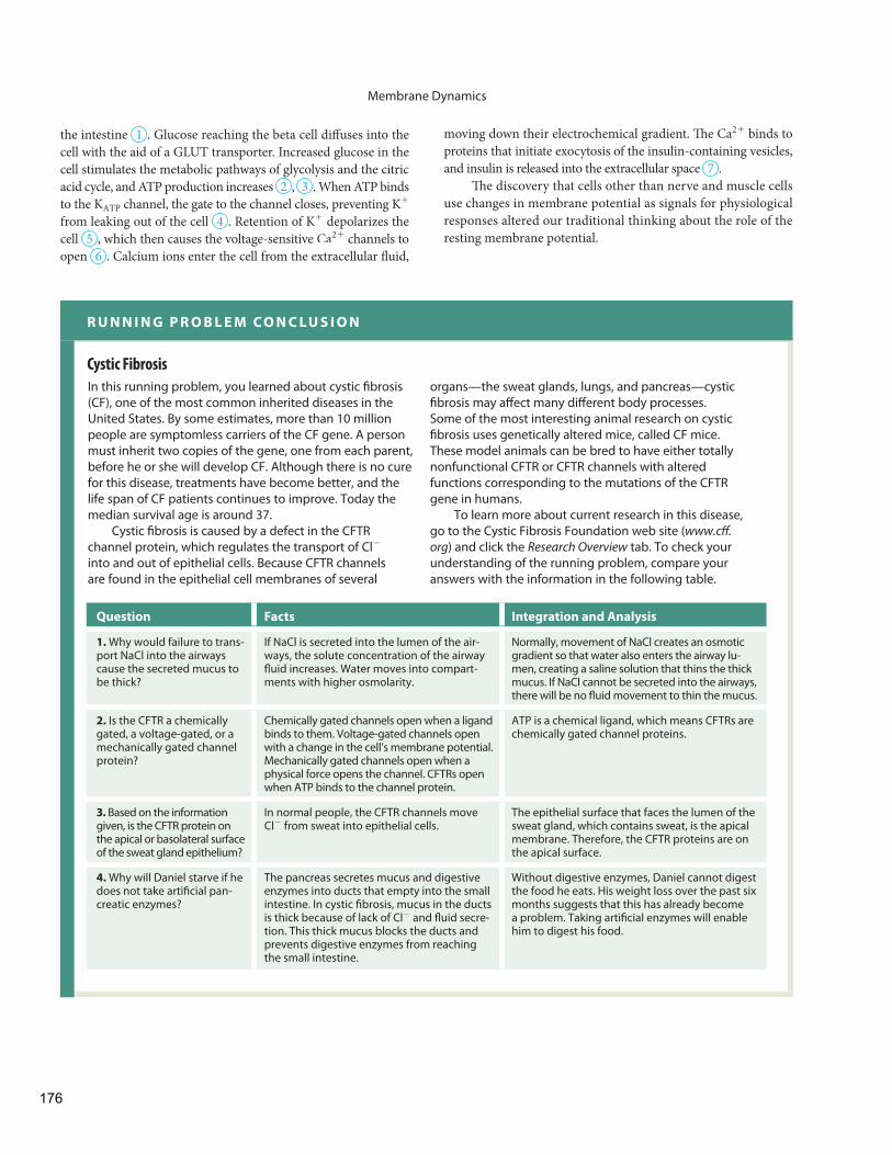
Membrane Dynamics
moving down their electrochemical gradient. Th e Ca2 + binds to proteins that initiate exocytosis of the insulin-containing vesicles, and insulin is released into the extracellular space 7 .
Th e discovery that cells other than nerve and muscle cells use changes in membrane potential as signals for physiological responses altered our traditional thinking about the role of the resting membrane potential.
the intestine 1 . Glucose reaching the beta cell diff uses into the cell with the aid of a GLUT transporter. Increased glucose in the cell stimulates the metabolic pathways of glycolysis and the citric acid cycle , and ATP production increases 2 , 3 . When ATP binds to the KATP channel, the gate to the channel closes, preventing K+ from leaking out of the cell 4 . Retention of K+ depolarizes the cell 5 , which then causes the voltage-sensitive Ca2 + channels to open 6 . Calcium ions enter the cell from the extracellular fl uid,
Cystic Fibrosis
In this running problem, you learned about cystic fi brosis (CF), one of the most common inherited diseases in the United States. By some estimates, more than 10 million people are symptomless carriers of the CF gene. A person must inherit two copies of the gene, one from each parent, before he or she will develop CF. Although there is no cure for this disease, treatments have become better, and the life span of CF patients continues to improve. Today the median survival age is around 37.
Cystic fi brosis is caused by a defect in the CFTR channel protein, which regulates the transport of Cl- into and out of epithelial cells. Because CFTR channels are found in the epithelial cell membranes of several
organs—the sweat glands, lungs, and pancreas—cystic fi brosis may aff ect many diff erent body processes. Some of the most interesting animal research on cystic fi brosis uses genetically altered mice, called CF mice. These model animals can be bred to have either totally nonfunctional CFTR or CFTR channels with altered functions corresponding to the mutations of the CFTR gene in humans.
To learn more about current research in this disease, go to the Cystic Fibrosis Foundation web site (www.cff .org) and click the Research Overview tab. To check your understanding of the running problem, compare your answers with the information in the following table.
R U N N I N G P R O B L E M C O N C L U S I O N
Question Facts Integration and Analysis
1. Why would failure to trans-port NaCl into the airways cause the secreted mucus to be thick?
If NaCl is secreted into the lumen of the air-ways, the solute concentration of the airway fl uid increases. Water moves into compart-ments with higher osmolarity.
Normally, movement of NaCl creates an osmotic gradient so that water also enters the airway lu-men, creating a saline solution that thins the thick mucus. If NaCl cannot be secreted into the airways, there will be no fl uid movement to thin the mucus.
2. Is the CFTR a chemically gated, a voltage-gated, or a mechanically gated channel protein?
Chemically gated channels open when a ligand binds to them. Voltage-gated channels open with a change in the cell’s membrane potential. Mechanically gated channels open when a physical force opens the channel. CFTRs open when ATP binds to the channel protein.
ATP is a chemical ligand, which means CFTRs are chemically gated channel proteins.
3. Based on the information given, is the CFTR protein on the apical or basolateral surface of the sweat gland epithelium?
In normal people, the CFTR channels move Cl- from sweat into epithelial cells.
The epithelial surface that faces the lumen of the sweat gland, which contains sweat, is the apical membrane. Therefore, the CFTR proteins are on the apical surface.
4. Why will Daniel starve if he does not take artifi cial pan-creatic enzymes?
The pancreas secretes mucus and digestive enzymes into ducts that empty into the small intestine. In cystic fi brosis, mucus in the ducts is thick because of lack of Cl- and fl uid secre-tion. This thick mucus blocks the ducts and prevents digestive enzymes from reaching the small intestine.
Without digestive enzymes, Daniel cannot digest the food he eats. His weight loss over the past six months suggests that this has already become a problem. Taking artifi cial enzymes will enable him to digest his food.
176
Membrane Dynamics
5
• PhysioExTM Lab Simulations • Interactive Physiology
Animations
Test your understanding with:
www.masteringaandp.com
• Practice Tests • Running Problem Quizzes • A&PFlixTM Animations
Several key themes come together in this chapter. You learned how the cell membrane acts as a barrier to create distinct intracellular and extracellular compartments, illustrating the theme of compartmenta-tion . Although the contents of the intracellular and extracellular com-partments differ, homeostasis keeps them in a dynamic steady state. Movement of materials between and within compartments is neces-sary for communication and is accomplished by bulk fl ow and biological transport . Flow of solutes and water across cell membranes occurs in
Chapter Summary
response to osmotic, chemical (concentration), or electrical gradients. Th e selectively permeable cell membrane creates resistance to fl ow that can be overcome by changing the composition of the membrane lipids or by inserting membrane proteins that act as channels or transporters. Biological transport in the body requires energy from diff erent sources: molecular motion, concentration gradients, or chemical bonds. Finally, the binding of substrates to transporters demonstrates the theme of protein interactions .
Osmosis and Tonicity
Fluids and Electrolytes: Introduction to Body Fluids
1. Most solutes are concentrated in either one compartment or the other, creating a state of chemical disequilibrium. ( Fig. 5.1 )
2. Cations and anions are not distributed equally between the body compartments, creating a state of electrical disequilibrium.
3. Water moves freely between the cells and extracellular fl uid, result-ing in a state of osmotic equilibrium.
4. Th e movement of water across a membrane in response to a con-centration gradient is called osmosis.
5. To compare solution concentrations, we express the concentra-tion in terms of osmolarity, the number of particles (ions or intact molecules) per liter of solution, expressed as milliosmoles per liter (mOsM).
6. Tonicity of a solution describes the cell volume change that oc-curs at equilibrium if the cell is placed in that solution. Cells swell in hypotonic solutions and shrink in hypertonic solutions. If the cell does not change size at equilibrium, the solution is isotonic.
7. Th e osmolarity of a solution cannot be used to determine the tonicity of the solution. Th e relative concentrations of nonpenetrating sol-utes in the cell and in the solution determine tonicity. Penetrating solutes contribute to the osmolarity of a solution but not to its tonic-ity. ( Fig. 5.4 )
Diff usion 8. In bulk fl ow a pressure gradient moves a fl uid along with its dis-
solved and suspended materials. 9. Th e cell membrane is a selectively permeable barrier that restricts
free exchange between the cell and the interstitial fl uid. Th e move-ment of a substance across the membrane depends on the perme-ability of the membrane to that substance.
10. Movement of molecules across membranes can be classifi ed either by energy requirements or by the physical means the molecule uses to cross the membrane. ( Fig. 5.5 )
11. Lipid-soluble substances can diffuse through the phospholipid bilayer. Less lipid-soluble molecules require the assistance of a membrane protein to cross the membrane.
12. Passive transport does not require the input of energy. 13. Diff usion is the passive movement of molecules down a chemical
(concentration) gradient from an area of higher concentration to an area of lower concentration. Net movement stops when the sys-tem reaches equilibrium, although molecular movement continues. ( Tbl. 5.6 )
14. Diff usion rate depends on the magnitude of the concentration gra-dient. Diffusion is slow over long distances, is directly related to temperature, and is inversely related to molecular size.
15. Simple diffusion across a membrane is directly proportional to membrane surface area, concentration gradient, and membrane per-meability, and inversely proportional to membrane thickness. ( Fig. 5.7 )
Protein-Mediated Transport 16. Most molecules cross membranes with the aid of membrane
proteins. 17. Membrane proteins have four functional roles: structural proteins
maintain cell shape and form cell junctions; membrane-associated enzymes catalyze chemical reactions and help transfer signals across the membrane; receptor proteins are part of the body’s signaling system; and transport proteins move many molecules into or out of the cell. ( Fig. 5.8 )
18. Channel proteins form water-fi lled channels that link the intra-cellular and extracellular compartments. Gated channels regulate movement of substances through them by opening and closing. Gated channels may be regulated by ligands, by the electrical state of the cell, or by physical changes such as pressure. ( Fig. 5.10 )
Fluids and Electrolytes: Introduction to Body Fluids
177
Membrane Dynamics
allows one-way movement of molecules across the epithelium. (Figs . 5.20, 5.21)
29. Molecules cross epithelia by moving between the cells by the para-cellular route or through the cells by the transcellular route. ( Fig. 5.20 )
30. Larger molecules cross epithelia by transcytosis, which includes vesicular transport. ( Fig. 5.22 )
The Resting Membrane Potential
Nervous I: The Membrane Potential
31. Although the total body is electrically neutral, diff usion and active transport of ions across the cell membrane create an electrical gra-dient, with the inside of cells negative relative to the extracellular fl uid. ( Fig. 5.23 )
32. Th e electrical gradient between the extracellular fl uid and the in-tracellular fluid is known as the resting membrane potential diff erence.
33. Th e movement of an ion across the cell membrane is infl uenced by the electrochemical gradient for that ion.
34. The membrane potential that exactly opposes the concentration gradient of an ion is known as the equilibrium potential ( Eion ) . Th e equilibrium potential for any ion can be calculated using the Nernst equation. ( Fig. 5.24 )
35. In most living cells, K+ is the primary ion that determines the rest-ing membrane potential.
36. Changes in membrane permeability to ions such as K+ , Na+ , Ca2 + ,or Cl- alter membrane potential and create electrical signals.
Integrated Membrane Processes: Insulin Secretion 37. Th e use of electrical signals to initiate a cellular response is a uni-
versal property of living cells. Pancreatic beta cells release insulin in response to a change in membrane potential. ( Fig. 5.27 )
19. Carrier proteins never form a continuous connection between the intracellular and extracellular fl uid. Th ey bind to substrates, then change conformation. ( Fig. 5.12 )
20. Protein-mediated diff usion is called facilitated diff usion. It has the same properties as simple diff usion. ( Tbl. 5.6; Fig. 5.13 )
21. Active transport moves molecules against their concentration gra-dient and requires an outside source of energy. In primary (direct) active transport , the energy comes directly from ATP. Secondary (indirect) active transport uses the potential energy stored in a concentration gradient and is indirectly driven by energy from ATP.
22. The most important primary active transporter is the sodium- potassium-ATPase ( Na+ - K+ -ATPase), which pumps Na+ out of the cell and K+ into the cell. ( Fig. 5.14 )
23. Most secondary active transport systems are driven by the sodium concentration gradient. ( Tbl. 5.8; Fig. 5.16 )
24. All carrier-mediated transport demonstrates specifi city, competi-tion, and saturation. Specifi city refers to the ability of a transporter to move only one molecule or a group of closely related molecules. Related molecules may compete for a single transporter. Saturation occurs when a group of membrane transporters are working at their maximum rate. ( Fig. 5.17 )
Vesicular Transport 25. Large macromolecules and particles are brought into cells by phago-
cytosis or endocytosis. Material leaves cells by exocytosis. When vesicles that come into the cytoplasm by endocytosis are returned to the cell membrane, the process is called membrane recycling. (Figs. 5.18, 5.19)
26. In receptor-mediated endocytosis, ligands bind to membrane recep-tors that concentrate in coated pits or caveolae . ( Fig. 5.19 )
27. In exocytosis, the vesicle membrane fuses with the cell membrane before releasing its contents into the extracellular space. Exocytosis requires ATP.
Epithelial Transport 28. Transporting epithelia have different membrane proteins
on their apical and basolateral surfaces. This polarization
Nervous I: The Membrane Potential
Questions
Level One Reviewing Facts and Terms 1. List the four functions of membrane proteins, and give an example
of each. 2. Distinguish between active transport and passive transport. 3. Which of the following processes are examples of active transport,
and which are examples of passive transport? Simple diffusion, phagocytosis, facilitated diff usion, exocytosis, osmosis, endocytosis.
4. List four factors that increase the rate of diff usion in air. 5. List the three physical methods by which materials enter cells. 6. A cotransporter is a protein that moves more than one molecule at
a time. If the molecules are moved in the same direction, the transporters are called carriers; if the molecules are transported in opposite directions, the transporters are called
carriers. A transport protein that moves only one sub-strate is called a(n) carrier.
7. Th e two types of active transport are , which derives en-ergy directly from ATP, and , which couples the kinetic energy of one molecule moving down its concentration gradient to the movement of another molecule against its concentration gradient.
8. A molecule that moves freely between the intracellular and extracel-lular compartments is said to be a(n) solute. A molecule that is not able to enter cells is called a(n) solute.
9. Rank the following individuals in order of how much body water they contain, from highest to lowest: (a) a 25-year-old, 70-kg male; (b) a 25-year-old, 50-kg female; (c) a 65-year-old, 50-kg female; and (d) a 1-year-old male toddler.
10. What determines the osmolarity of a solution? In what units is body osmolarity usually expressed?
11. What does it mean if we say that a solution is hypotonic to a cell? Hypertonic to the same cell? What determines the tonicity of a solu-tion relative to a cell?
178
5
12. Match the membrane channels with the appropriate descriptions. Answers may be used once, more than once, or not at all.
(a) chemically gated channel 1. channel that spends most (b) open pore of its time in the open (c) voltage-gated channel state (d) mechanically gated channel 2. channel that spends most
of its time in a closed state 3. channel that opens when
resting membrane poten-tial changes
4. channel that opens when a ligand binds to it
5. channel that opens in response to membrane stretch
6. channel through which water can pass
13. In your own words, state the four principles of electricity important in physiology.
14. Match each of the following items with its primary role in cellular activity.
(a) Na+ - K+ -ATPase 1. ion channel (b) protein 2. extracellular cation (c) unit of measurement for 3. source of energy membrane potential 4. intracellular anion (d) K+ 5. intracellular cation (e) Cl- 6. millivolts (f) ATP 7. electrogenic pump (g) Na+ 8. extracellular anion 9. milliosmoles
15. Th e membrane potential at which the electrical gradient exactly op-poses the concentration gradient for an ion is known as the .
16. A material that allows free movement of electrical charges is called a(n) , whereas one that prevents this movement is called a(n) .
Level Two Reviewing Concepts 17. Create a map of transport across cell membranes using the follow-
ing terms. You may add additional terms if you wish.
• active transport • ligand • carrier • Na+ - K+ -ATPase • caveolae • osmosis • channel • passive transport • clathrin-coated pit • phospholipid bilayer • concentration gradient • receptor-mediated endocytosis • electrochemical gradient • secondary active transport • exocytosis • simple diff usion • facilitated diff usion • small polar molecule • glucose • transcytosis • GLUT transporter • vesicle • ion • vesicular transport • large polar molecule • water
18. Draw a large rectangle to represent the total body volume. Using the information in Figure 5.1 b, divide the box proportionately into
Membrane Dynamics
compartments to represent the diff erent body compartments. Use the information in Figure 5.1 d and add solutes to the compart-ments. Use large letters for solutes with higher concentrations, and small letters for solutes with low concentrations. Label the cell membranes and the endothelial membrane.
19. What factors influence the rate of diffusion across a membrane? Briefl y explain each one.
20. Defi ne the following terms and explain how they diff er from one another: specifi city, competition, saturation. Apply these terms in a short explanation of facilitated diff usion of glucose.
21. Red blood cells are suspended in a solution of NaCl. Th e cells have an osmolarity of 300 mOsM, and the solution has an osmolarity of 250 mOsM. (a) Th e solution is (hypertonic, isotonic, or hypotonic) to the cells. (b) Water would move (into the cells, out of the cells, or not at all).
22. Two compartments are separated by a membrane that is perme-able to glucose. Each compartment is fi lled with 1 M glucose. Aft er 6 hours, compartment A contains 1.5 M glucose and compartment B contains 0.5 M glucose. What kind of transport occurred? Explain.
23. A 2 M NaCl solution is placed in compartment A and a 2 M glu-cose solution is placed in compartment B. Th e compartments are separated by a membrane that is permeable to water but not to NaCl or glucose. Complete the following statements. Defend your answers. (a) Th e salt solution is osmotic to the glucose solution. (b) True or false? Water will move from one compartment to another.
If water moves, it will move from compartment to compartment .
24. Explain the diff erences between a chemical gradient, an electrical gradient, and an electrochemical gradient.
Level Three Problem Solving 25. Sweat glands secrete into their lumen a fl uid that is identical to intersti-
tial fl uid. As the fl uid moves through the lumen on its way to the sur-face of the skin, the cells of the sweat gland’s epithelium make the fl uid hypotonic by removing Na+ and leaving water behind. Design an epi-thelial cell that will reabsorb Na+ but not water. You may place water pores, Na+ leak channels, K+ leak channels, and the Na+ - K+ -ATPase in the apical membrane, basolateral membrane, or both.
26. Insulin is a hormone that promotes the movement of glucose into many types of cells, thereby lowering blood glucose concentration. Propose a mechanism that explains how this occurs, using your knowledge of cell membrane transport.
27. Th e following terms have been applied to membrane carriers: speci-fi city, competition, saturation. Why can these terms also be applied to enzymes? What is the major diff erence in how enzymes and carri-ers carry out their work?
28. NaCl is a nonpenetrating solute and urea is a penetrating solute for cells. Red blood cells are placed in each of the solutions below. Th e intracellular concentration of nonpenetrating solute is 300 mOsM. What will happen to the cell volume in each solution? Label each solution with all the terms that apply: hypertonic, isotonic, hypo-tonic, hyperosmotic, hyposmotic, isosmotic. Watch units! Assume 1 M NaCl = 2 OsM for simplicity. (a) 150 mM NaCl plus 150 mM urea (b) 100 mM NaCl plus 50 mM urea (c) 100 mM NaCl plus 100 mM urea (d) 150 mM NaCl plus 100 mM urea (e) 100 mM NaCl plus 150 mM urea
179
Membrane Dynamics
(c) her ECF and ICF volumes (d) her ECF and ICF osmolarities
34. Th e following graph shows the results of an experiment in which a cell was placed in a solution of glucose. Th e cell had no glucose in it at the beginning, and its membrane can transport glucose. Which of the following processes is/are illustrated by this experiment?
[glu
cose
] in
cell
[glu
cose
] out
sid
e of
cel
l
0.5
1.0
1.5
Time (a) simple diff usion (b) saturation (c) competition (d) active transport
29. Integral membrane glycoproteins have sugars added as the proteins pass through the lumen of the endoplasmic reticulum and Golgi complex. Based on this information, where would you predict fi nd-ing the sugar “tails” of the proteins: on the cytoplasmic side of the membrane, the extracellular side, or both? Explain your reasoning.
Level Four Quantitative Problems 30. The addition of dissolved solutes to water lowers the freezing
point of water. A 1 OsM solution depresses the freezing point of water by 1.86 °C. If a patient’s plasma shows a freezing-point depression of 0.550 °C, what is her plasma osmolarity? (Assume that 1 kg water = 1 L.)
31. Th e patient in the previous question is found to have total body water volume of 42 L, ECF volume of 12.5 L, and plasma volume of 2.7 L. (a) What is her intracellular fluid (ICF) volume? Her interstitial
fl uid volume? (b) How much solute (osmoles) exists in her whole body? ECF?
ICF? plasma? ( Hint : concentration = solute amount/volume of solution)
32. What is the osmolarity of half-normal saline (= 0.45% NaCl)? As-sume that all NaCl molecules dissociate into two ions.
33. If you give 1 L of half-normal saline (see question 32) to the patient in question 31, what happens to each of the following at equilib-rium? ( Hint: NaCl is a nonpenetrating solute.) (a) her total body volume (b) her total body osmolarity
8. Prediction: 260 mOsM glucose is hyposmotic and hypotonic. Add-ing 260 mosmoles glucose and 1 L volume:
Total Body ECF ICF S (mosmol) 900 + 260 = 1160 300 600 + 260 = 860 V (L) 3 + 1 = 4 1.034 2.966 C (mOsM) 1160/4 = 290
mOsM S
290 S 290
9. (a) Sweat loss is hyposmotic. Body osmolarity will increase. (b) Cell volume will decrease. (c) Losing 0.5 L and 65 mosmoles NaCl. Yes, cell volume decreased and osmolarity increased.
Total Body ECF ICF S (mosmol) 900 - 65 = 835 300 - 65 = 235 600 V (L) 3 - 0.5 = 2.5 0.704 1.796 C (mOsM) 334 mOsM S 334 S 334
10. (a) Th e NaCl solution is better, even though both solutions are is-osmotic to the body ( Tbl. 5.8 ). Because blood is lost from the extra-cellular compartment, the best replacement solution would remain in the ECF. For this reason glucose is not as good a choice because it slowly enters cells, taking water with it. (b) If 1 L has been lost, you should replace at least 1 L.
1. Th e stem preceding the suffi x –ase is the name of the substrate on which the enzyme acts; therefore ATP is a substrate for this enzyme.
2. Intracellular fl uid has a high K+ concentration and low Na+ , Cl- , and Ca2 + concentrations.
3. Plasma is part of the ECF, so knowing the plasma concentra-tion of (a) Na+ and (b) K+ tells you the ECF concentration of these ions. The ICF ion concentrations are different from the ECF concentrations, so plasma concentration will not tell you the ICF values. (c) The body is in osmotic equilibrium, so water concentration is the same in all compartments. (d) Proteins are in the plasma but not in the interstitial fl uid, so plasma protein con-centration will not tell you ECF or ICF protein concentrations.
4. (a) total body water = 29 L; (b) ECF = 19.1, ICF = 38.9 L; (c) plasma = 4.8 L
5. Th e baby has lost 0.91 kg of water, which is 0.91 liter. 6. 1 M NaCl = 2 OsM NaCl. Th e 1 M (= 1 OsM) glucose and 1 OsM
NaCl have the most water. 7. (a) Water moves into A because A is 2 OsM; (b) no net move-
ment occurs because urea will diff use across the membrane until it reaches equilibrium; (c) water moves into A, because A has a higher concentration of nonpenetrating solutes.
Answers to Concept Check Questions
Answers
180
5
Membrane Dynamics
11. If distance triples, diff usion takes nine times as long. 12. Energy for diff usion comes from molecular motion. 13. Because it is lipophilic, the fatty acid is more likely to cross by sim-
ple diff usion. 14. Th e fl ux (a) decreases, (b) increases, (c) decreases. 15. Compartment A remains yellow, and compartment B turns green. 16. Th e skin’s thick extracellular matrix is generally impermeable to
oxygen. Also, oxygen needs a moist surface to effectively diffuse across a tissue membrane.
17. Positive ions are cations, and negative ions are anions. 18. Membrane proteins serve as structural proteins, receptors, en-
zymes, and transporters. 19. Ions and water molecules move through open channels. 20. Channel proteins form continuous connections between the two
sides of a membrane and transport molecules more quickly. 21. A channel lined with positive charges attracts anions, which in this
instance means Cl- . 22. Glucose is too large to pass through a channel. 23. The direction of facilitated diffusion of glucose reverses, and
glucose leaves the cell. 24. The ATPase is an antiporter, but the SGLT is a symporter. The
ATPase requires energy from ATP to change conformation, whereas the SGLT uses energy stored in the Na+ concentration gradient.
25. An antiporter moves substrates in opposite directions. 26. Larger doors could move more people. Th is would be analogous to
a cell’s synthesizing a new isoform of the transporter that would let the transporter move more substrate per second.
27. In phagocytosis, the cytoskeleton pushes the membrane out to engulf a particle in a large vesicle. In endocytosis, the membrane surface indents and the vesicle is much smaller.
28. Th e proteins associated with endocytosis are clathrin and caveolin. 29. Proteins move into cells by endocytosis and out of cells by
exocytosis. 30. Sodium movement out of the cell requires energy because the di-
rection of ion fl ow is against the concentration gradient. 31. Ouabain applied to the apical side would have no effect because
there are no Na+ - K+ -ATPase molecules on that side. Ouabain
applied to the basolateral side would stop the pump. Glucose trans-port would continue for a time until the Na+ gradient between the cell and the lumen disappears because Na+ has entered the cell.
32. Th e GLUT2 transporter is illustrated. 33. Transcytosis will stop because vesicular transport by the cytoskel-
eton depends on functioning microtubules. 34. Cl- will move into the cell down its concentration gradient, which
would make the inside of the cell negative. Th e positive charges left outside would attract Cl- back outside. Th e equilibrium potential would be negative.
35. Over time, Na+ would leak into the cell, and the resting membrane potential would become more positive.
Answers to Figure and Graph Questions
Figure 5.1 : 1. Plasma is 25% of 14 L = 3.5 L. Interstitial fluid is 75% = 10.5 L. 2. Total body water = 42 L. 3. Plasma is 3.5 L/42 L =8.3% of total body water; interstitial volume is 10.5 L/42 L =25%. 4. Total body weight is 121 lb * 1 kg/2.2 lb = 55 kg. If body water = 50% of body weight, total body water = 27.5 L. ICF is 67% of 27.5 L = 18.425 L. ECF is 33% of 27.5 L = 9.075 L. Plasma is 25% of ECF = 2.269 L. 5. Plasma contains proteins and large an-ions not present in interstitial fl uid. 6. Th e extracellular compartment contains more Na+ , Cl- , and bicarbonate than the intracellular com-partment, and fewer K+ .
Figure 5.4 : Example 2: (a) hyperosmotic; (b) 250 mOsM; (c) 300 mOsM; (d) Yes, into the cells; (e) hypotonic; (f) increased; hyperos-motic; (g) increased; hypotonic
Figure 5.17 : (c) You cannot tell if galactose is being transported because the curves only show glucose transport. (d) Th e cell could increase transport by adding more transporters to the membrane.
Figure 5.21 : 1 = b; 2 = a; 3 = b. Basolateral glucose transport is passive because the glucose moves down its concentration gradient. Na+ movement across the apical membrane does not require ATP because Na+ is moving down its concentration gradient.
Figure 5.24 : (b) ICF = -1 and ECF = + 1. (c) Th e cell membrane is not permeable to Na+ , Cl- , and proteins.
Figure 5.25 : Na+ leak into the cell is promoted by concentration and electrical gradients. K+ leak out of the cell is promoted by the concentration gradient.
Answers to Figure and Graph Questions
181
Membrane Dynamics
Level One Reviewing Facts and Terms
1. Structural proteins (link cell to matrix); transporters (water channels); recep-tors (hormone receptors); and enzymes (intestinal digestive enzymes)
2. Active: requires direct or indirect use of energy. Passive: uses energy stored in a concentration gradient.
3. Passive: simple and facilitated diffusion, osmosis. Active: phagocytosis, exo-cytosis, and endocytosis.
4. Greater concentration gradient, smaller distance, higher temperature, and smaller molecular size
5. Simple diffusion, protein-mediated transport, or vesicular transport
6. symport; antiport; uniport
7. primary ( direct) and secondary ( indirect)
8. penetrating; nonpenetrating 9. (d), (a), (b), (c). 10. Osmolarity: concentration of osmotically active particles, expressed as
osmol>L or milliosmoles per liter 11. Hypotonic: net influx of water into the cell at equilibrium. Hypertonic: net
water loss at equilibrium. Tonicity is determined by relative concentrations of nonpenetrating solutes in cell versus solution.
12. (a) 2, 4; (b) 1, 6; (c) 2, 3; (d) 2, 5 13. (1) Like charges repel; opposites attract. (2) Every positive ion has a match-
ing negative ion. (3) Energy must be used to separate ions or electrons and protons. (4) Conductors allow ions to move through them, while insulators keep ions separated.
14. (a) 7; (b) 1, 7; (c) 6; (d) 5; (e) 8; (f) 3; (g) 2
15. equilibrium potential 16. conductor; insulator
Level Two Reviewing Concepts
17. Use Figs. 5.5 , 5.8 , 5.10 , 5.21 , and 5.24 to create your map. 18. See Fig. 5.1 c and d. 19. Lipid solubility, so that a molecule can pass through the lipid core of the mem-
brane. Diffusion is slower for larger or heavier molecules and faster when there is more membrane surface area.
20. Specificity: enzyme or transporter works on one molecule or class of molecules. Competition: similar substrates can compete for the protein binding site. Satu-ration: rate reaches a maximum when all binding sites are filled. GLUT is spe-cific for hexose sugars. If glucose and fructose are both present, they compete for GLUT binding sites. If enough sugar is present, transport saturates.
21. (a) hypotonic (b) into the cells 22. Active transport. Must use energy to go from a state of equilibrium to one of
disequilibrium.
23. (a) hyperosmotic (convert molarity to osmolarity). (b) True. Water moves from B to A .
24. Chemical gradient = concentration gradient. Electrical gradient = separa-tion of electrical charge. Electrochemical gradient includes both concentra-tion and electrical gradients.
Level Three Problem Solving
25. Apical side: Na+ leak channels but no water pores. Basolateral side: Na+-K+ -ATPase and K+ leak channels. May also have water channels.
26. Insulin could increase the number or affinity of GLUT proteins or could act on cell metabolism to keep the intracellular glucose concentration low.
27. Both enzymes and transporters are proteins that bind ligands at a specific binding site. Enzymes alter their substrates. Transporters move substrates un-changed across a membrane.
28. Must convert units from mM to mOsM. (a) hyperosmotic, isotonic; (b) hyposmotic, hypotonic; (c) isosmotic, hypotonic; (d) hyperosmotic, isotonic; (e) hyperosmotic, hypotonic
29. Sugars are added to proteins inside the organelle>vesicle, therefore will face the ECF after being inserted into the membrane.
Level Four Quantitative Problems
30. 296 mOsM 31. (a) ICF = 29.5 L; interstitium = 9.8 L. (b) Total solute = 12.432 osmoles;
ECF = 3.7 osmoles; ICF = 8.732 osmoles; plasma = 0.799 osmoles. 32. 154 mOsM 33. (a) increases; (b) decreases; (c) increase; (d) decrease 34. Simple diffusion (a). Cannot be active transport because concentration in
never exceeds concentration out .
Answers to Review Questions
Photo Credits
CO: Marian Miller 5.4: Darren Hubley/iStockphoto.com.
182
Communication, Integration, and Homeostasis
From Chapter 6 of Human Physiology: An Integrated Approach, Sixth Edition. Dee Unglaub Silverthorn. Copyright © 2013 by Pearson Education, Inc. All rights reserved.
183