Embed Size (px)
Citation preview
cancers
Review
Trends in Research on Exosomes in Cancer Progression andAnticancer Therapy
Dona Sinha 1,*, Sraddhya Roy 1, Priyanka Saha 1, Nabanita Chatterjee 1 and Anupam Bishayee 2,*
�����������������
Citation: Sinha, D.; Roy, S.; Saha, P.;
Chatterjee, N.; Bishayee, A. Trends in
Research on Exosomes in Cancer
Progression and Anticancer Therapy.
Cancers 2021, 13, 326.
https://doi.org/10.3390/
cancers13020326
Received: 23 November 2020
Accepted: 14 January 2021
Published: 17 January 2021
Publisher’s Note: MDPI stays neu-
tral with regard to jurisdictional clai-
ms in published maps and institutio-
nal affiliations.
Copyright: © 2021 by the authors. Li-
censee MDPI, Basel, Switzerland.
This article is an open access article
distributed under the terms and con-
ditions of the Creative Commons At-
tribution (CC BY) license (https://
creativecommons.org/licenses/by/
4.0/).
1 Department of Receptor Biology and Tumour Metastasis, Chittaranjan National Cancer Institute,Kolkata 700 026, India; [email protected] (S.R.); [email protected] (P.S.);[email protected] (N.C.)
2 Lake Erie College of Osteopathic Medicine, Bradenton, FL 34211, USA* Correspondence: [email protected] or [email protected] (D.S.); [email protected] or
[email protected] (A.B.)
Simple Summary: Intensive research in the field of cancer biology has discovered a unique modeof interplay between cells via extracellular bioactive vesicles called exosomes. Exosomes serve asintermediators among cells via their cargoes that, in turn, contribute in the progression of cancer.They are ubiquitously present in all body fluids as they are secreted from both normal and tumorcells. These minuscules exhibit multiple unique properties that facilitate their migration to distantlocations and modulate the microenvironment for progression of cancer. This review summarizesthe multifarious role of exosomes in various aspects of cancer research with its pros and cons. Itdiscusses biogenesis of exosomes, their functional role in cancer metastasis, both protumorigenic andantitumorigenic, and also their applications in anticancer therapy.
Abstract: Exosomes, the endosome-derived bilayered extracellular nanovesicles with their contri-bution in many aspects of cancer biology, have become one of the prime foci of research. Exosomesderived from various cells carry cargoes similar to their originator cells and their mode of generationis different compared to other extracellular vesicles. This review has tried to cover all aspects ofexosome biogenesis, including cargo, Rab-dependent and Rab-independent secretion of endosomesand exosomal internalization. The bioactive molecules of the tumor-derived exosomes, by virtue oftheir ubiquitous presence and small size, can migrate to distal parts and propagate oncogenic sig-naling and epigenetic regulation, modulate tumor microenvironment and facilitate immune escape,tumor progression and drug resistance responsible for cancer progression. Strategies improvisedagainst tumor-derived exosomes include suppression of exosome uptake, modulation of exosomalcargo and removal of exosomes. Apart from the protumorigenic role, exosomal cargoes have beenselectively manipulated for diagnosis, immune therapy, vaccine development, RNA therapy, stem celltherapy, drug delivery and reversal of chemoresistance against cancer. However, several challenges,including in-depth knowledge of exosome biogenesis and protein sorting, perfect and pure isolationof exosomes, large-scale production, better loading efficiency, and targeted delivery of exosomes,have to be confronted before the successful implementation of exosomes becomes possible for thediagnosis and therapy of cancer.
Keywords: tumor-derived exosomes; exosomal cargoes; protumorigenic effect; drug resistance;anticancer therapy
1. Introduction
Exosomes are bilayered endosomal nanovesicles, first discovered in 1983, as transfer-rin conjugated vesicles (50 nm) released by reticulocytes [1]. Due to the increasing interestof scientists in exosome biology, a consensus guideline was proposed by board membersof International Society of Extracellular Vesicles under “minimal experimental require-ments for definition of extracellular vesicles and their functions” (MISEV2014) which was
Cancers 2021, 13, 326. https://doi.org/10.3390/cancers13020326 https://www.mdpi.com/journal/cancers
Cancers 2021, 13, 326 2 of 31
later updated in 2018 (MISEV2018). The guidelines advocated norms for nomenclature,isolation, separation, characterization, functional studies, and reporting requirements forproper identification of and experimentation with extracellular vesicles and exosomes [2,3].Exosomes are generally formed by inward budding of late endosomes, also known asmultivesicular bodies (MVBs). Intraluminal vesicles (ILVs) of MVBs engulf a variety ofbiomolecules which are released into extracellular space as exosomes. Exosomes are anucle-ated particles naturally released by cells, surrounded by lipid bilayer and are not capableof replication. Exosomes are identified by size (30–200 nm) and surface markers, suchas membrane-associated proteins, e.g., lysosome-associated membrane glycoprotein 3(LAMP3)/CD63; intercellular adhesion molecule (ICAM1)/CD81; and tetraspanin mem-brane protein/CD9. Exosomes are observed in various body fluids, such as blood, plasma,saliva, urine, synovial fluid, amniotic fluid, and breast milk [4,5].
All cellular types (normal and diseased) secrete exosomes, mediating intercellularcommunications [6]. Exosomes exhibit heterogeneity in size—Exo-Large (90–120 nm), Exo-Small (60–80 nm), and the membrane-less exomere (<50 nm). Exosome-mediated intercel-lular transfer of specific repertoire of proteins, lipids, RNA and DNA confer physiologicaland/or pathological functions to the recipient targets. Exosomes regulate physiologicalfunctions, such as neuronal communication, immune responses, reproductive activity, cellproliferation homeostasis, maturation and cellular waste disposition. They also contributein clinical disorders, including inflammation, cancer, cardiovascular diseases, neuronalpathologies and pathogenic infections [5].
Our review deals with exosomal contents, exosome-associated protumorigenic, antitu-morigenic effect and therapeutics, unlike other reviews, which discuss combinational rolesof all microvesicles in cancer progression [7,8] or have primarily focused on tumor-derivedexosomes (TEXs) with little information on therapeutics [9]. In contrast to reviews whichhave focused on specific exosomal cargoes and therapeutics [10,11], we have envisagedthe exosomal contents, the mechanisms influencing cancer progression and their ther-apeutic implications in cancer management. The inexplicable nature of exosomes hasraised concern about their role in the invasion and metastasis of cancer cells, encompassingepithelial-to-mesenchymal transition (EMT), angiogenesis, and immune regulation [12].Thus, instead of reviewing the isolated impact of exosomes, e.g., evasion of immunesurveillance [13] for cancer progression, we have tried to encompass exosome-mediatedpropagation of oncogenic signaling, epigenetic regulation, modulation of tumor microenvi-ronment (TME) and immune escape, EMT, angiogenesis, metastasis and drug resistance.Considering the clinical applications, the exosomes serve as potent diagnostic and prog-nostic biomarkers because of their bioavailability, low toxicity and differentiated surfacemarkers [5]. Recent reviews on exosomes have focused on therapeutic efficacy of exo-somes by addressing extracellular vesicular interaction with the host immune system [14],constraints and opportunities available with bioengineering of exosomes [15–17], successagainst multiple cancers [18] and exosome-based drug delivery [19–21]. Anticancer treat-ments sometimes experience shortfall in their efficacy due to unwanted side effects of thetherapeutic agents or shortened shelf-life, but exosomes serve as natural agents to overcomethese issues and become a potent therapeutic agent [22]. However, instead of perceivingspecific therapeutic potential of exosomes, the present review has tried to decipher theentire repertoire of exosomes, including both protumorigenic and antitumorigenic impact.
2. Cargo Composition of Exosomes
Exosomes are rich in enzymes, transcription factors, heat shock proteins (Hsps),major histocompatibility complex (MHC), cytoskeleton components, signal transducers,tetraspanins, lipids, RNAs and DNAs [6,23]. Detailed information about the exosomalcomponents can be accessed via databases, such as ExoCarta [www.exocarta.org], EVpe-dia [http://evpedia.info] and Vesiclepedia [www.microvesicles.org]. Though exosomesdiverge in size and biomolecular inclusions, some common components are observedin all types [5]. Lipid components are cholesterol, sphingomyelin, glycosphingolipids,
Cancers 2021, 13, 326 3 of 31
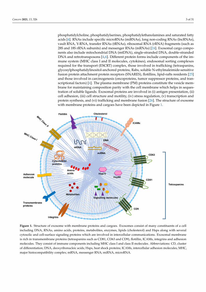
phosphatidylcholine, phosphatidylserines, phosphatidylethanolamines and saturated fattyacids [4]. RNAs include specific microRNAs (miRNAs), long non-coding RNAs (lncRNAs),vault RNA, Y-RNA, transfer RNAs (tRNAs), ribosomal RNA (rRNA) fragments (such as28S and 18S rRNA subunits) and messenger RNAs (mRNAs) [24]. Exosomal cargo compo-nents also include mitochondrial DNA (mtDNA), single-stranded DNA, double-strandedDNA and retrotransposons [4,6]. Different protein forms include components of the im-mune system (MHC class I and II molecules, cytokines), endosomal sorting complexesrequired for the transport (ESCRT) complex, those involved in trafficking (tetraspanins,glycosylphosphatidylinositol-anchored proteins, Rabs, soluble N-ethylmaleimide-sensitivefusion protein attachment protein receptors (SNARES), flotillins, lipid-rafts residents [25]and those involved in carcinogenesis (oncoproteins, tumor suppressor proteins, and tran-scriptional factors) [4]. The plasma membrane (PM) proteins constitute the vesicle mem-brane for maintaining composition parity with the cell membrane which helps in seques-tration of soluble ligands. Exosomal proteins are involved in (i) antigen presentation, (ii)cell adhesion, (iii) cell structure and motility, (iv) stress regulation, (v) transcription andprotein synthesis, and (vi) trafficking and membrane fusion [26]. The structure of exosomewith membrane proteins and cargoes have been depicted in Figure 1.
Cancers 2021, 13, x 3 of 31
diverge in size and biomolecular inclusions, some common components are observed in all types [5]. Lipid components are cholesterol, sphingomyelin, glycosphingolipids, phos-phatidylcholine, phosphatidylserines, phosphatidylethanolamines and saturated fatty ac-ids [4]. RNAs include specific microRNAs (miRNAs), long non-coding RNAs (lncRNAs), vault RNA, Y-RNA, transfer RNAs (tRNAs), ribosomal RNA (rRNA) frag-ments (such as 28S and 18S rRNA subunits) and messenger RNAs (mRNAs) [24]. Exoso-mal cargo components also include mitochondrial DNA (mtDNA), single-stranded DNA, double-stranded DNA and retrotransposons [4,6]. Different protein forms include com-ponents of the immune system (MHC class I and II molecules, cytokines), endosomal sort-ing complexes required for the transport (ESCRT) complex, those involved in trafficking (tetraspanins, glycosylphosphatidylinositol-anchored proteins, Rabs, soluble N-ethylma-leimide-sensitive fusion protein attachment protein receptors (SNARES), flotillins, lipid-rafts residents [25] and those involved in carcinogenesis (oncoproteins, tumor suppressor proteins, and transcriptional factors) [4]. The plasma membrane (PM) proteins constitute the vesicle membrane for maintaining composition parity with the cell membrane which helps in sequestration of soluble ligands. Exosomal proteins are involved in (i) antigen presentation, (ii) cell adhesion, (iii) cell structure and motility, (iv) stress regulation, (v) transcription and protein synthesis, and (vi) trafficking and membrane fusion [26]. The structure of exosome with membrane proteins and cargoes have been depicted in Figure 1.
Figure 1. Structure of exosome with membrane proteins and cargoes. Exosomes consist of many constituents of a cell including DNA, RNAs, amino acids, proteins, metabolites, enzymes, lipids (cholesterol) and Hsps along with several cy-tosolic and cell-surface signaling proteins which are involved in intercellular communications. Exosomal membrane is rich in transmembrane proteins (tetraspanins such as CD81, CD63 and CD9), flotillin, ICAMs, integrins and adhesion molecules. They consist of immune components including MHC class I and class II molecules. Abbreviations: CD, cluster of differentiation; DNA, deoxyribonucleic acids; Hsps, heat shock proteins; ICAMs, intercellular adhesion molecules; MHC, major histocompatibility complex; mRNA, messenger RNA; miRNA, microRNA.
Figure 1. Structure of exosome with membrane proteins and cargoes. Exosomes consist of many constituents of a cellincluding DNA, RNAs, amino acids, proteins, metabolites, enzymes, lipids (cholesterol) and Hsps along with severalcytosolic and cell-surface signaling proteins which are involved in intercellular communications. Exosomal membraneis rich in transmembrane proteins (tetraspanins such as CD81, CD63 and CD9), flotillin, ICAMs, integrins and adhesionmolecules. They consist of immune components including MHC class I and class II molecules. Abbreviations: CD, clusterof differentiation; DNA, deoxyribonucleic acids; Hsps, heat shock proteins; ICAMs, intercellular adhesion molecules; MHC,major histocompatibility complex; mRNA, messenger RNA; miRNA, microRNA.
Cancers 2021, 13, 326 4 of 31
3. Exosome Biogenesis
Endocytosis generates early endosomes via invagination of PM rich in lipid rafts. Thisinternalizes the PM receptors which are either recycled or degraded. The exosome biogene-sis involves a complex network of enzymatic actions and signal transductions. Early endo-somes mature to MVBs or late endosomes upon internal budding of endosomes, formingILVs [23]. MVB budding is primed with actin polymerization at PM lipid domains [27,28].ADP ribosylation factor 6 (ARF6), along with phospholipase D2 (PLD2), converts ILVsinto mature MVBs [29]. Heparanase enzyme stimulates the syndecan-syntenin-ALG-2interacting protein X (ALIX) axis, upregulating exosome formation [30]. ARF6-inducedactomyosin contractility and ESCRTs promote ILVs shedding from MVBs as exosomes [31].The MVBs undergo one of the three type consequences [23,32] mentioned below:
(i) Recycling through the trans-Golgi network (TGN) which may be subdivided into afast and a slow pathway, considering the duration taken by the specific proteins/lipidsfrom internalization to re-exposure at the cell surface or exocytosis.
(ii) Lysosomal degradation by hydrolytic enzymes which are able to digest complexmacromolecules.
(iii) Fusion of MVBs with the cell surface release exosomes via exocytosis. Additionalmaterials may be incorporated to the TGN at any juncture and processed through thecanonical secretory pathways.
4. Sorting of Exosomal Cargoes4.1. ESCRT-Dependent Sorting Pathway
The ESCRT pathway participates in sorting ubiquitinated proteins of exosome, afterbeing internalized within ILVs. The complex includes ESCRT-0, which identifies andprocesses ubiquitin-dependent cargo inside the vesicles; ESCRT-I and ESCRT-II evokebudding and ESCRT-III causes vesicle scission from endosomal membrane. Other accessoryproteins such as ALIX aid in vesicle budding and vacuolar protein sorting associated protein4 (VPS4) promotes scission [30,33].
4.2. ESCRT-Independent Exosomal Sorting
Ceramide and cholesterol, PLD2, or tetraspanins mediates ESCRT independent sortingmachinery. Tetraspanins may promote incorporation of specific cargoes into exosome,e.g., CD9 facilitates encapsulation of metalloproteinase CD10 and CD63. Even the lipidcomposition and membrane dynamics of the early endosome and MVBs may regulateexosomal cargoes. Ceramide and neutral sphingomyelinase 2 (nSMase2) play a pivotalrole in an ESCRT independent process of exosome formation, loading, and release [23].Podoplanin, a transmembrane glycoprotein, is another regulator of exosome biogenesisand cargo sorting [31].
5. Exocytosis and Secretion of Exosomes
Exocytosis is exosomal secretion into the extracellular matrix (ECM) which is regulatedby Rab GTPases, molecular motors, cytoskeletal proteins, SNAREs, intracellular Ca2+ levels(increased Ca2+ results in increased exosome secretion) and extracellular/intracellular pHgradients [23]. Vesicular SNAREs (v-SNARE) on the MVB bind with the target SNARE(t-SNARE), Syx 5, on the inner surface of the PM for mediating fusion of MVB with thecell membrane [34]. The fusion of exosome with PM occurs at the actin-rich zones of theinvadopodia, promoting ECM degradation and metastasis, followed by their exocytosisinto extracellular space [34]. Peptidyl arginine deiminases aid exosomal secretion bydeaminating actin [35]. A negative feedback mechanism limits excess exosome secretionfrom the same cells [34].
Rabs Control Endocytic Pathway
The Rab GTPases belong to a large family of highly conserved proteins with 60 mem-bers, which regulate vesicular trafficking in eukaryotes. Different Rab forms are involved
Cancers 2021, 13, 326 5 of 31
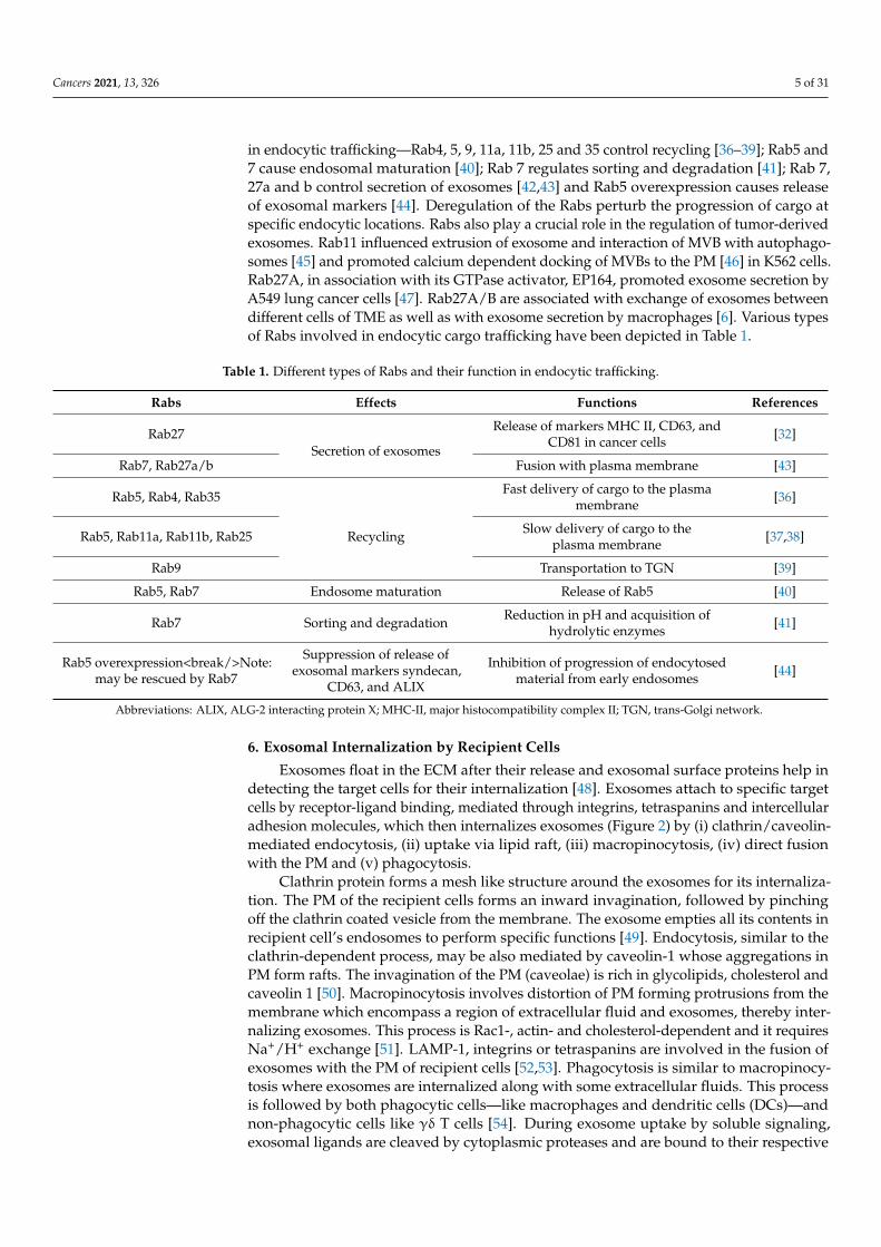
in endocytic trafficking—Rab4, 5, 9, 11a, 11b, 25 and 35 control recycling [36–39]; Rab5 and7 cause endosomal maturation [40]; Rab 7 regulates sorting and degradation [41]; Rab 7,27a and b control secretion of exosomes [42,43] and Rab5 overexpression causes releaseof exosomal markers [44]. Deregulation of the Rabs perturb the progression of cargo atspecific endocytic locations. Rabs also play a crucial role in the regulation of tumor-derivedexosomes. Rab11 influenced extrusion of exosome and interaction of MVB with autophago-somes [45] and promoted calcium dependent docking of MVBs to the PM [46] in K562 cells.Rab27A, in association with its GTPase activator, EP164, promoted exosome secretion byA549 lung cancer cells [47]. Rab27A/B are associated with exchange of exosomes betweendifferent cells of TME as well as with exosome secretion by macrophages [6]. Various typesof Rabs involved in endocytic cargo trafficking have been depicted in Table 1.
Table 1. Different types of Rabs and their function in endocytic trafficking.
Rabs Effects Functions References
Rab27Secretion of exosomes
Release of markers MHC II, CD63, andCD81 in cancer cells [32]
Rab7, Rab27a/b Fusion with plasma membrane [43]
Rab5, Rab4, Rab35
Recycling
Fast delivery of cargo to the plasmamembrane [36]
Rab5, Rab11a, Rab11b, Rab25 Slow delivery of cargo to theplasma membrane [37,38]
Rab9 Transportation to TGN [39]
Rab5, Rab7 Endosome maturation Release of Rab5 [40]
Rab7 Sorting and degradation Reduction in pH and acquisition ofhydrolytic enzymes [41]
Rab5 overexpression<break/>Note:may be rescued by Rab7
Suppression of release ofexosomal markers syndecan,
CD63, and ALIX
Inhibition of progression of endocytosedmaterial from early endosomes [44]
Abbreviations: ALIX, ALG-2 interacting protein X; MHC-II, major histocompatibility complex II; TGN, trans-Golgi network.
6. Exosomal Internalization by Recipient Cells
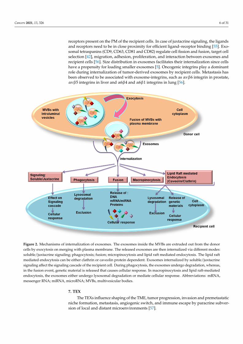
Exosomes float in the ECM after their release and exosomal surface proteins help indetecting the target cells for their internalization [48]. Exosomes attach to specific targetcells by receptor-ligand binding, mediated through integrins, tetraspanins and intercellularadhesion molecules, which then internalizes exosomes (Figure 2) by (i) clathrin/caveolin-mediated endocytosis, (ii) uptake via lipid raft, (iii) macropinocytosis, (iv) direct fusionwith the PM and (v) phagocytosis.
Clathrin protein forms a mesh like structure around the exosomes for its internaliza-tion. The PM of the recipient cells forms an inward invagination, followed by pinchingoff the clathrin coated vesicle from the membrane. The exosome empties all its contents inrecipient cell’s endosomes to perform specific functions [49]. Endocytosis, similar to theclathrin-dependent process, may be also mediated by caveolin-1 whose aggregations inPM form rafts. The invagination of the PM (caveolae) is rich in glycolipids, cholesterol andcaveolin 1 [50]. Macropinocytosis involves distortion of PM forming protrusions from themembrane which encompass a region of extracellular fluid and exosomes, thereby inter-nalizing exosomes. This process is Rac1-, actin- and cholesterol-dependent and it requiresNa+/H+ exchange [51]. LAMP-1, integrins or tetraspanins are involved in the fusion ofexosomes with the PM of recipient cells [52,53]. Phagocytosis is similar to macropinocy-tosis where exosomes are internalized along with some extracellular fluids. This processis followed by both phagocytic cells—like macrophages and dendritic cells (DCs)—andnon-phagocytic cells like γδ T cells [54]. During exosome uptake by soluble signaling,exosomal ligands are cleaved by cytoplasmic proteases and are bound to their respective
Cancers 2021, 13, 326 6 of 31
receptors present on the PM of the recipient cells. In case of juxtacrine signaling, the ligandsand receptors need to be in close proximity for efficient ligand–receptor binding [55]. Exo-somal tetraspanins (CD9, CD63, CD81 and CD82) regulate cell fission and fusion, target cellselection [42], migration, adhesion, proliferation, and interaction between exosomes andrecipient cells [56]. Size distribution in exosomes facilitates their internalization since cellshave a propensity for loading smaller exosomes [5]. Oncogenic integrins play a dominantrole during internalization of tumor-derived exosomes by recipient cells. Metastasis hasbeen observed to be associated with exosome-integrins, such as αvβ6 integrin in prostate,αvβ5 integrins in liver and α6β4 and α6β1 integrins in lung [56].
Cancers 2021, 13, x 6 of 31
exosomal ligands are cleaved by cytoplasmic proteases and are bound to their respective receptors present on the PM of the recipient cells. In case of juxtacrine signaling, the lig-ands and receptors need to be in close proximity for efficient ligand–receptor binding [55]. Exosomal tetraspanins (CD9, CD63, CD81 and CD82) regulate cell fission and fusion, tar-get cell selection [42], migration, adhesion, proliferation, and interaction between exo-somes and recipient cells [56]. Size distribution in exosomes facilitates their internalization since cells have a propensity for loading smaller exosomes [5]. Oncogenic integrins play a dominant role during internalization of tumor-derived exosomes by recipient cells. Me-tastasis has been observed to be associated with exosome-integrins, such as αvβ6 integrin in prostate, αvβ5 integrins in liver and α6β4 and α6β1 integrins in lung [56].
Figure 2. Mechanisms of internalization of exosomes. The exosomes inside the MVBs are extruded out from the donor cells by exocytosis on merging with plasma membrane. The released exosomes are then internalized via different modes: soluble/juxtacrine signaling; phagocytosis; fusion; micropinocytosis and lipid raft mediated endocytosis. The lipid raft mediated endocytosis can be either clathrin or caveolin protein dependent. Exosomes internalized by soluble/juxtacrine signaling affect the signaling cascade of the recipient cell. During phagocytosis, the exosomes undergo degradation, whereas, in the fusion event, genetic material is released that causes cellular response. In macropinocytosis and lipid raft-mediated endocytosis, the exosomes either undergo lysosomal degradation or mediate cellular response. Abbreviations: mRNA, messenger RNA; miRNA, microRNA; MVBs, multivesicular bodies.
7. TEX The TEXs influence shaping of the TME, tumor progression, invasion and premeta-
static niche formation, metastasis, angiogenic switch, and immune escape by paracrine subversion of local and distant microenvironments [57].
7.1. Oncogenic Signaling Involved in Exosomal Trafficking
Figure 2. Mechanisms of internalization of exosomes. The exosomes inside the MVBs are extruded out from the donorcells by exocytosis on merging with plasma membrane. The released exosomes are then internalized via different modes:soluble/juxtacrine signaling; phagocytosis; fusion; micropinocytosis and lipid raft mediated endocytosis. The lipid raftmediated endocytosis can be either clathrin or caveolin protein dependent. Exosomes internalized by soluble/juxtacrinesignaling affect the signaling cascade of the recipient cell. During phagocytosis, the exosomes undergo degradation, whereas,in the fusion event, genetic material is released that causes cellular response. In macropinocytosis and lipid raft-mediatedendocytosis, the exosomes either undergo lysosomal degradation or mediate cellular response. Abbreviations: mRNA,messenger RNA; miRNA, microRNA; MVBs, multivesicular bodies.
7. TEX
The TEXs influence shaping of the TME, tumor progression, invasion and premetastaticniche formation, metastasis, angiogenic switch, and immune escape by paracrine subver-sion of local and distant microenvironments [57].
Cancers 2021, 13, 326 7 of 31
7.1. Oncogenic Signaling Involved in Exosomal Trafficking
According to the genometastatic theory, complex biomolecules in exosomes transferoncogenic traits to target cells. Matrix cells in the TME interact with their oncogeniccounterparts through exosomes and mediate tumor evolution and progression. Exosomalcargoes confer oncogenic transformation, EMT, immune surveillance evasion, invasion,and metastatic properties to the recipient cells [58]. Hypoxia and extracellular acidityculminate in greater release of TEXs [58]. Cells having even one oncosuppressor mutationare more prone towards uptake of exosomal oncogenic factors. Mutations leading toupregulated mitogen-activated protein kinase (MAPK) signaling in cancer cells elevatedexosomes release [59]. Secretion of exosomes by activated platelets promoted MAPK andphosphoinositide 3-kinase (PI3K)/protein kinase B (Akt)/matrix metalloproteinase (MMP)signaling during cancer progression [31]. Expression of oncogenic RAS in non-tumorigenicepithelial cells promoted secretion of oncoprotein-rich exosomes [60]. Robust expression ofoncogenic and truncated forms of epidermal growth factor receptor (EGFR) vIII in gliomacells augmented exosomal secretion and transfer of oncogenic activity to other normalcells [61]. Mutation of liver kinase B1 (STK11), a tumor suppressor, increased exosomesecretion in lung cancer [62]. Secretion of exosomal mtDNA induced anaerobic metabolismand dormancy in cancer cells [31].
7.2. Exosomal miRNA-Mediated Cancer Promotion
Breast TEXs, enriched with Dicer, Protein Argonaut 2, and transactivation responseelement RNA-binding protein, processed precursor miRNAs into mature miRNAs forgene silencing in target cells and induced non-tumorigenic epithelial cells to form tu-mors [63]. Exosomal miRNAs suppressed cell proliferation by downregulating the C-X-C motif chemokine ligand 12 (CXCL12); exosomal-miR-23b augmented cell quiescenceby inhibiting myristoylated alanine-rich C-kinase substrate expression in the metastaticniche [64]; miR-10b molded the TME to promote tumor metastasis [65] of breast cancer (BC)cells. Astrocyte-derived exosomes suppressed phosphatase and tensin homolog (PTEN)by intracellular trafficking of miR-19a in metastatic BC and melanoma brain metasta-sis models [66]. Release of exosomal miR-1245 from mutant p53 cancer cells reorientedmacrophages to transforming growth factor-β (TGF-β)-rich tumor-associated macrophages(TAMs) which, in turn, propagated tumor progression [67]. Exosomal miR-105 and miR-939in BC and miR-181c in brain cancer dissolved tight junctions, caused vascular leakinessand induced metastasis [31].
7.3. Exosomes and TME
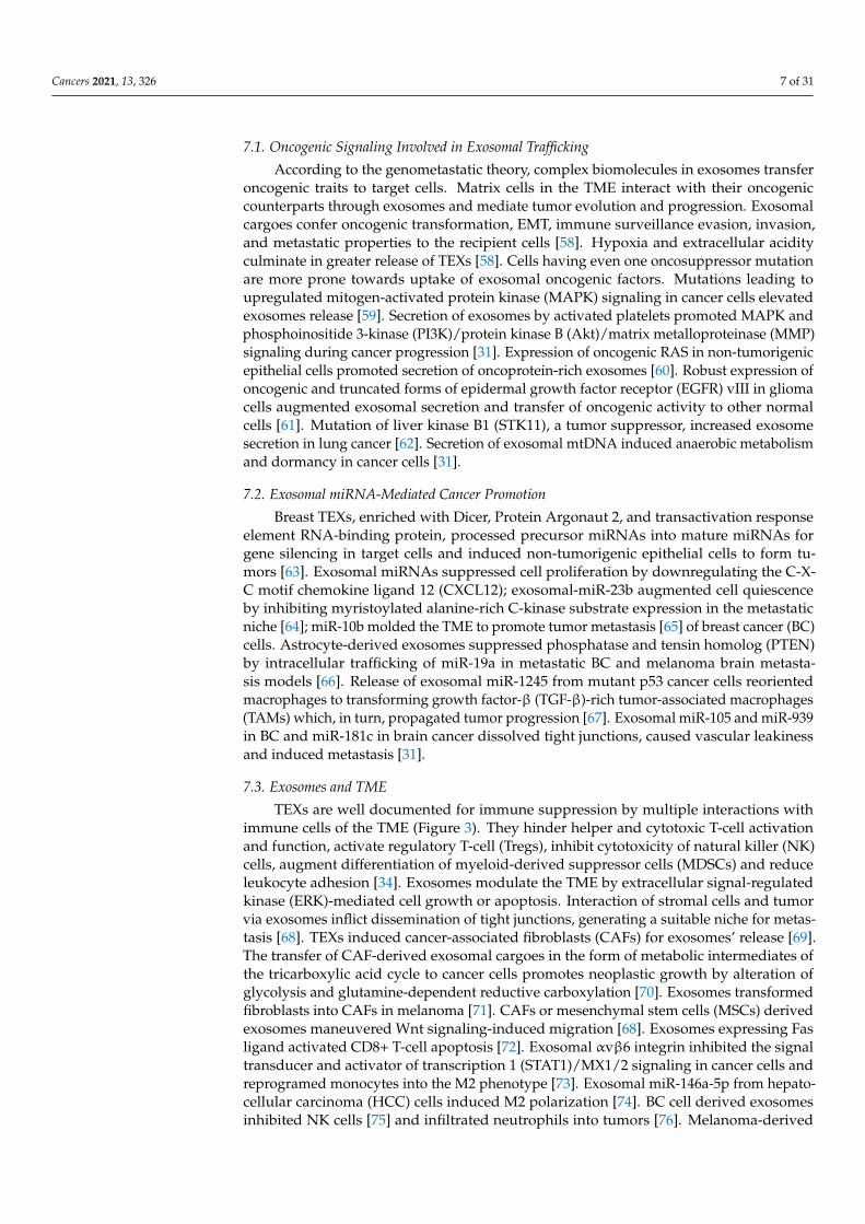
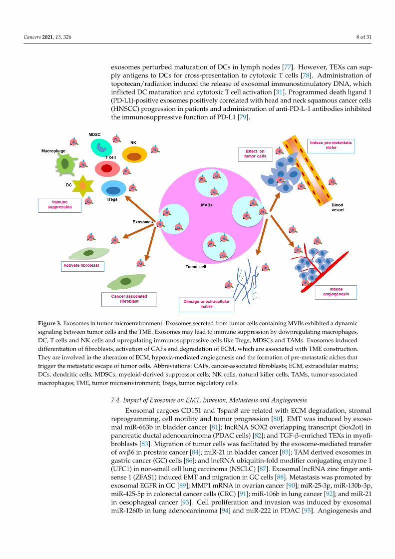
TEXs are well documented for immune suppression by multiple interactions withimmune cells of the TME (Figure 3). They hinder helper and cytotoxic T-cell activationand function, activate regulatory T-cell (Tregs), inhibit cytotoxicity of natural killer (NK)cells, augment differentiation of myeloid-derived suppressor cells (MDSCs) and reduceleukocyte adhesion [34]. Exosomes modulate the TME by extracellular signal-regulatedkinase (ERK)-mediated cell growth or apoptosis. Interaction of stromal cells and tumorvia exosomes inflict dissemination of tight junctions, generating a suitable niche for metas-tasis [68]. TEXs induced cancer-associated fibroblasts (CAFs) for exosomes’ release [69].The transfer of CAF-derived exosomal cargoes in the form of metabolic intermediates ofthe tricarboxylic acid cycle to cancer cells promotes neoplastic growth by alteration ofglycolysis and glutamine-dependent reductive carboxylation [70]. Exosomes transformedfibroblasts into CAFs in melanoma [71]. CAFs or mesenchymal stem cells (MSCs) derivedexosomes maneuvered Wnt signaling-induced migration [68]. Exosomes expressing Fasligand activated CD8+ T-cell apoptosis [72]. Exosomal αvβ6 integrin inhibited the signaltransducer and activator of transcription 1 (STAT1)/MX1/2 signaling in cancer cells andreprogramed monocytes into the M2 phenotype [73]. Exosomal miR-146a-5p from hepato-cellular carcinoma (HCC) cells induced M2 polarization [74]. BC cell derived exosomesinhibited NK cells [75] and infiltrated neutrophils into tumors [76]. Melanoma-derived
Cancers 2021, 13, 326 8 of 31
exosomes perturbed maturation of DCs in lymph nodes [77]. However, TEXs can sup-ply antigens to DCs for cross-presentation to cytotoxic T cells [78]. Administration oftopotecan/radiation induced the release of exosomal immunostimulatory DNA, whichinflicted DC maturation and cytotoxic T cell activation [31]. Programmed death ligand 1(PD-L1)-positive exosomes positively correlated with head and neck squamous cancer cells(HNSCC) progression in patients and administration of anti-PD-L-1 antibodies inhibitedthe immunosuppressive function of PD-L1 [79].
Cancers 2021, 13, x 8 of 31
exosomes perturbed maturation of DCs in lymph nodes [77]. However, TEXs can supply antigens to DCs for cross-presentation to cytotoxic T cells [78]. Administration of topo-tecan/radiation induced the release of exosomal immunostimulatory DNA, which in-flicted DC maturation and cytotoxic T cell activation [31]. Programmed death ligand 1 (PD-L1)-positive exosomes positively correlated with head and neck squamous cancer cells (HNSCC) progression in patients and administration of anti-PD-L-1 antibodies in-hibited the immunosuppressive function of PD-L1 [79].
Figure 3. Exosomes in tumor microenvironment. Exosomes secreted from tumor cells containing MVBs exhibited a dy-namic signaling between tumor cells and the TME. Exosomes may lead to immune suppression by downregulating mac-rophages, DC, T cells and NK cells and upregulating immunosuppressive cells like Tregs, MDSCs and TAMs. Exosomes induced differentiation of fibroblasts, activation of CAFs and degradation of ECM, which are associated with TME con-struction. They are involved in the alteration of ECM, hypoxia-mediated angiogenesis and the formation of pre-metastatic niches that trigger the metastatic escape of tumor cells. Abbreviations: CAFs, cancer-associated fibroblasts; ECM, extracel-lular matrix; DCs, dendritic cells; MDSCs, myeloid-derived suppressor cells; NK cells, natural killer cells; TAMs, tumor-associated macrophages; TME, tumor microenvironment; Tregs, tumor regulatory cells.
7.4. Impact of Exosomes on EMT, Invasion, Metastasis and Angiogenesis Exosomal cargoes CD151 and Tspan8 are related with ECM degradation, stromal re-
programming, cell motility and tumor progression [80]. EMT was induced by exosomal miR-663b in bladder cancer [81]; lncRNA SOX2 overlapping transcript (Sox2ot) in pancre-atic ductal adenocarcinoma (PDAC cells) [82]; and TGF-β-enriched TEXs in myofibro-blasts [83]. Migration of tumor cells was facilitated by the exosome-mediated transfer of αvβ6 in prostate cancer [84]; miR-21 in bladder cancer [85]; TAM derived exosomes in gastric cancer (GC) cells [86]; and lncRNA ubiquitin-fold modifier conjugating enzyme 1 (UFC1) in non-small cell lung carcinoma (NSCLC) [87]. Exosomal lncRNA zinc finger an-tisense 1 (ZFAS1) induced EMT and migration in GC cells [88]. Metastasis was promoted by exosomal EGFR in GC [89]; MMP1 mRNA in ovarian cancer [90]; miR-25-3p, miR-130b-3p, miR-425-5p in colorectal cancer cells (CRC) [91]; miR-106b in lung cancer [92]; and miR-21 in oesophageal cancer [93]. Cell proliferation and invasion was induced by exoso-mal miR-1260b in lung adenocarcinoma [94] and miR-222 in PDAC [95]. Angiogenesis and tumor progression were influenced by exosome mediated Wnt4/β-catenin signaling in CRC [96] and by vascular endothelial growth factor A (VEGF-A) enriched exosomes in
Figure 3. Exosomes in tumor microenvironment. Exosomes secreted from tumor cells containing MVBs exhibited a dynamicsignaling between tumor cells and the TME. Exosomes may lead to immune suppression by downregulating macrophages,DC, T cells and NK cells and upregulating immunosuppressive cells like Tregs, MDSCs and TAMs. Exosomes induceddifferentiation of fibroblasts, activation of CAFs and degradation of ECM, which are associated with TME construction.They are involved in the alteration of ECM, hypoxia-mediated angiogenesis and the formation of pre-metastatic niches thattrigger the metastatic escape of tumor cells. Abbreviations: CAFs, cancer-associated fibroblasts; ECM, extracellular matrix;DCs, dendritic cells; MDSCs, myeloid-derived suppressor cells; NK cells, natural killer cells; TAMs, tumor-associatedmacrophages; TME, tumor microenvironment; Tregs, tumor regulatory cells.
7.4. Impact of Exosomes on EMT, Invasion, Metastasis and Angiogenesis
Exosomal cargoes CD151 and Tspan8 are related with ECM degradation, stromalreprogramming, cell motility and tumor progression [80]. EMT was induced by exoso-mal miR-663b in bladder cancer [81]; lncRNA SOX2 overlapping transcript (Sox2ot) inpancreatic ductal adenocarcinoma (PDAC cells) [82]; and TGF-β-enriched TEXs in myofi-broblasts [83]. Migration of tumor cells was facilitated by the exosome-mediated transferof αvβ6 in prostate cancer [84]; miR-21 in bladder cancer [85]; TAM derived exosomes ingastric cancer (GC) cells [86]; and lncRNA ubiquitin-fold modifier conjugating enzyme 1(UFC1) in non-small cell lung carcinoma (NSCLC) [87]. Exosomal lncRNA zinc finger anti-sense 1 (ZFAS1) induced EMT and migration in GC cells [88]. Metastasis was promoted byexosomal EGFR in GC [89]; MMP1 mRNA in ovarian cancer [90]; miR-25-3p, miR-130b-3p,miR-425-5p in colorectal cancer cells (CRC) [91]; miR-106b in lung cancer [92]; and miR-21in oesophageal cancer [93]. Cell proliferation and invasion was induced by exosomalmiR-1260b in lung adenocarcinoma [94] and miR-222 in PDAC [95]. Angiogenesis and
Cancers 2021, 13, 326 9 of 31
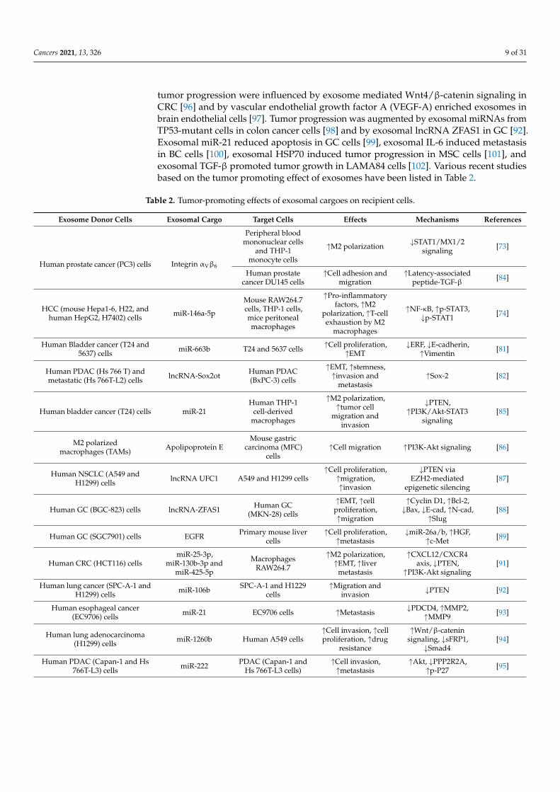
tumor progression were influenced by exosome mediated Wnt4/β-catenin signaling inCRC [96] and by vascular endothelial growth factor A (VEGF-A) enriched exosomes inbrain endothelial cells [97]. Tumor progression was augmented by exosomal miRNAs fromTP53-mutant cells in colon cancer cells [98] and by exosomal lncRNA ZFAS1 in GC [92].Exosomal miR-21 reduced apoptosis in GC cells [99], exosomal IL-6 induced metastasisin BC cells [100], exosomal HSP70 induced tumor progression in MSC cells [101], andexosomal TGF-β promoted tumor growth in LAMA84 cells [102]. Various recent studiesbased on the tumor promoting effect of exosomes have been listed in Table 2.
Table 2. Tumor-promoting effects of exosomal cargoes on recipient cells.
Exosome Donor Cells Exosomal Cargo Target Cells Effects Mechanisms References
Human prostate cancer (PC3) cells Integrin αVβ6
Peripheral bloodmononuclear cells
and THP-1monocyte cells
↑M2 polarization ↓STAT1/MX1/2signaling [73]
Human prostatecancer DU145 cells
↑Cell adhesion andmigration
↑Latency-associatedpeptide-TGF-β [84]
HCC (mouse Hepa1-6, H22, andhuman HepG2, H7402) cells miR-146a-5p
Mouse RAW264.7cells, THP-1 cells,mice peritonealmacrophages
↑Pro-inflammatoryfactors, ↑M2
polarization, ↑T-cellexhaustion by M2
macrophages
↑NF-κB, ↑p-STAT3,↓p-STAT1 [74]
Human Bladder cancer (T24 and5637) cells miR-663b T24 and 5637 cells ↑Cell proliferation,
↑EMT↓ERF, ↓E-cadherin,
↑Vimentin [81]
Human PDAC (Hs 766 T) andmetastatic (Hs 766T-L2) cells lncRNA-Sox2ot Human PDAC
(BxPC-3) cells
↑EMT, ↑stemness,↑invasion and
metastasis↑Sox-2 [82]
Human bladder cancer (T24) cells miR-21Human THP-1
cell-derivedmacrophages
↑M2 polarization,↑tumor cell
migration andinvasion
↓PTEN,↑PI3K/Akt-STAT3
signaling[85]
M2 polarizedmacrophages (TAMs) Apolipoprotein E
Mouse gastriccarcinoma (MFC)
cells↑Cell migration ↑PI3K-Akt signaling [86]
Human NSCLC (A549 andH1299) cells lncRNA UFC1 A549 and H1299 cells
↑Cell proliferation,↑migration,↑invasion
↓PTEN viaEZH2-mediated
epigenetic silencing[87]
Human GC (BGC-823) cells lncRNA-ZFAS1 Human GC(MKN-28) cells
↑EMT, ↑cellproliferation,↑migration
↑Cyclin D1, ↑Bcl-2,↓Bax, ↓E-cad, ↑N-cad,
↑Slug[88]
Human GC (SGC7901) cells EGFR Primary mouse livercells
↑Cell proliferation,↑metastasis
↓miR-26a/b, ↑HGF,↑c-Met [89]
Human CRC (HCT116) cellsmiR-25-3p,
miR-130b-3p andmiR-425-5p
MacrophagesRAW264.7
↑M2 polarization,↑EMT, ↑liver
metastasis
↑CXCL12/CXCR4axis, ↓PTEN,
↑PI3K-Akt signaling[91]
Human lung cancer (SPC-A-1 andH1299) cells miR-106b SPC-A-1 and H1229
cells↑Migration and
invasion ↓PTEN [92]
Human esophageal cancer(EC9706) cells miR-21 EC9706 cells ↑Metastasis ↓PDCD4, ↑MMP2,
↑MMP9 [93]
Human lung adenocarcinoma(H1299) cells miR-1260b Human A549 cells
↑Cell invasion, ↑cellproliferation, ↑drug
resistance
↑Wnt/β-cateninsignaling, ↓sFRP1,
↓Smad4[94]
Human PDAC (Capan-1 and Hs766T-L3) cells miR-222 PDAC (Capan-1 and
Hs 766T-L3 cells)↑Cell invasion,↑metastasis
↑Akt, ↓PPP2R2A,↑p-P27 [95]
Cancers 2021, 13, 326 10 of 31
Table 2. Cont.
Exosome Donor Cells Exosomal Cargo Target Cells Effects Mechanisms References
Hypoxic human CRC (HT29 andHCT116) cells Wnt4
Endothelial(HUVECs) and CRC
(HT29) cells
↑Proliferation,↑angiogenesis,↑migration
↑β-Catenin signaling [96]
TP53-mutant (HT29) coloncancer cells
miR-1249-5p,miR-6737-5p, and
miR-6819-5p
Human colonfibroblasts
(CCD-18Co) cells↑Tumor progression ↓TP53 [98]
Murine bone marrow–derivedmacrophages miR-21 Human GC (MFC,
MGC-803) cells
↓Apoptosis,↑resistance to
cisplatin
↑PI3K/AKTsignalling, ↓PTEN [99]
Co-culture of THP-1-derivedmacrophages exposed to
apoptotic human BC (MCF-7 orMDA-MB-231) cells
IL-6 Naive (MCF-7 orMDA-MB-231) cells
↑Proliferation,↑metastasis
↑p-STAT3, ↑cyclinD1, ↑MMP2, ↑MMP9 [100]
Human lung cancer (A549) cells HSP70 MSCs extracted fromhuman adipose tissue
Pro-inflammatoryMSCs, ↑tumor
growth
↑TLR-2/NF-κBsignaling, ↑IL-6,↑IL-8, ↑MCP-1
[101]
Human chronic myeloid leukemia(LAMA84) cells TGF-β LAMA84 cells
↑Proliferation,↓apoptosis, ↑tumor
growth
↑SMAD 2/3, ↑Bcl-w,↑Bcl-xL, ↑survivin,
↓BAD, ↓BAX,↓PUMA
[102]
Human BC (MCF-7) tamoxifenresistant cells miR-221/222 Human BC (MCF-7)
wild type cells↑Resistance to
tamoxifen ↓P27, ↓ERα, [103]
Human cisplatin resistantA549 cells miR-100-5p Human A549 cells ↑Resistance to
cisplatin ↑mTOR [104]
Gemcitabine treated humanPDAC CAFs Snail and miR-146a Human pancreatic
cancer L3.6pl cells
↑proliferation,↑resistance togemcitabine
↑Snail, ↑miR-146a [105]
Human HER-2-positive BCtrastuzumab resistant (SKBR-3
and BT474) cells
lncRNAAFAP1-AS1
SKBR-3 andBT474 cells
↑Resistance totrastuzumab ↑ERBB2 [106]
Tamoxifen resistant BC(LCC2) cells lncRNA UCA1 ER-positive BC
MCF-7 cells
↑Cell viability,↑resistance to
tamoxifen↓caspase-3 [107]
Human GC (MGC-803 andMKN-45) cisplatin resistant cells lncRNA HOTTIP MGC-803 and
MKN-45 cells↑Resistance to
cisplatin ↑HMGA1 [108]
Symbols: ↑, upregulated; ↓, downregulated; Abbreviations: AFAP1-AS1, actin filament associated protein1 antisense RNA 1; Akt, proteinkinase B; Bad, Bcl-2 associated agonist of cell death; Bax, Bcl-2-associated X protein; Bcl-2, B-cell lymphoma 2; c-Met, Mesenchymal-epithelialtransition factor; CXCL12, C-X-C motif chemokine ligand 12; CXCR4, C-X-C chemokine receptor type 4; Erα, estrogen receptor-α; ERF,Ets2-repressor factor; ERBB2, erythroblastic oncogene B; HGF, hepatocyte growth factor; HMGA1, High-mobility group A1; HOTTIP,HOXA transcript at the distal tip; MCP-1, monocyte chemoattractant protein-1; MMP, matrix metalloproteinase; NF-κB, nuclear factorkappa-light-chain-enhancer of activated B cells; PDCD4, programmed cell death 4; PI3K, phosphoinositide 3-kinase; PPP2R2A, proteinphosphatase 2 regulatory subunit B alpha; PTEN, phosphatase and tensin homolog; PUMA, p53 upregulated modulator of apoptosis; sFRP,secreted frizzled-related protein 1; STAT, signal transducer and activator of transcription; Sox-2, sex determining region Y-box 2; TGF-β,transforming growth factor-β; TLR-2, toll-like receptor 2; TP53, tumor protein p53.
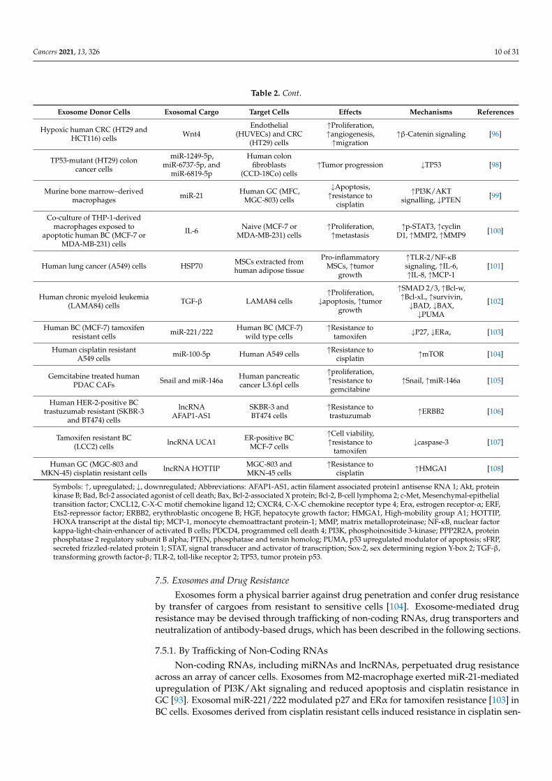
7.5. Exosomes and Drug Resistance
Exosomes form a physical barrier against drug penetration and confer drug resistanceby transfer of cargoes from resistant to sensitive cells [104]. Exosome-mediated drugresistance may be devised through trafficking of non-coding RNAs, drug transporters andneutralization of antibody-based drugs, which has been described in the following sections.
7.5.1. By Trafficking of Non-Coding RNAs
Non-coding RNAs, including miRNAs and lncRNAs, perpetuated drug resistanceacross an array of cancer cells. Exosomes from M2-macrophage exerted miR-21-mediatedupregulation of PI3K/Akt signaling and reduced apoptosis and cisplatin resistance inGC [93]. Exosomal miR-221/222 modulated p27 and ERα for tamoxifen resistance [103] inBC cells. Exosomes derived from cisplatin resistant cells induced resistance in cisplatin sen-
Cancers 2021, 13, 326 11 of 31
sitive A549 cells in a miR-100-5p-dependent manner [104]. In ovarian cancer cells, exosomalmiR-443 induced senescence and resistance against paclitaxel [109]. In prostate cancer, CAFderived exosomes conferred gemcitabine resistance via Snail and miR-146a [105]. Exoso-mal cargo-lncRNA UCA1 mediated tamoxifen resistance [107] and lncRNA actin filamentassociated protein1 antisense RNA 1 (AFAP1-AS1) conferred trastuzumab resistance bybinding to AU binding factor 1 and translating erythroblastic oncogene B2 (ERBB2) [106]in BC cells. MSC-derived exosomes aided the transfer of lncRNA PSMA3-AS1 to myelomacells and exerted resistance against proteasome inhibitor [110]. In GC, exosomal lncRNAHoxA transcript at a distal tip (HOTTIP) made sensitive GC cells cisplatin resistant [108].
7.5.2. By Trafficking of Drug Transporters and Neutralizing Antibody-Based Drugs
The exosome-mediated transfer of drug transporter molecules is intimately associatedwith the spread of drug resistance across diverse cancer forms. Exosomes transported P-glycoprotein (P-gp) from doxorubicin-resistant cells [68] and multidrug resistance protein-1(MDR-1) from docetaxel-resistant cells [111] to confer drug resistance in sensitive BC cells.Recently, it has been evidenced that exosome-mediated transfer of chloride intracellularchannel 1 upregulated P-gp and B cell lymphoma-2 (Bcl-2) and conferred vincristineresistance in GC cell line SGC-7901 [112].
B-cell lymphoma derived exosomes modulated ATP-binding cassette (ABC) trans-porter A3, carried CD20 antigen which shielded the cancer cells against therapeutic CD20antibodies and evaded immune surveillance [113]. Exocytosis of TEXs from human epider-mal growth factor receptor 2 (HER2) positive BC cells expressed specific decoy moleculesand conferred resistance against monoclonal antibody trastuzumab, thus depicting thatTEXs are also involved in neutralizing antibody based drugs [114].
8. Strategies against Tumor-Derived Exosomes
There have been, primarily, three approaches for the management of exosomes associ-ated with pathogenesis, as described below.
8.1. Suppression of Exosome Biogenesis and Trafficking
Genetic knockdown of tumor suppressor TSG1 (protein involved with exosome bio-genesis and trafficking) reduced Wnt5b-positive exosomes in colon cancer [115]. Sup-pression of annexin A1 (responsible for membrane contact sites, inward vesiculation andexosome biosynthesis) reduced the number of secreted exosomes in pancreatic cancercells [116]. Manumycin A was reported to inhibit ESCRT-dependent exosome biogenesisby modulating Ras/Raf/ERK1/2/heterogeneous nuclear ribonucleoprotein H1 axis inprostate cancer cells [117].
Small molecule inhibitor GW4869 against nSMase2 reduced secretion of ceramide en-riched exosomes [118] and sensitized breast tumors by inhibition of exosomal PD-L1 [119].Knockout of nSMase2 reduced exosome secretion, angiogenesis and metastasis in breasttumors [120]. Another inhibitor of lipid metabolism, pantethine, a pantothenic acid (vi-tamin B5) derivative, depleted the release of exosomes in MCF-7 variants and increaseddoxorubicin responsiveness [121]. Genetic silencing of Rab27A/B reduced exosomal se-cretion by HNSCC and macrophages, thereby minimizing metastasis in BC cells [76] andlung metastasis in melanoma [122]. PRAS40 downregulated Akt, downstream of TGF-β, and mediated antagonistic effects against exosome secretion and chemoresistance inbreast and lung cancer cells [123]. WEB2086, a platelet-activating factor receptor (PAFR)antagonist, was shown to reduce gemcitabine-induced exosome release in PAFR-positivepancreatic cancer cells [124]. Other exosome extrusion inhibitors, such as chloramidine,bisindolylmaleimide-I, imipramine, d-pantethine, and calpeptin, and calcium chelators,such as ethylene glycol bis (2-aminoethyl ether) tetra-acetic acid, increased responsive-ness toward 5-FU in prostate and BC cells [125]. The inhibition of protease-activatedreceptor 2 by an anticoagulant, apixaban, which binds to the tissue factor–factor VIIacomplex, downregulated the secretion of TF-bearing exosomes from pancreatic cancer
Cancers 2021, 13, 326 12 of 31
cells [126]. Dasitinib inhibited exosome release and beclin-1/Vps34 mediated autophagyin imatinib resistant K562 cells [127]. Reduced exosome secretion by synthetic peptide(constructed with a derivative of the secretion modification region of HIV-1 Nef protein, aN-terminus anchored polyethylene glycol residue and a c-terminus cluster in peptide) [128]and by Docosahexaenoic acid (a polyunsaturated fatty acid) [34] inhibited metastasis andangiogenesis, respectively, in BC cells.
8.2. Depletion of Exosome Uptake
A synthetic nanoparticle, which is a prototype of high-density lipoprotein, was usedas an agonist of the scavenger receptor type B-1 (SR-B1) which eliminated cholesterolfrom lipid rafts and prevented exosome uptake by SR-B1 expressing cancer cells [129].Other agents, such as heparin sulfate proteoglycans, methyl-β cyclodextrin (moleculeused for cholesterol removal from natural and artificial membranes) and dynasore (dy-namin inhibitor), have been reported to abrogate exosome endocytosis in cancer cells [130].Heparin and dynasore attenuated the uptake of multiple myeloma-derived exosome bybone marrow stromal cells and inhibited phosphorylation of STAT1, STAT3, and ERK1/2signaling pathways [131]. Radiation-derived exosomes made the recipient cancer cellsradiation-resistant and aggravated proliferation. Heparin and simvastatin attenuatedradiation-derived exosome uptake by recipient cells in in vitro and in vivo models ofglioblastoma [132].
8.3. Modulation of Harmful Exosomal Cargo and Inhibition of Exosome Dissemination
Alteration of exosomal cargoes was achieved by viral manipulation or by incorpo-ration of viral proteins/RNA into secreted exosomes [133]. Curcumin culminated theimmunosuppressive effect of exosomes in BC by deregulation of the ubiquitin-proteasomesystem and cargo sorting of ILVs [134]. Subscapular sinus CD169+ macrophages boundwith exosomes restricted their interaction with B cells, promoting tumor progression [135].Exosome release was inhibited by inhibitors like indometacin (COX2 inhibitor) in combina-tion with rapamycin (interfere with MVB biogenesis) in B lymphoma cells, by suppressingATP-binding cassette sub-family A member 3 expression of the lymphoma cells and in-duced the cells to undergo complement dependent cytolysis under the effect of drugrituximab [113].
8.4. Removal of Exosomes
A microfluidics-based technology-microscale acoustic standing wave technology facil-itates clearance of exosomes from circulation [136]. Innate immune system in co-operationwith opsonization effects of complement proteins may be used for elimination of exo-somes [137]. Opsonization of exosomal markers CD9 and CD63 by targeting anti CD9 andanti CD63 antibodies elevated exosomes representation to the macrophages, leading toexosomes’ elimination, which suppressed lung metastasis in vivo [138]. In colorectal can-cer, dimethyl amiloride depleted exosomes, thereby elevating cyclophosphamide efficacyagainst the cancer cells [139].
9. Cancer Management with Exosomes
Exosomes have emerged as a new arena of clinical interest due to their prospectiveuse in diagnostic applications as potential biomarkers, for carrying specific information oftheir progenitor cells, as well as for being ideal candidates for liquid biopsy [56].
9.1. Preclinical Studies on Anticancer Potential of Exosomal Cargoes
Uptake of exosomal contents does not always confer procarcinogenic signaling. Thereare instances where exosomal proteins promoted anticarcinogenic signaling pathways,e.g., exosomal uptake with payload of gastrokine1 suppressed H-Ras/Raf/MEK/ERK-mediated gastric carcinogenesis in gastric epithelial cells [140]. The miR-375 carried byexosomes inhibited cell proliferation and invasive capability in colon cancer cells through
Cancers 2021, 13, 326 13 of 31
Bcl-2 blocking [141]. Exosomal miR-520b derived from normal fibroblasts cells inhibitedproliferation and migration of pancreatic cancer cells [142]. The migratory behavior oflung cancer cells was reduced by exosomal miR-497 through suppression of growth fac-tors, cyclin E1 and VEGF [143]. Exosomal circulating RNA circ-0051443 inhibited tumorprogression through apoptosis induction in HCC cells [144]. In BC cells, exosomal miR-100derived from MSCs inhibited angiogenesis in vitro via modulating mTOR/HIF-1α/VEGFsignaling [145].
9.2. Exosomes as Biomarkers
Cancer cells secrete exosomes ten times higher than normal cells, which makes TEXsmajor potential candidates for liquid biopsy needed for cancer diagnosis and progno-sis [57]. The release of exosomes in the extracellular space also aids in cancer diagnosisby examining their increased levels in various body fluids, such as blood, ascites fluid,urine, and saliva [146]. Exosomal DNA represents the entire genome; therefore, liquidbiopsies of plasma aid in early detection of cancer-specific mutations. Exosomal CD63and caveolin-1 served as non-invasive markers of melanoma [121]. Exosomal lncRNA,either with miR-21 or alone, was correlated with tumor classification (III/IV), stage oftumor and lymph node/distant metastasis in many cancer types [5]. Differential expressionof exosomal miR-150, miR-155, and miR-1246 in serum of normal individuals and acutemyeloid leukemia patients detected minimal residual disease [147]. Phosphatidylserinepresent on the exosomal surface also serves as a biomarker for diagnosis of early-stagecancer [148]. However, exosomal biomarkers are often overshadowed by highly prevalentcomplex proteins of the body fluids. Exosome isolation from body fluids follows either ofthe three methods, namely differential centrifugation coupled with ultracentrifugation, im-munoaffinity pull-down, and density gradient separation. Mining of exosomal biomarkersfrom body fluid of cancer patients has been explored with fluorescence-based analyticaltechniques, electrochemical aptamer-based detection methods, localized surface plasmonresonance and surface-enhanced Raman scattering [149]. Though exosome biomarker anal-ysis has tremendous translational potential, a gold standard for exosome isolation underclinical settings is yet to be achieved [150]. Since there is no definite consensus for isolationof exosomes, the best suitable body fluid for exosome isolation is also under investigation.
9.3. Role of Exosomes in Immunotherapy and Vaccine Development
DCs and other antigen presenting cells (APCs) derived exosomes are loaded withspecific drugs; miRNAs of interest or even exosomes alone are implemented to triggerimmune response in the recipient individuals (Figure 4). DC-based exosomes, in therapy,are beneficial as they possess abundant surface lactadherin that helps in efficient exosomeuptake [151]. The functional moieties, such as MHC-I, MHC-II, CD40, CD80, CD86 TNF,FasL, TRAIL and natural killer group 2D (NKG2D) ligands on the surface of DC-derivedexosomes, facilitate in imparting innate and adaptive antitumor immune response [152].DC-derived exosomes activated NK cells in NKG2D and interkeukin (IL)-15Rα liganddependent mode, which restored 50% functionality of NK cells and was implemented as acell free vaccination strategy [153]. The administration of adjuvants, such as IFN-γ, Toll-like receptor agonists, and polyinosinic: polycyctidylic acid, was explored for productionof mature DC-derived exosomes which showed greater potential for activation of Th1cells [154,155]. Immunogenic cell death was induced by melphalan, an anticancer drug, inmultiple myeloma cells by increasing the damage-associated molecular pattern containingexosomes, thus triggering NK cell cytotoxicity [156]. A histone deacetylase inhibitor, MS-275, increased the release of Hsp70 and MHC-I polypeptide-related sequence B (MICB)-richexosomes which induced NK cytotoxicity and lymphocyte proliferation [157]. Heat shocktreatment increasing the immunostimulatory activities of TEXs has been demonstratedin A20 lymphoma/leukemia cells. Heat shock tumor derived exosomes were observedto possess more immune-stimulating activities due to elevated expression of MHC andincreased levels of cytokines, such as IL-1β, IL-12p40, and TNF-α [158].
Cancers 2021, 13, 326 14 of 31Cancers 2021, 13, x 14 of 31
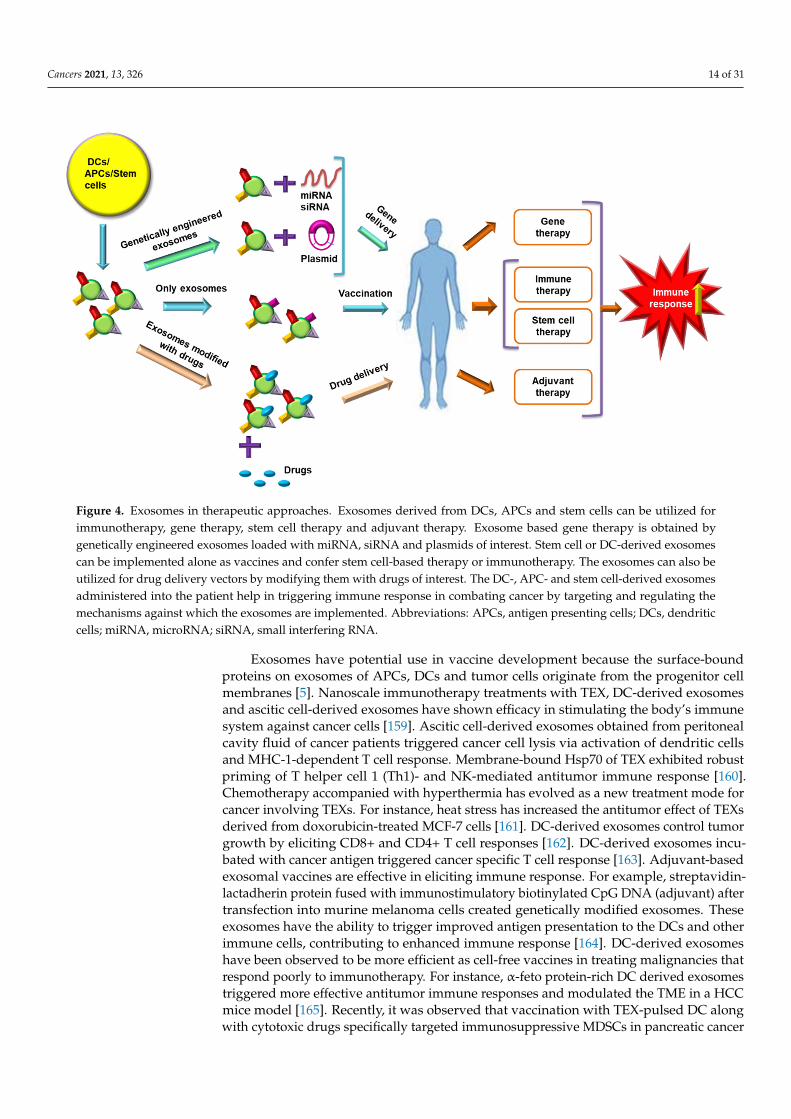
Figure 4. Exosomes in therapeutic approaches. Exosomes derived from DCs, APCs and stem cells can be utilized for im-munotherapy, gene therapy, stem cell therapy and adjuvant therapy. Exosome based gene therapy is obtained by genet-ically engineered exosomes loaded with miRNA, siRNA and plasmids of interest. Stem cell or DC-derived exosomes can be implemented alone as vaccines and confer stem cell-based therapy or immunotherapy. The exosomes can also be uti-lized for drug delivery vectors by modifying them with drugs of interest. The DC-, APC- and stem cell-derived exosomes administered into the patient help in triggering immune response in combating cancer by targeting and regulating the mechanisms against which the exosomes are implemented. Abbreviations: APCs, antigen presenting cells; DCs, dendritic cells; miRNA, microRNA; siRNA, small interfering RNA.
Exosomes have potential use in vaccine development because the surface-bound pro-teins on exosomes of APCs, DCs and tumor cells originate from the progenitor cell mem-branes [5]. Nanoscale immunotherapy treatments with TEX, DC-derived exosomes and ascitic cell-derived exosomes have shown efficacy in stimulating the body’s immune sys-tem against cancer cells [159]. Ascitic cell-derived exosomes obtained from peritoneal cav-ity fluid of cancer patients triggered cancer cell lysis via activation of dendritic cells and MHC-1-dependent T cell response. Membrane-bound Hsp70 of TEX exhibited robust priming of T helper cell 1 (Th1)- and NK-mediated antitumor immune response [160]. Chemotherapy accompanied with hyperthermia has evolved as a new treatment mode for cancer involving TEXs. For instance, heat stress has increased the antitumor effect of TEXs derived from doxorubicin-treated MCF-7 cells [161]. DC-derived exosomes control tumor growth by eliciting CD8+ and CD4+ T cell responses [162]. DC-derived exosomes incu-bated with cancer antigen triggered cancer specific T cell response [163]. Adjuvant-based exosomal vaccines are effective in eliciting immune response. For example, streptavidin-lactadherin protein fused with immunostimulatory biotinylated CpG DNA (adjuvant) af-ter transfection into murine melanoma cells created genetically modified exosomes. These exosomes have the ability to trigger improved antigen presentation to the DCs and other immune cells, contributing to enhanced immune response [164]. DC-derived exosomes have been observed to be more efficient as cell-free vaccines in treating malignancies that respond poorly to immunotherapy. For instance, α-feto protein-rich DC derived exo-somes triggered more effective antitumor immune responses and modulated the TME in a HCC mice model [165]. Recently, it was observed that vaccination with TEX-pulsed DC along with cytotoxic drugs specifically targeted immunosuppressive MDSCs in pancreatic cancer cells [166]. DNA vaccines prepared by fusing ovalbumin antigen with lactadherin
Figure 4. Exosomes in therapeutic approaches. Exosomes derived from DCs, APCs and stem cells can be utilized forimmunotherapy, gene therapy, stem cell therapy and adjuvant therapy. Exosome based gene therapy is obtained bygenetically engineered exosomes loaded with miRNA, siRNA and plasmids of interest. Stem cell or DC-derived exosomescan be implemented alone as vaccines and confer stem cell-based therapy or immunotherapy. The exosomes can also beutilized for drug delivery vectors by modifying them with drugs of interest. The DC-, APC- and stem cell-derived exosomesadministered into the patient help in triggering immune response in combating cancer by targeting and regulating themechanisms against which the exosomes are implemented. Abbreviations: APCs, antigen presenting cells; DCs, dendriticcells; miRNA, microRNA; siRNA, small interfering RNA.
Exosomes have potential use in vaccine development because the surface-boundproteins on exosomes of APCs, DCs and tumor cells originate from the progenitor cellmembranes [5]. Nanoscale immunotherapy treatments with TEX, DC-derived exosomesand ascitic cell-derived exosomes have shown efficacy in stimulating the body’s immunesystem against cancer cells [159]. Ascitic cell-derived exosomes obtained from peritonealcavity fluid of cancer patients triggered cancer cell lysis via activation of dendritic cellsand MHC-1-dependent T cell response. Membrane-bound Hsp70 of TEX exhibited robustpriming of T helper cell 1 (Th1)- and NK-mediated antitumor immune response [160].Chemotherapy accompanied with hyperthermia has evolved as a new treatment mode forcancer involving TEXs. For instance, heat stress has increased the antitumor effect of TEXsderived from doxorubicin-treated MCF-7 cells [161]. DC-derived exosomes control tumorgrowth by eliciting CD8+ and CD4+ T cell responses [162]. DC-derived exosomes incu-bated with cancer antigen triggered cancer specific T cell response [163]. Adjuvant-basedexosomal vaccines are effective in eliciting immune response. For example, streptavidin-lactadherin protein fused with immunostimulatory biotinylated CpG DNA (adjuvant) aftertransfection into murine melanoma cells created genetically modified exosomes. Theseexosomes have the ability to trigger improved antigen presentation to the DCs and otherimmune cells, contributing to enhanced immune response [164]. DC-derived exosomeshave been observed to be more efficient as cell-free vaccines in treating malignancies thatrespond poorly to immunotherapy. For instance, α-feto protein-rich DC derived exosomestriggered more effective antitumor immune responses and modulated the TME in a HCCmice model [165]. Recently, it was observed that vaccination with TEX-pulsed DC alongwith cytotoxic drugs specifically targeted immunosuppressive MDSCs in pancreatic cancer
Cancers 2021, 13, 326 15 of 31
cells [166]. DNA vaccines prepared by fusing ovalbumin antigen with lactadherin presenton exosomal surface diminished fibrosarcoma, thymoma and melanoma metastasis byactivating T lymphocytes [167].
9.4. Exosome-Based RNA Therapy
Exosome-based miRNA therapy exhibited immunosuppressive properties by control-ling the gene expressions [19]. An early study reported that exosomes derived from humanembryonic kidney cells were effective in regressing tumor growth by delivering miR-let7ain an EGFR-positive BC xenograft model [168]. The MSCs transfected with miR-124aenhanced exosomes carrying the RNA of interest production, which, when implementedagainst gliomas, reduced the cell viability and targeted FOXA2 that caused accumulationof lipids [169]. Transfer of lncRNA PTEN pseudogene 1 by exosomes derived from normalcells to bladder cancer cells reduced tumor progression in vitro and in vivo [170].
Exosomes also mediated targeted delivery of siRNA, e.g., siRNA transfected intoexosomes targeted RAD51 and RAD52 in Hela and fibrosarcoma cells, which inhibitedproliferation of the recipient cells [171]. Engineered exosomes containing IL-3 ligandor functional siRNA for BCR-ABL were successfully used against imatinib resistance inchronic myeloid leukemia patients [172]. Exosomes used for trafficking RNA interference(RNAi) mediators counteracted against oncogenic KRAS and improved overall survivalin mouse models of pancreatic cancer [173]. Delivery of engineered exosome mediatedsiRNA inhibited post-operative metastasis of BC, indicating a promising strategy againsttumor progression [174]. Successful delivery of antisense miRNA oligonucleotides againstmiR-21 by electroporating them in exosomal membrane improved the treatment efficacyfor glioblastoma by inducing the expression of PTEN and PDCD4, resulting in decreasedtumor size [175].
9.5. Exosomes in Stem Cell Therapy
Normal stem cell-derived exosomes are free of tumorigenic factors and are potentialcandidates for stem cell therapy [176]. MSC-derived exosomes can protect their cargoesfrom degradation, facilitate easier uptake by recipient cells, elicit low toxicity and immuno-genicity, and these exosomes can be modified to enhance cell type-specific targeting andmay be a prospective tool for cell-free based therapeutic approaches [177]. Exosomal miR-144 derived from bone marrow derived MSC retarded the spread of NSCLC by targetingcyclin E1 or E2 [178]. Exosomes released from miR-101-3p overexpressing MSCs negativelyaffected the proliferation and migration of oral cancer cells by targeting the collagen type Xα1 chain [179]. MSC-derived exosomes were genetically engineered by loading them withpolo-like kinase 1 (PLK-1)-siRNA and were utilized for PLK1 gene silencing in bladdercancer [180]. The primary hurdles of stem cell-based therapy, such as teratoma formationand embolization, are less frequent with exosome-based stem cell therapeutics. Exosomessecreted from induced pluripotent stem cells may exert better therapeutic effects [163].
9.6. Exosomes in Drug Delivery
Normal cell derived exosomes exhibit excellent biodistribution, biocompatibility, lowimmunogenicity, capacity to cross the blood–brain barrier and high target specificity, whichmake them potential candidates for drug delivery in cancer [5]. The exosomal surfaceproteins regulate efficient drug delivery because of their involvement in exosomes uptakeby the tumorigenic recipient cells [181]. Exosomes derived from androgen-sensitive hu-man prostate adenocarcinoma cells carrying paclitaxel negatively affect the cancer cells’viability [182]. DC-derived exosomes in BC and macrophage-derived exosomes in lungcancer were loaded with the drugs trastuzumab and paclitaxel, respectively, and success-fully delivered to the recipients [183,184]. Moreover, exosomes loaded with doxorubicinconjugated with gold nanoparticles showed anticancer effect against lung cancer cells [185].Exosomes with A disintegrin and metalloproteinase 15 (ADAM15) expression (A15-Exo)co-delivered with doxorubicin and cholesterol-modified miRNA 159 exhibited anticancer
Cancers 2021, 13, 326 16 of 31
effect in BC cells [186]. Paclitaxel loaded exosomes showed sensitivity towards MDRcancer cells via by-passing P-gp-mediated drug efflux and also inhibited metastasis ina lung cancer xenograft model [187]. Unmodified exosomes encapsulated with doxoru-bicin reduced tumor proliferation in a mouse mammary carcinoma xenograft model [137].Exosomal delivery of doxorubicin induced its therapeutic activity in xenograft modelsof breast and ovarian cancer [188]. Exosomes isolated from engineered immature DCs(expressed Lamp2b fused with αv integrin-specific iRGD peptide (CRGDKGPDC)) loadedwith doxorubicin successfully targeted αv integrin-positive breast tumor cells [189]. Exo-some encapsulated gemcitabine exhibited anticancer properties in autologous pancreaticcancer cells and in a xenograft model [190].
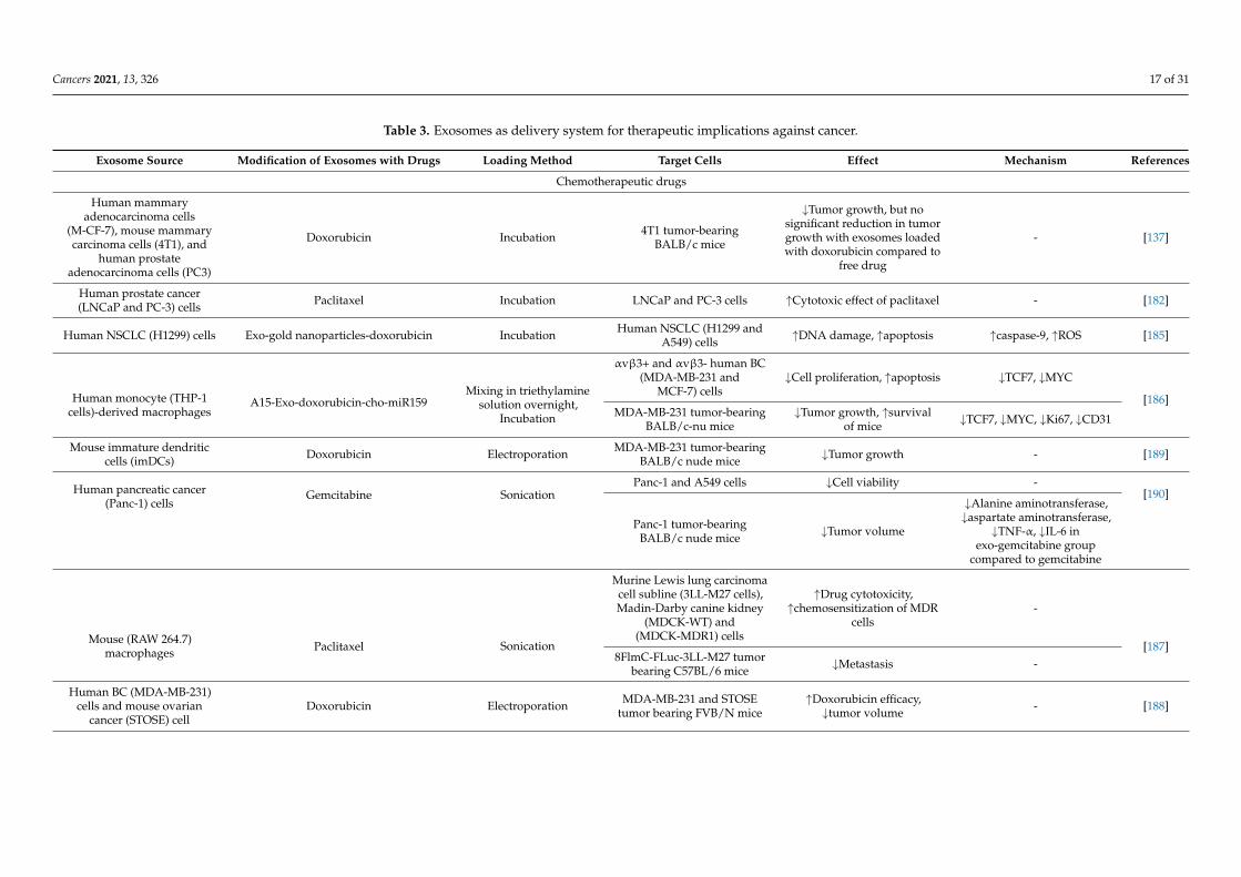
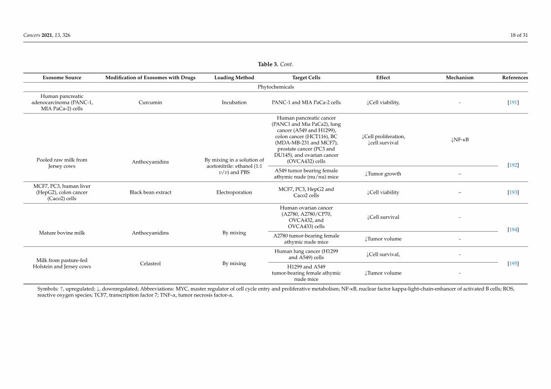
Phytochemicals, administered via an exosome-mediated drug delivery system, canprovide health benefits and anticancer properties [56]. Pancreatic adenocarcinoma cell-derived exosomes aided curcumin in inflicting its anticancer properties among tumorcells [191]. Milk-derived exosomes encapsulated with anthocyanidins exhibited antiprolif-erative effect in a xenograft lung carcinoma model [192]. Exosomal formulations of blackbean extract exhibited pronounced antiproliferative effect in many cancer cells [193]. Exo-somal formulations with berry anthocyanidins exhibited anticancer properties in ovariancancer with enhanced sensitivity in chemoresistant tumors [194]. Exosomal encapsulationof celastrol (a triterpenoid) exhibited antiproliferative effect in lung cancer cells and in axenograft model [195]. Recent studies on exosomal drug delivery of chemotherapeuticdrugs and phytochemicals are listed in Table 3.
9.7. Induction of Chemosensitivity with Exosomes
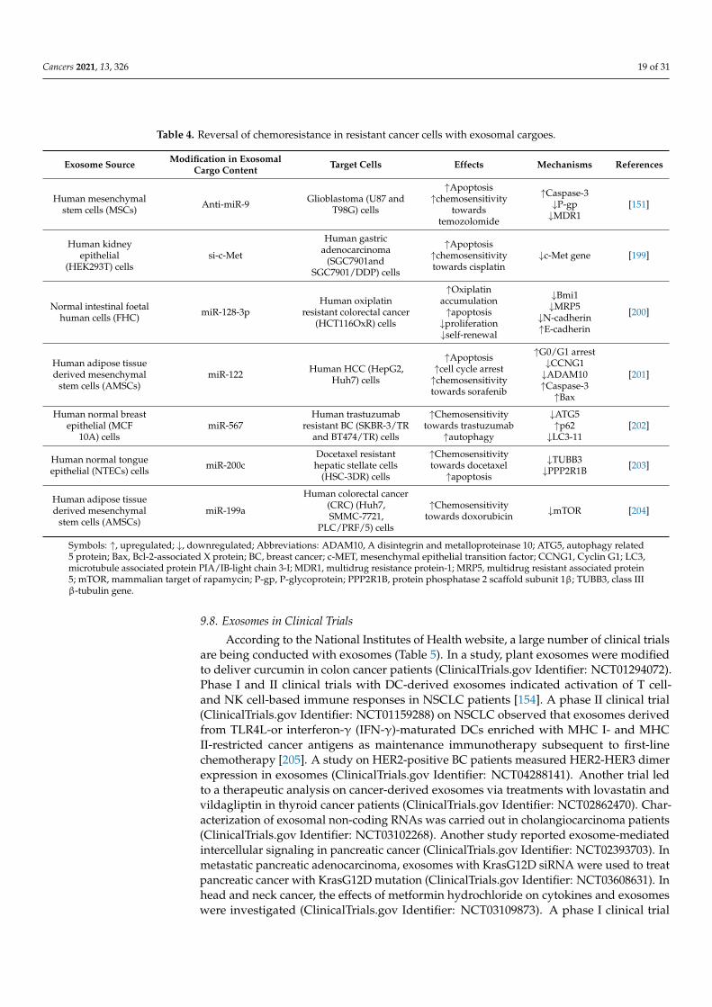
TEXs impart drug resistance but may also be used for inducing drug sensitivity.Dimethyl amiloride augmented ABC transporter containing exosome secretion revived thecyclophosphamide sensitivity of cancer cells [31]. Downregulation of the GAIP-interactingprotein C terminus mediated secretion of ABCG2 drug transporters containing exosomesand suppressed gemicitabine resistance in pancreatic cancer cells [196]. In oral squamouscell carcinoma, exosomal miR-155 increased chemoresistivity in cisplatin-sensitive cancercells [197]. The exosomes loaded with CRISPR/Cas9 induced apoptosis and cisplatinchemosensitivity in ovarian cancer cells [198]. An increase in apoptosis and chemosen-sitivity was observed in cisplatin-resistant human gastric adenocarcinoma cells throughtreatment with si-c-Met containing exosomes derived from human kidney epithelial cellline [199]. Normal intestinal FHC cell-derived exosomes transferred miR-128-3p intooxiplatin resistant CRC cells which induced their chemosensitivity and decreased motil-ity [200]. miR-122-transfected adipose tissue-derived MSCs (AMSCs) released exosomescarrying miR-122 and, when cocultured with hepatocyte carcinoma cells, induced sorafenibchemosensitivity [201]. miR-567 induced chemosensitivity in resistant BC cells towardstrastuzumab and blocked autophagy [202]. Exosomal miR-200c induced chemosensitivitytowards docetaxel and apoptosis in tongue squamous cell carcinoma [203]. Cocultureof miR-199a carrying exosomes derived from AMSCs with HCC cells downregulatedmammalian target of rapamycin (mTOR) pathway and induced chemosensitivity towardsdoxorubicin [204]. Various recent reports on exosome-mediated reversal of chemosensitiv-ity have been listed in Table 4.
Cancers 2021, 13, 326 17 of 31
Table 3. Exosomes as delivery system for therapeutic implications against cancer.
Exosome Source Modification of Exosomes with Drugs Loading Method Target Cells Effect Mechanism References
Chemotherapeutic drugs
Human mammaryadenocarcinoma cells
(M-CF-7), mouse mammarycarcinoma cells (4T1), and
human prostateadenocarcinoma cells (PC3)
Doxorubicin Incubation 4T1 tumor-bearingBALB/c mice
↓Tumor growth, but nosignificant reduction in tumorgrowth with exosomes loadedwith doxorubicin compared to
free drug
- [137]
Human prostate cancer(LNCaP and PC-3) cells Paclitaxel Incubation LNCaP and PC-3 cells ↑Cytotoxic effect of paclitaxel - [182]
Human NSCLC (H1299) cells Exo-gold nanoparticles-doxorubicin Incubation Human NSCLC (H1299 andA549) cells ↑DNA damage, ↑apoptosis ↑caspase-9, ↑ROS [185]
Human monocyte (THP-1cells)-derived macrophages
A15-Exo-doxorubicin-cho-miR159Mixing in triethylamine
solution overnight,Incubation
αvβ3+ and αvβ3- human BC(MDA-MB-231 and
MCF-7) cells↓Cell proliferation, ↑apoptosis ↓TCF7, ↓MYC
[186]MDA-MB-231 tumor-bearing
BALB/c-nu mice↓Tumor growth, ↑survival
of mice ↓TCF7, ↓MYC, ↓Ki67, ↓CD31
Mouse immature dendriticcells (imDCs) Doxorubicin Electroporation MDA-MB-231 tumor-bearing
BALB/c nude mice ↓Tumor growth - [189]
Human pancreatic cancer(Panc-1) cells
Gemcitabine SonicationPanc-1 and A549 cells ↓Cell viability -
[190]
Panc-1 tumor-bearingBALB/c nude mice ↓Tumor volume
↓Alanine aminotransferase,↓aspartate aminotransferase,
↓TNF-α, ↓IL-6 inexo-gemcitabine group
compared to gemcitabine
Mouse (RAW 264.7)macrophages Paclitaxel Sonication
Murine Lewis lung carcinomacell subline (3LL-M27 cells),Madin-Darby canine kidney
(MDCK-WT) and(MDCK-MDR1) cells
↑Drug cytotoxicity,↑chemosensitization of MDR
cells-
[187]8FlmC-FLuc-3LL-M27 tumor
bearing C57BL/6 mice ↓Metastasis -
Human BC (MDA-MB-231)cells and mouse ovarian
cancer (STOSE) cellDoxorubicin Electroporation MDA-MB-231 and STOSE
tumor bearing FVB/N mice↑Doxorubicin efficacy,
↓tumor volume - [188]
Cancers 2021, 13, 326 18 of 31
Table 3. Cont.
Exosome Source Modification of Exosomes with Drugs Loading Method Target Cells Effect Mechanism References
Phytochemicals
Human pancreaticadenocarcinoma (PANC-1,
MIA PaCa-2) cellsCurcumin Incubation PANC-1 and MIA PaCa-2 cells ↓Cell viability, - [191]
Pooled raw milk fromJersey cows
Anthocyanidins By mixing in a solution ofacetonitrile: ethanol (1:1
v/v) and PBS
Human pancreatic cancer(PANC1 and Mia PaCa2), lung
cancer (A549 and H1299),colon cancer (HCT116), BC(MDA-MB-231 and MCF7),prostate cancer (PC3 and
DU145), and ovarian cancer(OVCA432) cells
↓Cell proliferation,↓cell survival ↓NF-κB
[192]A549 tumor bearing femaleathymic nude (nu/nu) mice ↓Tumor growth –
MCF7, PC3, human liver(HepG2), colon cancer
(Caco2) cellsBlack bean extract Electroporation MCF7, PC3, HepG2 and
Caco2 cells ↓Cell viability – [193]
Mature bovine milk Anthocyanidins By mixing
Human ovarian cancer(A2780, A2780/CP70,
OVCA432, andOVCA433) cells
↓Cell survival -
[194]A2780 tumor-bearing female
athymic nude mice ↓Tumor volume -
Milk from pasture-fedHolstein and Jersey cows Celastrol By mixing
Human lung cancer (H1299and A549) cells ↓Cell survival, -
[195]H1299 and A549
tumor-bearing female athymicnude mice
↓Tumor volume -
Symbols: ↑, upregulated; ↓, downregulated; Abbreviations: MYC, master regulator of cell cycle entry and proliferative metabolism; NF-κB, nuclear factor kappa-light-chain-enhancer of activated B cells; ROS,reactive oxygen species; TCF7, transcription factor 7; TNF-α, tumor necrosis factor-α.
Cancers 2021, 13, 326 19 of 31
Table 4. Reversal of chemoresistance in resistant cancer cells with exosomal cargoes.
Exosome Source Modification in ExosomalCargo Content Target Cells Effects Mechanisms References
Human mesenchymalstem cells (MSCs) Anti-miR-9 Glioblastoma (U87 and
T98G) cells
↑Apoptosis↑chemosensitivity
towardstemozolomide
↑Caspase-3↓P-gp↓MDR1
[151]
Human kidneyepithelial
(HEK293T) cellssi-c-Met
Human gastricadenocarcinoma
(SGC7901andSGC7901/DDP) cells
↑Apoptosis↑chemosensitivitytowards cisplatin
↓c-Met gene [199]
Normal intestinal foetalhuman cells (FHC) miR-128-3p
Human oxiplatinresistant colorectal cancer
(HCT116OxR) cells
↑Oxiplatinaccumulation↑apoptosis
↓proliferation↓self-renewal
↓Bmi1↓MRP5
↓N-cadherin↑E-cadherin
[200]
Human adipose tissuederived mesenchymal
stem cells (AMSCs)miR-122 Human HCC (HepG2,
Huh7) cells
↑Apoptosis↑cell cycle arrest↑chemosensitivitytowards sorafenib
↑G0/G1 arrest↓CCNG1↓ADAM10↑Caspase-3
↑Bax
[201]
Human normal breastepithelial (MCF
10A) cellsmiR-567
Human trastuzumabresistant BC (SKBR-3/TR
and BT474/TR) cells
↑Chemosensitivitytowards trastuzumab
↑autophagy
↓ATG5↑p62
↓LC3-11[202]
Human normal tongueepithelial (NTECs) cells miR-200c
Docetaxel resistanthepatic stellate cells
(HSC-3DR) cells
↑Chemosensitivitytowards docetaxel
↑apoptosis
↓TUBB3↓PPP2R1B [203]
Human adipose tissuederived mesenchymal
stem cells (AMSCs)miR-199a
Human colorectal cancer(CRC) (Huh7,SMMC-7721,
PLC/PRF/5) cells
↑Chemosensitivitytowards doxorubicin ↓mTOR [204]
Symbols: ↑, upregulated; ↓, downregulated; Abbreviations: ADAM10, A disintegrin and metalloproteinase 10; ATG5, autophagy related5 protein; Bax, Bcl-2-associated X protein; BC, breast cancer; c-MET, mesenchymal epithelial transition factor; CCNG1, Cyclin G1; LC3,microtubule associated protein PIA/IB-light chain 3-I; MDR1, multidrug resistance protein-1; MRP5, multidrug resistant associated protein5; mTOR, mammalian target of rapamycin; P-gp, P-glycoprotein; PPP2R1B, protein phosphatase 2 scaffold subunit 1β; TUBB3, class IIIβ-tubulin gene.
9.8. Exosomes in Clinical Trials
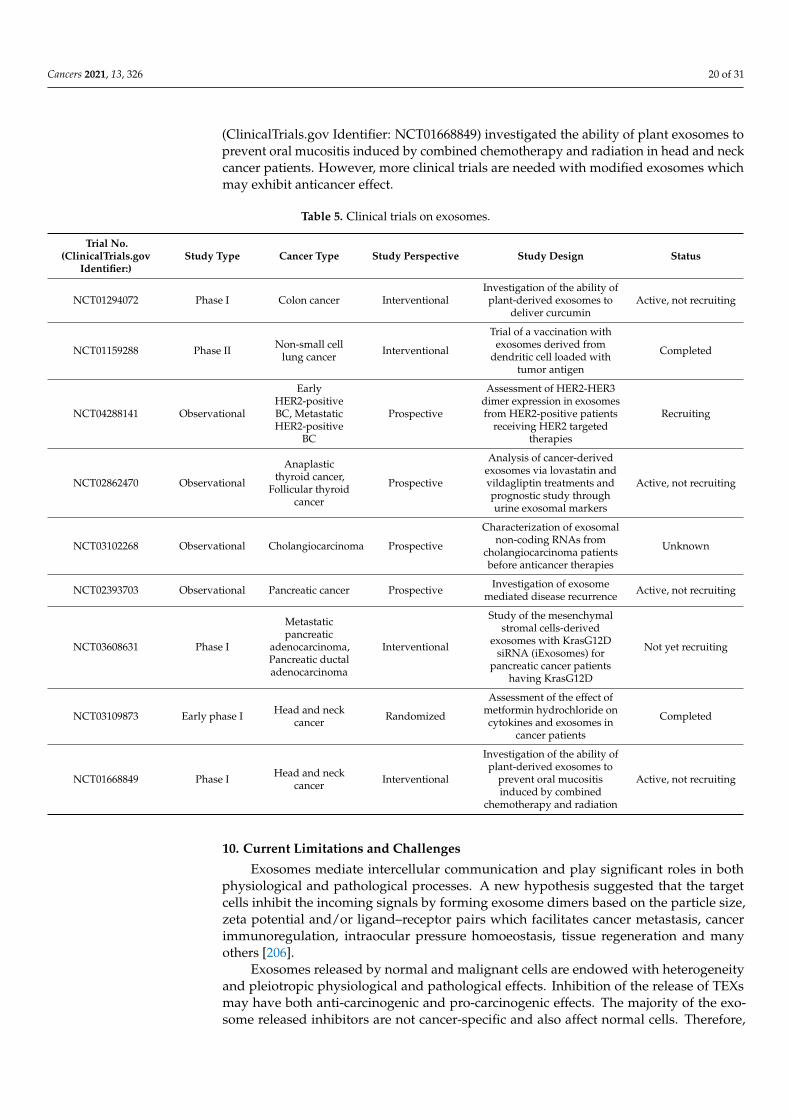
According to the National Institutes of Health website, a large number of clinical trialsare being conducted with exosomes (Table 5). In a study, plant exosomes were modifiedto deliver curcumin in colon cancer patients (ClinicalTrials.gov Identifier: NCT01294072).Phase I and II clinical trials with DC-derived exosomes indicated activation of T cell-and NK cell-based immune responses in NSCLC patients [154]. A phase II clinical trial(ClinicalTrials.gov Identifier: NCT01159288) on NSCLC observed that exosomes derivedfrom TLR4L-or interferon-γ (IFN-γ)-maturated DCs enriched with MHC I- and MHCII-restricted cancer antigens as maintenance immunotherapy subsequent to first-linechemotherapy [205]. A study on HER2-positive BC patients measured HER2-HER3 dimerexpression in exosomes (ClinicalTrials.gov Identifier: NCT04288141). Another trial ledto a therapeutic analysis on cancer-derived exosomes via treatments with lovastatin andvildagliptin in thyroid cancer patients (ClinicalTrials.gov Identifier: NCT02862470). Char-acterization of exosomal non-coding RNAs was carried out in cholangiocarcinoma patients(ClinicalTrials.gov Identifier: NCT03102268). Another study reported exosome-mediatedintercellular signaling in pancreatic cancer (ClinicalTrials.gov Identifier: NCT02393703). Inmetastatic pancreatic adenocarcinoma, exosomes with KrasG12D siRNA were used to treatpancreatic cancer with KrasG12D mutation (ClinicalTrials.gov Identifier: NCT03608631). Inhead and neck cancer, the effects of metformin hydrochloride on cytokines and exosomeswere investigated (ClinicalTrials.gov Identifier: NCT03109873). A phase I clinical trial
Cancers 2021, 13, 326 20 of 31
(ClinicalTrials.gov Identifier: NCT01668849) investigated the ability of plant exosomes toprevent oral mucositis induced by combined chemotherapy and radiation in head and neckcancer patients. However, more clinical trials are needed with modified exosomes whichmay exhibit anticancer effect.
Table 5. Clinical trials on exosomes.
Trial No.(ClinicalTrials.gov
Identifier:)Study Type Cancer Type Study Perspective Study Design Status
NCT01294072 Phase I Colon cancer InterventionalInvestigation of the ability ofplant-derived exosomes to
deliver curcuminActive, not recruiting
NCT01159288 Phase II Non-small celllung cancer Interventional
Trial of a vaccination withexosomes derived from
dendritic cell loaded withtumor antigen
Completed
NCT04288141 Observational
EarlyHER2-positiveBC, MetastaticHER2-positive
BC
Prospective
Assessment of HER2-HER3dimer expression in exosomesfrom HER2-positive patients
receiving HER2 targetedtherapies
Recruiting
NCT02862470 Observational
Anaplasticthyroid cancer,
Follicular thyroidcancer
Prospective
Analysis of cancer-derivedexosomes via lovastatin andvildagliptin treatments andprognostic study throughurine exosomal markers
Active, not recruiting
NCT03102268 Observational Cholangiocarcinoma Prospective
Characterization of exosomalnon-coding RNAs from
cholangiocarcinoma patientsbefore anticancer therapies
Unknown
NCT02393703 Observational Pancreatic cancer Prospective Investigation of exosomemediated disease recurrence Active, not recruiting
NCT03608631 Phase I
Metastaticpancreatic
adenocarcinoma,Pancreatic ductaladenocarcinoma
Interventional
Study of the mesenchymalstromal cells-derived
exosomes with KrasG12DsiRNA (iExosomes) for
pancreatic cancer patientshaving KrasG12D
Not yet recruiting
NCT03109873 Early phase I Head and neckcancer Randomized
Assessment of the effect ofmetformin hydrochloride oncytokines and exosomes in
cancer patients
Completed
NCT01668849 Phase I Head and neckcancer Interventional
Investigation of the ability ofplant-derived exosomes to
prevent oral mucositisinduced by combined
chemotherapy and radiation
Active, not recruiting
10. Current Limitations and Challenges
Exosomes mediate intercellular communication and play significant roles in bothphysiological and pathological processes. A new hypothesis suggested that the targetcells inhibit the incoming signals by forming exosome dimers based on the particle size,zeta potential and/or ligand–receptor pairs which facilitates cancer metastasis, cancerimmunoregulation, intraocular pressure homoeostasis, tissue regeneration and manyothers [206].
Exosomes released by normal and malignant cells are endowed with heterogeneityand pleiotropic physiological and pathological effects. Inhibition of the release of TEXsmay have both anti-carcinogenic and pro-carcinogenic effects. The majority of the exo-some released inhibitors are not cancer-specific and also affect normal cells. Therefore,
Cancers 2021, 13, 326 21 of 31
inhibition of exosome release may act as a double-edged sword which should be carefullymanipulated for minimal adverse effects [34].
Isolation of pure and specific exosomes is limited by technical constraints, the availabil-ity of suitable biomarkers for specific exosomes, and expensive technologies [5]. A majorhurdle in the execution of liquid biopsy is isolation of exosomes by an economic user-friendly tool. Protein contaminated and heterogenous exosome pool is obtained usingultracentrifugation. Asymmetric flow field-flow fractionation, though a prospective tool,needs technical expertise and requires a huge amount of initial sample. Other exosome iso-lation methods like microfluidic devices, sucrose gradients, size exclusion chromatography,and affinity-based exosome isolation kits are accompanied with both advantages and dis-advantages like lack of robustness and specificity [31]. A perfect exosome isolation methodshould be robust, reproducible, specific, economic and user friendly as a diagnostic tool.
Detailed research of exosome biogenesis, functional diversity of exosomes and theidentification of cancer specific biomarkers may be effective for exosome-based therapeuticapproaches with minimum adverse effects [34]. Determination of exosomal cargo sortingand releasing mechanisms holds great potential for the development of various applicationsin cancer research [31].
Normally, less than 1 µg of exosomal protein is yielded from 1 mL of culture medium,whereas the majority of studies have reported 10–100 µg of exosomal protein as an effectivedose for in vivo models [163]. The introduction of exosome-mimetic vesicles (100–200 nmin diameter) has conquered exosomal limitations like low loading efficiency and low yields.These nanovesicles have been used for delivery of chemotherapeutic drugs [204,205] andRNAi [207] to target cancer cells. Hybrid nanocarriers formed by the fusion of exosomeswith liposomes changed the exogenous lipid composition and was effective in the deliveryof chemotherapeutic drugs [208].
11. Conclusions
It may be deciphered that the intercellular communication via exosomes is evidentthroughout cancer progression. Apart from cancer pathogenesis, exosome biology heraldsthe future arena of non-invasive diagnostic tools for cancer management, especially inthe spheres of liquid biopsy, immunotherapy and vaccine development, RNA therapy,stem cell therapy, drug delivery, and reversal of chemoresistance. Preclinical studieshave undoubtedly proven the immense potential of exosomes in cancer therapeutics,but a number of clinical trials have failed to achieve this success. These inconsistentresults indicate major challenges including in-depth knowledge of exosome biogenesisand protein sorting, perfect and pure isolation of exosomes, large scale production, betterloading efficiency targeted delivery of exosomes. These hurdles have to be confrontedbefore successful implementation of exosomes for the diagnosis and therapy of cancer.This review has attempted to envisage the implication of exosomes in cancer pathogenesisand cancer therapeutics along with the current limitations so that researchers may bemade aware of the existing lacunae with regard to exosomes in their use against cancer.This knowledge may help scientists to improvise innovative technologies for successfultranslation of the exosome-mediated diagnosis and treatment of malignant neoplasms.
Author Contributions: Conceptualization and writing, D.S.; writing and figure preparation, S.R., P.S.and N.C.; editing and suggestion of improvements, A.B. All authors have read and agreed to thepublished version of the manuscript.
Funding: This research received no external source of funding.
Institutional Review Board Statement: Not applicable.
Informed Consent Statement: Not applicable.
Data Availability Statement: Not applicable.
Cancers 2021, 13, 326 22 of 31
Acknowledgments: D.S., S.R., P.S., and N.C. are deeply grateful to Jayanta Chakrabarti, Director,Chittaranjan National Cancer Institute, Kolkata, for providing the infrastructural facilities. P.S. isthankful to Jayanta Chakrabarti for granting an institutional fellowship. S.R. is indebted to theUniversity Grants Commission, New Delhi, India, for providing a fellowship.
Conflicts of Interest: The authors declare no conflict of interest.
Abbreviations
ABC ATP-binding cassette;ABCA3 ATP-binding cassette sub-family A member 3;ADAM10 A disintegrin and metalloproteinase 10;AFAP1-AS1 actin filament associated protein1 antisense RNA 1;Akt protein kinase B;ALIX ALG-2 interacting protein X;AMSCs adipose tissue-derived MSCs;APC antigen presenting cell;ARF6 ADP ribosylation factor 6;ATG5 autophagy related 5 protein;CAFs cancer-associated fibroblasts;CCNG1 cyclin G1;CRC colorectal cancer cells;CXCR4 C-X-C chemokine receptor type 4;DCs dendritic cells;ECM extracellular matrix;EGFR epidermal growth factor receptor;EMT epithelial mesenchymal transition;ERBB2 erythroblastic oncogene B2;ERF Ets2-repressor factor;ERK extracellular signal-regulated kinase;ERα estrogen receptor-α;ESCRT endosomal sorting complexes required for the transport;GC gastric cancer;HCC hepatocellular carcinoma;HER2 human epidermal growth factor receptor 2;HGF hepatocyte growth factor;HNSCC head and neck squamous cell carcinoma;HOTTIP HoxA transcript at a distal tip;Hsps heat shock proteins;ICAM intercellular adhesion molecule;IL-6 interleukin-6;ILV intraluminal vesicles;LAMP-1 lysosome-associated membrane glycoprotein-1 LncRNAsLncRNAs long non-coding RNAs;MAPK mitogen activated protein kinase;MDR-1 multidrug resistance protein-1;MDSCs myeloid-derived suppressor cells;MHC major histocompatibility complex;miRNAs microRNA;MMP matrix metalloproteinase;mRNA messenger RNA;MSCs mesenchymal stem cells;mtDNA mitochondrial DNA;MVB multivesicular bodies;NF-κB nuclear factor κ-light-chain-enhancer of activated B cells;NK natural killer cells;NKG2D natural killer group 2D;
Cancers 2021, 13, 326 23 of 31
NSCLC non-small cell lung carcinoma;PAFR a platelet-activating factor receptor;PDAC pancreatic ductal adenocarcinoma;PDCD4 programmed cell death 4;PD-L1 programmed death ligand 1;P-gp P-glycoprotein;PI3K phosphoinositide 3-kinase;PLD2 phospholipase D2;PLK-1 polo-like kinase 1;PM plasma membrane;PPP2R1B protein phosphatase 2 scaffold subunit 1β;PTEN phosphatase and tensin homolog;rRNA ribosomal RNA;SNARES soluble NSF attachment protein receptors;Sox2ot SOX2 overlapping transcript;SR-B1 scavenger receptor type B-1;STAT1 signal transducer and activator of transcription 1;TAMs tumor-associated macrophages;TEX tumor derived exosomes;TGF-β transforming growth factor-β;TGN trans-Golgi network;TLR-2 toll like receptor-2;TME tumor microenvironment;Tregs T regulatory cells;TUBB3 class III β-tubulin gene;UFC1 Ubiquitin-fold modifier conjugating enzyme 1;VEGF-A vascular endothelial growth factor A;Vps4 vacuolar protein sorting associated protein 4;ZFAS1 zinc finger antisense 1.
References1. Harding, C.; Stahl, P. Transferrin Recycling in Reticulocytes: pH and Iron Are Important Determinants of Ligand Binding and
Processing. Biochem. Biophys. Res. Commun. 1983, 113, 650–658. [CrossRef]2. Lötvall, J.; Hill, A.F.; Hochberg, F.; Buzás, E.I.; Vizio, D.D.; Gardiner, C.; Gho, Y.S.; Kurochkin, I.V.; Mathivanan, S.; Quesenberry,
P.; et al. Minimal experimental requirements for definition of extracellular vesicles and their functions: A position statement fromthe International Society for Extracellular Vesicles. J. Extracell. Vesicles 2014, 3, 26913. [CrossRef]
3. Théry, C.; Witwer, K.W.; Aikawa, E.; Alcaraz, M.J.; Anderson, J.D.; Andriantsitohaina, R.; Antoniou, A.; Arab, T.; Archer, F.; Atkin-Smith,G.K.; et al. Minimal information for studies of extracellular vesicles 2018 (MISEV2018): A position statement of the International Societyfor Extracellular Vesicles and update of the MISEV2014 guidelines. J. Extracell. Vesicles 2018, 7, 1535750. [CrossRef]
4. Zhang, C.; Ji, Q.; Yang, Y.; Li, Q.; Wang, Z. Exosome: Function and role in cancer metastasis and drug resistance. Technol. CancerRes. Treat. 2018, 17. [CrossRef]
5. He, C.; Zheng, S.; Luo, Y.; Wang, B. Exosome theranostics: Biology and translational medicine. Theranostics 2018, 8, 237–255. [CrossRef]6. McAndrews, K.M.; Kalluri, R. Mechanisms associated with biogenesis of exosomes in cancer. Mol. Cancer 2019, 18, 52. [CrossRef]7. Jurj, A.; Zanoaga, O.; Braicu, C.; Lazar, V.; Tomuleasa, C.; Irimie, A.; Berindan-neagoe, I. A comprehensive picture of extracellular
vesicles and their contents. Molecular transfer to cancer cells. Cancers 2020, 12, 298. [CrossRef]8. Lucchetti, D.; Tenore, C.R.; Colella, F.; Sgambato, A. Extracellular Vesicles and Cancer: A Focus on Metabolism, Cytokines, and
Immunity. Cancers 2020, 12, 171. [CrossRef]9. Tang, Z.; Li, D.; Hou, S.; Zhu, X. The cancer exosomes: Clinical implications, applications and challenges. Int. J. Cancer 2020,
146, 2946–2959. [CrossRef]10. Mohammadi, S.; Yousefi, F.; Shabaninejad, Z.; Movahedpour, A.; Mahjoubin Tehran, M.; Shafiee, A.; Moradizarmehri, S.;
Hajighadimi, S.; Savardashtaki, A.; Mirzaei, H. Exosomes and cancer: From oncogenic roles to therapeutic applications. IUBMBLife 2020, 72, 724–748. [CrossRef]
11. Parayath, N.N.; Padmakumar, S.; Amiji, M.M. Extracellular vesicle-mediated nucleic acid transfer and reprogramming in thetumor microenvironment. Cancer Lett. 2020, 482, 33–43. [CrossRef]
12. Wee, I.; Syn, N.; Sethi, G.; Goh, B.C.; Wang, L. Role of tumor-derived exosomes in cancer metastasis. Biochim. Biophys. Acta Rev.Cancer 2019, 1871, 12–19. [CrossRef]
13. Raimondo, S.; Pucci, M.; Alessandro, R.; Fontana, S. Extracellular vesicles and tumor-immune escape: Biological functions andclinical perspectives. Int. J. Mol. Sci. 2020, 21, 2286. [CrossRef]
14. Gilligan, K.E.; Dwyer, R.M. Extracellular Vesicles for Cancer Therapy: Impact of Host Immune Response. Cells 2020, 9, 224. [CrossRef]
Cancers 2021, 13, 326 24 of 31
15. Li, X.; Corbett, A.L.; Taatizadeh, E.; Tasnim, N.; Little, J.P.; Garnis, C.; Daugaard, M.; Guns, E.; Hoorfar, M.; Li, I.T.S. Challenges andopportunities in exosome research—Perspectives from biology, engineering, and cancer therapy. APL Bioeng. 2019, 3, 011503. [CrossRef]
16. Srivastava, A.; Amreddy, N.; Pareek, V.; Chinnappan, M.; Ahmed, R.; Mehta, M.; Razaq, M.; Munshi, A.; Ramesh, R. Progressin extracellular vesicle biology and their application in cancer medicine. Wiley Interdiscip. Rev. Nanomed. Nanobiotechnol. 2020,12, e1621. [CrossRef]
17. Taghikhani, A.; Farzaneh, F.; Sharifzad, F.; Mardpour, S.; Ebrahimi, M.; Hassan, Z.M. Engineered Tumor-Derived ExtracellularVesicles: Potentials in Cancer Immunotherapy. Front. Immunol. 2020, 11, 221. [CrossRef]
18. Stevic, I.; Buescher, G.; Ricklefs, F.L. Monitoring Therapy Efficiency in Cancer through Extracellular Vesicles. Cells 2020, 9, 130. [CrossRef]19. Pullan, J.E.; Confeld, M.I.; Osborn, J.K.; Kim, J.; Sarkar, K.; Mallik, S. Exosomes as Drug Carriers for Cancer Therapy. Mol. Pharm.
2019, 16, 1789–1798. [CrossRef]20. Yong, T.; Wang, D.; Li, X.; Yan, Y.; Hu, J.; Gan, L.; Yang, X. Extracellular vesicles for tumor targeting delivery based on five features
principle. J. Control. Release 2020, 322, 555–565. [CrossRef]21. Zhao, X.; Wu, D.; Ma, X.; Wang, J.; Hou, W.; Zhang, W. Exosomes as drug carriers for cancer therapy and challenges regarding
exosome uptake. Biomed. Pharmacother. 2020, 128, 110237. [CrossRef]22. Pirisinu, M.; Pham, T.C.; Zhang, D.X.; Hong, T.N.; Nguyen, L.T.; Le, M.T. Extracellular vesicles as natural therapeutic agents and
innate drug delivery systems for cancer treatment: Recent advances, current obstacles, and challenges for clinical translation.Semin. Cancer Biol. 2020. [CrossRef]
23. Milane, L.; Singh, A.; Mattheolabakis, G.; Suresh, M.; Amiji, M.M. Exosome Mediated Communication within the TumorMicroenvironment. J. Control. Release 2015, 10, 278–294. [CrossRef]
24. Jenjaroenpun, P.; Kremenska, Y.; Nair, V.M.; Kremenskoy, M.; Joseph, B.; Kurochkin, I.V. Characterization of RNA in exosomessecreted by human breast cancer cell lines using next-generation sequencing. PeerJ 2013, 1, e201. [CrossRef]
25. Li, S.P.; Lin, Z.X.; Jiang, X.Y.; Yu, X.Y. Exosomal cargo-loading and synthetic exosome-mimics as potential therapeutic tools. ActaPharmacol. Sin. 2018, 39, 542–551. [CrossRef]
26. Zhang, Y.; Liu, Y.; Liu, H.; Tang, W.H. Exosomes: Biogenesis, biologic function and clinical potential. Cell Biosci. 2019, 9, 19. [CrossRef]27. Souza-Schorey, C.; Schorey, J.S. Regulation and mechanisms of extracellular vesicle biogenesis and secretion. Essays Biochem.
2018, 62, 125–133. [CrossRef]28. Antonyak, M.A.; Cerione, R.A. Microvesicles as mediators of intercellular communication in cancer. Methods Mol. Biol. 2014,
1165, 147–173. [CrossRef]29. Ghossoub, R.; Lembo, F.; Rubio, A.; Gaillard, C.B.; Bouchet, J.; Vitale, N.; Slavík, J.; Machala, M.; Zimmermann, P. Syntenin-ALIX
exosome biogenesis and budding into multivesicular bodies are controlled by ARF6 and PLD2. Nat. Commun. 2014, 5, 3477. [CrossRef]30. Kowal, J.; Tkach, M.; Théry, C. Biogenesis and secretion of exosomes. Curr. Opin. Cell Biol. 2014, 29, 116–125. [CrossRef]31. Wortzel, I.; Dror, S.; Kenific, C.M.; Lyden, D. Exosome-Mediated Metastasis: Communication from a Distance. Dev. Cell 2019,
49, 347–360. [CrossRef]32. Alenquer, M.; Amorim, M.J. Exosome biogenesis, regulation, and function in viral infection. Viruses 2015, 7, 5066–5083. [CrossRef]33. Stoorvogel, W. Resolving sorting mechanisms into exosomes. Cell Res. 2015, 25, 531–532. [CrossRef]34. Moloudizargari, M.; Asghari, M.H.; Abdollahi, M. Modifying exosome release in cancer therapy: How can it help? Pharmacol. Res.
2018, 134, 246–256. [CrossRef]35. Lange, S.; Gallagher, M.; Kholia, S.; Kosgodage, U.S.; Hristova, M.; Hardy, J.; Inal, J.M. Peptidylarginine deiminases—Roles in
cancer and neurodegeneration and possible avenues for therapeutic intervention via modulation of exosome and microvesicle(EMV) release? Int. J. Mol. Sci. 2017, 18, 1196. [CrossRef]
36. Mohrmann, K.; Leijendekker, R.; Gerez, L.; Van Sluijs, P. Der rab4 regulates transport to the apical plasma membrane inMadin-Darby canine kidney cells. J. Biol. Chem. 2002, 277, 10474–10481. [CrossRef]
37. Schlierf, B.; Fey, G.H.; Hauber, J.; Hocke, G.M.; Rosorius, O. Rab11b is essential for recycling of transferrin to the plasmamembrane. Exp. Cell Res. 2000, 259, 257–265. [CrossRef]
38. Casanova, J.E.; Wang, X.; Kumar, R.; Bhartur, S.G.; Navarre, J.; Woodrum, J.E.; Altschuler, Y.; Ray, G.S.; Goldenring, J.R.Association of Rab25 and Rab11a with the apical recycling system of polarized Madin-Darby Canine Kidney cells. Mol. Biol. Cell1999, 10, 47–61. [CrossRef]
39. Lombardi, D.; Soldati, T.; Riederer, M.A.; Goda, Y.; Zerial, M.; Pfeffer, S.R. Rab9 functions in transport between late endosomesand the trans Golgi network. EMBO J. 1993, 12, 677–682. [CrossRef]
40. Guerra, F.; Bucci, C. Multiple Roles of the Small GTPase Rab7. Cells 2016, 5, 34. [CrossRef]41. Vitelli, R.; Santillo, M.; Lattero, D.; Chiariello, M.; Bifulco, M.; Bruni, C.B.; Bucci, C. Role of the small GTPase RAB7 in the late
endocytic pathway. J. Biol. Chem. 1997, 272, 4391–4397. [CrossRef]42. Chen, J.; Chopp, M. Exosome therapy for stroke. Stroke 2018, 49, 1083–1090. [CrossRef]43. Ostrowski, M.; Carmo, N.B.; Krumeich, S.; Fanget, I.; Raposo, G.; Savina, A.; Moita, C.F.; Schauer, K.; Hume, A.N.; Freitas, R.P.;
et al. Rab27a and Rab27b control different steps of the exosome secretion pathway. Nat. Cell Biol. 2010, 12, 19–30. [CrossRef]44. Baietti, M.F.; Zhang, Z.; Mortier, E.; Melchior, A.; Degeest, G.; Geeraerts, A.; Ivarsson, Y.; Depoortere, F.; Coomans, C.; Vermeiren,
E.; et al. Syndecan-syntenin-ALIX regulates the biogenesis of exosomes. Nat. Cell Biol. 2012, 14, 677–685. [CrossRef]45. Savina, A.; Vidal, M.; Colombo, M.I. The exosome pathway in K562 cells is regulated by Rab11. J. Cell Sci. 2002, 115, 2505–2515.
Cancers 2021, 13, 326 25 of 31
46. Savina, A.; Fader, C.M.; Damiani, M.T.; Colombo, M.I. Rab11 promotes docking and fusion of multivesicular bodies in acalcium-dependent manner. Traffic 2006, 6, 131–143. [CrossRef]
47. Li, W.; Hu, Y.; Jiang, T.; Han, Y.; Han, G.; Chen, J.; Li, X. Rab27A regulates exosome secretion from lung adenocarcinoma cellsA549: Involvement of EPI64. APMIS 2014, 122, 1080–1087. [CrossRef]
48. Heusermann, W.; Hean, J.; Trojer, D.; Steib, E.; von Bueren, S.; Graff-Meyer, A.; Genoud, C.; Martin, K.; Pizzato, N.; Voshol, J.; et al.Exosomes surf on filopodia to enter cells at endocytic hot spots, traffic within endosomes, and are targeted to the ER. J. Cell Biol.2016, 213, 173–184. [CrossRef]
49. Tian, T.; Zhu, Y.L.; Zhou, Y.Y.; Liang, G.F.; Wang, Y.Y.; Hu, F.H.; Xiao, Z.D. Exosome uptake through clathrin-mediated endocytosisand macropinocytosis and mediating miR-21 delivery. J. Biol. Chem. 2014, 289, 22258–22267. [CrossRef]
50. Nabi, I.R.; Le, P.U. Caveolae/raft-dependent endocytosis. J. Cell Biol. 2003, 161, 673–677. [CrossRef]51. Gonda, A.; Kabagwira, J.; Senthil, G.N.; Wall, N.R. Internalization of exosomes through receptor-mediated endocytosis. Mol.
Cancer Res. 2019, 17, 337–347. [CrossRef]52. Joshi, B.S.; de Beer, M.A.; Giepmans, B.N.G.; Zuhorn, I.S. Endocytosis of Extracellular Vesicles and Release of Their Cargo from
Endosomes. ACS Nano 2020, 14, 4444–4455. [CrossRef]53. Andreu, Z.; Yáñez-Mó, M. Tetraspanins in extracellular vesicle formation and function. Front. Immunol. 2014, 5, 442. [CrossRef]54. Wu, Y.; Wu, W.; Wong, W.M.; Ward, E.; Thrasher, A.J.; Goldblatt, D.; Osman, M.; Digard, P.; Canaday, D.H.; Gustafsson, K. Human
γδ T Cells: A Lymphoid Lineage Cell Capable of Professional Phagocytosis. J. Immunol. 2009, 183, 5622–5629. [CrossRef]55. McKelvey, K.J.; Powell, K.L.; Ashton, A.W.; Morris, J.M.; McCracken, S.A. Exosomes: Mechanisms of Uptake. J. Circ. Biomark.
2015, 4, 7. [CrossRef]56. Farooqi, A.A.; Desai, N.N.; Qureshi, M.Z.; Librelotto, D.R.N.; Gasparri, M.L.; Bishayee, A.; Nabavi, S.M.; Curti, V.; Daglia, M. Exosome
biogenesis, bioactivities and functions as new delivery systems of natural compounds. Biotechnol. Adv. 2018, 36, 328–334. [CrossRef]57. Datta, A.; Kim, H.; McGee, L.; Johnson, A.E.; Talwar, S.; Marugan, J.; Southall, N.; Hu, X.; Lal, M.; Mondal, D.; et al. High-
Throughput screening identified selective inhibitors of exosome biogenesis and secretion: A drug repurposing strategy foradvanced cancer. Sci. Rep. 2018, 8, 8161. [CrossRef]
58. Logozzi, M.; Spugnini, E.; Mizzoni, D.; Di Raimo, R.; Fais, S. Extracellular acidity and increased exosome release as keyphenotypes of malignant tumors. Cancer Metastasis Rev. 2019, 38, 93–101. [CrossRef]
59. Agarwal, K.; Saji, M.; Lazaroff, S.M.; Palmer, A.F.; Ringel, M.D.; Paulaitis, M.E. Analysis of exosome release as a cellular responseto MAPK pathway inhibition. Langmuir 2015, 31, 5440–5448. [CrossRef]
60. Lee, T.H.; Chennakrishnaiah, S.; Audemard, E.; Montermini, L.; Meehan, B.; Rak, J. Oncogenic ras-driven cancer cell vesiculationleads to emission of double-stranded DNA capable of interacting with target cells. Biochem. Biophys. Res. Commun. 2014,451, 295–301. [CrossRef]
61. Al-Nedawi, K.; Meehan, B.; Micallef, J.; Lhotak, V.; May, L.; Guha, A.; Rak, J. Intercellular transfer of the oncogenic receptorEGFRvIII by microvesicles derived from tumour cells. Nat. Cell Biol. 2008, 10, 619–624. [CrossRef]
62. Zhang, C.; Xiao, X.; Chen, M.; Aldharee, H.; Chen, Y.; Long, W. Liver kinase B1 restoration promotes exosome secretion andmotility of lung cancer cells. Oncol. Rep. 2018, 39, 376–382. [CrossRef]
63. Melo, S.A.; Sugimoto, H.; O’Connell, J.T.; Kato, N.; Villanueva, A.; Vidal, A.; Qiu, L.; Vitkin, E.; Perelman, L.T.; Melo, C.A.; et al. CancerExosomes Perform Cell-Independent MicroRNA Biogenesis and Promote Tumorigenesis. Cancer Cell 2014, 26, 707–721. [CrossRef]
64. Jia, Y.; Chen, Y.; Wang, Q.; Jayasinghe, U.; Luo, X.; Wei, Q.; Wang, J.; Xiong, H.; Chen, C.; Xu, B.; et al. Exosome: Emergingbiomarker in breast cancer. Oncotarget 2017, 8, 41717–41733. [CrossRef]
65. Singh, R.; Pochampally, R.; Watabe, K.; Lu, Z.; Mo, Y.Y. Exosome-mediated transfer of miR-10b promotes cell invasion in breastcancer. Mol. Cancer 2014, 13, 256. [CrossRef]
66. Zhang, L.; Zhang, S.; Yao, J.; Lowery, F.J.; Zhang, Q.; Huang, W.C.; Li, P.; Li, M.; Wang, X.; Zhang, C.; et al. Microenvironment-induced PTEN loss by exosomal microRNA primes brain metastasis outgrowth. Nature 2015, 527, 100–104. [CrossRef]
67. Cooks, T.; Pateras, I.S.; Jenkins, L.M.; Patel, K.M.; Robles, A.I.; Morris, J.; Forshew, T.; Appella, E.; Gorgoulis, V.G.; Harris, C.C.Mutant p53 cancers reprogram macrophages to tumor supporting macrophages via exosomal miR-1246. Nat. Commun. 2018,9, 771. [CrossRef]
68. Zhang, H.D.; Jiang, L.H.; Hou, J.C.; Zhong, S.L.; Zhu, L.P.; Wang, D.D.; Zhou, S.Y.; Yang, S.J.; Wang, J.Y.; Zhang, Q.; et al. Exosome:A novel mediator in drug resistance of cancer cells. Epigenomics 2018, 10, 1499–1509. [CrossRef]
69. Yang, X.; Li, Y.; Zou, L.; Zhu, Z. Role of Exosomes in Crosstalk Between Cancer-Associated Fibroblasts and Cancer Cells. Front.Oncol. 2019, 9, 356. [CrossRef]
70. Zhao, H.; Yang, L.; Baddour, J.; Achreja, A.; Bernard, V.; Moss, T.; Marini, J.C.; Tudawe, T.; Seviour, E.G.; San Lucas, F.A.; et al.Tumor microenvironment derived exosomes pleiotropically modulate cancer cell metabolism. Elife 2016, 5, e10250. [CrossRef]
71. Hu, T.; Hu, J. Melanoma-derived exosomes induce reprogramming fibroblasts into cancer-associated fibroblasts via Gm26809delivery. Cell Cycle 2019, 18, 3085–3094. [CrossRef]
72. Abusamra, A.J.; Zhong, Z.; Zheng, X.; Li, M.; Ichim, T.E.; Chin, J.L.; Min, W.P. Tumor exosomes expressing Fas ligand mediateCD8+ T-cell apoptosis. Blood Cells Mol. Dis. 2005, 35, 169–173. [CrossRef]
73. Lu, H.; Bowler, N.; Harshyne, L.A.; Craig Hooper, D.; Krishn, S.R.; Kurtoglu, S.; Fedele, C.; Liu, Q.; Tang, H.Y.; Kossenkov, A.V.;et al. Exosomal αvβ6 integrin is required for monocyte M2 polarization in prostate cancer. Matrix Biol. 2018, 70, 20–35. [CrossRef]
Cancers 2021, 13, 326 26 of 31
74. Yin, C.; Han, Q.; Xu, D.; Zheng, B.; Zhao, X.; Zhang, J. SALL4-mediated upregulation of exosomal miR-146a-5p drives T-cellexhaustion by M2 tumor-associated macrophages in HCC. Oncoimmunology 2019, 8, 1601479. [CrossRef]
75. Liu, C.; Yu, S.; Zinn, K.; Wang, J.; Zhang, L.; Jia, Y.; Kappes, J.C.; Barnes, S.; Kimberly, R.P.; Grizzle, W.E.; et al. Murine MammaryCarcinoma Exosomes Promote Tumor Growth by Suppression of NK Cell Function. J. Immunol. 2006, 176, 1375–1385. [CrossRef]
76. Bobrie, A.; Krumeich, S.; Reyal, F.; Recchi, C.; Moita, L.F.; Seabra, M.C.; Ostrowski, M.; Théry, C. Rab27a supports exosome-dependent and -independent mechanisms that modify the tumor microenvironment and can promote tumor progression. CancerRes. 2012, 72, 4920–4930. [CrossRef]
77. Maus, R.L.G.; Jakub, J.W.; Nevala, W.K.; Christensen, T.A.; Noble-Orcutt, K.; Sachs, Z.; Hieken, T.J.; Markovic, S.N. Humanmelanoma-derived extracellular vesicles regulate dendritic cell maturation. Front. Immunol. 2017, 8, 358. [CrossRef]
78. Wolfers, J.; Lozier, A.; Raposo, G.; Regnault, A.; Théry, C.; Masurier, C.; Flament, C.; Pouzieux, S.; Faure, F.; Tursz, T.; et al. Tumor-derived exosomes are a source of shared tumor rejection antigens for CTL cross-priming. Nat. Med. 2001, 7, 297–303. [CrossRef]
79. Theodoraki, M.N.; Yerneni, S.S.; Hoffmann, T.K.; Gooding, W.E.; Whiteside, T.L. Clinical significance of PD-L1 þ exosomes inplasma of head and neck cancer patients. Clin. Cancer Res. 2018, 24, 896–905. [CrossRef]
80. Yue, S.; Mu, W.; Erb, U.; Zöller, M. The tetraspanins CD151 and Tspan8 are essential exosome components for the crosstalkbetween cancer initiating cells and their surrounding. Oncotarget 2015, 6, 2366–2384. [CrossRef]
81. Yin, X.; Zheng, X.; Liu, M.; Wang, D.; Sun, H.; Qiu, Y.; Chen, J.; Shi, B. Exosomal miR-663b targets Ets2-repressor factor to promoteproliferation and the epithelial–mesenchymal transition of bladder cancer cells. Cell Biol. Int. 2020, 44, 958–965. [CrossRef]
82. Li, Z.; Jiang, P.; Li, J.; Peng, M.; Zhao, X.; Zhang, X.; Chen, K.; Zhang, Y.; Liu, H.; Gan, L.; et al. Tumor-derived exosomal lnc-Sox2otpromotes EMT and stemness by acting as a ceRNA in pancreatic ductal adenocarcinoma. Oncogene 2018, 37, 3822–3838. [CrossRef]
83. Suchorska, W.M.; Lach, M.S. The role of exosomes in tumor progression and metastasis (Review). Oncol. Rep. 2016,35, 1237–1244. [CrossRef]
84. Fedele, C.; Singh, A.; Zerlanko, B.J.; Iozzo, R.V.; Languino, L.R. The αvβ6 integrin is transferred intercellularly via exosomes.J. Biol. Chem. 2015, 290, 4545–4551. [CrossRef]
85. Lin, F.; Yin, H.B.; Li, X.Y.; Zhu, G.M.; He, W.Y.; Gou, X. Bladder cancer cell-secreted exosomal miR-21 activates the PI3K/AKTpathway in macrophages to promote cancer progression. Int. J. Oncol. 2020, 56, 151–164. [CrossRef]
86. Zheng, P.; Luo, Q.; Wang, W.; Li, J.; Wang, T.; Wang, P.; Chen, L.; Zhang, P.; Chen, H.; Liu, Y.; et al. Tumor-associated macrophages-derived exosomes promote the migration of gastric cancer cells by transfer of functional Apolipoprotein E. Cell Death Dis. 2018,9, 434. [CrossRef]
87. Zang, X.; Gu, J.; Zhang, J.; Shi, H.; Hou, S.; Xu, X.; Chen, Y.; Zhang, Y.; Mao, F.; Qian, H.; et al. Exosome-transmitted lncRNAUFC1 promotes non-small-cell lung cancer progression by EZH2-mediated epigenetic silencing of PTEN expression. Cell DeathDis. 2020, 11, 215. [CrossRef]
88. Pan, L.; Liang, W.; Fu, M.; Huang, Z.H.; Li, X.; Zhang, W.; Zhang, P.; Qian, H.; Jiang, P.C.; Xu, W.R.; et al. Exosomes-mediated transferof long noncoding RNA ZFAS1 promotes gastric cancer progression. J. Cancer Res. Clin. Oncol. 2017, 143, 991–1004. [CrossRef]
89. Zhang, H.; Deng, T.; Liu, R.; Bai, M.; Zhou, L.; Wang, X.; Li, S.; Wang, X.; Yang, H.; Li, J.; et al. Exosome-delivered EGFR regulatesliver microenvironment to promote gastric cancer liver metastasis. Nat. Commun. 2017, 8, 15016. [CrossRef]
90. Yokoi, A.; Yoshioka, Y.; Yamamoto, Y.; Ishikawa, M.; Ikeda, S.I.; Kato, T.; Kiyono, T.; Takeshita, F.; Kajiyama, H.; Kikkawa, F.; et al.Malignant extracellular vesicles carrying MMP1 mRNA facilitate peritoneal dissemination in ovarian cancer. Nat. Commun. 2017,8, 14470. [CrossRef]
91. Wang, D.; Wang, X.; Si, M.; Yang, J.; Sun, S.; Wu, H.; Cui, S.; Qu, X.; Yu, X. Exosome-encapsulated miRNAs contribute toCXCL12/CXCR4-induced liver metastasis of colorectal cancer by enhancing M2 polarization of macrophages. Cancer Lett. 2020,474, 36–52. [CrossRef]
92. Sun, S.; Chen, H.; Xu, C.; Zhang, Y.; Zhang, Q.; Chen, L.; Ding, Q.; Deng, Z. Exosomal miR-106b serves as a novel marker for lungcancer and promotes cancer metastasis via targeting PTEN. Life Sci. 2020, 244, 117297. [CrossRef]
93. Liao, J.; Liu, R.; Shi, Y.J.; Yin, L.H.; Pu, Y.P. Exosome-shuttling microRNA-21 promotes cell migration and invasion-targetingPDCD4 in esophageal cancer. Int. J. Oncol. 2016, 48, 2567–2579. [CrossRef]
94. Xia, Y.; Wei, K.; Hu, L.Q.; Zhou, C.R.; Lu, Z.B.; Zhan, G.S.; Pan, X.L.; Pan, C.F.; Wang, J.; Wen, W.; et al. Exosome-mediatedtransfer of miR-1260b promotes cell invasion through Wnt/β–catenin signaling pathway in lung adenocarcinoma. J. Cell. Physiol.2020, 235, 6843–6853. [CrossRef]
95. Li, Z.; Tao, Y.; Wang, X.; Jiang, P.; Li, J.; Peng, M.; Zhang, X.; Chen, K.; Liu, H.; Zhen, P.; et al. Tumor-secreted exosomal miR-222promotes tumor progression via regulating P27 expression and re-localization in pancreatic cancer. Cell. Physiol. Biochem. 2018,51, 610–629. [CrossRef]
96. Huang, Z.; Feng, Y. Exosomes derived from hypoxic colorectal cancer cells promote angiogenesis through Wnt4-Induced β-cateninsignaling in endothelial cells. Oncol. Res. 2017, 25, 651–661. [CrossRef]
97. Treps, L.; Perret, R.; Edmond, S.; Ricard, D.; Gavard, J. Glioblastoma stem-like cells secrete the pro-angiogenic VEGF-A factor inextracellular vesicles. J. Extracell. Vesicles 2017, 6, 1359479. [CrossRef]
98. Yoshii, S.; Hayashi, Y.; Iijima, H.; Inoue, T.; Kimura, K.; Sakatani, A.; Nagai, K.; Fujinaga, T.; Hiyama, S.; Kodama, T.; et al.Exosomal microRNAs derived from colon cancer cells promote tumor progression by suppressing fibroblast TP53 expression.Cancer Sci. 2019, 110, 2396–2407. [CrossRef]
Cancers 2021, 13, 326 27 of 31
99. Zheng, P.; Chen, L.; Yuan, X.; Luo, Q.; Liu, Y.; Xie, G.; Ma, Y.; Shen, L. Exosomal transfer of tumor-associated macrophage-derivedmiR-21 confers cisplatin resistance in gastric cancer cells. J. Exp. Clin. Cancer Res. 2017, 36, 53. [CrossRef]
100. Yu, X.; Zhang, Q.; Zhang, X.; Han, Q.; Li, H.; Mao, Y.; Wang, X.; Guo, H.; Irwin, D.M.; Niu, G.; et al. Exosomes from macrophagesexposed to apoptotic breast cancer cells promote breast cancer proliferation and metastasis. J. Cancer 2019, 10, 2892–2906. [CrossRef]
101. Li, X.; Wang, S.; Zhu, R.; Li, H.; Han, Q.; Zhao, R.C. Lung tumor exosomes induce a pro-inflammatory phenotype in mesenchymalstem cells via NFκB-TLR signaling pathway. J. Hematol. Oncol. 2016, 9, 42. [CrossRef]
102. Raimondo, S.; Saieva, L.; Corrado, C.; Fontana, S.; Flugy, A.; Rizzo, A.; De Leo, G.; Alessandro, R. Chronic myeloid leukemia-derived exosomes promote tumor growth through an autocrine mechanism. Cell Commun. Signal. 2015, 13, 8. [CrossRef]
103. Wei, Y.; Lai, X.; Yu, S.; Chen, S.; Ma, Y.; Zhang, Y.; Li, H.; Zhu, X.; Yao, L.; Zhang, J. Exosomal miR-221/222 enhances tamoxifenresistance in recipient ER-positive breast cancer cells. Breast Cancer Res. Treat. 2014, 147, 423–431. [CrossRef]
104. Qin, X.; Yu, S.; Zhou, L.; Shi, M.; Hu, Y.; Xu, X.; Shen, B.; Liu, S.; Yan, D.; Feng, J. Cisplatin-resistant lung cancer cell–derivedexosomes increase cisplatin resistance of recipient cells in exosomal miR-100–5p-dependent manner. Int. J. Nanomed. 2017,12, 3721–3733. [CrossRef]
105. Richards, K.E.; Zeleniak, A.E.; Fishel, M.L.; Wu, J.; Littlepage, L.E.; Hill, R. Cancer-associated fibroblast exosomes regulatesurvival and proliferation of pancreatic cancer cells. Oncogene 2017, 36, 1770–1778. [CrossRef]
106. Han, M.; Gu, Y.; Lu, P.; Li, J.; Cao, H.; Li, X.; Qian, X.; Yu, C.; Yang, Y.; Yang, X.; et al. Exosome-mediated lncRNA AFAP1-AS1 pro-motes trastuzumab resistance through binding with AUF1 and activating ERBB2 translation. Mol. Cancer 2020, 19, 26. [CrossRef]
107. Xu, C.G.; Yang, M.F.; Ren, Y.Q.; Wu, C.H.; Wang, L.Q. Exosomes mediated transfer of lncRNA UCA1 results in increasedtamoxifen resistance in breast cancer cells. Eur. Rev. Med. Pharmacol. Sci. 2016, 20, 4362–4368.
108. Wang, J.; Lv, B.; Su, Y.; Wang, X.; Bu, J.; Yao, L. Exosome-mediated transfer of lncRNA HOTTIP promotes cisplatin resistance ingastric cancer cells by regulating HMGA1/miR-218 axis. OncoTargets Ther. 2019, 12, 11325–11338. [CrossRef]
109. Weiner-Gorzel, K.; Dempsey, E.; Milewska, M.; Mcgoldrick, A.; Toh, V.; Walsh, A.; Lindsay, S.; Gubbins, L.; Cannon, A.; Sharpe,D.; et al. Overexpression of the microRNA miR-433 promotes resistance to paclitaxel through the induction of cellular senescencein ovarian cancer cells. Cancer Med. 2015, 4, 745–758. [CrossRef]
110. Xu, H.; Han, H.; Song, S.; Yi, N.; Qian, C.; Qiu, Y.; Zhou, W.; Hong, Y.; Zhuang, W.; Li, Z.; et al. Exosome-transmitted PSMA3 andPSMA3-AS1 promote proteasome inhibitor resistance in multiple myeloma. Clin. Cancer Res. 2019, 25, 1923–1935. [CrossRef]
111. Giallombardo, M.; Taverna, S.; Alessandro, R.; Hong, D.; Rolfo, C. Exosome-mediated drug resistance in cancer: The near futureis here. Ther. Adv. Med. Oncol. 2016, 8, 320–322. [CrossRef] [PubMed]
112. Zhao, K.; Wang, Z.; Li, X.; Liu, J.L.; Tian, L.; Chen, J.Q. Exosome-mediated transfer of CLIC1 contributes to the vincristine-resistance in gastric cancer. Mol. Cell. Biochem. 2019, 462, 97–105. [CrossRef] [PubMed]
113. Aung, T.; Chapuy, B.; Vogel, D.; Wenzel, D.; Oppermann, M.; Lahmann, M.; Weinhage, T.; Menck, K.; Hupfeld, T.; Koch, R.; et al.Exosomal evasion of humoral immunotherapy in aggressive B-cell lymphoma modulated by ATP-binding cassette transporterA3. Proc. Natl. Acad. Sci. USA 2011, 108, 15336–15341. [CrossRef] [PubMed]
114. Ciravolo, V.; Huber, V.; Ghedini, G.C.; Venturelli, E.; Bianchi, F.; Campiglio, M.; Morelli, D.; Villa, A.; Mina, P. Della Menard, S.; et al.Potential Role of Exosomes in Countering Trastuzumab-Based Therapy. J. Cell. Physiol. 2012, 227, 658–667. [CrossRef] [PubMed]
115. Harada, T.; Yamamoto, H.; Kishida, S.; Kishida, M.; Awada, C.; Takao, T.; Kikuchi, A. Wnt5b-associated exosomes promote cancercell migration and proliferation. Cancer Sci. 2017, 108, 42–52. [CrossRef] [PubMed]
116. Pessolano, E.; Belvedere, R.; Bizzarro, V.; Franco, P.; De Marco, I.; Porta, A.; Tosco, A.; Parente, L.; Perretti, M.; Petrella, A. AnnexinA1 may induce pancreatic cancer progression as a key player of extracellular vesicles effects as evidenced in the in vitro MIAPaCa-2 model system. Int. J. Mol. Sci. 2018, 19, 3878. [CrossRef] [PubMed]
117. Datta, A.; Kim, H.; Lal, M.; McGee, L.; Johnson, A.; Moustafa, A.A.; Jones, J.C.; Mondal, D.; Ferrer, M.; Abdel-Mageed, A.B.Manumycin A suppresses exosome biogenesis and secretion via targeted inhibition of Ras/Raf/ERK1/2 signaling and hnRNPH1 in castration-resistant prostate cancer cells. Cancer Lett. 2017, 408, 73–81. [CrossRef] [PubMed]
118. Trajkovic, K.; Hsu, C.; Chiantia, S.; Rajendran, L.; Wenzel, D.; Wieland, F.; Schwille, P.; Brügger, B.; Simons, M. Ceramide triggersbudding of exosome vesicles into multivesicular endosomes. Science 2008, 319, 1244–1247. [CrossRef]
119. Yang, Y.; Li, C.W.; Chan, L.C.; Wei, Y.; Hsu, J.M.; Xia, W.; Cha, J.H.; Hou, J.; Hsu, J.L.; Sun, L.; et al. Exosomal PD-L1 harbors activedefense function to suppress t cell killing of breast cancer cells and promote tumor growth. Cell Res. 2018, 28, 862–864. [CrossRef]
120. Kosaka, N.; Iguchi, H.; Hagiwara, K.; Yoshioka, Y.; Takeshita, F.; Ochiya, T. Neutral sphingomyelinase 2 (nSMase2)-dependentexosomal transfer of angiogenic micrornas regulate cancer cell metastasis. J. Biol. Chem. 2013, 288, 10849–10859. [CrossRef]
121. Roseblade, A.; Luk, F.; Ung, A.; Bebawy, M. Targeting Microparticle Biogenesis: A Novel Approach to the Circumvention ofCancer Multidrug Resistance. Curr. Cancer Drug Targets 2015, 15, 205–214. [CrossRef] [PubMed]
122. Peinado, H.; Aleckovic, M.; Lavotshkin, S.; Matei, I.; Costa-Silva, B.; Moreno-Bueno, G.; Hergueta-Redondo, M.; Williams, C.;García-Santos, G.; Ghajar, C.; et al. Melanoma exosomes educate bone marrow progenitor cells. Nat. Med. 2013, 18, 883–891.[CrossRef] [PubMed]
123. Guo, J.; Jayaprakash, P.; Dan, J.; Wise, P.; Jang, G.-B.; Liang, C.; Chen, M.; Woodley, D.T.; Fabbri, M.; Li, W. PRAS40 ConnectsMicroenvironmental Stress Signaling to Exosome-Mediated Secretion. Mol. Cell. Biol. 2017, 37, e00171-17. [CrossRef] [PubMed]
124. Thyagarajan, A.; Kadam, S.M.; Liu, L.; Kelly, L.E.; Rapp, C.M.; Chen, Y.; Sahu, R.P. Gemcitabine induces microvesicle particlerelease in a platelet-activating factor-receptor-dependent manner via modulation of the MAPK pathway in pancreatic cancer cells.Int. J. Mol. Sci. 2019, 20, 32. [CrossRef]
Cancers 2021, 13, 326 28 of 31
125. Kosgodage, U.S.; Trindade, R.P.; Thompson, P.R.; Inal, J.M. Chloramidine / Bisindolylmaleimide-I-Mediated Inhibition ofExosome and Microvesicle Release and Enhanced Efficacy of Cancer Chemotherapy. Int. J. Mol. Sci. 2017, 18, 1007. [CrossRef]
126. Featherby, S.; Madkhali, Y.; Maraveyas, A.; Ettelaie, C. Apixaban suppresses the release of TF-positive microvesicles and restrainscancer cell proliferation through directly inhibiting TF-fVIIa activity. Thromb. Haemost. 2019, 119, 1419–1432. [CrossRef]
127. Liu, J.; Zhang, Y.; Liu, A.; Wang, J.; Li, L.; Chen, X.; Gao, X. Distinct Dasatinib-Induced Mechanisms of Apoptotic Response andExosome Release in Imatinib-Resistant Human Chronic Myeloid Leukemia Cells. Int. J. Mol. Sci. 2016, 17, 531. [CrossRef]
128. Huang, M.B.; Gonzalez, R.R.; Lillard, J.; Bond, V.C. Secretion modification region-derived peptide blocks exosome release andmediates cell cycle arrest in breast cancer cells. Oncotarget 2017, 8, 11302–11315. [CrossRef]
129. Plebanek, M.P.; Mutharasan, R.K.; Volpert, O.; Matov, A.; Gatlin, J.C.; Thaxton, C.S. Nanoparticle targeting and cholesterol fluxthrough scavenger receptor type B-1 inhibits cellular exosome uptake. Sci. Rep. 2015, 5, 15724. [CrossRef]
130. Bastos, N.; Ruivo, C.F.; da Silva, S.; Melo, S.A. Exosomes in cancer: Use them or target them? Semin. Cell Dev. Biol. 2018,78, 13–21. [CrossRef]
131. Zheng, Y.; Tu, C.; Zhang, J.; Wang, J. Inhibition of multiple myeloma-derived exosomes uptake suppresses the functional responsein bone marrow stromal cell. Int. J. Oncol. 2019, 54, 1061–1070. [CrossRef] [PubMed]
132. Mrowczynski, O.D.; Madhankumar, A.B.; Sundstrom, J.M.; Zhao, Y.; Kawasawa, Y.I.; Slagle-Webb, B.; Mau, C.; Payne, R.A.; Rizk,E.B.; Zacharia, B.E.; et al. Exosomes impact survival to radiation exposure in cell line models of nervous system cancer. Oncotarget2018, 9, 36083–36101. [CrossRef] [PubMed]
133. Sampey, G.C.; Meyering, S.S.; Asad Zadeh, M.; Saifuddin, M.; Hakami, R.M.; Kashanchi, F. Exosomes and their role in CNS viralinfections. J. Neurovirol. 2014, 20, 199–208. [CrossRef] [PubMed]
134. Zhang, H.G.; Kim, H.; Liu, C.; Yu, S.; Wang, J.; Grizzle, W.E.; Kimberly, R.P.; Barnes, S. Curcumin reverses breast tumor exosomesmediated immune suppression of NK cell tumor cytotoxicity. Biochim. Biophys. Acta Mol. Cell Res. 2007, 1773, 1116–1123.[CrossRef] [PubMed]
135. Pucci, F.; Garris, C.; Lai, C.P.; Newton, A.; Pfirschke, C.; Engblom, C.; Alvarez, D.; Sprachman, M.; Evavold, C.; Magnuson, A.; et al.SCS macrophages suppress melanoma by restricting tumor-derived vesicle-B cell interactions. Science 2016, 352, 242–246. [CrossRef]
136. Evander, M.; Gidlöf, O.; Olde, B.; Erlinge, D.; Laurell, T. Non-contact acoustic capture of microparticles from small plasmavolumes. Lab Chip 2015, 15, 2588–2596. [CrossRef]
137. Smyth, T.; Kullberg, M.; Malik, N.; Smith-Jones, P.; Graner, M.W.; Anchordoquy, T.J. Biodistribution and delivery efficiency ofunmodified tumor-derived exosomes. J. Control. Release 2015, 199, 145–155. [CrossRef]
138. Nishida-Aoki, N.; Tominaga, N.; Takeshita, F.; Sonoda, H.; Yoshioka, Y.; Ochiya, T. Disruption of Circulating Extracellular Vesiclesas a Novel Therapeutic Strategy against Cancer Metastasis. Mol. Ther. 2017, 25, 181–191. [CrossRef]
139. Bttpdjbufe, F.; Gspn, T.Q.; Chalmin, F.; Ladoire, S.; Mignot, G.; Vincent, J.; Bruchard, M.; Boireau, W.; Rouleau, A.; Simon, B.; et al.Membrane-associated Hsp72 from tumor-derived exosomes mediates STAT3-dependent immunosuppressive function of mouseand human myeloid-derived suppressor cells Fanny. J. Clin. Investig. 2010, 120, 457–471. [CrossRef]
140. Yoon, J.H.; Ashktorab, H.; Smoot, D.T.; Nam, S.W.; Hur, H.; Park, W.S. Uptake and tumor-suppressive pathways of exosome-associated GKN1 protein in gastric epithelial cells. Gastric Cancer 2020, 23, 848–862. [CrossRef]
141. Zaharie, F.; Muresan, M.S.; Petrushev, B.; Berce, C.; Gafencu, G.A.; Selicean, S.; Jurj, A.; Cojocneanu-Petric, R.; Lisencu, C.I.;Pop, L.A.; et al. Exosome-carried microRNA-375 inhibits cell progression and dissemination via Bcl-2 blocking in colon cancer.J. Gastrointest. Liver Dis. 2015, 24, 435–443. [CrossRef] [PubMed]
142. Shi, H.; Li, H.; Zhen, T.; Dong, Y.; Pei, X.; Zhang, X. The Potential Therapeutic Role of Exosomal MicroRNA-520b Derived fromNormal Fibroblasts in Pancreatic Cancer. Mol. Ther. Nucleic Acids 2020, 20, 373–384. [CrossRef] [PubMed]
143. Jeong, K.; Yu, Y.J.; You, J.Y.; Rhee, W.J.; Kim, J.A. Exosome-mediated microRNA-497 delivery for anti-cancer therapy in amicrofluidic 3D lung cancer model. Lab Chip 2020, 20, 548–557. [CrossRef] [PubMed]
144. Chen, W.; Quan, Y.; Fan, S.; Wang, H.; Liang, J.; Huang, L.; Chen, L.; Liu, Q.; He, P.; Ye, Y. Exosome-transmitted circular RNAhsa_circ_0051443 suppresses hepatocellular carcinoma progression. Cancer Lett. 2020, 475, 119–128. [CrossRef]
145. Pakravan, K.; Babashah, S.; Sadeghizadeh, M.; Mowla, S.J.; Mossahebi-Mohammadi, M.; Ataei, F.; Dana, N.; Javan, M. MicroRNA-100 shuttled by mesenchymal stem cell-derived exosomes suppresses in vitro angiogenesis through modulating the mTOR/HIF-1α/VEGF signaling axis in breast cancer cells. Cell. Oncol. 2017, 40, 457–470. [CrossRef]
146. Simpson, R.J.; Lim, J.W.E.; Moritz, R.L.; Mathivanan, S. Exosomes: Proteomic insights and diagnostic potential. Expert Rev.Proteomics 2009, 6, 267–283. [CrossRef]
147. Hornick, N.I.; Huan, J.; Doron, B.; Goloviznina, N.A.; Lapidus, J.; Chang, B.H.; Kurre, P. Serum Exosome MicroRNA as aminimally-invasive early biomarker of AML. Sci. Rep. 2015, 5, 11295. [CrossRef]
148. Sharma, R.; Huang, X.; Brekken, R.A.; Schroit, A.J. Detection of phosphatidylserine-positive exosomes for the diagnosis ofearly-stage malignancies. Br. J. Cancer 2017, 117, 545–552. [CrossRef]
149. Bu, H.; He, D.; He, X.; Wang, K. Exosomes: Isolation, Analysis, and Applications in Cancer Detection and Therapy. ChemBioChem2019, 20, 451–461. [CrossRef]
150. Boukouris, S.; Mathivanan, S. Exosomes in bodily fluids are a highly stable resource of disease biomarkers. Proteomics Clin. Appl.2015, 9, 358–367. [CrossRef]
Cancers 2021, 13, 326 29 of 31
151. Munoz, J.L.; Bliss, S.A.; Greco, S.J.; Ramkissoon, S.H.; Ligon, K.L.; Rameshwar, P. Delivery of functional anti-miR-9 by mes-enchymal stem cell-derived exosomes to glioblastoma multiforme cells conferred chemosensitivity. Mol. Ther. Nucleic Acids 2013,2, e126. [CrossRef] [PubMed]
152. Syn, N.L.; Wang, L.; Chow, E.K.H.; Lim, C.T.; Goh, B.C. Exosomes in Cancer Nanomedicine and Immunotherapy: Prospects andChallenges. Trends Biotechnol. 2017, 35, 665–676. [CrossRef] [PubMed]
153. Viaud, S.; Terme, M.; Flament, C.; Taieb, J.; André, F.; Novault, S.; Escudier, B.; Robert, C.; Caillat-Zucman, S.; Tursz, T.; et al.Dendritic cell-derived exosomes promote natural killer cell activation and proliferation: A role for NKG2D ligands and IL-15Rα.PLoS ONE 2009, 4, e4942. [CrossRef] [PubMed]
154. Pitt, J.M.; André, F.; Amigorena, S.; Soria, J.C.; Eggermont, A.; Kroemer, G.; Zitvogel, L. Dendritic cell-derived exosomes forcancer therapy. J. Clin. Investig. 2016, 126, 1224–1232. [CrossRef]
155. Viaud, S.; Ploix, S.; Lapierre, V.; Théry, C.; Commere, P.H.; Tramalloni, D.; Gorrichon, K.; Virault-Rocroy, P.; Tursz, T.; Lantz, O.;et al. Updated technology to produce highly immunogenic dendritic cell-derived exosomes of clinical grade: A critical role ofinterferon-γ. J. Immunother. 2011, 34, 65–75. [CrossRef] [PubMed]
156. Vulpis, E.; Cecere, F.; Molfetta, R.; Soriani, A.; Fionda, C.; Peruzzi, G.; Caracciolo, G.; Palchetti, S.; Masuelli, L.; Simonelli, L.;et al. Genotoxic stress modulates the release of exosomes from multiple myeloma cells capable of activating NK cell cytokineproduction: Role of HSP70/TLR2/NF-kB axis. Oncoimmunology 2017, 6, e1279372. [CrossRef] [PubMed]
157. Xiao, W.; Dong, W.; Zhang, C.; Saren, G.; Geng, P.; Zhao, H.; Li, Q.; Zhu, J.; Li, G.; Zhang, S.; et al. Effects of the epigenetic drugMS-275 on the release and function of exosome-related immune molecules in hepatocellular carcinoma cells. Eur. J. Med. Res.2013, 18, 61. [CrossRef] [PubMed]
158. Chen, W.; Wang, J.; Shao, C.; Liu, S.; Yu, Y.; Wang, Q.; Cao, X. Efficient induction of antitumor T cell immunity by exosomesderived from heat-shocked lymphoma cells. Eur. J. Immunol. 2006, 36, 1598–1607. [CrossRef] [PubMed]
159. Tan, A.; de la Peña, H.; Seifalian, A.M. The application of exosomes as a nanoscale cancer vaccine. Int. J. Nanomed. 2010, 5, 889–900.160. Xie, Y.; Bai, O.; Zhang, H.; Yuan, J.; Zong, S.; Chibbar, R.; Slattery, K.; Qureshi, M.; Wei, Y.; Deng, Y.; et al. Membrane-bound
HSP70-engineered myeloma cell-derived exosomes stimulate more efficient CD8+ CTL- and NK-mediated antitumourimmunity than exosomes released from heat-shocked tumour cells expressing cytoplasmic HSP70. J. Cell. Mol. Med. 2010,14, 2655–2666. [CrossRef]
161. Yang, Y.; Chen, Y.; Zhang, F.; Zhao, Q.; Zhong, H. Increased anti-tumour activity by exosomes derived from doxorubicin-treatedtumour cells via heat stress. Int. J. Hyperth. 2015, 31, 498–506. [CrossRef] [PubMed]
162. Hao, S.; Bai, O.; Li, F.; Yuan, J.; Laferte, S.; Xiang, J. Mature dendritic cells pulsed with exosomes stimulate efficient cytotoxicT-lymphocyte responses and antitumour immunity. Immunology 2007, 120, 90–102. [CrossRef] [PubMed]
163. Yamashita, T.; Takahashi, Y.; Takakura, Y. Possibility of exosome-based therapeutics and challenges in production of exosomeseligible for therapeutic application. Biol. Pharm. Bull. 2018, 41, 835–842. [CrossRef] [PubMed]
164. Morishita, M.; Takahashi, Y.; Matsumoto, A.; Nishikawa, M.; Takakura, Y. Exosome-based tumor antigens–adjuvant co-deliveryutilizing genetically engineered tumor cell-derived exosomes with immunostimulatory CpG DNA. Biomaterials 2016, 111, 55–65.[CrossRef] [PubMed]
165. Lu, Z.; Zuo, B.; Jing, R.; Gao, X.; Rao, Q.; Liu, Z.; Qi, H.; Guo, H.; Yin, H.F. Dendritic cell-derived exosomes elicit tumor regressionin autochthonous hepatocellular carcinoma mouse models. J. Hepatol. 2017, 67, 739–748. [CrossRef]
166. Xiao, L.; Erb, U.; Zhao, K.; Hackert, T.; Zöller, M. Efficacy of vaccination with tumor-exosome loaded dendritic cells combinedwith cytotoxic drug treatment in pancreatic cancer. Oncoimmunology 2017, 6. [CrossRef]
167. Sedlik, C.; Vigneron, J.; Torrieri-Dramard, L.; Pitoiset, F.; Denizeau, J.; Chesneau, C.; de la Rochere, P.; Lantz, O.; Thery, C.; Bellier,B. Different immunogenicity but similar antitumor efficacy of two DNA vaccines coding for an antigen secreted in differentmembrane vesicle-associated forms. J. Extracell. Vesicles 2014, 3. [CrossRef]
168. Ohno, S.I.; Takanashi, M.; Sudo, K.; Ueda, S.; Ishikawa, A.; Matsuyama, N.; Fujita, K.; Mizutani, T.; Ohgi, T.; Ochiya, T.;et al. Systemically injected exosomes targeted to EGFR deliver antitumor microrna to breast cancer cells. Mol. Ther. 2013,21, 185–191. [CrossRef]
169. Lang, F.M.; Hossain, A.; Gumin, J.; Momin, E.N.; Shimizu, Y.; Ledbetter, D.; Shahar, T.; Yamashita, S.; Parker Kerrigan, B.; Fueyo, J.;et al. Mesenchymal stem cells as natural biofactories for exosomes carrying miR-124a in the treatment of gliomas. Neuro-Oncology2018, 20, 380–390. [CrossRef]
170. Zheng, R.; Du, M.; Wang, X.; Xu, W.; Liang, J.; Wang, W.; Lv, Q.; Qin, C.; Chu, H.; Wang, M.; et al. Exosome-transmitted longnon-coding RNA PTENP1 suppresses bladder cancer progression. Mol. Cancer 2018, 17, 143. [CrossRef]
171. Shtam, T.A.; Kovalev, R.A.; Varfolomeeva, E.Y.; Makarov, E.M.; Kil, Y.V.; Filatov, M.V. Exosomes are natural carriers of exogenoussiRNA to human cells in vitro. Cell Commun. Signal. 2013, 11, 88. [CrossRef] [PubMed]
172. Bellavia, D.; Raimondo, S.; Calabrese, G.; Forte, S.; Cristaldi, M.; Patinella, A.; Memeo, L.; Manno, M.; Raccosta, S.; Diana, P.; et al.Interleukin 3- receptor targeted exosomes inhibit in vitro and in vivo chronic myelogenous Leukemia cell growth. Theranostics2017, 7, 1333–1345. [CrossRef] [PubMed]
173. Kamerkar, S.; LeBleu, V.; Sugimoto, H.; Yang, S.; Ruivo, C.; Melo, S.A.; Lee, J.; Kalluri, R. Exosomes Facilitate TherapeuticTargeting of Oncogenic Kras in Pancreatic Cancer. Nature 2017, 546, 498–503. [CrossRef] [PubMed]
174. Zhao, L.; Gu, C.; Gan, Y.; Shao, L.; Chen, H.; Zhu, H. Exosome-mediated siRNA delivery to suppress postoperative breast cancermetastasis. J. Control. Release 2020, 318, 1–15. [CrossRef] [PubMed]
Cancers 2021, 13, 326 30 of 31
175. Kim, G.; Kim, M.; Lee, Y.; Byun, J.W.; Hwang, D.W.; Lee, M. Systemic delivery of microRNA-21 antisense oligonucleotides to thebrain using T7-peptide decorated exosomes. J. Control. Release 2020, 317, 273–281. [CrossRef]
176. Fatima, F.; Nawaz, M. Stem cell-derived exosomes: Roles in stromal remodeling, tumor progression, and cancer immunotherapy.Chin. J. Cancer 2015, 34, 541–553. [CrossRef]
177. Mendt, M.; Rezvani, K.; Shpall, E. Mesenchymal stem cell-derived exosomes for clinical use. Bone Marrow Transplant. 2019,54, 789–792. [CrossRef]
178. Liang, Y.; Zhang, D.; Li, L.; Xin, T.; Zhao, Y.; Ma, R.; Du, J. Exosomal microRNA-144 from bone marrow-derived mesenchymal stem cellsinhibits the progression of non-small cell lung cancer by targeting CCNE1 and CCNE2. Stem Cell Res. Ther. 2020, 11, 87. [CrossRef]
179. Xie, C.; Du, L.Y.; Guo, F.; Li, X.; Cheng, B. Exosomes derived from microRNA-101-3p-overexpressing human bone marrow mesenchymalstem cells suppress oral cancer cell proliferation, invasion, and migration. Mol. Cell. Biochem. 2019, 458, 11–26. [CrossRef]
180. Greco, K.A.; Franzen, C.A.; Foreman, K.E.; Flanigan, R.C.; Kuo, P.C.; Gupta, G.N. PLK-1 Silencing in Bladder Cancer by siRNADelivered with Exosomes. Urology 2016, 91, 241.e1–241.e7. [CrossRef]
181. Hu, Q.; Su, H.; Li, J.; Lyon, C.; Tang, W.; Wan, M.; Hu, T.Y. Clinical applications of exosome membrane proteins. Precis. Clin. Med.2020, 3, 54–66. [CrossRef] [PubMed]
182. Saari, H.; Lázaro-Ibáñez, E.; Viitala, T.; Vuorimaa-Laukkanen, E.; Siljander, P.; Yliperttula, M. Microvesicle- and exosome-mediateddrug delivery enhances the cytotoxicity of Paclitaxel in autologous prostate cancer cells. J. Control. Release 2015, 220, 727–737.[CrossRef] [PubMed]
183. Wang, L.; Xie, Y.; Ahmed, K.A.; Ahmed, S.; Sami, A.; Chibbar, R.; Xu, Q.; Kane, S.E.; Hao, S.; Mulligan, S.J.; et al. ExosomalpMHC-I complex targets T cell-based vaccine to directly stimulate CTL responses leading to antitumor immunity in transgenicFVBneuN and HLA-A2/HER2 mice and eradicating trastuzumab-resistant tumor in athymic nude mice. Breast Cancer Res. Treat.2013, 140, 273–284. [CrossRef] [PubMed]
184. Kim, M.S.; Haney, M.J.; Zhao, Y.; Yuan, D.; Deygen, I.; Klyachko, N.L.; Kabanov, A.V.; Batrakova, E.V. Engineering macrophage-derived exosomes for targeted paclitaxel delivery to pulmonary metastases: In vitro and in vivo evaluations. Nanomed. Nanotech-nol. Biol. Med. 2018, 14, 195–204. [CrossRef] [PubMed]
185. Srivastava, A.; Amreddy, N.; Babu, A.; Panneerselvam, J.; Mehta, M.; Muralidharan, R.; Chen, A.; Zhao, Y.D.; Razaq, M.; Riedinger,N.; et al. Nanosomes carrying doxorubicin exhibit potent anticancer activity against human lung cancer cells. Sci. Rep. 2016,6, 38541. [CrossRef]
186. Gong, C.; Tian, J.; Wang, Z.; Gao, Y.; Wu, X.; Ding, X.; Qiang, L.; Li, G.; Han, Z.; Yuan, Y.; et al. Functional exosome-mediated co-delivery of doxorubicin and hydrophobically modified microRNA 159 for triple-negative breast cancer therapy. J. Nanobiotechnol.2019, 17, 93. [CrossRef]
187. Kim, M.S.; Haney, M.J.; Zhao, Y.; Mahajan, V.; Deygen, I.; Klyachko, N.L.; Inskoe, E.; Piroyan, A.; Sokolsky, M.; Hingtgen, S.D.; et al.Development of Exosome-encapsulated Paclitaxel to Overcome MDR in Cancer cells. Nanomedicine 2017, 12, 655–664. [CrossRef]
188. Hadla, M.; Palazzolo, S.; Corona, G.; Caligiuri, I.; Canzonieri, V.; Toffoli, G.; Rizzolio, F. Exosomes increase the therapeutic indexof doxorubicin in breast and ovarian cancer mouse models. Nanomedicine 2016, 11, 2431–2441. [CrossRef]
189. Tian, Y.; Li, S.; Song, J.; Ji, T.; Zhu, M.; Anderson, G.J.; Wei, J.; Nie, G. A doxorubicin delivery platform using engineered naturalmembrane vesicle exosomes for targeted tumor therapy. Biomaterials 2014, 35, 2383–2390. [CrossRef]
190. Li, Y.J.; Wu, J.Y.; Wang, J.M.; Hu, X.B.; Cai, J.X.; Xiang, D.X. Gemcitabine loaded autologous exosomes for effective and safechemotherapy of pancreatic cancer. Acta Biomater. 2020, 101, 519–530. [CrossRef]
191. Osterman, C.J.D.; Lynch, J.C.; Leaf, P.; Gonda, A.; Bennit, H.R.F.; Griffiths, D.; Wall, N.R. Curcumin modulates pancreaticadenocarcinoma cell-derived exosomal function. PLoS ONE 2015, 10, e0132845. [CrossRef] [PubMed]
192. Munagala, R.; Aqil, F.; Jeyabalan, J.; Agrawal, A.K.; Mudd, A.M.; Kyakulaga, A.H.; Singh, I.P.; Vadhanam, M.V.; Gupta, R.C.Exosomal formulation of anthocyanidins against multiple cancer types. Cancer Lett. 2017, 393, 94–102. [CrossRef] [PubMed]
193. Donoso-Quezada, J.; Guajardo-Flores, D.; González-Valdez, J. Exosomes as nanocarriers for the delivery of bioactive compoundsfrom black bean extract with antiproliferative activity in cancer cell lines. Mater. Today Proc. 2019, 13, 362–369. [CrossRef]
194. Aqil, F.; Jeyabalan, J.; Agrawal, A.K.; Kyakulaga, A.H.; Munagala, R.; Parker, L.; Gupta, R.C. Exosomal delivery of berryanthocyanidins for the management of ovarian cancer. Food Funct. 2017, 8, 4100–4107. [CrossRef]
195. Aqil, F.; Kausar, H.; Agrawal, A.K.; Jeyabalan, J.; Kyakulaga, A.H.; Munagala, R.; Gupta, R. Exosomal formulation enhancestherapeutic response of celastrol against lung cancer. Exp. Mol. Pathol. 2016, 101, 12–21. [CrossRef]
196. Bhattacharya, S.; Pal, K.; Sharma, A.K.; Dutta, S.K.; Lau, J.S.; Yan, I.K.; Wang, E.; Elkhanany, A.; Alkharfy, K.M.; Sanyal, A.;et al. GAIP interacting protein C-Terminus regulates autophagy and exosome biogenesis of pancreatic cancer through metabolicpathways. PLoS ONE 2014, 9, e114409. [CrossRef]
197. Kirave, P.; Gondaliya, P.; Kulkarni, B.; Rawal, R.; Garg, R.; Jain, A.; Kalia, K. Exosome mediated miR-155 delivery confers cisplatinchemoresistance in oral cancer cells via epithelial-mesenchymal transition. Oncotarget 2020, 11, 1157–1171. [CrossRef]
198. Kim, S.M.; Yang, Y.; Oh, S.J.; Hong, Y.; Seo, M.; Jang, M. Cancer-derived exosomes as a delivery platform of CRISPR/Cas9 confercancer cell tropism-dependent targeting. J. Control. Release 2017, 266, 8–16. [CrossRef]
199. Zhang, Q.; Zhang, H.; Ning, T.; Liu, D.; Deng, T.; Liu, R.; Bai, M.; Zhu, K.; Li, J.; Fan, Q.; et al. Exosome-Delivered c-Met siRNACould Reverse Chemoresistance to Cisplatin in Gastric Cancer. Int. J. Nanomed. 2020, 15, 2323–2335. [CrossRef]
200. Liu, T.; Zhang, X.; Du, L.; Wang, Y.; Liu, X.; Tian, H.; Wang, L.; Li, P.; Zhao, Y.; Duan, W.; et al. Exosome-transmitted miR-128-3pincrease chemosensitivity of oxaliplatin-resistant colorectal cancer. Mol. Cancer 2019, 18, 43. [CrossRef]
Cancers 2021, 13, 326 31 of 31
201. Lou, G.; Song, X.; Yang, F.; Wu, S.; Wang, J.; Chen, Z.; Liu, Y. Exosomes derived from MIR-122-modified adipose tissue-derivedMSCs increase chemosensitivity of hepatocellular carcinoma. J. Hematol. Oncol. 2015, 8, 122. [CrossRef] [PubMed]
202. Han, M.; Hu, J.; Lu, P.; Cao, H.; Yu, C.; Li, X.; Qian, X.; Yang, X.; Yang, Y.; Han, N.; et al. Exosome-transmitted miR-567 reversestrastuzumab resistance by inhibiting ATG5 in breast cancer. Cell Death Dis. 2020, 11, 43. [CrossRef] [PubMed]
203. Cui, J.; Wang, H.; Zhang, X.; Sun, X.; Zhang, J.; Ma, J. Exosomal miR-200c suppresses chemoresistance of docetaxel in tonguesquamous cell carcinoma by suppressing TUBB3 and PPP2R1B. Aging (Albany N. Y.) 2020, 12, 6756–6773. [CrossRef]
204. Lou, G.; Chen, L.; Xia, C.; Wang, W.; Qi, J.; Li, A.; Zhao, L.; Chen, Z.; Zheng, M.; Liu, Y. MiR-199a-modified exosomes fromadipose tissue-derived mesenchymal stem cells improve hepatocellular carcinoma chemosensitivity through mTOR pathway.J. Exp. Clin. Cancer Res. 2020, 39, 4. [CrossRef] [PubMed]
205. Besse, B.; Charrier, M.; Lapierre, V.; Dansin, E.; Lantz, O.; Planchard, D.; Le Chevalier, T.; Livartoski, A.; Barlesi, F.; Laplanche, A.;et al. Dendritic cell-derived exosomes as maintenance immunotherapy after first line chemotherapy in NSCLC. Oncoimmunology2016, 5, 1071008. [CrossRef] [PubMed]
206. Beit-Yannai, E.; Tabak, S.; Stamer, W.D. Physical exosome:exosome interactions. J. Cell. Mol. Med. 2018, 22, 2001–2006.[CrossRef] [PubMed]
207. Lunavat, T.R.; Jang, S.C.; Nilsson, L.; Park, H.T.; Repiska, G.; Lässer, C.; Nilsson, J.A.; Gho, Y.S.; Lötvall, J. RNAi delivery byexosome-mimetic nanovesicles—Implications for targeting c-Myc in cancer. Biomaterials 2016, 102, 231–238. [CrossRef]
208. Sato, Y.T.; Umezaki, K.; Sawada, S.; Mukai, S.A.; Sasaki, Y.; Harada, N.; Shiku, H.; Akiyoshi, K. Engineering hybrid exosomes bymembrane fusion with liposomes. Sci. Rep. 2016, 6, 21933. [CrossRef]