Embed Size (px)
Citation preview
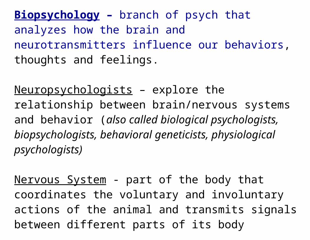
Biopsychology – branch of psych that analyzes how the brain and neurotransmitters influence our behaviors, thoughts and feelings.
Neuropsychologists – explore the relationship between brain/nervous systems and behavior (also called biological psychologists, biopsychologists, behavioral geneticists, physiological psychologists)
Nervous System - part of the body that coordinates the voluntary and involuntary actions of the animal and transmits signals between different parts of its body
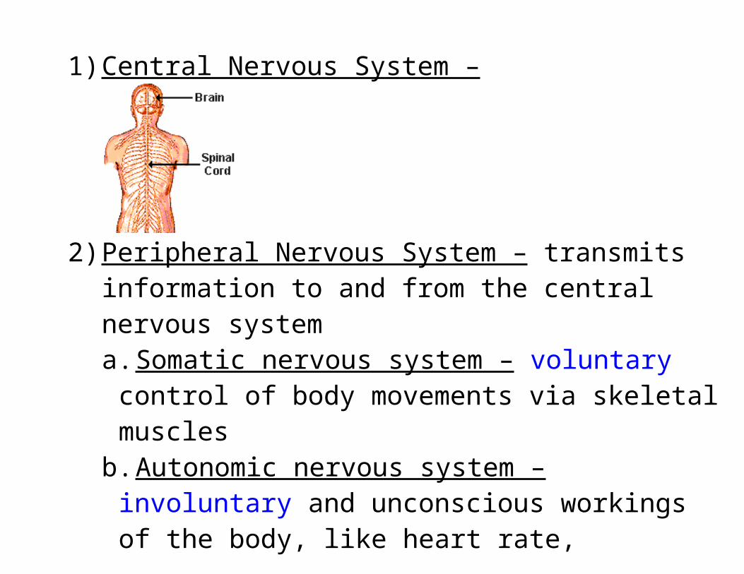
1) Central Nervous System –
2) Peripheral Nervous System – transmits information to and from the central nervous systema. Somatic nervous system – voluntary control of body
movements via skeletal musclesb. Autonomic nervous system – involuntary and
unconscious workings of the body, like heart rate, swallowing, pupil dilation, perspiration, respiratory ratei. Sympathetic – activates “fight or flight” responses,
preps body for traumatic event, mobilizes the body for action and energy output (think Superman)
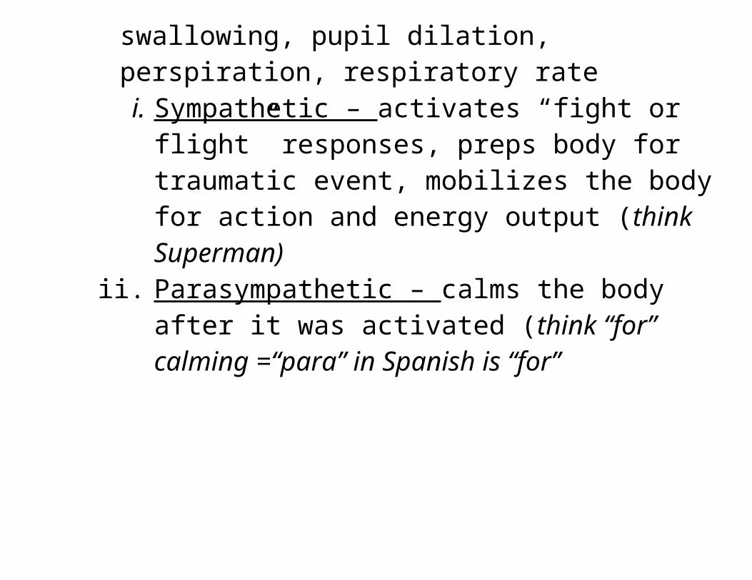
ii. Parasympathetic – calms the body after it was activated (think “for” calming =“para” in Spanish is “for”
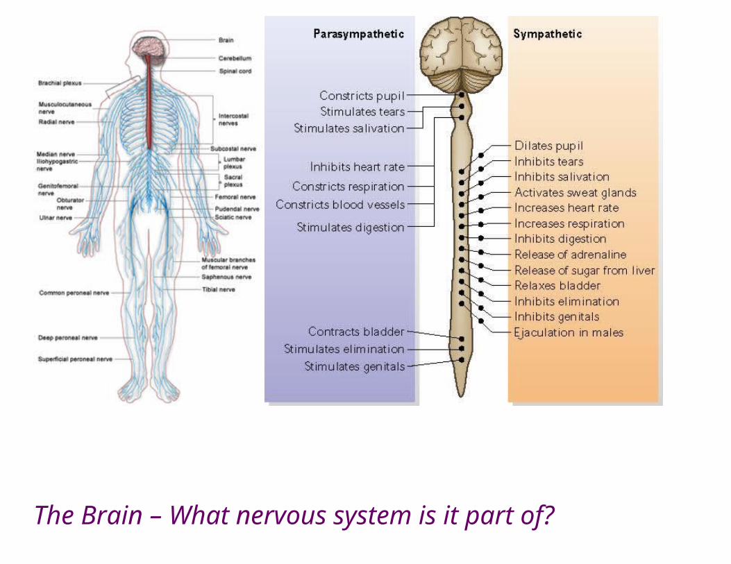
The Brain – What nervous system is it part of?
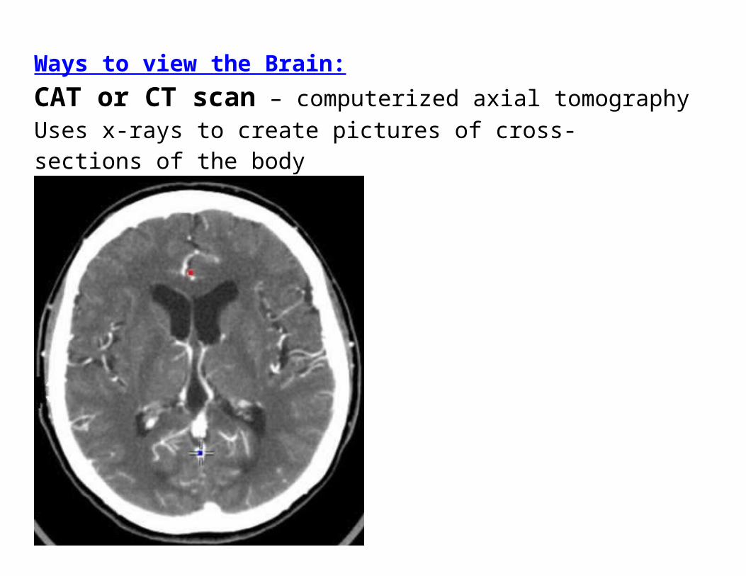
Ways to view the Brain:CAT or CT scan – computerized axial tomographyUses x-rays to create pictures of cross-sections of the body
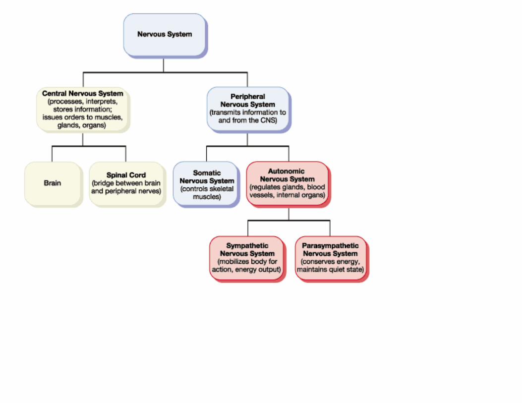
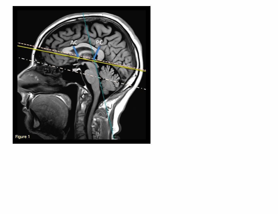
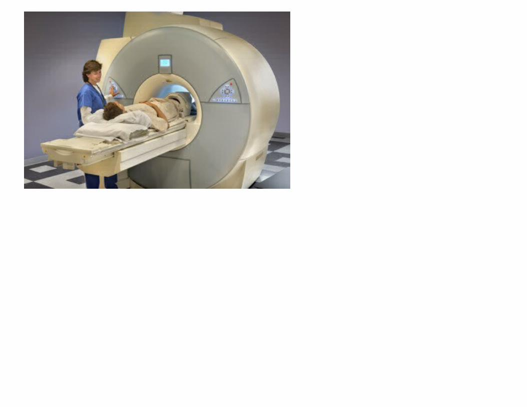
MRI – magnetic resonance imaging – does not use radiationuses powerful magnets and radio waves to create pictures of the body
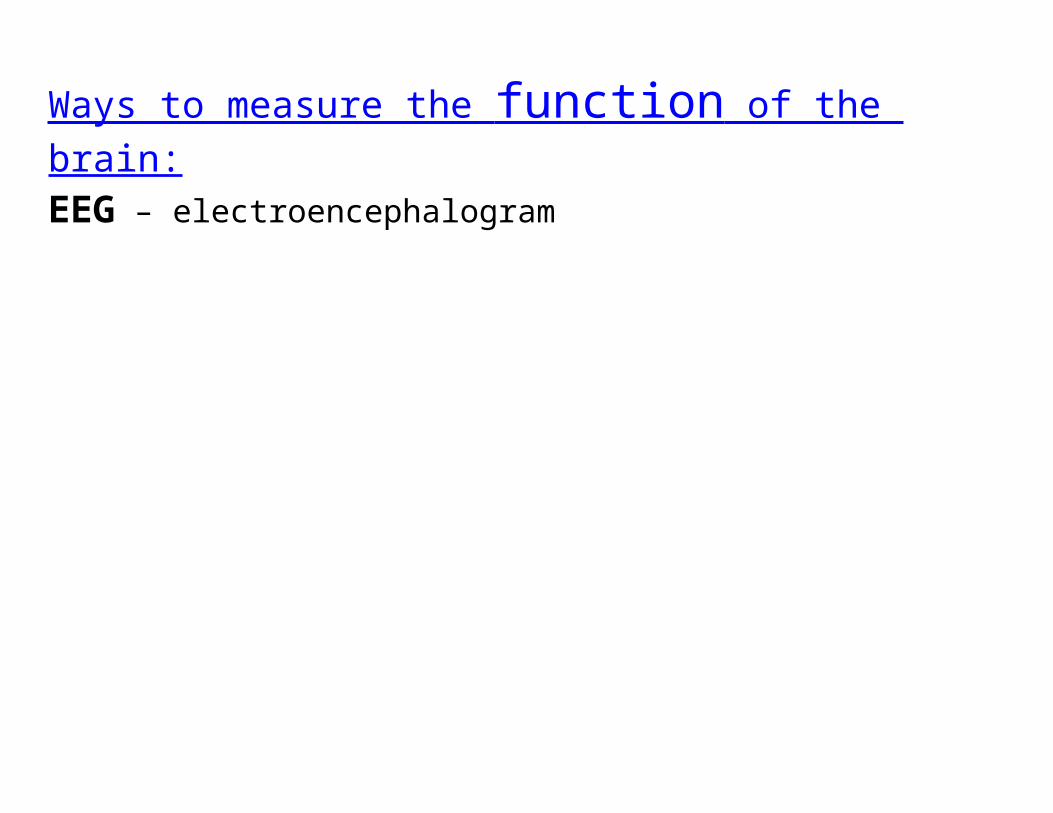
Ways to measure the function of the brain: EEG – electroencephalogram
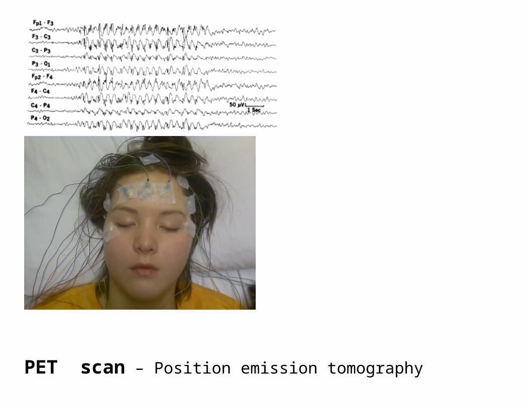
PET scan – Position emission tomographyuses a radioactive substance called a tracer to measure activity of the body, shows how organs function
www.dementiadiet.com
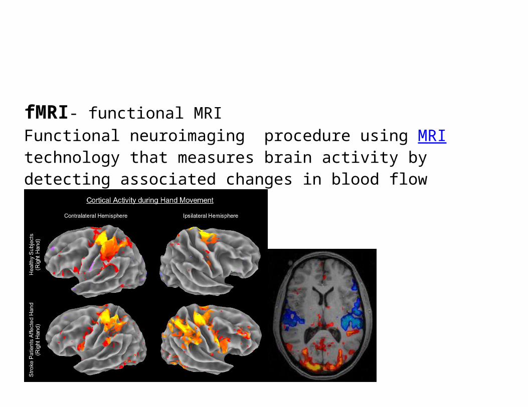
fMRI- functional MRIFunctional neuroimaging procedure using MRI technology that measures brain activity by detecting associated changes in blood flow
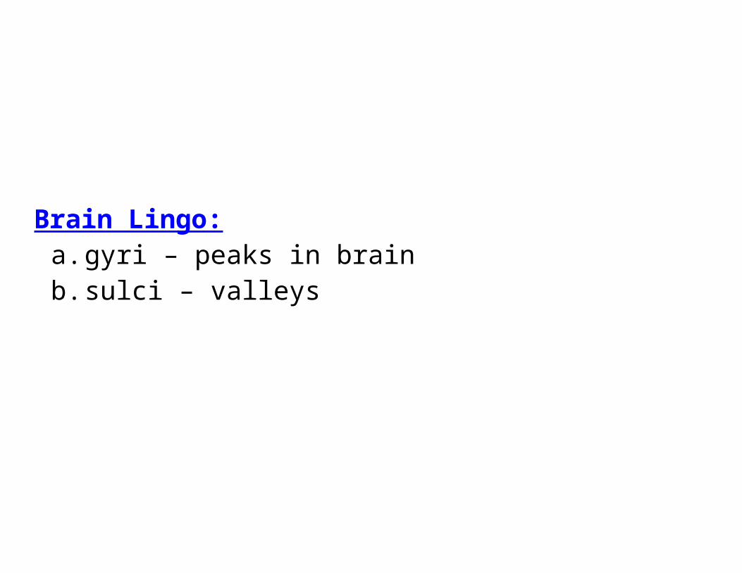
Brain Lingo:a. gyri – peaks in brainb. sulci – valleys
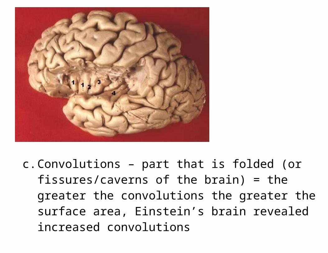
c. Convolutions – part that is folded (or fissures/caverns of the brain) = the greater the convolutions the greater the surface area, Einstein’s brain revealed increased convolutions
“Einstein Literally Had Unusual Brain”
Major structures of the Brain:
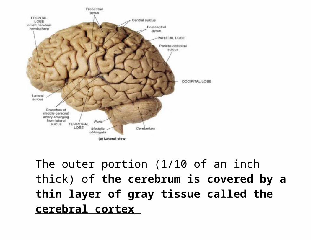
1. Cerebrum: largest part of the brain, resp. for higher brain functioning, most highly developed part of the brain
Makes up about two-thirds of the brain mass and lies over and around most of the structures of the brain.
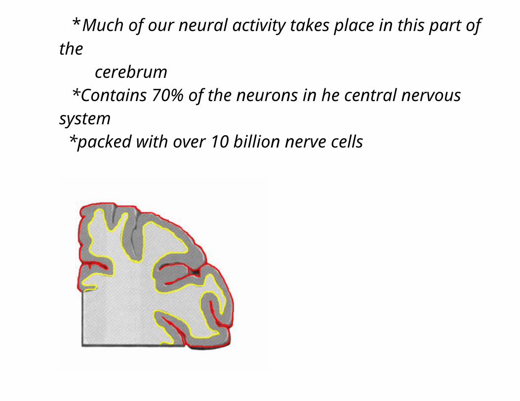
The outer portion (1/10 of an inch thick) of the cerebrum is covered by a thin layer of gray tissue called the cerebral cortex
*Much of our neural activity takes place in this part of the cerebrum
*Contains 70% of the neurons in he central nervous system
*packed with over 10 billion nerve cells
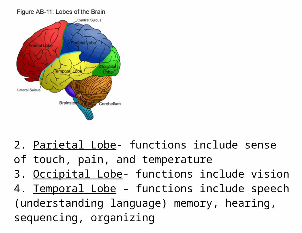
Each hemisphere of cerebrum is divided into four lobes:
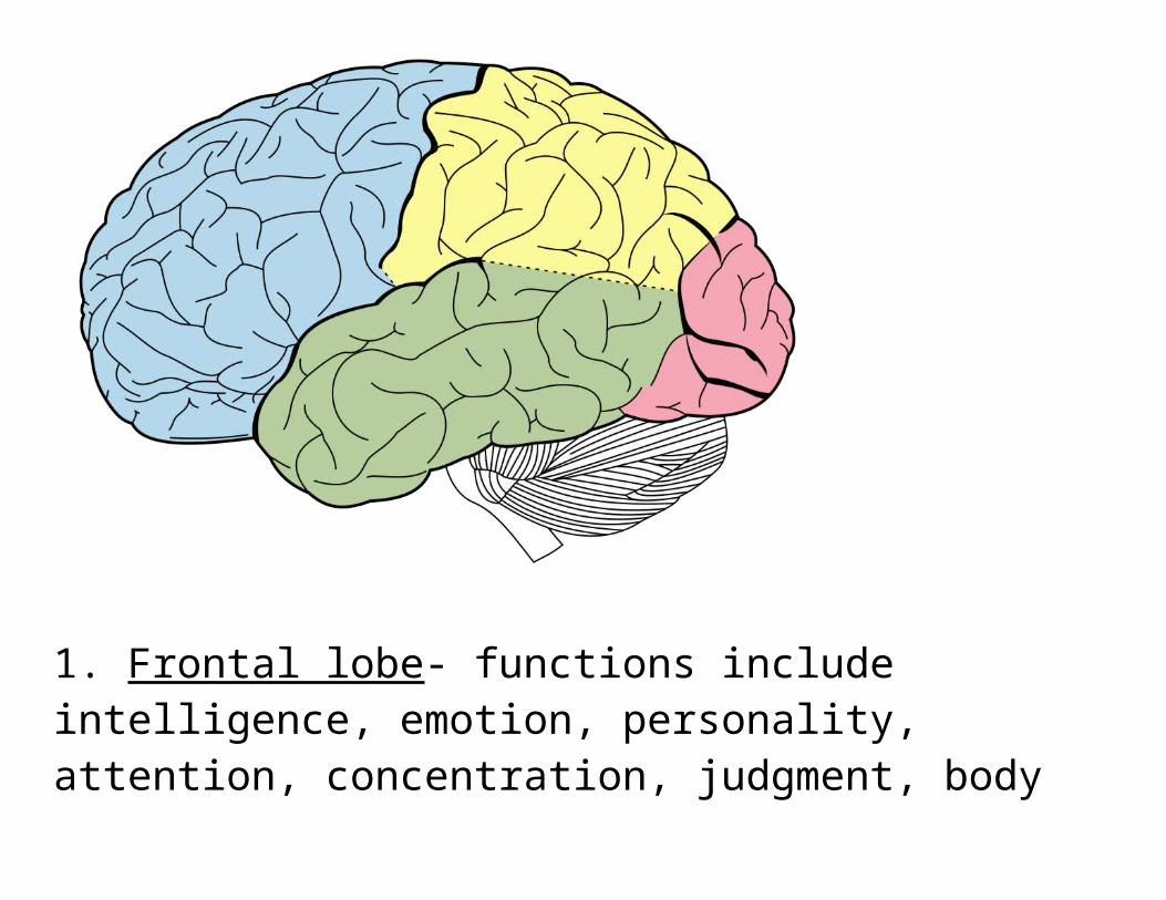
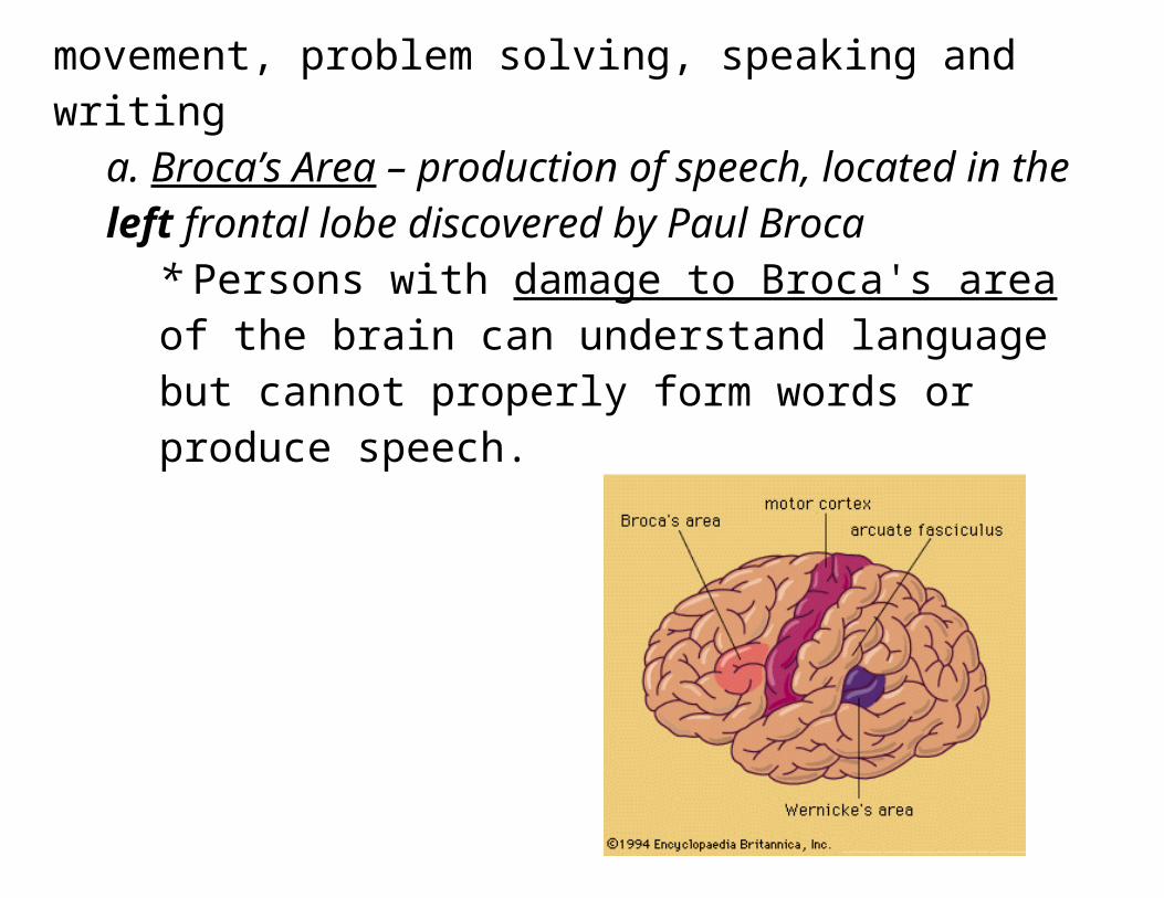
1. Frontal lobe- functions include intelligence, emotion, personality, attention, concentration, judgment, body movement, problem solving, speaking and writing
a. Broca’s Area – production of speech, located in the left frontal lobe discovered by Paul Broca
* Persons with damage to Broca's area of the brain can understand language but cannot properly form words or produce speech.
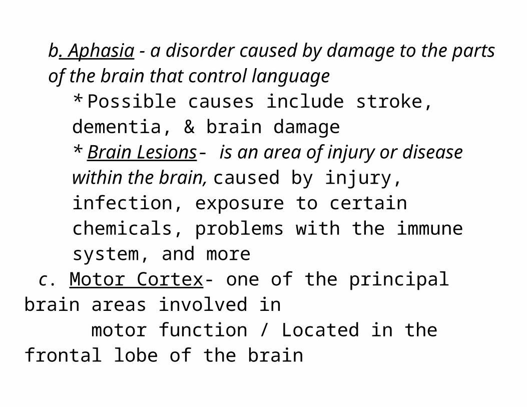
b. Aphasia - a disorder caused by damage to the parts of the brain that control language
* Possible causes include stroke, dementia, & brain damage* Brain Lesions- is an area of injury or disease within the brain, caused by injury, infection, exposure to certain chemicals, problems with the immune system, and more
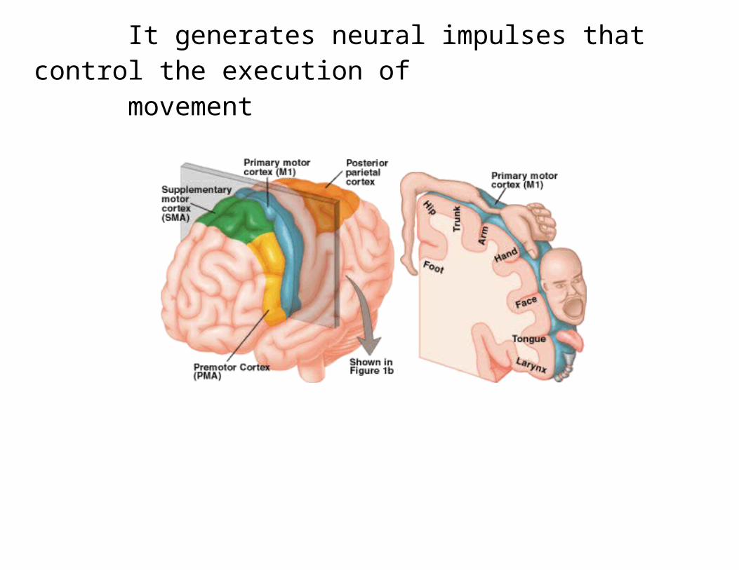
c. Motor Cortex- one of the principal brain areas involved in motor function / Located in the frontal lobe of the brain It generates neural impulses that control the execution of movement
2. Parietal Lobe- functions include sense of touch, pain, and temperature3. Occipital Lobe- functions include vision4. Temporal Lobe – functions include speech (understanding language) memory, hearing, sequencing, organizing
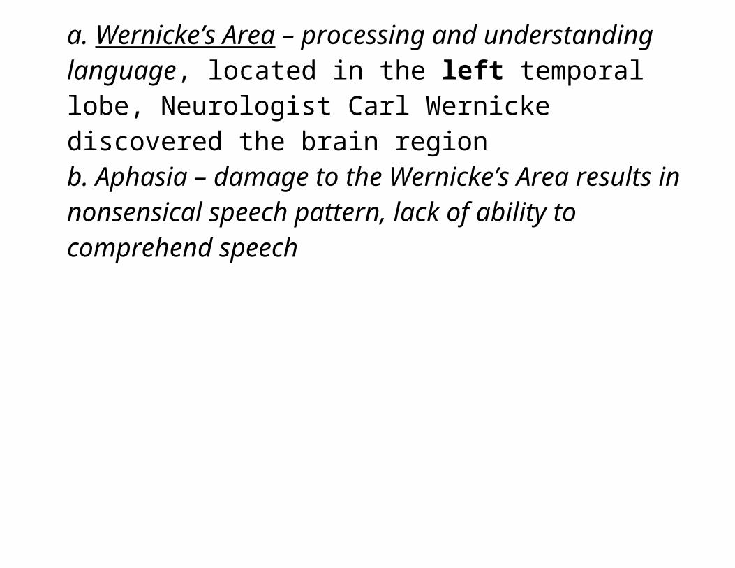
a. Wernicke’s Area – processing and understanding language, located in the left temporal lobe, Neurologist Carl Wernicke discovered the brain regionb. Aphasia – damage to the Wernicke’s Area results in nonsensical speech pattern, lack of ability to comprehend speech
The cerebrum is divided into right and left hemispheres that are connected by the corpus callosum. List some functions of each in your notes
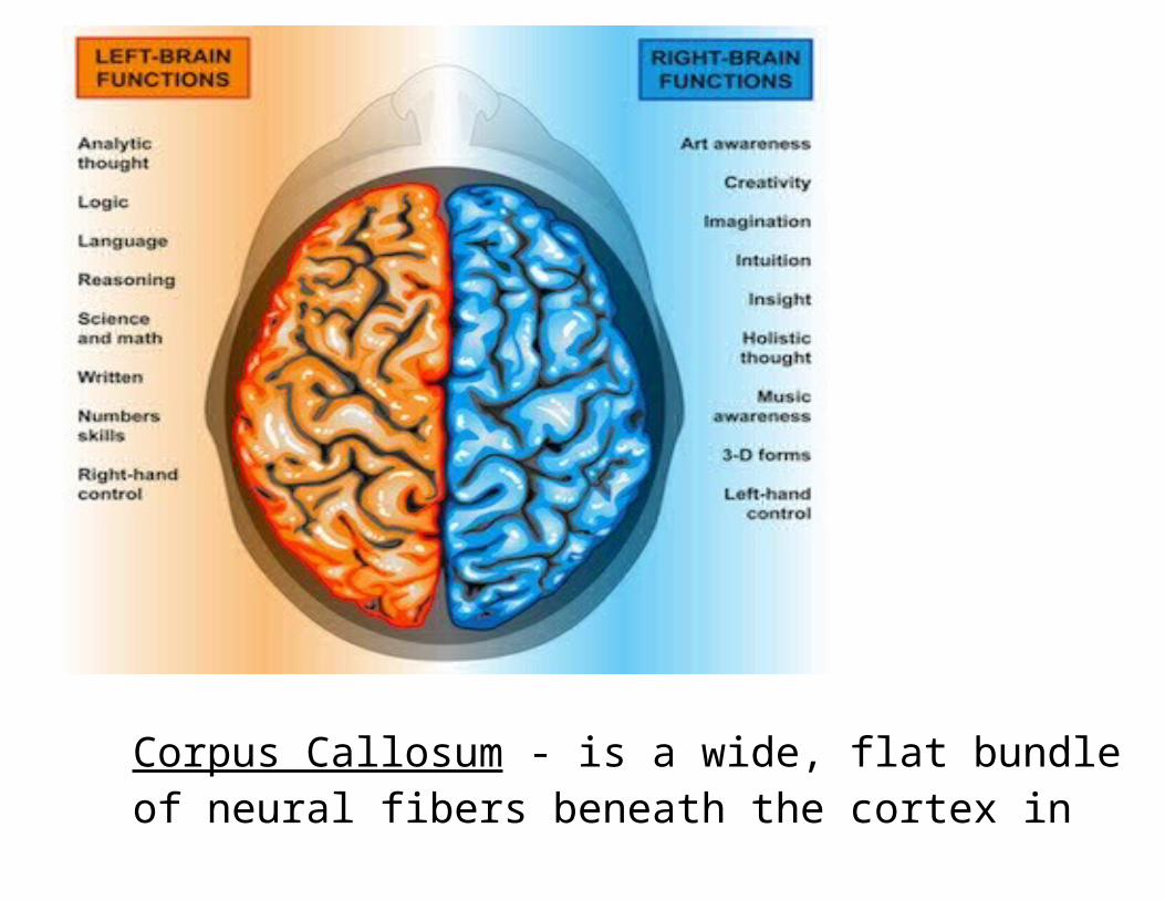
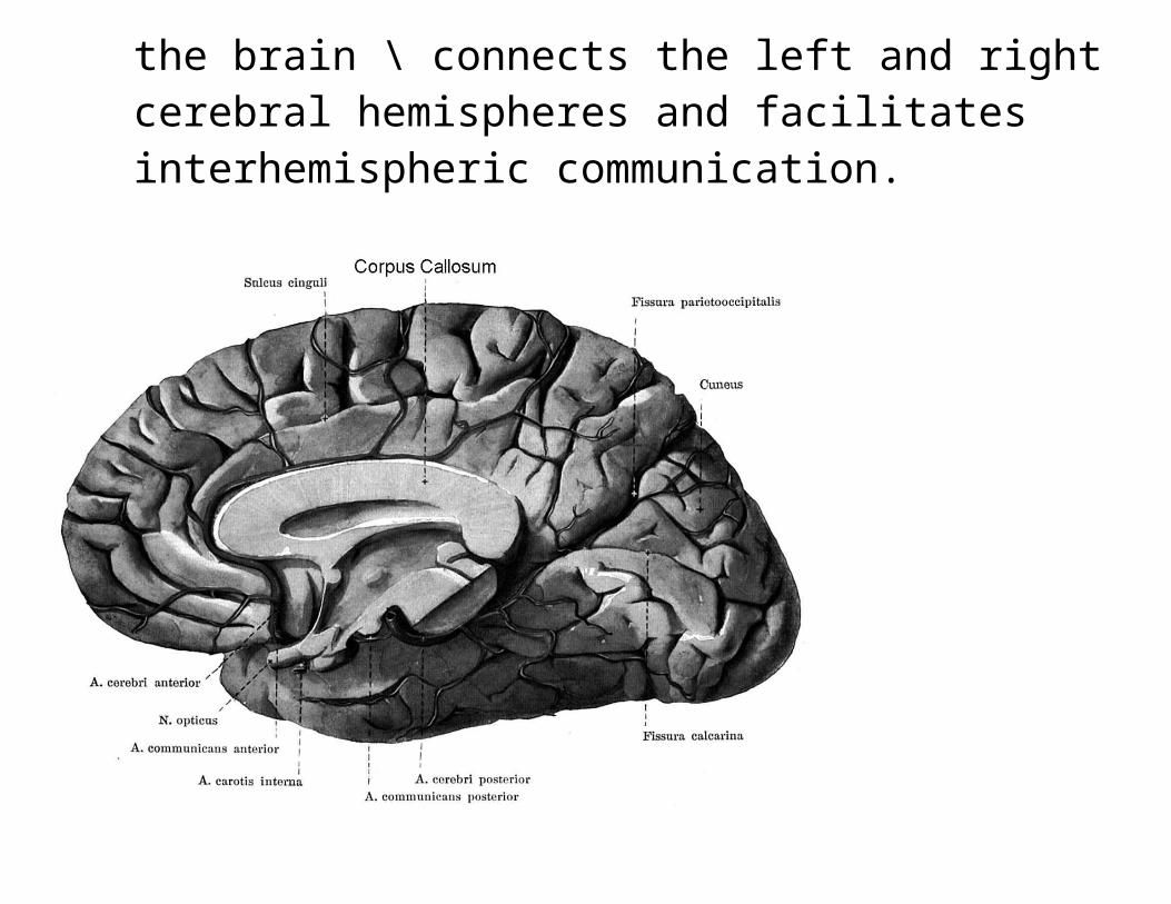
Corpus Callosum - is a wide, flat bundle of neural fibers beneath the cortex in the brain \ connects the left and right
cerebral hemispheres and facilitates interhemispheric communication.
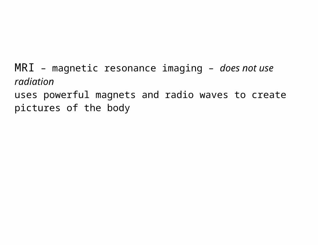
Contralaterality- one side of the body is controlled by the opposite side of the brain
Example: if one has a stroke in their right hemisphere, they won’t be able to move the ______________ side of their face.
“Girl Living With Half Her Brain” you tube
Split Brain Procedure is when he corpus callosum is severed, it is used to treat and minimize epilepsy. If done before age 10 the hemispheres of the brain learn to “communicate” in spite of splitPlasticity – brain reorganizes and learns to compensates for the damaged or missing structures in the brain Greater plasticity in early years! Plasticity diminishes with age.
“Split-brain patient 'Joe'
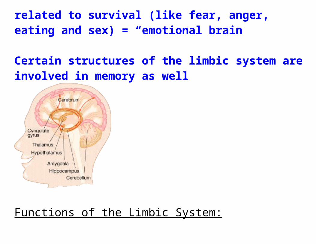
2. Limbic System: Evolutionarily primitive brain structures located on top of the brainstem
Functions include emotions, motivations, and pleasure particularly those that are related to survival (like fear, anger, eating and sex) = “emotional brain”
Certain structures of the limbic system are involved in memory as well
Functions of the Limbic System:
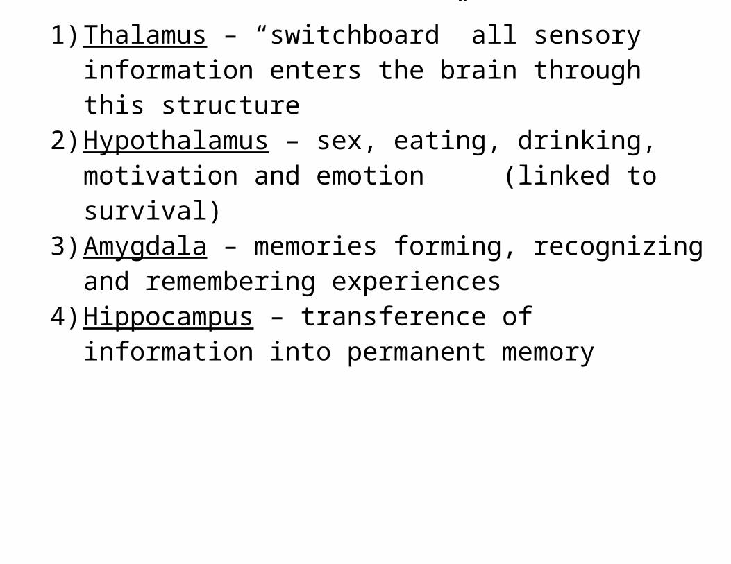
1) Thalamus – “switchboard” all sensory information enters the brain through this structure
2) Hypothalamus – sex, eating, drinking, motivation and emotion (linked to survival)
3) Amygdala – memories forming, recognizing and remembering experiences
4) Hippocampus – transference of information into permanent memory
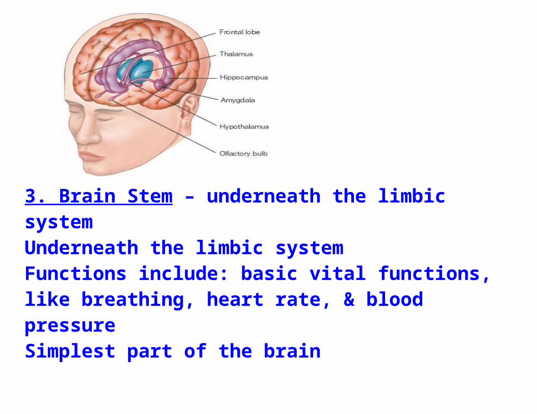
3. Brain Stem – underneath the limbic systemUnderneath the limbic system
Functions include: basic vital functions, like breathing, heart rate, & blood pressureSimplest part of the brainLike structures make up the entire brain of more simple creatures (reptiles)
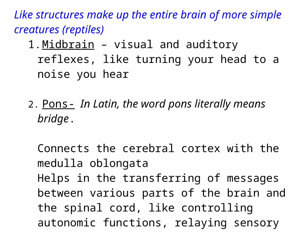
1.Midbrain – visual and auditory reflexes, like turning your head to a noise you hear
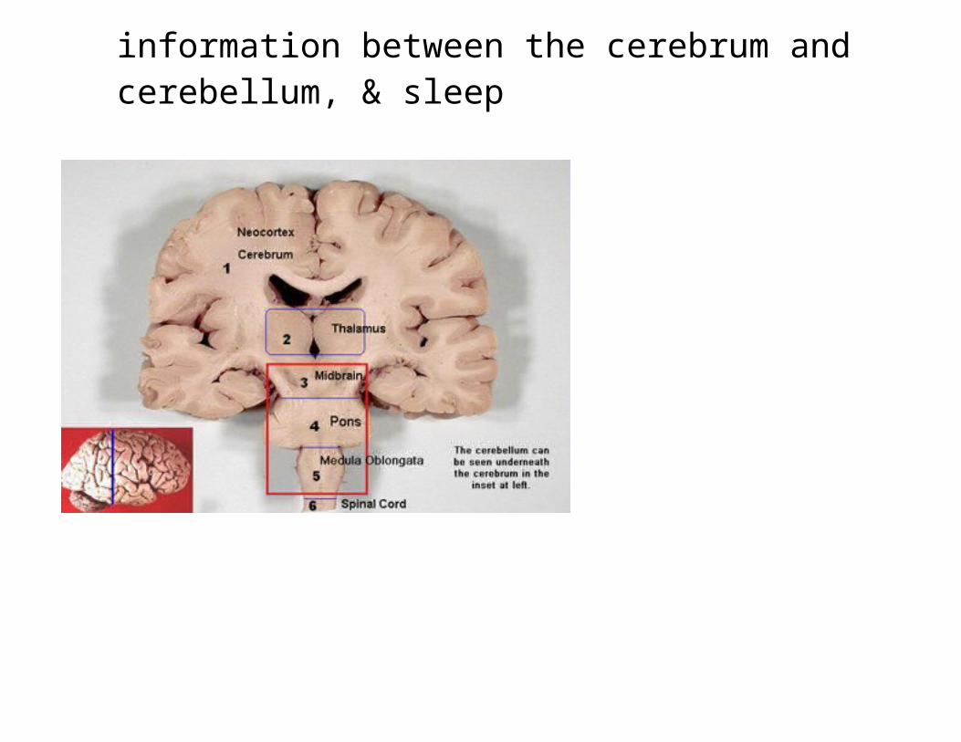
2. Pons- In Latin, the word pons literally means bridge.
Connects the cerebral cortex with the medulla oblongataHelps in the transferring of messages between various parts of the brain and the spinal cord, like controlling autonomic functions, relaying sensory information between the cerebrum and cerebellum, & sleep
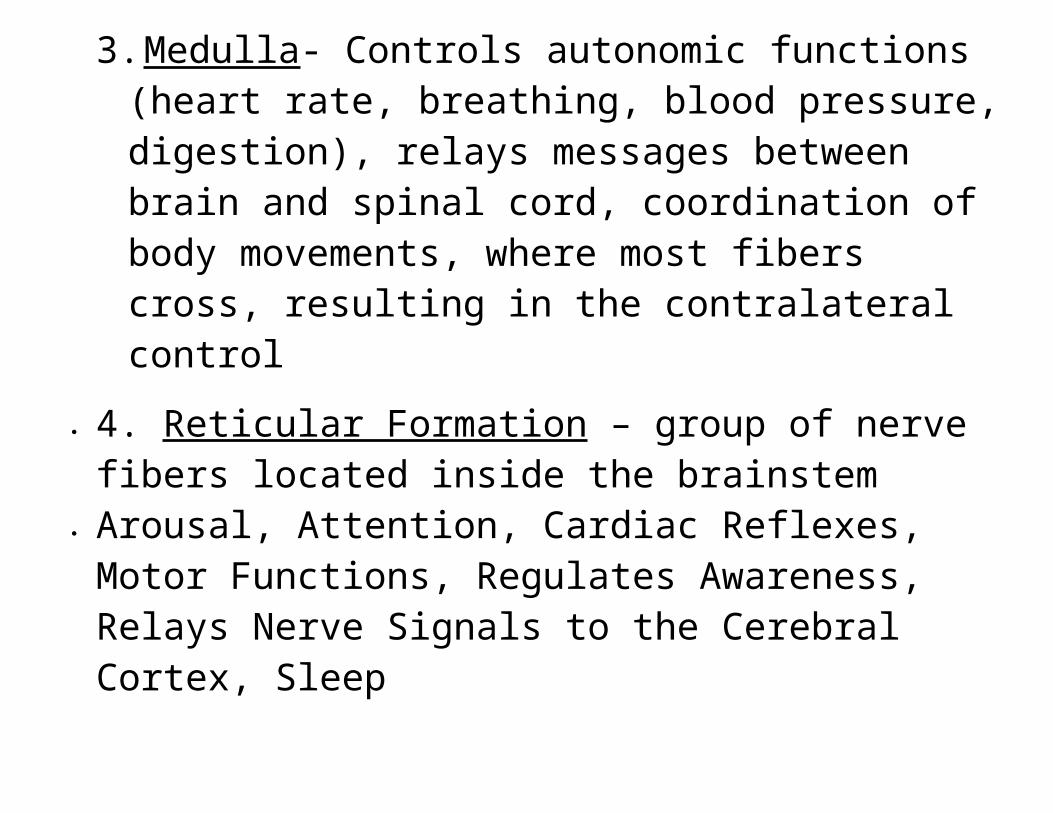
3.Medulla - Controls autonomic functions (heart rate, breathing, blood pressure, digestion), relays messages between brain and spinal cord, coordination of body movements, where most fibers cross, resulting in the contralateral control
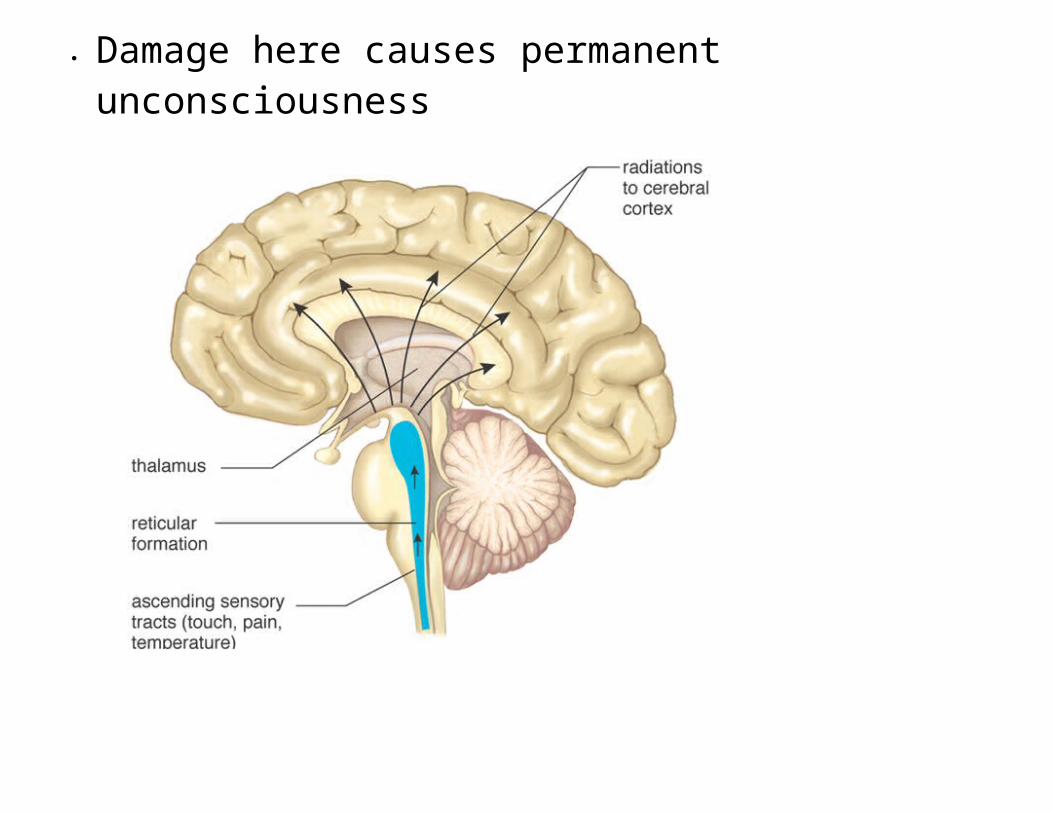
4. Reticular Formation – group of nerve fibers located inside the brainstem
Arousal, Attention, Cardiac Reflexes, Motor Functions, Regulates Awareness, Relays Nerve Signals to the Cerebral Cortex, Sleep
Damage here causes permanent unconsciousness
4. Cerebellum – “little brain” Has two hemispheres and convolutionsRegulation, coordination of movement, posture and balanceOlder, primitive structure of the brain“bellum = balance”
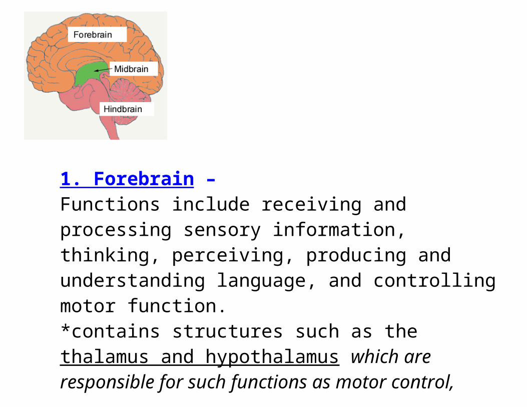
There are three major divisions of the brain:
1. Forebrain – Functions include receiving and processing sensory information, thinking, perceiving, producing and understanding language, and controlling motor function. *contains structures such as the thalamus and hypothalamus which are responsible for such functions as motor control, relaying sensory information, and controlling autonomic functions*Also contains the largest part of the brain, the cerebrum
Most of the actual information processing in the brain takes place in the cerebral cortex
2. Midbrain- *connects the hindbrain and the forebrain. *involved in auditory and visual responses as well as motor function.
The midbrain and the hindbrain together make up the brainstem
3. Hindbrain – extends from the spinal cord *contains structures such as the pons and cerebellum *These regions assists in maintaining balance and equilibrium, movement coordination, and the conduction of sensory information. Other structure included medulla oblongata
*responsible for controlling such autonomic functions as breathing, heart rate, and digestion.
Neuron: Individual nerve cell; an electrically excitable cell that processes & transmits information through electrical and chemical signals3 functions: 1) receive info 2) process it 3)transmit it to body
100 billion in brain at birth, generated at ¼ million per minute during gestation
Dendrites: Receive messages from other neuronsSoma: “Cell body” body of the neuron. Receives messages and sends messages down axonAxon: Carries information away from the cell bodyAxon Terminals/ Terminal Buttons: Branches that link the dendrites and somas of other neuronsMyelin Sheath: Fatty layer that coats some axons
Multiple Sclerosis (MS) occurs when myelin layer is destroyed; numbness, weakness, and paralysis occur
Glial cells - provide support and protection for neurons. They are thus known as the "supporting cells" of the nervous system. The four main functions of glial cells are:
1) to surround neurons and hold them in place2) to supply nutrients and oxygen to neurons 3) to insulate one neuron from another4) to destroy and remove the carcasses of dead neurons = clean up
Nerve Impulse - each neuron is like a tiny biological battery
Fig. 2.1 An example of a neuron, or nerve cell, showing several of its important features. The right foreground shows a nerve cell fiber in cross section, and the upper left inset gives a more realistic picture of the shape of neurons. The nerve impulse usually travels from the dendrites and soma to the branching ends of the axon. The neuron shown here is a motor neuron. Motor neurons originate in the brain or spinal cord and send their axons
Resting Potential : Electrical charge of an inactive neuron / At rest, the inside of an axon is about –60 to –70 millivolts, compared with the outside.
Threshold : Trigger point for a neuron’s firing Action Potential : When positively charged sodium ions
(Na+) rush into the cell, its interior briefly becomes positive = nerve impulse
Ion Channels : Axon membrane has these tiny holes or
tunnels
Negative After-Potential : After the action potential, an outward flow of positive potassium ions (K+) restores the negative charge inside the axon = neuron is less willing to fire
All or nothing principle: – the neuron either fire or it doesn’t
Fig. 2.2 Activity in an axon can be measured by placing electrical probes inside and outside the axon. (The scale is exaggerated here. Such measurements require ultra-small electrodes, as described later in this chapter.) At rest, the inside of an axon is about –60 to –70 millivolts, compared with the outside. Electrochemical changes in a nerve cell generate an action potential. When positively charged sodium ions (Na+) rush into the cell, its interior briefly becomes positive. This is the action potential. After the action potential, an outward flow of positive potassium ions (K+) restores the negative charge inside the axon. (See Figure 2.3 for further explanation.)
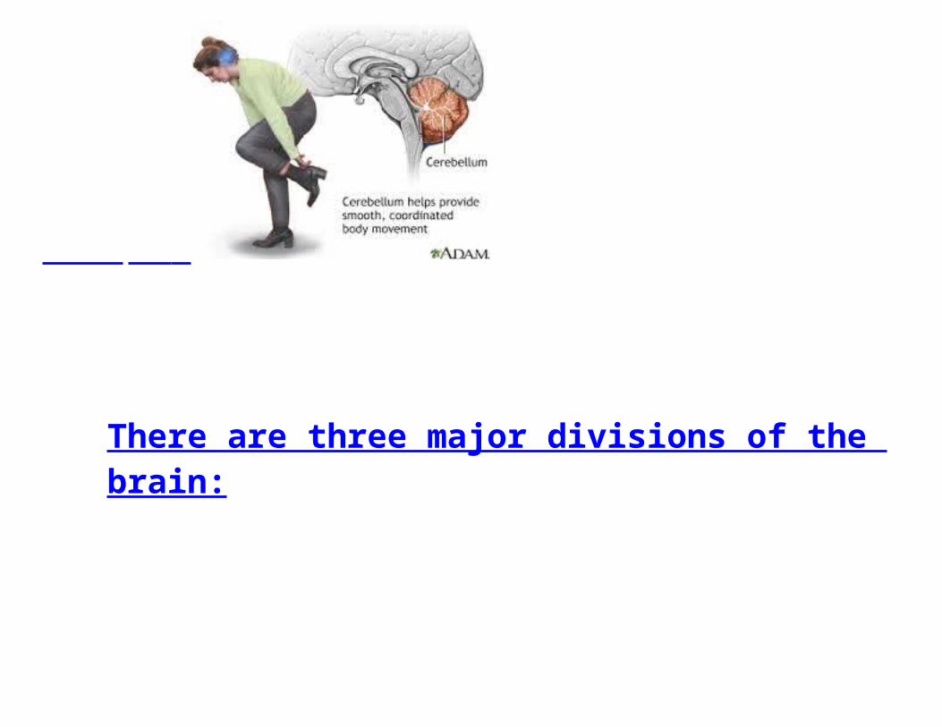
Fig. 2.5 A highly magnified view of the synapse shown in Fig. 2.1. Neurotransmitters are stored in tiny sacs called synaptic vesicles. The size of the gap is exaggerated here; it is actually only about one millionth of an inch. Transmitter molecules vary in their effects
Synapse: The microscopic space between two neurons, over with messages pass
Neurotransmitter s: Chemicals that alter activity in neurons; brain chemicals (some excite and other inhibit activity)
Receptor Site : Areas on the surface of neurons and other cells that are sensitive to neurotransmitters
Types of Neurotransmitters you will be tested on:
Acetylcholine (ACh): causes contractions of skeletal muscles, regulates heart muscles, involved in memory, helps transmit message to and from brain and spinal cord
Lack of ACh is associated with Alzheimer’s diseaseDopamine: Muscle control and movement, synthesizes hormones, affects alertness
addictive drugs, including stimulants such as cocaine, amphetamine, and methamphetamine, act by amplifying the effects of dopamine
Dysfunctions of the dopamine system: Parkinson's disease, a degenerative condition causing tremor and motor impairment, is caused by loss of dopamine-secreting neurons
Schizophrenia involves altered levels of dopamine activity, and the antipsychotic drugs (neuroleptics) that are frequently used to treat it have a primary effect of attenuating dopamine activity / Increased dopamine leads to positive symptoms of disorder = hallucinations and delusions
Attention deficit hyperactivity disorder (ADHD) are also believed to be associated with decreased dopamine activity.
Serotonin: Mood, appetite control, sex activity, attention and emotion, lack of it linked to depression
Drugs which alter serotonin levels are used in treating depression, generalized anxiety disorder and social phobia.
Anti-depressants prevent the breakdown of neurotransmitters = increase concentrations of the serotonin in the brain
Glutamate – excitatory neurotransmitter involved in info processing and memory formation in hippocampus, linked to both Alzheimer’s and SchizophreniaEndorphins: Released by pituitary gland; also help to relieve painGABA – inhibits firing of neurons
associated with Huntington’s disease, effects coordination & movement (neurodegenerative genetic disorder)
Seizures associated with malfunctioning GABA Current Meds utilize GABA to help control chronic
nerve painAgonists- mimic neurotransmitters, bind to receptors and produce effect of neurotransmitterAntagonists – blocks receptors and inhibit effect of neurotransmitters


![How Science Affects People’s Lives Health Medical Imaging X-Rays C(A)T [Computerized (Axial) Tomography] Scanning PET [Positron Emission Tomography]](https://img.pdfslide.us/doc/110x75/56649ec95503460f94bd6a9a/how-science-affects-peoples-lives-health-medical-imaging-x-rays.jpg)