Embed Size (px)
Citation preview
12/2/08
1
NEURORADIOLOGY Angela Lignelli, MD
Neuroradiology
Plain radiographs
CT
MRI
Cerebral Angiogram
Myelograms
Neuroradiology
Computerized Axial Tomography (CT) CT without and with contrast
CTA – CT angiogram
CTP - CT perfusion
Magnetic Resonance Imaging (MRI)
MR without and with contrast
MRA – MR angiogram/MRV –MR venogram
MRP – MR perfusion
MRS- MR spectroscopy
MR tractography (DTI)
fMRI – functional MRI
Computerized Axial
Tomography CT images are reconstructed from sets of quantitative x-ray measurements obtained through the head at multiple angles
X-ray source rotates around the head and divides x-ray attenuation into compartments called pixels.
The computer assigns a number to each pixel and by using a gray scale, reconstructs an image.
Adv: very quick, less expensive
Disadv: good but not great in delineation of soft tissue anatomy and pathology
uses x-ray radiation
CT noncontrast uses
Intial evaluation of
Head injury – acute intracranial hemorrhage especially subarchnoid hemorrhage – superior in evaluating cortical bone structures of bone and spine
Stroke
Less sensitive than MRI during first 48 hours
Posterior fossa infarcts difficult to see due to beam hardening artifacts (artifacts caused by x-ray attenuation by thick osseus structures eg at skull base.)
Computerized Axial
Tomography Contrast enhanced CT
Iodinated water soluble contrast agents can be
given intravenously to enhance differences in
tissue density
Used to detect lesions that involve breakdown of
the blood brain barrier eg: certain tumors,
infections or inflammatory conditions
Intravenous CT contrast agents are based on
iodine – high osmolar contrast media vs low
osmolar contrast media (nonionic)
12/2/08
2
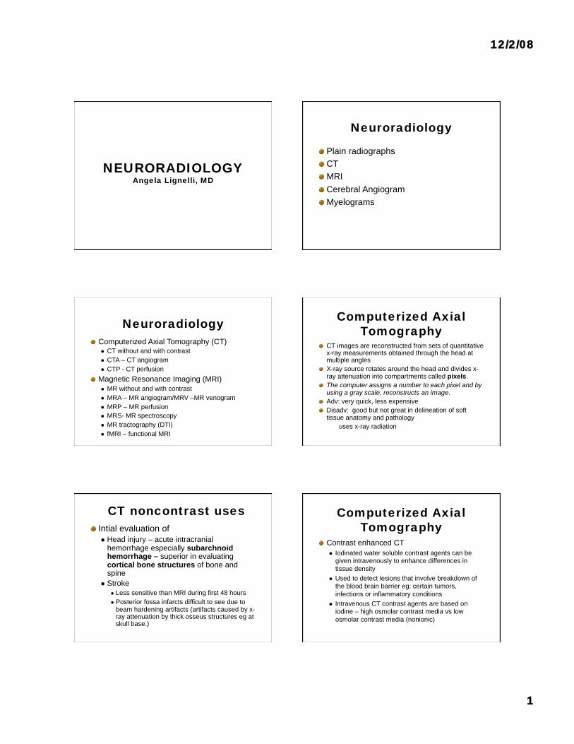
Normal Head CT
Gray
matter
White
matter Frontal
Horns of
lateral
vents Internal
capsule Basal
ganglia
Head CT
INFERIOR SLICE AT LEVEL OF POSTERIOR
FOSSA
Bone Windows Subdural windows
12/2/08
3
MRI Magnetic field causes alignment of atomic nuclei of 2 or more magnetic states.
Proton based MRI - application of radiowaves of the hydrogen specific resonance frequency to biologic tissues excites some protons into a higher energy state.
Following the pulse the relaxation of these protons back to their original energy state is accompanied by emission of radiowaves that are characteristic of the particular tissue. Two tissue specific relaxation constants known as T1 and T2 as well as proton density can be measured
The difference in proton density, T1 and T2 relaxation enable MRI to distinguish fat, muscle, bone marrow and gray or white matter of the brain.
MRI
magnetic resonance
imaging Adv
Superior to CT for the detection of most CNS diseases due to its high soft tissue contrast resolution
Multiplanar capability
Lack of ionizing radiation
Better visualization of the posterior fossa
Disadv
Typical brain MR study takes approx. 30 min
Patient must be able to hold still
Multiple sequences are obtained
CI- Swan Ganz/ Pacemaker
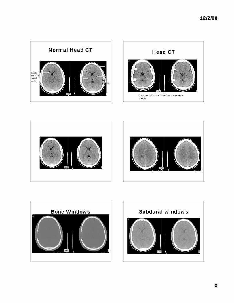
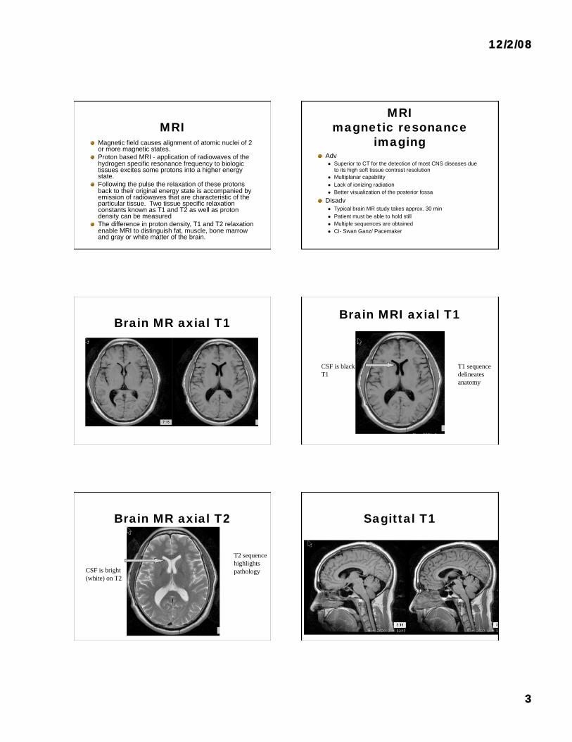
Brain MR axial T1 Brain MRI axial T1
T1 sequence
delineates
anatomy
CSF is black on
T1
Brain MR axial T2
CSF is bright
(white) on T2
T2 sequence
highlights
pathology
Sagittal T1
12/2/08
4
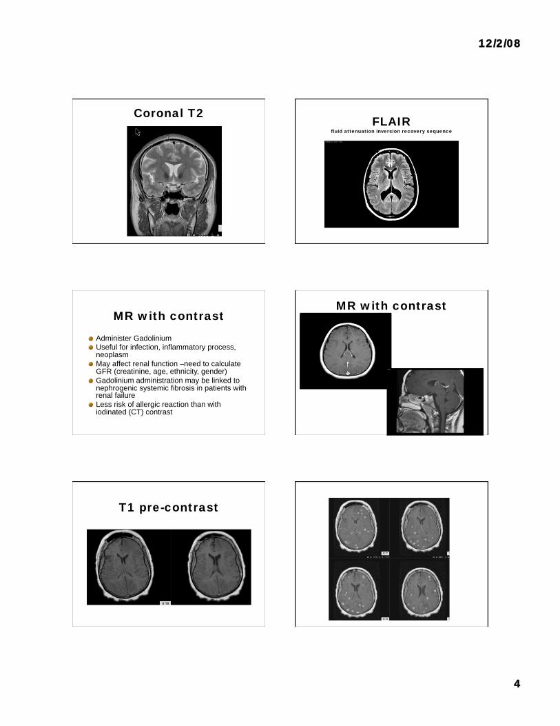
Coronal T2 FLAIR
fluid attenuation inversion recovery sequence
MR with contrast
Administer Gadolinium Useful for infection, inflammatory process, neoplasm
May affect renal function –need to calculate GFR (creatinine, age, ethnicity, gender)
Gadolinium administration may be linked to nephrogenic systemic fibrosis in patients with renal failure
Less risk of allergic reaction than with iodinated (CT) contrast
MR with contrast
T1 pre-contrast
12/2/08
5
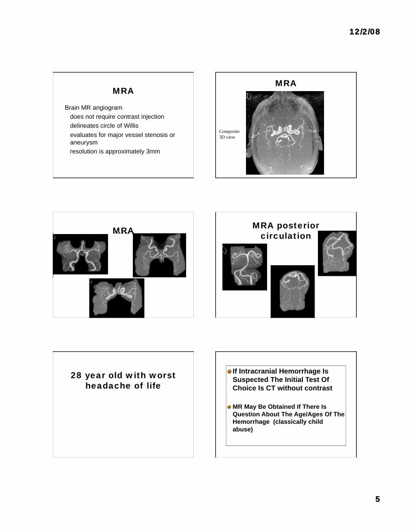
MRA
Brain MR angiogram
does not require contrast injection
delineates circle of Willis
evaluates for major vessel stenosis or
aneurysm
resolution is approximately 3mm
MRA
Composite
3D view
MRA MRA posterior
circulation
28 year old with worst
headache of life
If Intracranial Hemorrhage Is
Suspected The Initial Test Of
Choice Is CT without contrast
MR May Be Obtained If There Is
Question About The Age/Ages Of The
Hemorrhage (classically child
abuse)
12/2/08
6
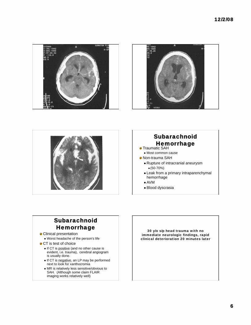
Subarachnoid
Hemorrhage Traumatic SAH
Most common cause
Non-trauma SAH
Rupture of intracranial aneurysm
(50-70%)
Leak from a primary intraparenchymal hemorrhage
AVM
Blood dyscrasia
Subarachnoid
Hemorrhage Clinical presentation
Worst headache of the person’s life
CT is test of choice
If CT is positive (and no other cause is evident, i.e. trauma), cerebral angiogram is usually done.
If CT is negative, an LP may be performed next to look for xanthocromia
MR is relatively less sensitive/obvious to SAH. (Although some claim FLAIR imaging works relatively well)
30 y/o s/p head trauma with no
immediate neurologic findings, rapid
clinical deterioration 20 minutes later
12/2/08
7
Epidural Hemorrhage
Usually secondary to trauma
Arterial epidural
Most common
From laceration of the middle meningeal
artery
Associated with a temporal bone fracture
Venous epidural Less common
From tear of middle meningeal vein
Laceration of a venous sinus (posterior fossa,
more common in children)
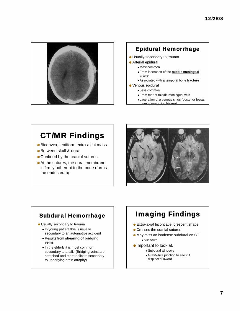
CT/MR Findings Biconvex, lentiform extra-axial mass
Between skull & dura
Confined by the cranial sutures
At the sutures, the dural membrane
is firmly adherent to the bone (forms
the endosteum)
Subdural Hemorrhage
Usually secondary to trauma
In young patient this is usually
secondary to an automotive accident
Results from shearing of bridging
veins
In the elderly it is most common
secondary to a fall. (Bridging veins are
stretched and more delicate secondary
to underlying brain atrophy)
Imaging Findings
Extra-axial biconcave, crescent shape
Crosses the cranial sutures
May miss an isodense subdural on CT
Subacute
Important to look at: Subdural windows
Gray/white junction to see if it
displaced inward
12/2/08
8
Cerebral Aneurysm
Besides subarachnoid hemorrhage,
an aneurysm may present
secondary to mass effect.
PCA or Superior Cerebellar Artery
aneurysm may press on the third
nerve causing a palsy
Angiography
Gold standard for diagnosis of an aneurysm
Is however an invasive procedure
NPO
Well hydrated
Off coumadin, if on heparin d/c 4 hrs prior
Need recent PT/PTT, Platelet count, BUN,
creatinine
Off Glucophage
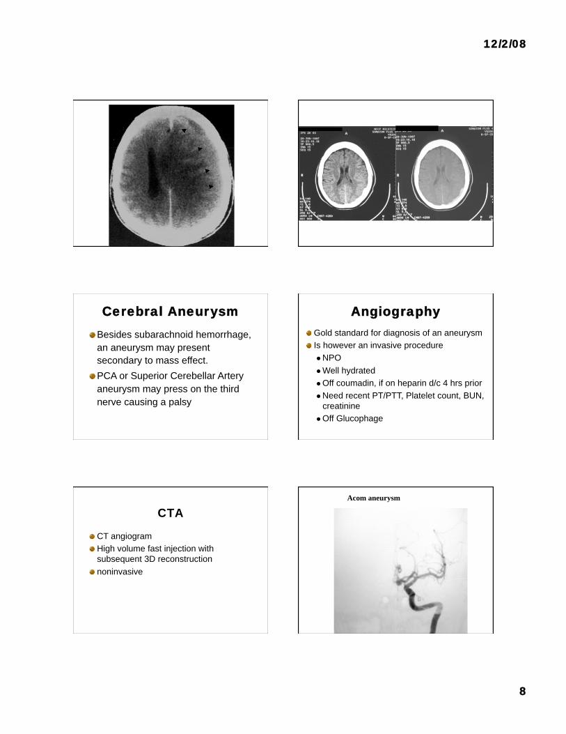
CTA
CT angiogram
High volume fast injection with
subsequent 3D reconstruction
noninvasive
Acom aneurysm
12/2/08
9
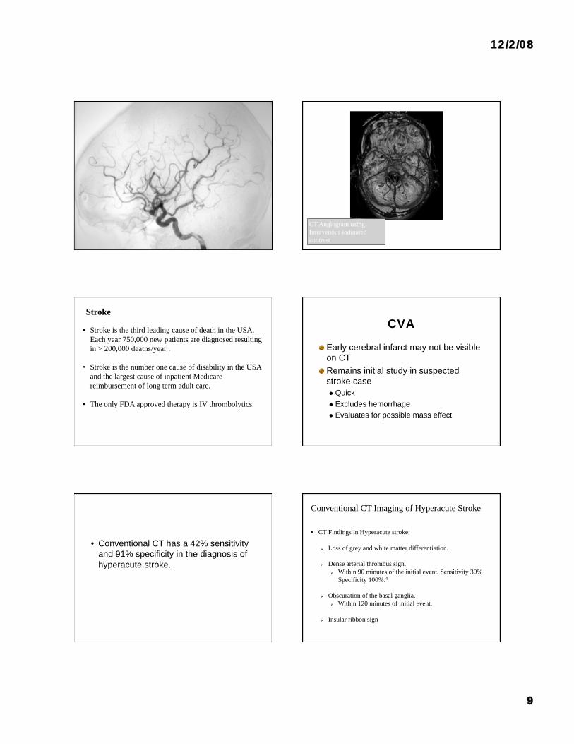
CT Angiogram using
Intravenous iodinated
contrast
Stroke
• Stroke is the third leading cause of death in the USA.
Each year 750,000 new patients are diagnosed resulting
in > 200,000 deaths/year .
• Stroke is the number one cause of disability in the USA
and the largest cause of inpatient Medicare
reimbursement of long term adult care.
• The only FDA approved therapy is IV thrombolytics.
CVA
Early cerebral infarct may not be visible
on CT
Remains initial study in suspected
stroke case
Quick
Excludes hemorrhage
Evaluates for possible mass effect
• Conventional CT has a 42% sensitivity
and 91% specificity in the diagnosis of
hyperacute stroke.
Conventional CT Imaging of Hyperacute Stroke
• CT Findings in Hyperacute stroke:
Loss of grey and white matter differentiation.
Dense arterial thrombus sign.
Within 90 minutes of the initial event. Sensitivity 30%
Specificity 100%.4
Obscuration of the basal ganglia.
Within 120 minutes of initial event.
Insular ribbon sign
12/2/08
10
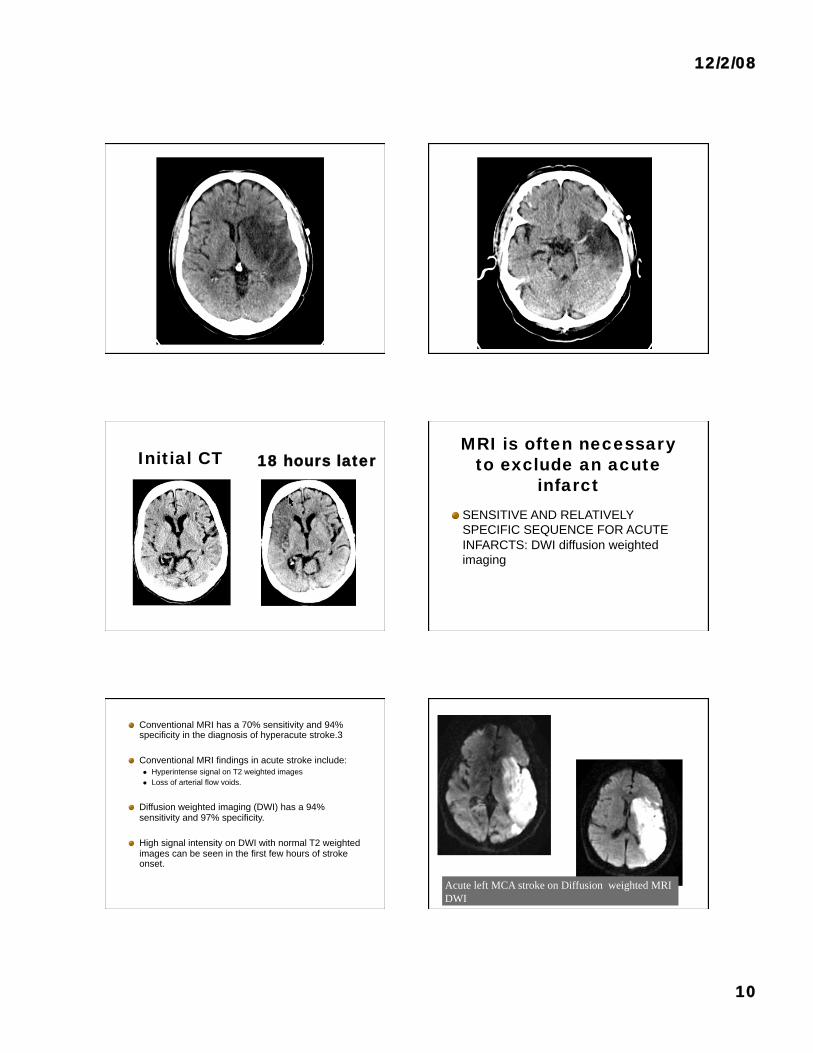
Initial CT 18 hours later MRI is often necessary
to exclude an acute
infarct
SENSITIVE AND RELATIVELY
SPECIFIC SEQUENCE FOR ACUTE
INFARCTS: DWI diffusion weighted
imaging
Conventional MRI has a 70% sensitivity and 94% specificity in the diagnosis of hyperacute stroke.3
Conventional MRI findings in acute stroke include:
Hyperintense signal on T2 weighted images
Loss of arterial flow voids.
Diffusion weighted imaging (DWI) has a 94% sensitivity and 97% specificity.
High signal intensity on DWI with normal T2 weighted images can be seen in the first few hours of stroke onset.
Acute left MCA stroke on Diffusion weighted MRI
DWI
12/2/08
11
Left hemianopsia White Matter Diseases
Microvascular Ischemic Disease Primary Demyelinating diseases
Multiple Sclerosis
Secondary demyelinating diseases
Infectious agents/vaccinations
Nutritional/vitamin deficiency
Physical/chemical agents or therapy
Vascular
Genetic abnormality
Big Two
Microvascular ischemic
disease
Multiple sclerosis (MS)
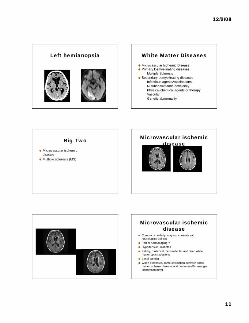
Microvascular ischemic
disease
Microvascular ischemic
disease Common in elderly; may not correlate with
neurological deficits
Part of normal aging ?
Hypertension, diabetes
Patchy, multifocal, periventicular and deep white
matter optic radiations
Basal ganglia
When extensive, some correlation between white
matter ischemic disease and dementia (Binswanger
encephalopathy)
12/2/08
12
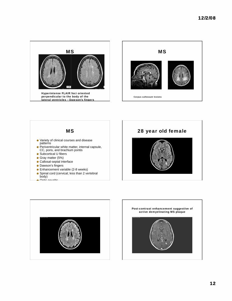
MS
Hyperintense FLAIR foci oriented
perpendicular to the body of the
lateral ventricles – Dawson’s fingers
MS
Corpus callossum lesions
MS
Variety of clinical courses and disease patterns
Periventricular white matter, internal capsule, CC, pons, and brachium pontis
Subcortical U fibers
Gray matter (5%)
Callosal-septal interface
Dawson’s fingers
Enhancement variable (2-8 weeks)
Spinal cord (cervical; less than 2 vertebral body)
Optic neuritis
28 year old female
Post-contrast enhancement suggestive of
active demyelinating MS plaque
12/2/08
13
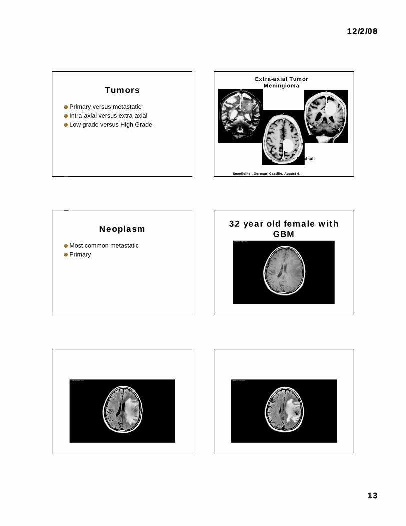
Tumors
Primary versus metastatic
Intra-axial versus extra-axial
Low grade versus High Grade
Extra-axial Tumor
Meningioma
Emedicine , German Castillo, August 6,
20044
Dural tail
Neoplasm
Most common metastatic
Primary
32 year old female with
GBM
12/2/08
14
4 months earlier
Infiltrative High Grade
Tumor Severe mass effect
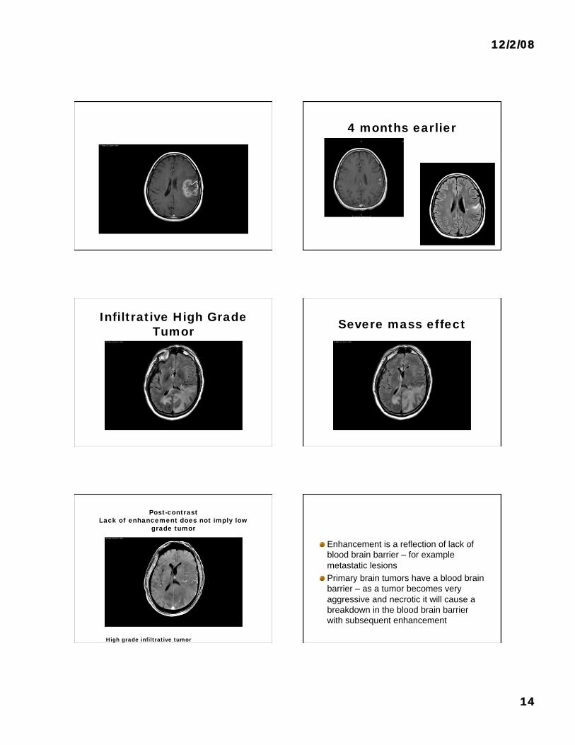
Post-contrast
Lack of enhancement does not imply low
grade tumor
High grade infiltrative tumor
Enhancement is a reflection of lack of
blood brain barrier – for example
metastatic lesions
Primary brain tumors have a blood brain
barrier – as a tumor becomes very
aggressive and necrotic it will cause a
breakdown in the blood brain barrier
with subsequent enhancement
12/2/08
15
Case #8
Post-contrast
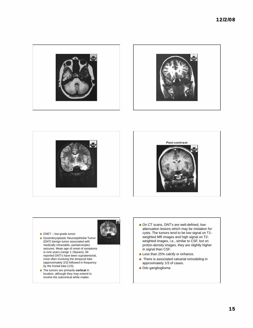
DNET – low grade tumor
Dysembryoplastic Neuroepithelial Tumor
(DNT) benign tumor associated with
medically intractable, partialcomplex
seizures. Mean age of onset of symptoms
is nine years (range 1-19years). All
reported DNT's have been supratentorial, most often involving the temporal lobe
(approximately 2/3) followed in frequency
by the frontal lobe (1/3).
The tumors are primarily cortical in
location, although they may extend to
involve the subcortical white matter.
On CT scans, DNT's are well-defined, low-
attenuation lesions which may be mistaken for
cysts. The tumors tend to be low signal on T1-
weighted MR images and high signal on T2-
weighted images, i.e., similar to CSF, but on
proton-density images, they are slightly higher
in signal than CSF.
Less than 25% calcify or enhance.
There is associated calvarial remodeling in
approximately 1/3 of cases.
Ddx ganglioglioma
12/2/08
16
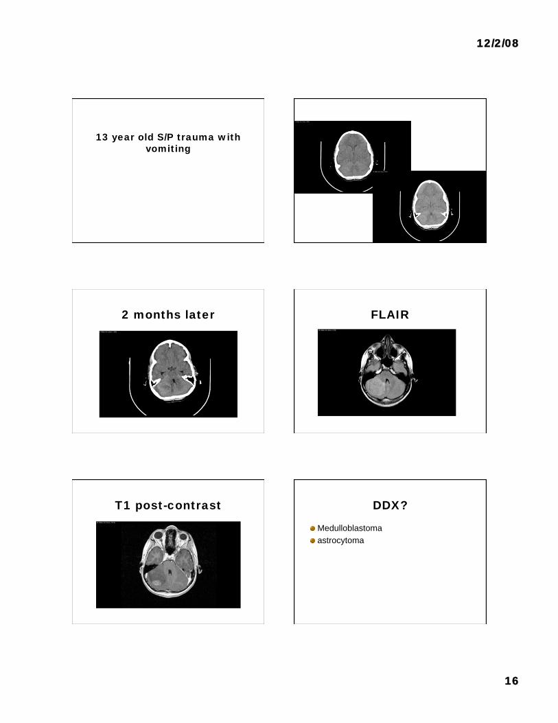
13 year old S/P trauma with
vomiting
2 months later FLAIR
T1 post-contrast DDX?
Medulloblastoma
astrocytoma
12/2/08
17
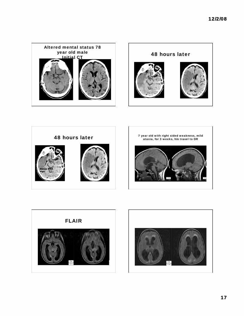
Altered mental status 78
year old male
Initial CT 48 hours later
48 hours later
Dense MCA
sign
7 year old with right sided weakness, mild
ataxia, for 3 weeks, h/o travel to DR
FLAIR
12/2/08
18
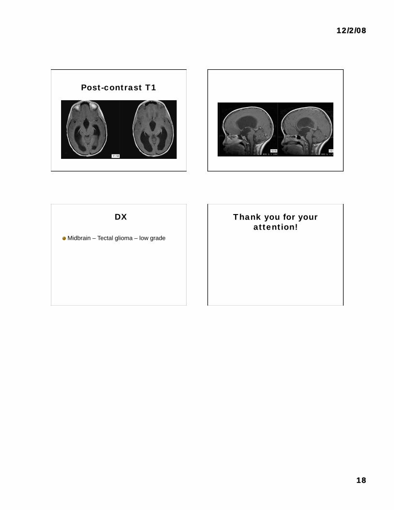
Post-contrast T1
DX
Midbrain – Tectal glioma – low grade
Thank you for your
attention!