Embed Size (px)
Citation preview
���������� ����� ������ ������ ����� ������������� ��� ���� �������� ����������� ��� ����� ����� �� �� ���
����� ����
Neritopsis represents the only surviving genus of an ancient group of the Neritimorpha that has no internal dissolution ofits shell walls. While the two known living species have lecithotrophic early development without larval shell,Neritopsis aqabaensis n. sp. from the Gulf of Aqaba, Jordan, has a planktotrophic veliger. These living species of the ge-nus differ in their protoconch shape as well as teleoconch morphology and ornament from the Triassic species that can beconsidered related to Neritopsis. Neritopsidae with the modern Neritopsinae is distinguished from the TriassicCassianopsinae n. subfam. based on the genus Cassianopsis n. gen. with three species by features of their protoconch aswell as the different characters of the operculum. Zardiniopsis n. gen. differs from these by higher shell shape and asmaller more complexly ornamented protoconch. Fossariopsis has a more angular shell shape. Colubrellopsinae n.subfam. with Colubrellopsis n. gen. resembles Cassianopsis n. gen. in respect to protoconch and features of the apertureof the teleoconch, but the former has rounded whorls and an ornament of axial ribs. Among the Fedaiellidae n. fam. withsmooth shells two species of Fedaiella are redefined. The characters of the inner lip of their aperture connect them withthe Neritopsidae, whereas the operculum in the Fedaiellidae with concentric structure on the outside distinguishes themfrom neritopsids.In distinction to the groups of the Neritopsoidea members of the Dephinulopsidae have a smooth inner lip of the aperture.Here Delphinulopsinae and Platychilininae n. subfam. differ from each other in the shape and ornament of theirteleoconch. Schwardtopsis n. gen. resembles a juvenile Delphinulopsis grown to a larger size. The large concave callusof the inner lip and almost open coiling defines Delphinulopsis. Rows of nodes, low initial shell and rapid growth in shelldiameter to an almost limpet shape characterises Platychilina, and lamellar growth increments on an almost limpet-likeshell with flat initial part is present in Marmolatella. The Palaeonaricidae n. fam. contains two species of Palaeonarica,which have a Nerita-like shell with simple aperture.The Naticopsidae of the Carboniferous and Permian has its continuation in the Ampezzonaticopsinae n. subfam. of theTriassic, being connected to each other by the sinuous ornament of ribs on their larval shells. The genera are distin-guished by teleoconch shape and differences in the ornament of their larval shell. Ampezzonaticopsis n. gen. has whorlswell separated by deep sutures, Cortinaticopsis n. gen. has a simple aperture and a callus covering the umbilicus. TheHologyrinae n. subfam. with Hologyra have a ridge in the columellar furrow and an operculum that resembles that attrib-uted to Carboniferous Naticopsis. Their protoconch has a characteristic chevron ornament on its larval whorls.The new family Tricolnaticopsidae is proposed to hold Tricolnaticopsis n. gen. with a smooth shell and convex inner lipand color pattern of dots. Into the aperture an operculum of bean-like shape may be fitted and this operculum has quite inde-pendent characters. It would also fit with Pachyomphalus with shallow sutures and two species, one with short roundedconical shape and the other with elongate shell. Both resemble Tricolnaticopsis n. gen. as does Rinaldopsis n. gen. with itswide flat inner lip. Laubopsis n. gen. has an open umbilicus and has no clear connection with similar species.The new family Scalaneritinidae is based on Scalaneritina n. gen. with an elongate shape and axial ribs. The protoconchis rounded with its larval shell having an ornament of low, fold-like collabral ribs. Ladinaticella n. gen. has three species:two have similar shell shape, in one case with a few strong ribs as ornament, while in another case there are many, and athird has a more rounded apex. In the case of Lancedellopsis n. gen., the shell is low, has a rapid increase in diameter anda low tooth in its aperture. Its relation to the other Neritimorpha remains undetermined.Among the species of Neritoidea from St Cassian Formation the members of the Neritariidae are arranged into the gen-era Neritaria, Ruganeritaria n. gen., Dentineritaria n. gen., Oncochilus, and Cassianpisulina n. gen. The genusNeritaria has a complexly ornamented protoconch, and the related Ruganeritaria n. gen. differs by the protoconch hav-ing only axial ribs. The operculum has the general shape of that found in modern Nerita and its relatives. Dentineritarian. gen. has a central tooth on its inner lip and its protoconch is relatively large with straight axial ribs. The Oncochilinaeis distinguished notably by projections of the inner lip into the aperture, which are two in Oncochilus and single largerone in Cassianpisulina n. gen. The latter is defined and compared with modern Pisulina. The Trachineritariinaen. subfam. with Trachyneritaria n. gen. is distinguished by characters of the teleoconch that has the inner whorls dis-solved. Its relation to Trachynerita remains problematic. • Key words: Gastropoda, Neritimorpha, Triassic, Tethys, Italy,Jordan, new taxa (fossil, living).
��

BANDEL, K. 2007. Description and classification of Late Triassic Neritimorpha (Gastropoda, Mollusca) from the StCassian Formation, Italian Alps. Bulletin of Geosciences 82(3), 215–274 (14 figures). Czech Geological Survey, Prague.ISSN 1214-1119. Manuscript received April 27, 2007; accepted in revised form July 30, 2007; issued September 30,2007. • DOI 10.3140/bull.geosci.2007.03.215
Klaus Bandel, Universität Hamburg, Geologisch Paläontologisches Institut und Museum, Bundesstrasse 55, 20146Hamburg, Germany; [email protected]
The number of species of the Neritimorpha that lived inthe environment of the tropical reef of the Late TriassicSt Cassian Formation in the Tethys Ocean is amazinglyhigh, compared to that found in this kind of environmenttoday. Thirty six species can be recognized and this diver-sity distinguishes this Tethyan reef from modern, tropicalIndo-Pacific reefs where only about 10 species may be pre-sent including the nearby shore and the lagoon. In regardsto taxonomic groups within the Neritimorpha only Neri-topsis among these modern genera and species can be re-garded as relatively closely related to some of the speciesfrom the Cassian Reef described here. Some others can beplaced with the superfamily Neritopsoidea, and some withthe superfamily Neritioidea but not to any of the modernfamilies. A few species are clearly representatives of thepredominantly Paleozoic Naticopsidae, and quite a numberof taxa appear to be quite unrelated to any surviving unit ofthe Neritimorpha. This difference is not so amazing consi-dering the long time interval of about 220 million years thathas passed between the populations of both tropical reef as-semblages. The large diversity of fossil species recognizedcan be considered evidence for the limitations into whichany DNA analysis would run when it is used in the recon-struction of a phylogenetic tree of the Neritimorpha. Mostmodern Neritimorpha form more closely related groupsand appear to be more related to each other than to severalof the groups recognized from the Late Triassic St CassianFormation. As will be shown below, there is evidence thatthe diversity of the modern species of the Neritimorpha ref-lects only a fraction of what had been present in the past.Larger units recognized in the Triassic have not survived inany of the species living today and can, therefore, not bestudied in regard to their genomic composition.
������!����������������������������� ��� ����� ��
In the St Cassian Formation of the Italian Alps, Dolomitesnear Cortina d’Ampezzo and St Cassian the deposits of Lo-wer Carnian age include lagoonal sediments of well bed-ded, often dolomitic limestone, and rarely also along theirmargins deposits of deeper water consisting of marls, shaleand tuffaceous deposits. This margin of the carbonate plat-
form consisted of bioconstructed reefoid sediments. It isusually transformed into dolomite in those places wherethe actual reef is still exposed in its original position ofgrowth. From that area material had broken off and beenwashed out by currents and transported down-slope. In thecase it came to rest in the fine-grained sediment of the ba-sin, which included volcanic ashes and mud derived fromnearby volcanism, it was well preserved. This calcareousmaterial was sometimes so well preserved that even arago-nitic shells were not changed into calcite during diagenesis.Of the molluscan fauna that lived in the shallow water ju-venile shells may preserve the early ontogenetic part oftheir shell, which even during life is usually destroyed bycorrosion and boring. These assemblages may form layerswhich contain quite different gastropods, most of whichhave come from shallow water. Some of the shells havebeen covered with algal crusts before they came to rest inthe mud of the deep water environment.
The gastropods of the St Cassian Formation lived duringearly Carnian (Cordevolian) time of the Late Triassic(Urlichs 1974, 1994). The environment was that of reefs, thelagoon behind them and their slope to the open sea whichwas the tropical Tethys Ocean. Here carbonate platformsforming islands in the Tethys Ocean were not very stable intheir position through time and moved (Blendinger & Blen-dinger 1989), probably connected to earthquakes and volca-nic eruptions. The Neritimorpha here discussed have livedprimarily in or near the reefs which consisted of algal mats,calcareous and stromatoporoid sponges and corals (Wendt1982). Reefs are only exceptionally preserved in situ as inthe Richthofen reef at Settsass near St Cassian (Mojsisovics1879). In those places where parts of the reef broke off andmoved down the fore-slope the reef community is well doc-umented, for example at the localities Misurina and Alpe diSpecie (Fürsich & Wendt 1977). A map of the localitiesfrom which gastropods have been extracted from the beds ofthe St Cassian Formation has been provided by Zardini(1978).
Note. – All the specimens studied are housed in theBayrische Staatssammlung für Paläontologie und Geo-logie München, BSPG, Germany. Other material cited is tobe found in the Naturhistorisches Museum in Vienna, Aus-tria (NHMW), and the Museum of the region Ampezzo“Ciasa de ra Regoles” at Cortina d’Ampezzo, Italy.
���
����������� ������ �������������
�"��� ���� � ���#"
Subclass Neritimorpha Golikov & Starobogatov, 1975
Remarks. – In the members of this group the shell of itsspecies differs in ornament and shape, but is generally lowand consists of few whorls (Thiele 1929–1931; Wenz1938–1944; Knight et al. 1960). The outer layer of the shellis usually calcitic and the inner layer consists of aragoniticcrossed lamellar structure (Boggild 1930, Bandel 1990, Sa-saki 2001). The anatomy differs from that of other groups ofthe Gastropoda in several details (Fretter & Graham 1962).That of Neritopsis is closest to that of Nerita and its relatives(Holthuis 1995). The radula has many teeth in each row andresembles in that manner the Archaeogastropoda, and wastherefore considered rhipidoglossate (Troschel 1856, Tros-chel & Thiele 1865–1893). The protoconch is strongly invo-lute with whorls overlapping and an egg-shaped embryonicshell (Robertson 1971, Bandel 1982).
The Neritimorpha holds the aquatic Neritoina Rafinesque,1815 as well as the terrestrial Helicinina Thompson, 1980 andHydrocenina Troschel, 1856. Within the Neritoina the stemgroup of the Neritopsidae Gray, 1847 appears with a numberof species in the Devonian (Heidelberger & Bandel 1999,Frýda 2000, Bandel & Heidelberger 2001, Heidelberger2001). Probably the history of the Neritimorpha can be tracedto the Early Paleozoic (Bandel & Frýda 1999).
The Neritimorpha represents the most ancient and pri-mitive of the living groups (subclasses) of the Gastropodawith a larval shell in addition to the embryonic one. Re-garding the organization of the teeth on the ribbon of theradula they resemble those of the Rhipidoglossa Mörch,1865 (here used for Archaeogastropoda without inclusionof the Docoglossa Troschel, 1856 and Neritopsina Cox &Knight, 1960). In shell and gill structure they are similar tothe Docoglossa, which represent the most primitive orderof the subclass Archaeogastropoda (Haszprunar 1988,Ponder & Lindberg 1997, Sasaki 1998). Regarding themode of ontogeny of their early shell as demonstrated byBandel (1982), the Archaeogastropoda represents a sub-class of the living Gastropoda with its species providedonly with the embryonic and without larval shell.
Based on cleavage patterns (Biggelaer & Haszprunar1996) and on computer-based cladograms, Ponder &Lindberg (1997) arrived at similar conclusions to Bourne(1908) based on anatomical analysis and Bandel (1982)based on the pattern of formation of the early ontogeneticshell. These data indicate that the Neritimorpha is an inde-pendent group within the Gastropoda and should not beseen in close relation with the Archaeogastropoda, as hadbeen suggested by Thiele (1929–1931) and repeated eversince in most studies on the subject, and also confirmed bymolecular data (Kano et al. 2002).
Superorder Cycloneritimorpha Bandel & Frýda, 1999
Remarks. – Neritimorpha can be divided into the PaleozoicCyrtoneritimopha with openly coiled protoconch and theCycloneritimopha with tightly coiled protoconch.
Order Neritoina Rafinesque, 1815
Remarks. – Here can be included the aquatic Neritimorphawith the cycloneritimorph protoconch morphology. Anato-mically Neritimorpha, the Neritoina is distinguished fromother Gastropoda, as had been recognized by Bourne(1908) and Fretter & Graham (1962). They observed thatliving species have one left kidney, a single left feather-likectenidium (except in lung-bearing terrestrial species), aheart with one or two auricles, internal fertilization, pairedretractor muscles, and a rhipidoglossate radula. The cteni-dium has filaments on both sides which are attached only atits base, similar to the gill of Acmaea. It is thus consideredto represent the most primitive type found among moderngastropods (Haszprunar 1988). The nerves connecting thectenidia with each other and also the ctenidia themselvesare different from those found in other gastropod groupsand are therefore considered to have evolved indepen-dently from them (Thiele 1929–1931). Internal fertilizationand the production of egg capsules have evolved also whilein most Archaeogastropoda external fertilization is practi-ced. Bourne (1908) suggested that the Neritimorpha hascome from primitive gastropod stock which is supportedby paleontological evidence according to which pre-mid-Devonian Neritimorpha and their stem group representati-ves with uncoiled embryonic shell and openly coiled larvalshell (Frýda & Bandel 1997, Bandel 1997) range back tothe early Ordovician and thus have developed indepen-dently from the rest of the Gastropoda for about 500 mil-lion years.
Bouchet & Rocroi (2005) provided a recent classifica-tion scheme of the “clade” Neritimorpha, which differsconsiderably from the one suggested here, especially re-garding the taxa based on fossil species.
Superfamily Neritopsoidea Gray, 1847 (= Rafinesque,1815)
Diagnosis. – Inner walls of shell not dissolved. Calcareousoperculum not spirally arranged (Wenz 1938–1944).
Remarks. – Unlike the Neritoidea the shell inner walls arenot dissolved. Within the Neritimopha species belongingto the Neritopsoidea Rafinesque, 1815 do not dissolve theinner walls of their shell (Cossmann 1925, Wenz1938–1944). This superfamily is based on members of Ne-ritopsidae and interpreted to consist of quite a few generain the Late Paleozoic and the Early Mesozoic (Knight et al.
���
���� ������ ��������� ���������� !�"�� �� ��#���$��%� ���&������
1960). The genus Neritopsis Grateloup, 1832 is reported tohave existed since Jurassic time (Bandel & Kiel 2003).Cossmann (1925) suggested that it also had Triassic repre-sentatives. Since the beginning of the Tertiary only repre-sentatives of the genus Neritopsis have survived among theNeritopsoidea.
Family Neritopsidae Gray, 1847
Diagnosis. – Globular shell with low spire and large lastwhorl. Inner lip of aperture broad and smooth forming acallus ribbon that has on its inner side a central depressioninto which fits the angular inner projection of the opercu-lum. On the outer side of the callus a groove often forms thecontinuation of the umbilicus. Inner walls of shell not dis-solved; operculum without spiral growth increments. Pro-toconch consists of low and tightly coiled rounded whorls.
Remarks. – The family is based on the genus Neritopsis.
Subfamily Neritopsinae Gray, 1847
Diagnosis. – Whorls of shell are rounded. Smooth proto-conch and angular operculum of trapezoidal shape withan almost smooth thickened rounded exterior and angulargrooved attachment on its inner side.
Remarks. – The taxon is based on modern Neritopsis.
Genus Neritopsis Grateloup, 1832
Diagnosis. – Medium-sized shell with rounded whorls, amoderately protruding spire, and globose last whorl withevenly convex flanks. Sculpture consists of thick, granulatedspiral cords that may be intersected by axial ribs and form areticulate pattern. Protoconch consists of smooth whorls.
Teleoconch aperture round with a moderately thickenedconcave inner lip and median quadrangular depression intowhich fits the trapeziform, solid operculum that is minerali-zed with calcite and has a quadrangular projection at the sideheld next to the inner lip when the aperture is closed.
Remarks. – The type of the genus is Neritopsis monilifor-mis Grateloup, 1832 from the Miocene of the Aquitaine inFrance, which closely resembles the still living Neritopsisradula Linné, 1757 from the Indo-Pacific as illustrated byWilson (1993).
The protoconch is rounded with smooth whorls whichin the case of an individual of Neritopsis cf. radula fromthe Indo-Pacific Ocean of Mauritius consist of more thantwo whorls (Bandel & Frýda 1999, pl. 1, figs 1, 2), as isalso the case in Neritopsis aqabaensis described belowfrom the Gulf of Aqaba. Neritopsis parisiensis Deshayes,1864 from the Eocene of the Paris Basin has only a littlemore than one whorl (Bandel & Frýda 1999, pl. 1, figs 3, 4),as is usual case in living Neritopsis radula from theIndo-Pacific (Kano & Kase 2000, Kaim & Sztajner 2005,fig. 4C). The protoconch of Neritopsis atlantica Sarasua,1973 from the Caribbean Sea is not known.
The operculum of Neritopsis is of characteristic trape-zoidal shape and has calcitic composition. Jagt & Janssen(1988, pl. 2, figs 1–3) noted opercula of Neritopsis in thePaleocene of the Province Limburg in Belgium. Theoperculum of living Neritopsis has a convex outer surfaceand a concave inner side with an inner projection that is an-gular and fits into a groove present on the inner lip of theaperture (Zittel 1895, Scott & Kenny 1998); even though itresembles in shape that of Cassianopsis n. gen., it differs indetail. The Triassic species of Cassianopsis n. gen. of theSt Cassian Formation of Cortina d’Ampezzo (Zardini1978, pl. 15, figs 11–15) have an operculum of similarshape to that of a not more closely specified Neritopsisfrom the Jurassic (Kaim & Sztajner 2005, fig. 1). In thecase of Neritopsis on the inner side the margin is smooth
���
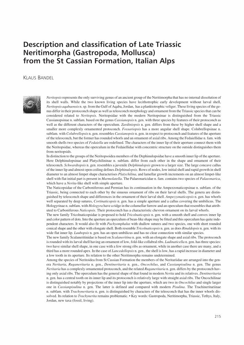
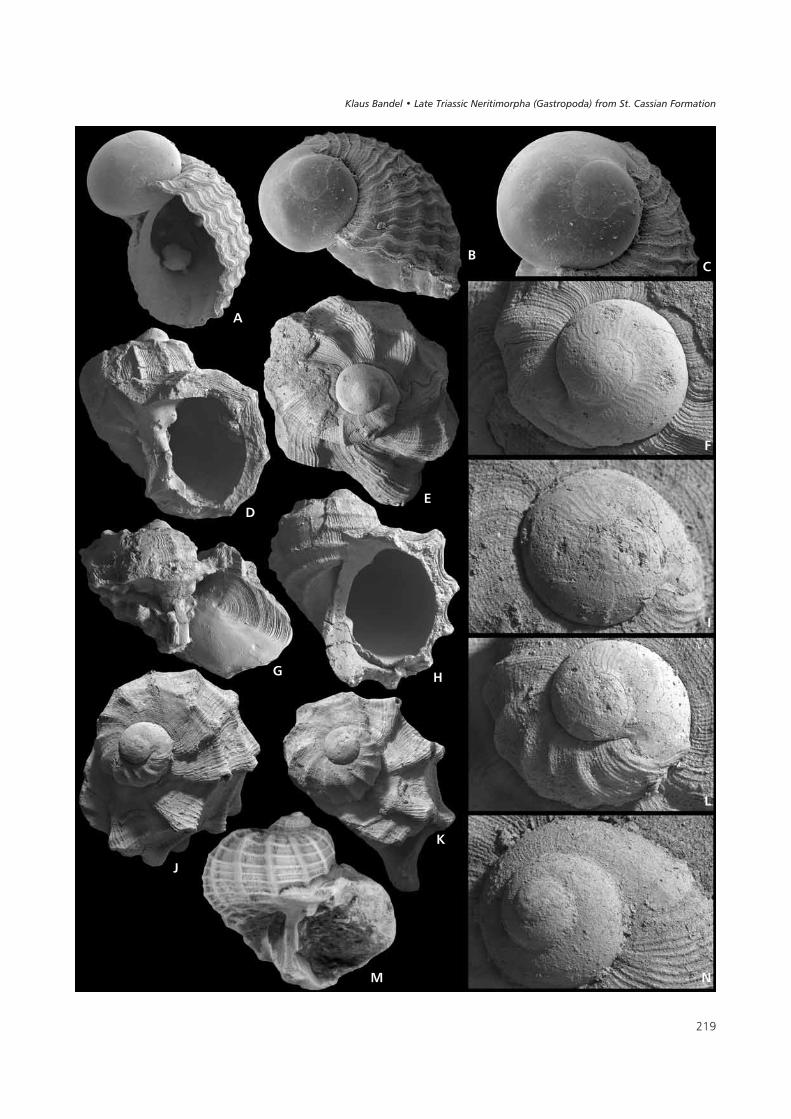
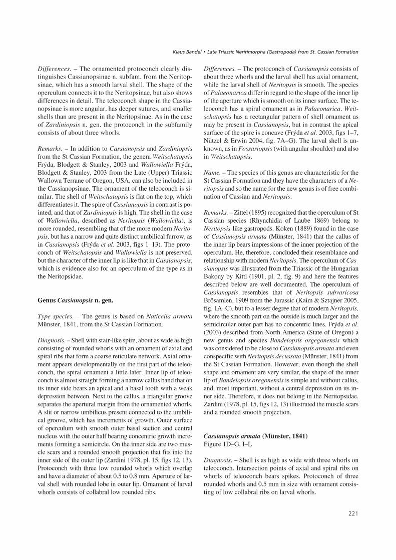
��#����$% A – juvenile shell of Neritopsis aqabaensis n. sp. representing the holotype with a rounded protoconch and slit-like umbilical groove in theteleoconch. The shell is 1 mm high (BSPG 2007 XVI 1). • B – a more apical view of Neritopsis aqabaensis n. sp. as in Fig. 1A. • C – protoconch ofNeritopsis aqabaensis n. sp. having the embryonic shell succeeded by the smooth whorl of the larval shell. • D – the aperture of Cassianopsis armata(Münster, 1841) has an angular depression on the inner side of its inner lip. The shell is 1.8 mm high (BSPG 2007 XVI 2). • E – apical view ofCassianopsis armata (Münster, 1841) with the rounded protoconch succeeded by an ornamented teleoconch with a pattern of axial and spiral ribs. Theshell is 1.8 mm wide. • F – the protoconch of Cassianopsis armata (Münster, 1841) has a fine sinuous axial ornament on its larval whorls and is about0.5 mm wide. Detailed view of Fig. 1E. • G – Cassianopsis armata (Münster, 1841) with the operculum preserved in the aperture with is outer part orna-mented by concentric lines and its inner part smooth. The shell is about 5 mm wide (BSPG 2007 XVI 3). • H – the inner lip of Cassianopsis ornata(Münster, 1841) with its characteristic angular depression. The shell is 3.2 mm high (BSPG 2007 XVI 6). • I – the protoconch of Cassianopsis armata(Münster, 1841) has an ornament that reflects its lobed margin. The protoconch is about 0.5 mm wide. Detailed view of Fig. 1K. • J – apical view ofthe shell of Cassianopsis armata (Münster, 1841) with the protoconch shown in Fig.1L. The shell is about 1 mm wide (BSPG 2007 XVI 4).• K – Cassianopsis armata (Münster, 1841) with an ornament of axial ribs that form spines at their intersection with the larger spiral ribs. The shell isabout 2 mm wide (BSPG 2007 XVI 5). • L – this protoconch of Cassianopsis armata (Münster, 1841) is about 0.5 mm wide with the marginal lobe of thelarval shell reflected in the sinuous ornament. Detailed view of Fig. 1J. • M – this shell with about three whorls of the teleoconch of Cassianopsis ornata(Münster, 1841) is about 11 mm wide with an ornament of axial and spiral ribs and a wide umbilical furrow (BSPG 2007 XVI 7). • N – the protoconch ofCassianopsis ornata (Münster, 1841) has more than three whorls and distinct sutures. 0.8 mm in width.
����������� ������ �������������
���
�&
�
�
�
'
(
�
)
*
� �
�
���� ������ ��������� ���������� !�"�� �� ��#���$��%� ���&������
with an angular inner projection that is accompanied bytwo pits. A pattern of elongate grooves covers the inner ex-tension. This resembles the operculum of Naticopsis fromthe Carboniferous and of Hologyra from the Triassic butthe latter does not have the inner rectangular projection onthe inner side as is found in Neritopsis and Cassianopsis(see below).
The shell structure of Neritopsis radula was analyzedwith the scanning electronic microscope and x-ray diffrac-tion, and accordingly the outer layer is calcitic. Its thick-ness differs in relation to the ornament. In ribs it is thickerthan between ribs. The aragonitic layer added below theouter calcitic layer was deposited on the smooth and evenlyrounded surface of the inner whorl. Deposition at the mar-gin of the shell aperture during shell growth is by calciticsheets on top of each other. These platy layers have the sin-gle calcitic biocrystallites growing in different directionsbut about parallel to shell surface. When fractured thecalcitic layer appears to have an irregular structure sincethe direction of crystallites in these sheets differs from eachother. Below the calcitic outer layer a fractured shell dem-onstrates crossed lamellar structure with the largest sheetsarranged parallel to the margin of the outer lip in very regu-lar orientation. Smaller sheet-like lamellae composingthese and the third order needle-like lamellae are character-istic of shells of the Neritimorpha (Bandel 1990). The shellof Neritopsis is thus not composed of two whollyaragonitic layers as was suggested by Kaim & Sztajner(2005) who relied on a report by Suzuki et al. (1991), butconsists of a calcitic outer layer and aragonitic inner layer.
Neritopsis aqabaensis n. sp.Figure 1A–C
Holotype. – The specimen illustrated here is deposited inthe collection of the Bayrische Staatssammlung für Paläon-tologie und Geologie München, BSPG 2007 XVI 1.
Diagnosis. – Teleoconch shape as in Neritopsis radula.Protoconch consists of two rounded whorls. Embryonicwhorls surrounded and largely covered by larval shell.
Derivation of the name and type locality. – This specieswas found in the fringing reef at Aqaba, in front of theMarine Science Station and it is named according to itstype locality, the Gulf of Aqaba near Aqaba, Jordan. Indi-viduals live within the fringing reef in crevices and cavi-ties within shallow water of less than 3 m in depth. Herewater in summer may be more then 30 °C, in winter itsinks below 20 °C.
Description. – The protoconch is well rounded and smoothand measures about 0.4 mm in diameter. The embryonic
whorl is egg-shaped with growth lines and the later larvalshell consists of one smooth rounded whorl that overlapsstrongly onto the embryonic whorl. The margin of the lar-val shell expands into a sinuous rim. Change to the orna-mented teleoconch is abrupt. The fully-grown teleoconchis of medium size with upright spire and has a globose lastwhorl with evenly convex flanks. Sculpture consists ofthick granulated spiral cords which are intersected by nar-row axial ribs. At these intersections are formed roundedtubercles. The orbicular aperture has a moderately thicke-ned concave inner lip that has a quadrangular depression inthe middle into which the inner projection of the operculumis fitted. Next to the callus of the inner lip of the juvenileshell lies a narrow cleft-like umbilical furrow. The adultshell as well as the operculum are the same as is found inNeritopsis radula.
Differences. – The protoconch of Neritopsis aqabaensis n.sp. has two whorls in contrast to the one present in the caseof Neritopsis radula as shown by Kano & Kase (2000) andKaim & Sztajner (2005). A similar protoconch as that inNeritopsis aqabaensis had been described by Bandel &Frýda (1999, pl. 1, figs 1, 2). Neritopsis radula (Linné,1758) as illustrated and described by Wilson (1993) fromAustralia is the characteristic Indo-West Pacific form and ithas the same shape of teleoconch as does Neritopsis aqaba-ensis from the Red Sea. Adult shells found at Aqaba arerare and it was not possible to check if they belong to thenew species or instead represent Neritopsis radula sincetheir protoconch is not preserved, as is usually the case inlarger shells. The embryonic shell of Neritopsis aqabaen-sis has growth lines, as is characteristic of those of the Neri-tidae as well as in contrast to those of most other Gastro-poda that hatch as planktotrophic veligers from their eggmass (Bandel 1982).
Subfamily Cassianopsinae n. subfam.
Type genus. – Cassianopsis n. gen.
Diagnosis. – Shell whorls ornamented by axial and spiralribs, with aperture rounded. A basal and an apical tooth arepresent on inner side of inner lip and between these a con-cave zone, into which the inner side of operculum may befitted. Protoconch low, rounded and ornamented by lowsomewhat undulating axial ribs and spiral lines consistingof three whorls. Operculum trapezoidal, its outer surfacewith a smooth inner side and a concentric outer side. Twoattachment scars present on its interior surface.
Composition. – The subfamily is based on Cassianopsis n.gen. and also includes Zardiniopsis n. gen., both from theLate Triassic St Cassian Formation.
���
����������� ������ �������������
Differences. – The ornamented protoconch clearly dis-tinguishes Cassianopsinae n. subfam. from the Neritop-sinae, which has a smooth larval shell. The shape of theoperculum connects it to the Neritopsinae, but also showsdifferences in detail. The teleoconch shape in the Cassia-nopsinae is more angular, has deeper sutures, and smallershells than are present in the Neritopsinae. As in the caseof Zardiniopsis n. gen. the protoconch in the subfamilyconsists of about three whorls.
Remarks. – In addition to Cassianopsis and Zardiniopsisfrom the St Cassian Formation, the genera WeitschatopsisFrýda, Blodgett & Stanley, 2003 and Wallowiella Frýda,Blodgett & Stanley, 2003 from the Late (Upper) TriassicWallowa Terrane of Oregon, USA, can also be included inthe Cassianopsinae. The ornament of the teleoconch is si-milar. The shell of Weitschatopsis is flat on the top, whichdifferentiates it. The spire of Cassianopsis in contrast is po-inted, and that of Zardiniopsis is high. The shell in the caseof Wallowiella, described as Neritopsis (Wallowiella), ismore rounded, resembling that of the more modern Nerito-psis, but has a narrow and quite distinct umbilical furrow, asin Cassianopsis (Frýda et al. 2003, figs 1–13). The proto-conch of Weitschatopsis and Wallowiella is not preserved,but the character of the inner lip is like that in Cassianopsis,which is evidence also for an operculum of the type as inthe Neritopsidae.
Genus Cassianopsis n. gen.
Type species. – The genus is based on Naticella armataMünster, 1841, from the St Cassian Formation.
Diagnosis. – Shell with stair-like spire, about as wide as highconsisting of rounded whorls with an ornament of axial andspiral ribs that form a coarse reticulate network. Axial orna-ment appears developmentally on the first part of the teleo-conch, the spiral ornament a little later. Inner lip of teleo-conch is almost straight forming a narrow callus band that onits inner side bears an apical and a basal tooth with a weakdepression between. Next to the callus, a triangular grooveseparates the apertural margin from the ornamented whorls.A slit or narrow umbilicus present connected to the umbili-cal groove, which has increments of growth. Outer surfaceof operculum with smooth outer basal section and centralnucleus with the outer half bearing concentric growth incre-ments forming a semicircle. On the inner side are two mus-cle scars and a rounded smooth projection that fits into theinner side of the outer lip (Zardini 1978, pl. 15, figs 12, 13).Protoconch with three low rounded whorls which overlapand have a diameter of about 0.5 to 0.8 mm. Aperture of lar-val shell with rounded lobe in outer lip. Ornament of larvalwhorls consists of collabral low rounded ribs.
Differences. – The protoconch of Cassianopsis consists ofabout three whorls and the larval shell has axial ornament,while the larval shell of Neritopsis is smooth. The speciesof Palaeonarica differ in regard to the shape of the inner lipof the aperture which is smooth on its inner surface. The te-leoconch has a spiral ornament as in Palaeonarica. Weit-schatopsis has a rectangular pattern of shell ornament asmay be present in Cassianopsis, but in contrast the apicalsurface of the spire is concave (Frýda et al. 2003, figs 1–7,Nützel & Erwin 2004, fig. 7A–G). The larval shell is un-known, as in Fossariopsis (with angular shoulder) and alsoin Weitschatopsis.
Name. – The species of this genus are characteristic for theSt Cassian Formation and they have the characters of a Ne-ritopsis and so the name for the new genus is of free combi-nation of Cassian and Neritopsis.
Remarks. – Zittel (1895) recognized that the operculum of StCassian species (Rhynchidia of Laube 1869) belong toNeritopsis-like gastropods. Koken (1889) found in the caseof Cassianopsis armata (Münster, 1841) that the callus ofthe inner lip bears impressions of the inner projection of theoperculum. He, therefore, concluded their resemblance andrelationship with modern Neritopsis. The operculum of Cas-sianopsis was illustrated from the Triassic of the HungarianBakony by Kittl (1901, pl. 2, fig. 9) and here the featuresdescribed below are well documented. The operculum ofCassianopsis resembles that of Neritopsis subvaricosaBrösamlen, 1909 from the Jurassic (Kaim & Sztajner 2005,fig. 1A–C), but to a lesser degree that of modern Neritopsis,where the smooth part on the outside is much larger and thesemicircular outer part has no concentric lines. Frýda et al.(2003) described from North America (State of Oregon) anew genus and species Bandelopsis orgegonensis whichwas considered to be close to Cassianopsis armata and evenconspecific with Neritopsis decussata (Münster, 1841) fromthe St Cassian Formation. However, even though the shellshape and ornament are very similar, the shape of the innerlip of Bandelopsis oregonensis is simple and without callus,and, most important, without a central depression on its in-ner side. Therefore, it does not belong in the Neritopsidae.Zardini (1978, pl. 15, figs 12, 13) illustrated the muscle scarsand a rounded smooth projection.
Cassianopsis armata (Münster, 1841)Figure 1D–G, I–L
Diagnosis. – Shell is as high as wide with three whorls onteleoconch. Intersection points of axial and spiral ribs onwhorls of teleoconch bears spikes. Protoconch of threerounded whorls and 0.5 mm in size with ornament consis-ting of low collabral ribs on larval whorls.
���
���� ������ ��������� ���������� !�"�� �� ��#���$��%� ���&������
Description. – The shell is about as wide as high and has arelatively high spire. Ornament of the teleoconch consistsof 6–10 strong axial ribs which represent former aperturalthickenings and 6–9 spiral ribs which have a variable num-ber of finer spiral lines between them. A shell with about2.5 whorls of the teleoconch is about 6 mm high and wide.The ornament of the whorls varies from early to laterwhorls, with fine spiral ribs successively appearing betweenthe larger spiral ribs. The overall ornament is reticulatewith a network of squares with spiny corners developedwhere larger axial and spiral ribs cross. Sutures are deepand whorls clearly distinct from each other. The apical sideof whorls is a little flattened and the periphery is occupiedby a strong spiral rib. The aperture is semicircular. The nar-row callus ribbon of the inner lip covers the umbilicus andis accompanied by a wide deep furrow next to it that beginsin the umbilicus. The inner side of the inner lip has the cha-racteristic rectangular median depression with teeth onboth its sides. The outer lip is rounded and ends in a sharpedge. Shell width increases rapidly just behind the interiorside of the apertural edge. An inner rim is thus produced onwhich the margin of the operculum had rested.
The protoconch has a rounded shape with the threewhorls overlapping and the suture consisting of only a linethat is not impressed. Ornament of the larval whorls con-sists of collabral low axial ribs which reflect a low lobe onthe outer lip of the aperture. The size of the protoconch isless than 0.5 mm. There is a noticeable and strong changein the ornament from the protoconch to the teleoconch asthe collabral ribs in the latter are more lamellar and closerto each other without a lobe. In addition, the outer layer ofthe teleoconch has a calcitic construction and its surfaceappears rougher.
Differences. – Cassianopsis armata resembles Cassianop-sis ornata and Cassianopsis decussata in the shape of theshell and the organization of the apertural margin. Also,their peotoconchs are similar to each other in shape,although it is larger in Cassianopsis ornata. Differences lielargely in the ornament of the teleoconch which is reticu-late with axial ribs not much stronger than the spiral ribs inCassianopsis armata, while they are stronger and morerounded in Cassianopsis ornata. In the case of Cassianop-sis decussata, the spiral ribs are more irregular and have tu-bercles.
The whorls of Neritopsis are more rounded, sutures areless deep, and the fully-grown shell is larger. Theoperculum resembles that of Neritopsis, but differs in re-gards to the outer side. Here the part that fits with the outerlip of the aperture is occupied by a semicircle with well-de-veloped growth lines and nucleus at its central base. Such asemicircle is not found in the operculum of Neritopsis,which has a narrow rim without growth lines. In addition,the inner side also differs with two rounded attachment
scars present in Cassianopsis, whereas these are repre-sented by narrow pits at the base of the angular depressionin Neritopsis. Also, the inner projection of Cassianopsis isrounded and smooth, while it is angular and covered bylongitudinal furrows in Neritopsis. The groove on the innerlip is deeper in Neritopsis than it is in Cassianopsis.
Remarks. – The original name Naticella armata was provi-ded for this species by Münster (1841). Neritopsis waageniLaube, 1868 represents a variety of Cassianopsis armata,and is the same as Naticella cancellata Münster, 1841. Na-ticella plicata Giebel, 1852 is also the same species as wasnoted by Kittl (1892). In addition, Leonardi & Fiscon(1959) noted several varieties of their Neritopsis armata.The different species and subspecies reflect the large va-riability in the ornament of individuals of Cassianopsisarmata.
Cassianopsis ornata (Münster, 1841)Figure 1H, M, N
Diagnosis. – Round shell with three whorls in teleoconch.Dominant ornament of rounded strong axial ribs crossedby many smaller spiral ribs. Umbilicus closed or slit-like.First teleoconch whorl ornamented with fine lamellar axialribs; stronger ribs appear later. Protoconch measures about0.8 mm with more than three whorls with larval whorls or-namented with weak collabral ribs.
Description. – The shell shape closely resembles that ofCassianopsis armata, round with deep sutures and a lowspire, which has a rapid increase in whorl diameter. How-ever, unlike Cassianopsis armata, there are 7 to 11 varix-like axial ribs crossed by 18–20 spiral ribs of different size.The umbilicus is narrow, slit-like or closed. The aperture iscircular and the outer lip is simple and thin at its margin,but thickened further inward, forming a rim on which theoperculum rested. The inner lip forms a narrow callus rib-bon with a central depression on its inner side accompaniedby ridges on each end. A shell with more than three whorlson the teleoconch is almost fully-grown and about 9 mmhigh and 11 mm wide.
The protoconch consists of more than three whorls withlarval whorls ornamented by low axial ribs. A large part ofthe embryonic shell is covered by the larval whorls whichoverlap onto it. Their ornament consists of about 30collabral ribs on one whorl. Ribs follow the outline of themargin of the outer lip that has a rounded lobe. With the ex-ception of the embryonic and first larval whorl a distinctsuture is developed. Ornament of the first whorl of theteleoconch is quite similar to that in Cassianopsis armata,consisting of fine lamellar collabral straight ribs, which ap-pear with the change from the aragonitic shell of the
���
����������� ������ �������������
protoconch to the calcitic construction of the outer shelllayer of the teleoconch.
Differences. – Cassianopsis armata has stronger axial ribsthan Cassianopsis ornata, and spikes where the ribs crosseach other. Cassianopsis decussata has a denser ornamentof fine spiral lines, has tubercles, and reaches larger sizewhen fully-grown. Zardiniopsis subornata has a relativelyhigher shell and a smaller protoconch with ornament alsoconsisting of spiral ribs.
Remarks. – Kittl (1892, pl. 5, figs 10–12) and Zardini(1978, pl. 14, fig. 18) documented the species as Neritopsisornata. Part of the individuals determined as Neritopsis ar-mata and Neritopsis decussata by Zardini (1978, pl. 15,figs 4–6) are also close to Cassianopsis ornata.
Cassianopsis decussata (Münster, 1841)Figure 2E, G–J
Diagnosis. – Broadly globular shell with strong spiral ribsand fine spiral ribs crossed by axial ribs forming tubercleswith each other. Ornament of larval whorls in rounded pro-toconch consists of short, narrow axial ribs well developednext to the suture.
Description. – The protoconch measures about 0.5 mm andhas a rounded shape. The aperture of the larval shell is pro-vided with a rounded low median lobe. The embryonicshell is succeeded by about two larval whorls, which are or-namented by fine collabral ribs.
The juvenile teleoconch has sinuous growth lines for ahalf whorl before the spiral ornament appears. The periph-ery is angular and here as well as on the sides and base spi-ral ribs are present with 12 to 13 tubercles found on them ineach whorl. The aperture is somewhat oblique, of ovalshape, and higher than wide. The callous inner lip has aconcave columellar edge with a rounded tooth below andabove. Its callus may cover the slit-like narrow umbilicus.A shell with 8 mm in height is about 7 mm wide and con-sists of three whorls of the teleoconch. The body whorl hasabout 15 axial ribs which form rounded tubercles wherethey are crossed by spiral ribs. These later are quite vari-able in size and thus different individuals appear to have adifferent ornament.
Differences. – Cassianopsis decussata differs from Cassia-nopsis ornata and Cassianopsis armata in having an orna-ment of finer and more numerous spiral lines. Its proto-conch is similar to that of Cassianopsis armata in size andshape. Ornament of the larval whorls is finer and axialribs are arranged more densely. The shell of Zardiniopsissubornata is higher and has a smaller protoconch with an
ornament also composed of spiral ribs. The first whorl ofthe teleoconch has an ornament of only collabral lines, andaxial ornament, which differs from the species of Cassia-nopsis with earlier origin of spiral ribs, appears later.
Remarks. – The species was originally called Naticella de-cussata by Münster (1841) but also Naticella nodulosaMünster, 1841 (Münster 1841, pl. 10, figs 20–22), and Na-ticella cincta Klipstein, 1843, while Laube (1869, pl. 17,fig. 31) called it Neritopsis decussata, as did Kittl (1892,pl. 5, figs 17–23). The latter author, however, also createdPalaeonarica cancellata Kittl, 1892 that probably repre-sents the same species. It was called Palaeonarica hology-riformis Blaschke, 1905 from the Seiser Alp (Blaschke1905, pl. 19, fig. 20) repeated by Broili (1907, pl. 7,figs 40, 41). Zardini (1978, pl. 15, fig. 8) determined it asNeritopsis subornata; Zardini (1980, pl. 3, figs 11–13) asPalaeonarica pyrulaeformis cf. nodosa; and then as Paleo-narica mortisanensis Zardini, 1985 (Zardini 1985, pl. 5,fig. 13). Transitions connect with Neritopsis rumerloensisthat has been proposed by Leonardi & Fiscon (1959) andmay simply represent varieties.
Genus Zardiniopsis n. gen.
Type species. – The genus is based on Naticella subornataMünster, 1841 from the St Cassian Formation.
Diagnosis. – Slender and conical shell with smaller proto-conch. Larval shell with ornament of sinuous axial ribscrossed by fine spiral ribs provided with a sinuous outer lip.Early teleoconch ornamented only by fine collabral ribs.Spiral ornament initiates at the end of first whorl and axialornament even later. Flat ribbon of callus of inner lip has arectangular indentation on its inner side and is accompa-nied by a long narrow columellar furrow. Shell consists ofvery fine calcitic outer layer and thick aragonitic inner layercomposed of crossed lamellar structure.
Name. – This new genus, with apertural features resem-bling those of Neritopsis is named in honor of Rinaldo Zar-dini, who collected and described many of the St Cassiangastropods. Zardiniopsis n. gen. represents a free combina-tion of both names with Zardini connected to the end ofNeritopsis.
Differences. – The shells of Zardiniopsis n. gen. are higherthan wide with a conical initial part of the teleoconch, dif-fering from Cassianopsis n. gen. in which the shell is aswide as high and in which the first whorls of the teleoconchare low. Features of the aperture are like those of Cassia-nopsis n. gen. Ornament resembles that of Cassianopsis or-nata, but ribs appear later. In addition, the smaller and
���
���� ������ ��������� ���������� !�"�� �� ��#���$��%� ���&������

more ornamented protoconch of Zardiniopsis n. gen. dis-tinguishes it from other similar genera. Details of the oper-culum in Zardiniopsis n. gen. are still not known. Perhapsthe operculum described by Zardini (1978, pl. 15, fig. 15a, b)belongs here, with its wide, evenly rounded outer side andan inner side with two deep lateral depressions next to themedian lobe.
Zardiniopsis subornata (Münster, 1841)Figure 2A–D, F
Diagnosis. – Shell higher than wide with spiral ribs narrowerthan the rounded axial ribs. Globular protoconch measuring0.35 mm is ornamented by collabral ribs that reflect the appe-arance of a sinus in the outer lip of the larval shell and by spi-ral ribs. Ornament of larval shell appears to be reticulated.Rounded conical early teleoconch with an ornament of fine,straight collabral lines. Spiral ornament beginning on secondwhorl of teleoconch; broad axial ribs appear later.
Description. – The shell is relatively high and of fusiformshape. The aperture is oval, higher than wide and the callusof the inner lip is a narrow ribbon with the deep slit-likeumbilicus open or covered. The callus is widest in the api-cal region and of even width below. Here it is accompaniedon its outer side by the umbilical furrow. This groove has akeel on its outer margin in which the axial ribs of the orna-ment on the whorls end. The ornament consists of roundedlarge varix-like axial ribs crossed by irregular and lowerspiral ribs. While the spiral ribs initiate at the end of thefirst whorl of the teleoconch, the axial ribs appear on thethird whorl. The shell is relatively high and of fusiformshape. A fully-grown shell consists of about 3 whorls in theteleoconch and is about 8–10 mm high and 5 mm wide. Theumbilicus may be wide, but it may also be closed by thecallus ribbon of the inner lip. The teeth on the inner side ofthe inner lip are small and distant from one another, one to-oth is next to the apical end and the other tooth is next to thebasal end of the inner lip. The angular groove between theteeth is shallow but distinctly developed.
The protoconch is evenly rounded globular and mea-sures about 0.35 mm in diameter and consist of a little morethan 2.5 whorls. Its ornament consists of many collabralrounded ribs which are crossed by fine spiral lines to form areticulate pattern. A sinus is developed that is well recog-nized in the transition to the teleoconch. The outer lip of thefully-grown larval shell is upturned and has a thickenedmargin.
As noted above, it may be that the operculum illus-trated by Zardini (1978, pl. 15, fig. 15; 1985, pl. 3, fig. 19)belongs to Zardiniopsis n. gen. In that case it resemblesthat of Cassianopsis n. gen., but is wider and has an orna-mented inner side of its inner projection, and is also that ofmodern Neritopsis. Like the latter it has a trapezoidalshape with the inner side towards the inner lip withgrooves, similar to that illustrated by Knight et al. (1960,fig. 182, 9b), but with the sides and the outer margin lessrounded.
Differences. – The shell is relatively higher than that ofCassianopsis n. gen., but resembles it in the style of the or-nament that consists of fine lamellar growth lines on itsbase; these are part of the calcitic outer shell layer. The or-nament of the protoconch and its smaller size distinguishesit from that in Cassianopsis n. gen. The reticulate pattern ofthe ribs on the larval shell distinguishes it from the sinuousornament of that in Hologyra. There is no corner on theshoulder of the body whorl, as is the case in Fossariopsis,which also has a relatively high shell. Nützel & Erwin(2004, fig. 7L–Q) suggested that Nuetzelopsis Frýda, Blod-gett & Stanley, 2003 from the Late Triassic from westernUSA might be related to Cassianopsis n. gen. and Nuetze-lopsis tozeri may even represent the same genus as Cassia-nopsis subornata. This is clearly not so as their illustrationdocuments, since the inner lip of their species is roundedand smooth. Where to put Nuetzelopsis in the taxonomicsystem is still quite unknown, and this genus may evenbelong to some other gastropod group, rather than the Ne-ritimorpha. Neither shell structure nor protoconch mor-phology of Nuetzelopsis is known (Frýda et al. 2003,figs 25–38).
���
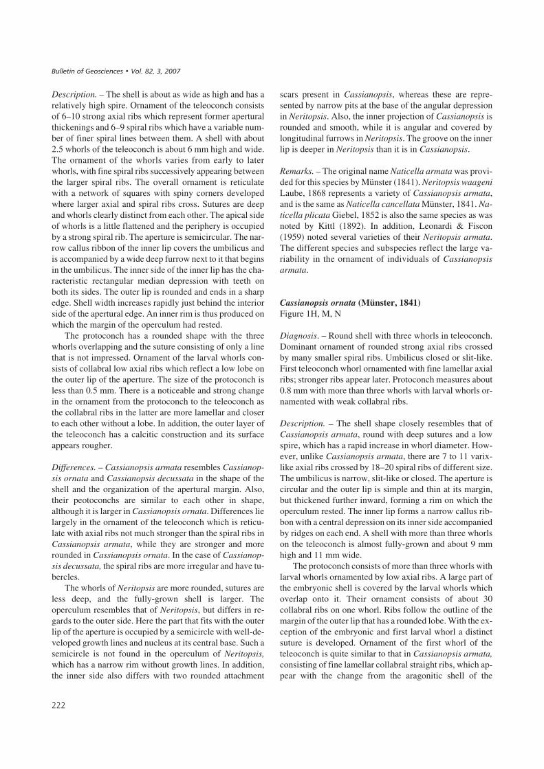
��#����+% A – the inner lip of Zardiniopsis subornata (Münster, 1841) with an angular depression on its inner side. The shell is about 1.8 mm high(BSPG 2007 XVI 8). • B – this juvenile shell of Zardiniopsis subornata (Münster, 1841) with ornament and a callus plate is about 4 mm high (BSPG 2007XVI 9). • C – the juvenile shell of Zardiniopsis subornata (Münster, 1841) with the beginnings of the spiral ornament. The shell is about 1 mm high in-cluding the rounded protoconch (BSPG 2007 XVI 10). • D – the protoconch of Zardiniopsis subornata (Münster, 1841) has an ornament of axial and spi-ral ribs and measures about 0.35 mm in diameter. • E – protoconch of Cassianopsis decussata seen from above, same as in Fig. 2H. • F – larval shell andjuvenile teleoconch of Zardiniopsis subornata (Münster, 1841) showing change in ornament at the lobed margin of the protoconch. The shell is about1 mm high (BSPG 2007 XVI 11). • G – this protoconch of Cassianopsis decussata (Münster, 1841) has collabral ornament and measures about 0.5 mm.• H – protoconch of Cassianopsis decussata (Münster, 1841) seen from the side with its ornament restricted to the apical part of the larval shell. Theprotoconch is about 0.5 mm wide. • I – a juvenile shell with protoconch of Cassianopsis decussata (Münster, 1841) with a diameter about 1.2 mm wide(BSPG 2007 12). • J – protoconch of Cassianopsis decussata (Münster, 1841) measuring about 0.5 mm and as a fine ornament of short axial ribs on the apicalside of larval whorls. Detailed view in Fig. 2I. • K – a juvenile shell of Fossariopsis rugosocarinata Laube, 1868 with the inner lip and in which its upper in-ner ridge can be seen (BSPG 2007 XVI 13). • L – the same shell (Fossariopsis rugosocarinata Laube, 1868) seen from the back. The shell is 15 mm high.
����������� ������ �������������

��
(
�
&
��
'
� �
*
)
�
���� ������ ��������� ���������� !�"�� �� ��#���$��%� ���&������

Remarks. – The species had originally been named Nati-cella subornata by Münster (1841) and was determined asNeritopsis subornata by Laube (1869). The species studiedand illustrated by Kittl (1892, pl. 6, fig. 13) was based onspecimens from NHMW, which agree with the individualscollected from Seelandalpe (Alpe di Specie) and studiedhere. The characteristic shell was documented by Zardini(1978, pl. 15, figs 7–10).
Genus Fossariopsis Laube, 1869
Type species. – The genus is based on Fossariopsis rugoso-carinata (Klipstein, 1843) from the St Cassian Formation.
Diagnosis. – Shell angular with a flattened apical side onits whorls and rounded transition to the base. Ornamentconsists of axial rows of tubercles and narrow spiral ribs.Of the rows of tubercles, one situated at the upper cornermay form short, hollow, gutter-like spines. Inner side ofinner lip of aperture has two teeth and a median depres-sion. Protoconch unknown. Inner whorls of shell are notdissolved.
Differences. – Fossariopsis differs from Zardiniopsis n.gen. by its larger size and a distinct apical corner on itswhorls. Its shell is more elongate and has angular whorls incomparison to Cassianopsis n. gen. Palaeonarica differsby its ornament of dominating keels, a relatively widershell and smooth inner side of the inner lip. Whorls expandmore rapidly in Platychilina. Fossariopsis is distinguishedfrom Delphinulopsis by having its whorls not detachedfrom each other in the later part of the teleoconch and bynot forming a depressed and extended callus of the innerlip. The latter two genera, in addition, also differ in havinga smooth inner side of the inner lip.
Remarks. – Fossariopsis as described here is based on theindividuals determined as Palaeonarica rugosocarinataby Zardini (1978, pl. 17, figs 11, 12), not on those of Zar-dini (1978, pl. 17, figs 13–15), which represent juvenile in-dividuals of Palaeonarica concentrica. Laube (1869) re-cognized in Pleurotomaria binodosa Münster, 1841 twodifferent species one of which he assigned to Fossariopsisand the other to Delphinulopsis. Kittl (1892, pl. 6, fig. 18)suggested that Fossariopsis rugosocarinata represents aPalaeonarica. The type of Fossariopsis rugoso-carinatawas based on a poorly preserved specimen, and Zittel(1895) united both genera in the genus Delphinulopsis.Koken (1889), in contrast, suggested replacing Delphinu-lopsis with Fossariopsis, as was adopted by Wenz(1938–1944), who stated that Fossariopsis and Delphinu-lopsis are based on the same type species and therefore aresynonyms. In fact Fossariopsis muensteri Laube, 1869 is
the same species as Delphinulopsis binodosa (Münster,1841). Yin & Yochelson (1983) noted the stair-like shellshape of Fossariopsis and a rapid increase in shell diame-ter. They suggested that the ornament of two spiral keels,one at the apical edge and a weaker keel on the corner to thebase could be regarded as characters with which to reevalu-ate the genera of Laube (1869). Even though their diagnos-tic character does not deviate from that of Wenz(1938–1944), it is based on a different type species. Yin &Yochelson (1983) regarded Delphinulopsis binodosa, andFossariopsis rugosocarinata as representing different ge-nera and retained Fossariopsis as an independent genuswith the type species Fossariopsis rugosocarinata.
Fossariopsis rugosocarinata (Klipstein, 1843)Figure 2K, L
Diagnosis. – Shell with an angular apical margin and a rowof nodes or hollow spines. Ornament consists of numerousnarrow spiral tubercle-bearing ribs where crossed by thewide and rounded axial ribs. Inner lip is accompanied byouter columellar furrow and two teeth and a median de-pression on its inner side.
Remarks. – The species has the characters of the genus.
Description. – A shell with four whorls of the teleoconch isabout 16 mm high and 15 mm wide. It has a low spire andwhorls are arranged in a stair-like manner increasing ra-pidly in diameter. The apical corner of whorls is enforcedby axial ribs that carry tubercles where they cross the cor-ner. These can also be developed as hollow spines (Zardini1978, pl. 17, figs 11, 12). A shell with 2.5 whorls of the te-leoconch is about 8 mm high and wide. Its peripheral edgecarries about 13 coarse tubercles or gutter-like spines. Theedge to the base has weaker tubercles. On the rounded basethree tubercle-bearing spiral ribs are present which havefine spiral lines between them. Growth lines are straightand may be strong and lamellar. The aperture is semicircu-lar with a straight inner lip that forms a narrow continuouscallus ribbon. The umbilical depression may be covered bythe callus, or it leaves a narrow deep open slit. On the innerside of the inner lip two teeth indicate the area into whichthe projection of the operculum was fitted. An umbilicalgroove with growth increments ends in the base at the outerlip. The margin of the outer lip is acute. A little further intothe shell interior the wall increases in thickness thus for-ming an internal rim which provided a good support onwhich the operculum rested. The protoconch is still un-known, as well as the operculum. Predictably, it would pro-bably resemble that of Cassianopsis n. gen. in generalshape and outline. The inner walls of the shell are not dis-solved.
���
����������� ������ �������������
Differences. – Fossariopsis rimbianchi (Zardini, 1985) andFossariopsis sinense (Yin & Yochelson, 1983) both have afine pattern of spiral ornament on their surface, as can alsobe present in individuals of Fossariopsis rugosocarinata;all three species may represent the same or very similarspecies. Palaeonarica concentrica and Palaeonarica pyru-laeformis differ not only by having a lower shell shape, butalso in having a smooth simple inner lip without central de-pression and inner teeth.
Subfamily Colubrellopsinae n. subfam.
Type genus. – The subfamily is based on the genus Colub-rellopsis n. gen. from St Cassian Formation.
Diagnosis. – Ornament on rounded whorls consists ofcoarse straight axial ribs with fine axial and spiral linesbetween them. Rounded whorls form a flat coil on theapex, while their diameter increases rapidly. Protoconch isrounded, about 0.5 mm in size consisting of up to 3 whorlswith a basically smooth surface. Aperture of teleoconchwith median shallow groove on inner side of inner lip. Nextto the flat callus of the inner lip lies an umbilical furrowwith a ridge on its outer margin in which the axial ornamentof whorls ends.
Differences. – Colubrellopsinae differ from Cassianopsi-nae by the ornament of the teleoconch. The flattened apicalregion as found in Colubrellopsis n. gen. may also be notedin Weitschatopsis Frýda, Blodgett & Stanley, 2003, but thereticulate ornament of axial and spiral ribs as well as lessrapid increase in shell width distinguishes the latter genusfrom Colubrellopsis n. gen.
Genus Colubrellopsis n. gen.
Type species. – Naticella acuticostata Klipstein, 1843 fromthe St Cassian Formation, as newly defined below.
Diagnosis. – Apical shell portion appears flatly coiled,while basal shell indicates that whorl diameter increases ra-pidly. Protoconch consists of rounded whorls with only aweak axial ornament and well impressed sutures. Apertureof teleoconch high with a rounded outer lip and straight in-ner lip with Neritopsis-like depression and well-developedtubercle-like ends. Flattened callus ribbon ends abruptly atmargin of umbilical groove, which has also a sharp cornerto the ornamented side of the whorl. Ornament consists ofstrong, straight axial ribs with fine lines between and evenfiner spiral lines. Shell composed of thin but solid outercalcitic layer and thick aragonitic layer with bulk formed ofcrossed lamellar structure.
Name. – The ornament is like that Colubrella while theshell construction and features of the inner lip place the ge-nus near Neritopsis. The name represents a free combina-tion of Colubrella and Neritopsis.
Differences. – Colubrella Koken, 1897 has very similar or-nament and a planispiral top to Colubrellopsis, but is alsocharacterized by a planispiral base, which differs from thatin the latter genus. The Paleozoic genera Spirina Kaiser,1898 from the Middle Devonian and Natiria from the Car-boniferous may have a similar ornament and shape of theshell, but attain a larger size in fully-grown specimens(Knight 1941, pls 84, 85). Natiria Koninck, 1881 from theEarly Carboniferous has a quite similar ornament with lar-ger axial ribs and fine axial line between them, but detailsregarding the protoconch or the character of the aperture ofthe teleoconch are not known from that genus.
Colubrellopsis acuticostata (Klipstein, 1843)Figure 3A–I, K, L
Diagnosis. – Inner lip of aperture bearing the characters ofNeritopsis, with on its inner side a median depression and arounded tooth on either side of that. Narrow callus plate ac-companied by a narrow umbilical furrow along its lowerpart. Ornament of teleoconch consists of larger narrowaxial ribs which have a rounded posterior side and a steepfront. Between them are many very fine axial lines that arecrossed by even finer spiral lines. In second whorl orna-ment may end and shell becomes smooth or wrinkled. Pro-toconch is about 0.5 mm wide consisting of a little morethan 3 whorls which overlap each other but display a wellrecognized suture. Larval whorls are smooth with only fewaxial lines near the sutures. Final end of larval shell canhave a rim or sometimes a fractured margin.
Description. – With three whorls the shell measures about10 mm in height and width. The ornament consists ofbroad, slightly rounded and lamellar axial ribs with a finepattern of axial and spiral lines in the interspaces. About15 to 30 ribs are on each whorl, but this ornament may di-sappear with the second whorl of the teleoconch. The ribsoriginate in the umbilicus or at the margin of the basal gro-ove in the continuation of the umbilicus. This furrow is or-namented by fine growth lines. Next to it lies the narrowcallus plate of the inner lip, which rests on the umbilicuswithout closing it or covering it. The aperture is of charac-teristic shape with a straight inner lip and evenly roundedouter lip. On the inner side of the inner lip there are two te-eth with a straight inner depression between them.
Differences. – Colubrellopsis acuticostata closely resem-bles Scalaneritina n. gen. in shape and ornament but the
���
���� ������ ��������� ���������� !�"�� �� ��#���$��%� ���&������

axial ribs of its teleoconch ornament do not end in the mid-dle and below the callus cover, but rather terminate on themargin of the umbilical groove. The inner side of the innerlip of Scalaneritina n. gen. is simple and smooth withoutthe characters of Neritopsis as are present in Colubrellopsisacuticostata. The larval whorls of the protoconch of Cassi-anopsis n. gen. are ornamented and the apertural marginends with a lobe while the protoconch of Colubrellopsis n.gen. is smooth and has a straight margin in the larval shell.
Remarks. – This species of Colubrellopsis was called Nati-cella acutecostata by Kittl (1892, pl. 6, figs 22–24) as isdocumented by a specimen kept at NHMW. However,among the specimens that he described and illustrated notonly this species is present, but also Scalaneritina münste-riana (Orbigny, 1849) with very similar ornament andshell shape, but with a different morphology of the inner lipand higher aperture. In the case of the individuals deter-mined by Zardini (1978, pl. 20, figs 5, 6) as Naticella acu-tecostata, one (fig. 6) belongs to Colubrellopsis, whereasthat in his fig. 5 is a Scalaneritina n. gen., since the ribs endat the callus and there is no umbilical furrow. Naticellaacutecostata as determined by Zardini (1978) was describedby Klipstein (1843) as Naticella acuticostata; Kittl (1892)changed the original spelling into acutecostata.
The genus Colubrella Koken, 1897 has as its diagnosticcharacters a shell that is planispiral with its whorls barelytouching. Its ornament consists of axial ribs of whichcoarser ones have a number of finer ones intercalated.Colubrella nautiliformis (Broili, 1907) was characterizedas having a low spire and a planar coiled shell in which thediameter of the whorl increased rapidly. A secondarilycompressed shell called Colubrella (Keration) nautili-formis (Broili) was described by Zardini (1978, pl. 2, fig. 1)and may very well represent Colubrellopsis acuticostata. Itwas also found at Alpe di Specie (Seelandalpe) as were theindividuals studied here. The specimen from the Seiser Alpupon which the species was based by Broili (1907, pl. 7,fig. 9) was obviously reconstructed, since he described it asonly partly preserved, especially regarding its apicalwhorls. The original to Colubrella of Koken (1897) wasalso crushed. It may be that Colubrella is actually not a
planispiral shell, but represents a crushed higher shell, andin that case it might prove to be Colubrellopsis.
Family Fedaiellidae n. fam.
Type genus. – Fedaiella Kittl, 1894.
Diagnosis. – Shell ornament composed only of growth linesand collabral folds. Whorls are rounded and sutures well ex-pressed. The last whorl encompasses most of the earliershell. Inner lip may extend over much of the umbilical area.Inner side of the inner lip with two folds or teeth in apicaland basal positions accompanied by a straight and concavemedian zone, as in Cassianopsis n. gen. Operculum an ovoidshape with a concentric construction with its nucleus lyingclose to the middle; two rounded scars present on its innerside. Protoconch a rounded shape with distinct sutures andabout 2.5 rounded whorls with little or no ornament on thelarval shell. Outer lip has a sinuous or straight margin.
Remarks. – The family is based on the genus Fedaiella asit occurs in the St Cassian Formation near Cortinad’Ampezzo.
Differences. – Regarding shell shape, Fedaiella resemblesHologyra, the latter having a pillar in its umbilical furrowand no teeth and no groove on the inner side of the inner lip.The Fedaiellidae differs from the members of the Hology-rinae in the shape and ornament of the protoconch and ofthe operculum. The taxa have no sinuous ornament of theirlarval shell, and their operculum has no triangular base.The shell shape similar to that of Fedaiella is also found inNeritaria which has the inner whorls of its shell dissolved,which is not the case in Fedaiella. In contrast to the con-centric outer surface ornament of the Fedaiellidae, theNeritariidae have an operculum as in the Neritidae with asemicircular shape and a nucleus near the side, and withspiral increments of growth.
Remarks. – The species included in the Fedaiellidae n. fam.were originally considered as members of the Naticidae
���
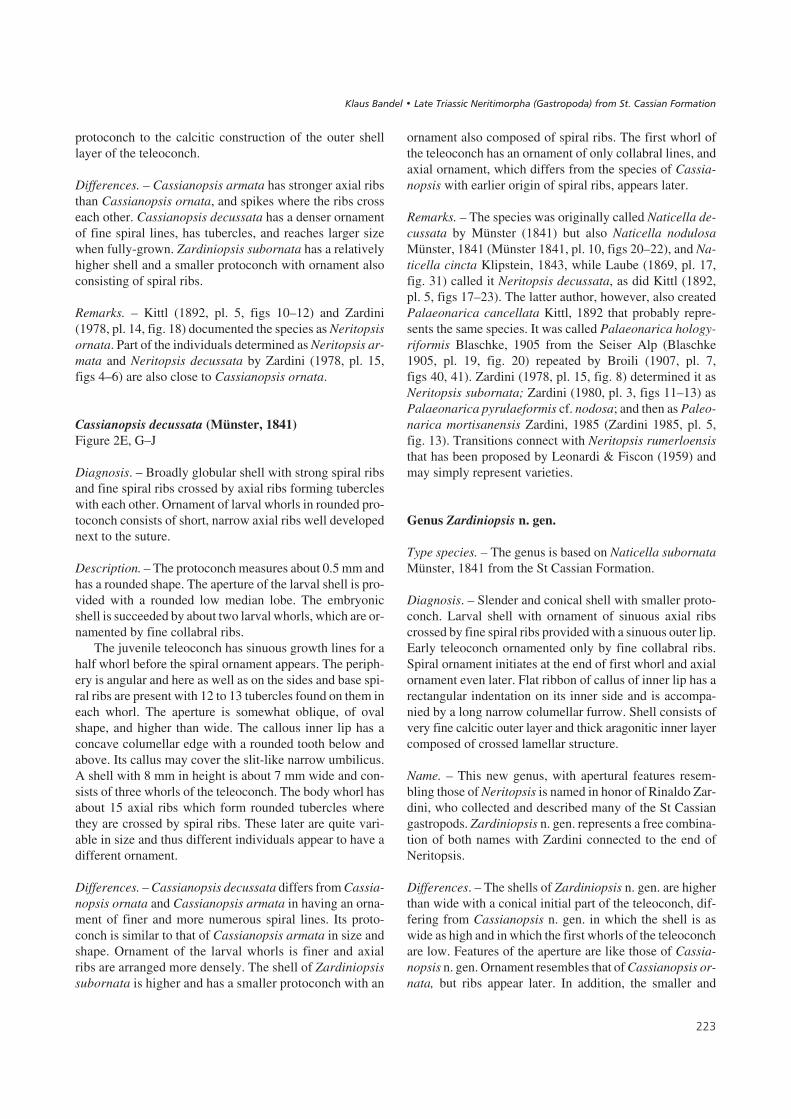
��#����,% A – this protoconch of Colubrellopsis acuticostata (Klipstein, 1843) is almost smooth and 0.5 mm in width and ends with the axial ornamentof the teleoconch. • B – Colubrellopsis acuticostata (Klipstein, 1843) showing the change of ornament in the late teleoconch and with a shell width ofabout 4 mm (BSPG 2007 XVI 14). • C – the inner lip of Colubrellopsis acuticostata (Klipstein, 1843) has a flat central groove. This shell is about 5 mmhigh. • D – the protoconch of Colubrellopsis acuticostata (Klipstein, 1843) consist of more than three whorls and is about 0.5 mm wide. • E – apical viewof Colubrellopsis acuticostata (Klipstein, 1843). Same shell as in Fig. 2D. • F – the callus of the inner lip of Colubrellopsis acuticostata (Klipstein, 1843)with its median depression in the 5 mm high shell (BSPG 2007 XVI 15). • G – shell of Colubrellopsis acuticostata (Klipstein, 1843) with the structure ofthe thin calcitic layer with the growth lines imprinted on the shell surface and crossed lamellar structure below. The shell is about 0.3 mm thick. • H – shellof Colubrellopsis acuticostata (Klipstein, 1843) with umbilical groove and about 8 mm high (BSPG 2007 XVI 16). • I – Colubrellopsis acuticostata(Klipstein, 1843) with a 10 mm high shell (BSPG 2007 XVI 17). • J – convergent shell shape of Ladinaticella costata with a 10 mm high shell (BSPG2007 XVI 72). • K – Colubrellopsis acuticostata (Klipstein, 1843), same as in Fig. 3L from the back. • L – Colubrellopsis acuticostata (Klipstein, 1843)of Kittl type with a shell about 10 mm wide.
����������� ������ �������������

���
(
�
�&
�
�
'
�
)
*
�
���� ������ ��������� ���������� !�"�� �� ��#���$��%� ���&������

(Münster 1841, Laube 1869). Zittel (1895) recognized thatsome individuals that now belong in the new family had anoperculum of the type found in Neritopsis and therefore heremoved them from Natica and its relatives and placedthem in Naticopsis. Kittl (1892) discussed the possibilitythat Naticopsis might represent a precursor to Natica,which can be rejected based on shell structure, operculumshape, and protoconch morphology. Shell composition andshape indicates a relationship with Cassianopsis n. gen. aswell as with Colubrellopsis n. gen. The genus DicosmosCanavari, 1890 had been based on quite undeterminablematerial, as was discussed by Böhm (1895), while the simi-lar genus Fedaiella has as its type species Fedaiella cuc-censis (Mojsisovics, 1873) which is synonymous with Fe-daiella maculosa (Klipstein, 1843) and which earlier hasbeen described as Natica neritacea by Münster (1841). Thespecies Fedaiella neritacea (Münster, 1841) is documentedin detail below.
Genus Fedaiella Kittl, 1894
Type species. – Natica neritacea Münster, 1841 from StCassian Formation.
Diagnosis. – Genus with characters of the family Fedaielli-dae. Smooth and rounded shell may grow to relativelylarge size. Aperture rounded with inner lip forming a callusribbon accompanied by an umbilical furrow while the ac-tual umbilicus is covered. Inner side of the inner lip withcentral depression. Concentric operculum well seen exter-nally with two rounded muscle attachment pits near the in-ner margin of its inner side. The shell is composed of a thincalcitic outer layer and a thick aragonitic inner layer arran-ged in crossed lamellar structure.
Remarks. – Wenz (1938–1944, fig. 988) characterized thegenus Dicosmos as having a globular shell with a smoothsurface and a rounded apex and base. The genus had origi-nally been based on the Ladinian age Dicosmos pulcherCanavari, 1890 from Italy and the holotype is represented
by shell that cannot be further determined (Böhm 1895).The specimen may well be related or belong to Fedaiella,but could just as well represent Hologyra or even Nerita-ria. All these have a similar rounded shape with a smoothsurface. Individuals having an operculum with concentriclines on the outer surface were placed in Naticopsis zitteliKittl, 1892, which is synonymous with Fedaiella nerita-cea. Zardini (1978, pl. 22, figs 3–5) determined a form Di-cosmos maculosa (Klipstein, 1843), which is synonymouswith Naticopsis neritacea (Münster, 1841), according tothe analysis of Kittl (1892). It has also been called Fedai-ella maculosa (= cuccensis Mojsisovics, 1873) when thegenus was first proposed by Kittl (1894) from the Marmo-lata Limestone. This Fedaiella neritacea (= maculosa= cuccensis) can be more clearly recognized and betterdefined with the material from the St Cassian Formationproviding an improved understanding of the type species.Zardini (1978, pl. 21, figs 11a, b, 13a, b) illustrated the ty-pical muscle attachments pits.
Fedaiella neritacea (Münster, 1841)Figure 4A–F, M
Diagnosis. – Broadly globular shell with a low but pointedspire. Sutures impressed and ornament consisting of moreor less distinct growth increments. Protoconch of roundedlow outline with 2.7 apparently smooth whorls. Margin oflarval shell has a rounded lobe on its outer lip. Teleoconchof up to five whorls reaching almost 40 mm; aperture roun-ded with callus of the inner lip accompanied by an umbili-cal furrow. Inner side of the inner lip has ridges with anangular depression between. Operculum concave and ofconcentric composition with nucleus displaced a little fromthe center; two shallow grooves on the inner side.
Remarks. – Zardini (1978, pl. 21, figs 11a, b, 13a, b) illus-trated the shallow grooves of the operculum.
Description. – The solid shell has a rounded shape, is usu-ally a little broader than high, has a rapidly increasing
���
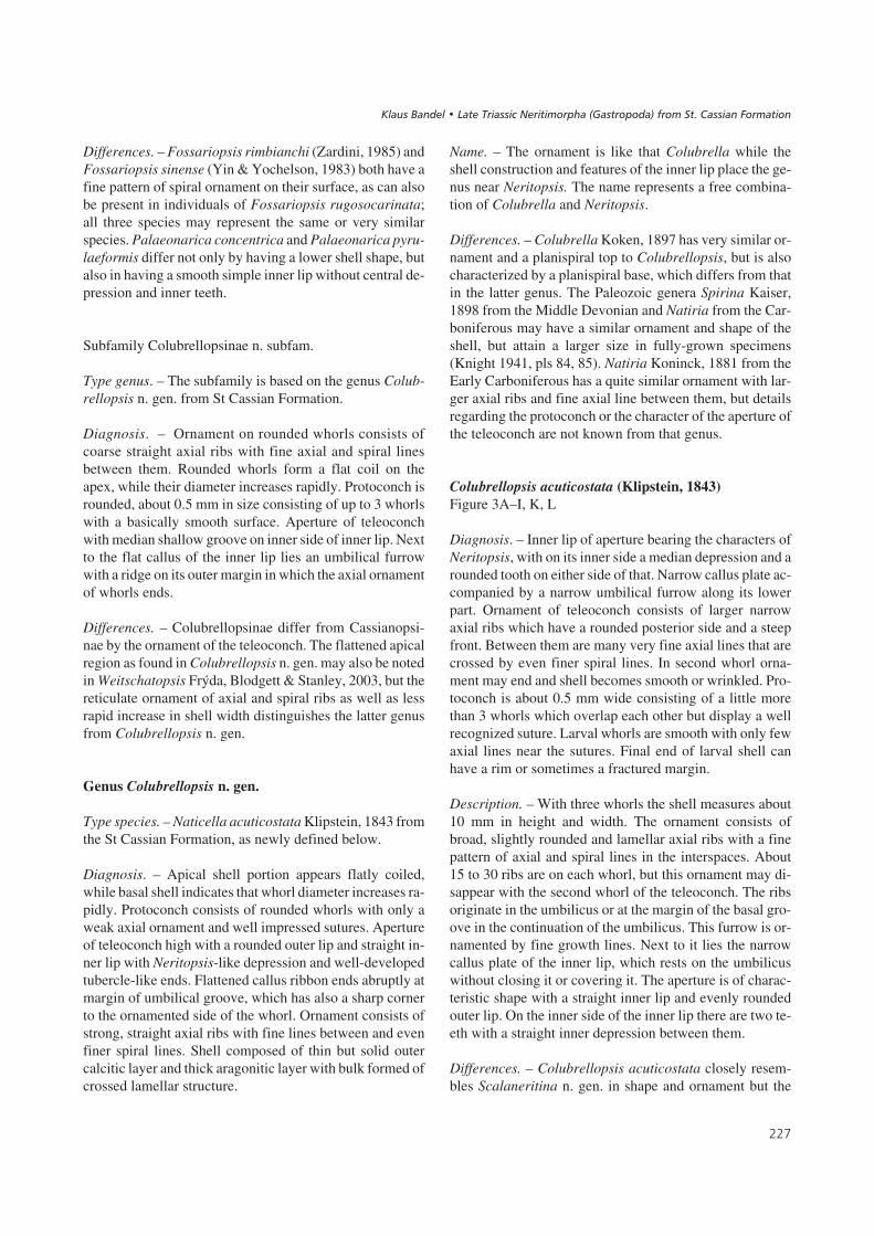
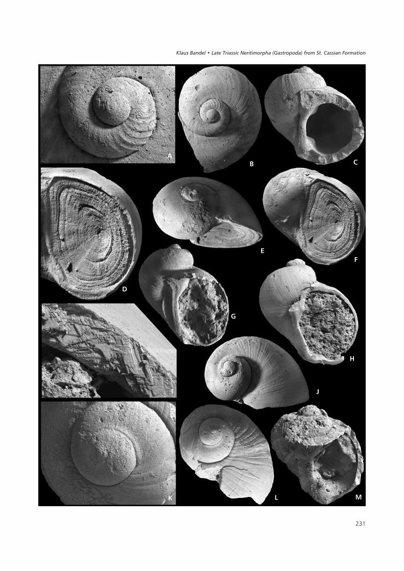
��#����-% A – protoconch of Fedaiella neritacea (Münster, 1841) about 0.5 mm wide and showing the teleoconch with initial axial ribs. • B – shell ofFedaiella neritacea (Münster, 1841). Same shell as protoconch shown in Fig. 4A. The shell is about 2 mm in diameter (BSPG 2007 XVI 19). • C – the in-ner lip of Fedaiella neritacea (Münster, 1841) has a central groove on its inner side. The shell is about 2 mm high. • D – this operculum still in the apertureof Fedaiella neritacea (Münster, 1841) is of concentric construction. Detail of Fig. 4E, F. • E – side view of Fedaiella neritacea (Münster, 1841) as seen inFig. 4D (BSPG 2007 XVI 20). • F – specimen of Fedaiella neritacea (Münster, 1841) with the operculum in place, as in Fig. 4D. The shell is about 8 mmin diameter. • G – the aperture of Fedaiella elongata (Münster, 1841) has a concave inner lip and its callus is accompanied by the umbilical groove. Theshell is about 2.5 mm high (BSPG 2007 XVI 21). • H – Fedaiella elongata (Münster, 1841). Shell about 3 mm high (BSPG 2007 XVI 22). • I – this frac-tured shell of Fedaiella elongata (Münster, 1841) shows the very thin outer calcitic layer and a thick crossed lamellar layer below it. Shell thickness isabout 0.1 mm. • J – Fedaiella elongata (Münster, 1841). Apical view of the shell in Fig. 4H. Shell width about 2.5 mm. • K – the protoconch of Fedaiellaelongata (Münster, 1841) is smooth with a sinuous margin of the larval shell that is about 0.5 mm wide. • L – teleoconch has increments of growth in itsearly part in case of Fedaiella elongata (Münster, 1841). Same shell as shown in detail in Fig. 4K. Shell width about 1.5 mm (BSPG 2007 XVI 23).• M – the apex of Fedaiella neritacea (Münster, 1841) with the characteristic inner lip. The shell is about 11 mm high (BSPG 2007 XVI 24).
����������� ������ �������������
���
( �
&
�
�
�
'
� )
* � �
���� ������ ��������� ���������� !�"�� �� ��#���$��%� ���&������
whorl diameter and a large body whorl. The protoconchmeasures a little less than 0.5 mm. Its embryonic whorlis succeeded by about 1.5 larval whorls, which appear tobe smooth. Apertural margin of the larval shell is sinuouswith a rounded lobe in the middle of the outer lip. The ini-tial part of the teleoconch attached to the protoconch maybe ornamented by some rounded low axial ribs which thenbecome discontinuous. The teleoconch consists of 4–5rounded whorls with a thickened shell and separated by in-cised sutures, which are deeper in juvenile than in maturewhorls. One shell with 1.25 whorls measures 3.3 mm inwidth and 2.9 mm in height, and a shell of 37 mm in widthis 31 mm high (Zardini 1978, pl. 22, fig. 5). Color patternsare commonly preserved, mostly represented by elongatedark brown dots of variable size among individuals andalso within different zones of growth in the same indivi-dual. The aperture is egg-shaped and oblique with straightinner and evenly rounded outer lips. The outer lip is thin atthe margin and becomes thick further inward; a ledgeis thus developed on the inner margin of the aperturallip. The inner lip forms a flat callus band that covers theumbilicus. Tubercles accompany the central depressionof the inner side on either side. The inner walls of the shellare not dissolved. This is the case in juvenile shells, aswell as in a fully-grown shell of more than 30 mm in dia-meter.
An operculum found in place closing the shell isslightly convex and of oval shape with its surface coveredby concentric growth lines. Growth increments consist ofcalcitic needle-like crystallites arranged so that they pointtoward the outside of the operculum. The slightly eccentricnucleus lies closer to the outer lip than to the inner lip andtwo depressions diverge from it running to the corners ofthe operculum. The inner side of the operculum has a thick-ened margin and two low depressions, which are dividedby a central swelling.
Differences. – The shell shape resembles that of Hologyracassiana, which differs in the characteristic column-likeridge on the umbilical furrow, and the sinuous ornament ofits larval shell. In addition, the operculum in Fedaiella ne-ritacea has a triangular base.
Remarks. – Klipstein (1843) named Natica maculosa,which, according to Kittl (1892, pl. 6, figs 29–32, pl. 7,fig. 1), should be placed into the genus Naticopsis, and ac-tually represents the same species as Natica neritaceaMünster, 1841. A problem with this interpretation is theabsence of the specimen on which Münster (1841, pl. 10,fig. 2) based his species, which apparently has been lost.Kittl (1892) used part of the type material of Natica macu-losa to describe his Naticopsis neritacea and most laterauthors accepted that suggestion, as is also done here. Ti-chy (1980, pl. 3, fig. 3) in his description of color patterns
used the figure of Klipstein (1843) and determined it as Di-cosmos maculosus (Klipstein, 1843), and next to it in Tichy(1980, pl. 3, fig. 2) named an individual Naticopsis (N.)neritacea (Münster, 1841) based on a figure from Kittl(1892, pl. 6, figs 29–32), apparently interpreting these twoas different species. Broili (1907, pl. 8, figs 17–19) sugges-ted that in the case of Naticopsis maculosa from St Cassianthere might be a depression below the suture of the lastwhorl. However, such a depression may or may not appearin different individuals of the same species.
Blaschke (1905, pl. 20, fig. 1) recognized the formFedaiella neritacea as Dicosmos (Fedaiella?) seisiensis(aff. Dicosmos declivis) n. f. from the Seiser Alp, consider-ing it a local variety of Naticopsis maculosa. In the case ofDicosmos applanatus Kutassy, 1937, the author also usedas a characteristic the flattening of the subsutural ribbon todifferentiate it from Naticopsis maculosa. Actually thisspecies is probably identical with Fedaiella neritacea. Itwas determined as Dicosmos maculosus by Leonardi &Fiscon (1959, pl. 2, figs 24, 25, pl. 3, fig. 1) and Zardini(1978, pl. 20, figs 9, 10, pl. 22, fig. 5; 1985, pl. 7, figs 2, 3),but none of the illustrated individuals show the inner foldson the inner lip of the aperture. The operculum was docu-mented as belonging to Neritopsis by Zardini (1978, pl. 21,fig. 13; 1985, pl. 4, fig. 9).
Fedaiella elongata (Münster, 1841)Figure 4G–L
Diagnosis. – Shell higher than wide up to about 11 mmwith a pointed spire. Whorls rounded. Inner side of the in-ner lip has a rectangular median depression. Oval aperturewith a narrow apical portion; thick callus of the inner lipcovers the umbilical region. Narrow callus band at the baseand wide at the apical side of aperture. Rounded proto-conch consists of about 2.5 whorls with the suture of thelast whorl quite distinct; larval whorls have a straight outerlip at the aperture.
Remarks. – The characters of the inner lip and general shellfeatures resemble those of Fedaiella neritacea.
Description. – The shell is short, Lymnaea-shaped withfour whorls in the teleoconch. The ornament consists ofsimple collabral growth increments, some more stronglydeveloped than others. The callus of the inner lip broadenstowards its middle and forms a pad, accompanied by a gro-ove basal to it. This depression has growth increments ofthe former margins, of which the outermost forms a sharpedge to the whorl. This edge is continuous into the evenlyrounded outer lip of the semicircular aperture. On the innerside of the inner lip two folds are developed of which theapical fold is inclined and the basal fold is pointed. A shell
���
����������� ������ �������������

with 2.7 whorls in the teleoconch is 3.5 mm wide and 4 mmhigh. It has an angular groove between the tubercles on theinner lip, which is well developed in juveniles but becomesshallow and indistinct in more mature shells with some4 whorls in the teleoconch and typically 11 mm high by10 mm wide. The outer calcitic layer of the shell is verythin and the shell surface is smooth.
The protoconch measuring about 0.5 mm in diameterhas a globular embryonic whorl that is covered to a largeextent by the first larval whorl. In the larval part of theprotoconch whorls increase in width and the suture be-comes more distinct. The margin of the larval shell isstraight. The changeover to the teleoconch is indicated bycoarser growth lines. The ornament is not well preservedbut seems to consist of fine and low axial ribs.
Differences. – The two folds on the inner side of the innerlip of the aperture resemble those found in Fedaiella neri-tacea, but the high spire differentiates them from one anot-her.
Remarks. – The species exhibits its lower inclined ridgeon the inner side of the inner lip as illustrated by Zardini(1978, pl. 19, fig. 15), determined by him as Naticopsiscassiana. The description of the species presented byKittl (1892, pl. 7, figs 3, 4) is not conclusive with the ex-ception of his remarks about the callus of the inner lipwith its thickened upper part and a depression below it,that is characteristic of the specimens studied here. Theholotype of Naticopsis elongata var. chromatica Kittl,1892 coincides its morphology with Fedaiella elongata.It has a similar size and shape, but belongs to a relativelyworn specimen, so that the features of the callus of the in-ner lip are not well preserved. The specimen illustrated byKittl (1892, pl. 7, figs 3, 4) and determined as Naticopsissubelongata Orbigny has indications of the folds or tuber-cles on the inner side of their inner lip, but the shells ap-pear to be too short to fit with Fedaiella elongata. A betterfitting shell shape and ornament is preserved in Naticop-sis gaderana Kittl, 1892 as seen in the holotype in the col-lection at NHMW, which has an egg-shaped shell butdoes not expose the inner lip (Kittl 1892, pl. 7, figs 9, 10).Another individual determined as Naticopsis gaderanaby Kittl (1892) has folds on its inner lip. It is quite po-ssible that the species determined as Naticopsis limnaei-forme (Laube, 1869) is Fedaiella elongata as well (Kittl1892, pl. 7, fig. 8). Neither the original description provi-ded by Laube (1869, pl. 22, fig. 3), nor that of Kittl (1892,pl. 7, fig. 8) is conclusive.
Böhm (1895, figs 33, 34) described Fedaiella cuc-censis (Mojsosovics, 1873) as well as Fedaiella benneckiBöhm, 1895 and clearly documented the two teeth on theinner side of the inner lip of their aperture connected to anangular depression as in Neritopsis.
Family Delphinulopsidae Blodgett, Frýda & Stanley, 2001
Diagnosis. – Shell diameter increases rapidly and laterwhorls may discontinue the earlier tight coiling mode ofthe shell; whorls angular. Aperture increases in size untilan almost limpet-like form is achieved with straight innerand a well-rounded outer lip. Inner side of the inner lip issmooth. Ornament consists of spiral keels or rows of tu-bercles or spines. Outer shell layer thin and calcitic andunderlain by a thick aragonitic layer in crossed lamellarstructure.
Remarks. – The family is based on Delphinulopsis Laube,1868 and was created to find a place for some Triassic spe-cies with a goat-horn-like shell resembling that of Delphi-nulopsis binodosa from the St Cassian Formation. Blodgettet al. (2001) suggested that the taxon should also includespecies belonging to Paradelphinulopsis Blodgett, Frýda& Stanley, 2001 from the Late Triassic of western NorthAmerica.
Subfamily Delphinulopsinae Blodgett, Frýda & Stanley,2001
Diagnosis. – Inner lip expanded to form a concave callus;whorls have spiral ribs with tubercles or spines.
Remarks. – The subfamily has the character of the genusDelphinulopsis from the St Cassian Formation. Here thegenus Delphinulopsis is characteristic and a new genusSchwardtopsis n. gen. (see below) is included because itclosely resembles young stages of Delphinulopsis binodo-sus but with its shell attaining a larger size. All are from theSt Cassian Formation.
Genus Delphinulopsis Laube, 1868
Type species. – The genus is based on Delphinulopsis bino-dosa (Münster, 1841) from the St Cassian Formation.
Diagnosis. – Shell shaped like the horn of a goat with who-rls of the teleoconch detaching from each other, but remai-ning in a spiral coil. A concave callus plate of the inner lipincreases in size as whorls become less tightly coiled. Or-nament consists of four spiral rows of large tubercles ofwhich the basal one is covered by the succeeding whorl.
Remarks. – The genus has been considered to be a syno-nym of Fossariopsis by Wenz (1938–1944, fig. 1006) (seethere). Delphinulopsis has its early whorls similar toSchwardtopsis n. gen. but differs from the latter by its moreopenly coiled shell with a characteristic wide, flattened cal-
���
���� ������ ��������� ���������� !�"�� �� ��#���$��%� ���&������
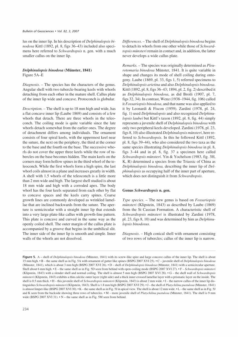
lus on the inner lip. In his description of Delphinulopsis bi-nodosa Kittl (1892, pl. 8, figs 36–43) included also speci-mens here referred to Schwardtopsis n. gen. with a muchsmaller callus on the inner lip.
Delphinulopsis binodosa (Münster, 1841)Figure 5A–E
Diagnosis. – The species has the characters of the genus.Angular shell with two tubercle-bearing keels with whorlsdetaching from each other in the mature shell. Callus plateof the inner lip wide and concave. Protoconch is globular.
Description. – The shell is up to 18 mm high and wide, hasa flat concave inner lip (Laube 1869) and consists of a fewwhorls that detach. There are three whorls in the teleo-conch. The coiling mode is quite variable since the latewhorls detach somewhat from the earlier ones. The degreeof detachment differs among individuals. The ornamentconsists of four spiral keels, with the uppermost keel nearthe suture, the next on the periphery, the third at the cornerto the base and the fourth on the base. The successive who-rls do not cover the upper three keels while the row of tu-bercles on the base becomes hidden. The main keels on thecorners may form hollow spines in the third whorl of the te-leoconch. While the first whorls form a high spire, the lastwhorl coils almost in a plane and increases greatly in width.A shell with 1.5 whorls of the teleoconch is a little morethan 2 mm wide and high. The largest shell studied is about18 mm wide and high with a corroded apex. The bodywhorl has the four keels separated from each other by flatto concave spaces and the keels carry spines. Coarsegrowth lines are commonly developed as wrinkled lamel-lae that are inclined backwards from the suture. The aper-ture is semicircular with a straight inner lip that extendsinto a very large plate-like callus with growth-line pattern.This plate is concave and curved in the same way as theopenly coiled shell. The outer margin of the callus plate isaccompanied by a groove that begins in the umbilical slit.The inner side of the inner lip is smooth and simple. Innerwalls of the whorls are not dissolved.
Differences. – The shell of Delphinulopsis binodosa beginsto detach its whorls from one other while those of Schward-topsis münsteri remain in contact and, in addition, the latternever develops a wide callus plate.
Remarks. – The species was originally determined as Pleu-rotomaria binodosa Münster, 1841. It is quite variable inshape and changes its mode of shell coiling during onto-geny. Laube (1869, pl. 33, figs 1, 5) referred specimens toDelphinulopsis arietina and also Delphinulopsis binodosa.Kittl (1892, pl. 8, figs 36–43; 1894, pl. 2, fig. 2) described itas Delphinulopsis binodosa, as did Broili (1907, pl. 7,figs 32, 34). In contrast, Wenz (1938–1944, fig. 106) calledit Fossariopsis binodosa, and that name was also applied toit by Leonardi & Fiscon (1959). Zardini (1978, pl. 24,fig. 1) used Delphinulopsis and also recognized Delphinu-lopsis laubei but Kittl’s taxon (1892, pl. 8, fig. 44) simplyrepresents a juvenile shell of Delphinulopsis binodosa withonly two peripheral keels developed. Zardini (1978, pl. 23,figs 8, 10) also illustrated Delphinulopsis münsteri, here re-ferred to Schwardtopsis. In this he followed Kittl (1892,pl. 8, figs 39–44), who also considered the two taxa as thesame species illustrating Delphinulopsis binodosa in pl. 8,figs 3–44 and in pl. 8, fig. 37 a specimen here calledSchwardtopsis münsteri. Yin & Yochelson (1983, fig. 3H,K, R) determined a species from the Triassic of China asDelphinulopsis binodosa, describing the inner lip of Del-phinulopsis as occupying half of the inner part of aperture,which does not distinguish it from Schwardtopsis.
Genus Schwardtopsis n. gen.
Type species. – The new genus is based on Fossariopsismünsteri (Klipstein, 1843) as described by Laube (1869)from the St Cassian Formation. A characteristic shell ofSchwardtopsis münsteri is illustrated by Zardini (1978,pl. 23, figs 8, 10) and was determined by him as Delphinu-lopsis binodosus.
Diagnosis. – High conical shell with ornament consistingof two rows of tubercles; callus of the inner lip is narrow.
���
��#����.% A – shell of Delphinulopsis binodosa (Münster, 1841) with its screw-like spire and large concave callus of the inner lip. The shell is about15 mm high. • B – the same shell as in Fig. 5A with ornament of gutter like spines (BSPG 2007 XVI 25). • C – juvenile shell of Delphinulopsis binodosa(Münster, 1841), which is about 3 mm high (BSPG 2007 XVI 26). • D – shell of Delphinulopsis binodosa (Münster, 1841) with a semicircular aperture.Shell about 6 mm high. • E – the same shell as in Fig. 5D seen from behind with open-coiling mode (BSPG 2007 XVI 27). • F – Schwardtopsis münsteri(Klipstein, 1843) with a slender shell and normal coiling. The shell is almost 5 mm high (BSPG 2007 XVI 28). • G – the shell wall of Schwardtopsismünsteri (Klipstein, 1843) exhibits a thin calcitic outer layer (right side) and a thick inner crossed lamellar layer with a prismatic layer on the inside. Theshell is 0.5 mm thick. • H – this juvenile shell of Schwardtopsis münsteri (Klipstein, 1843) is about 2 mm wide. • I – the narrow callus of the inner lip dis-tinguishes Schwardtopsis münsteri (Klipstein, 1843). Shell is 1.8 mm high (BSPG 2007 XVI 29). • J – the shell of Platychilina pustulosa (Münster, 1841)is almost limpet-like (BSPG 2007 XVI 30). • K – the same shell as in Fig. 5J in apical view. The shell is about 12 mm wide. • L – the same shell as in Fig. 5Jand K seen from the backside showing three rows of tubercles. • M – more juvenile shell of Platychilina pustulosa (Münster, 1841). The shell is 9 mmwide (BSPG 2007 XVI 31). • N – the same shell as in Fig. 5M seen from behind.
����������� ������ �������������
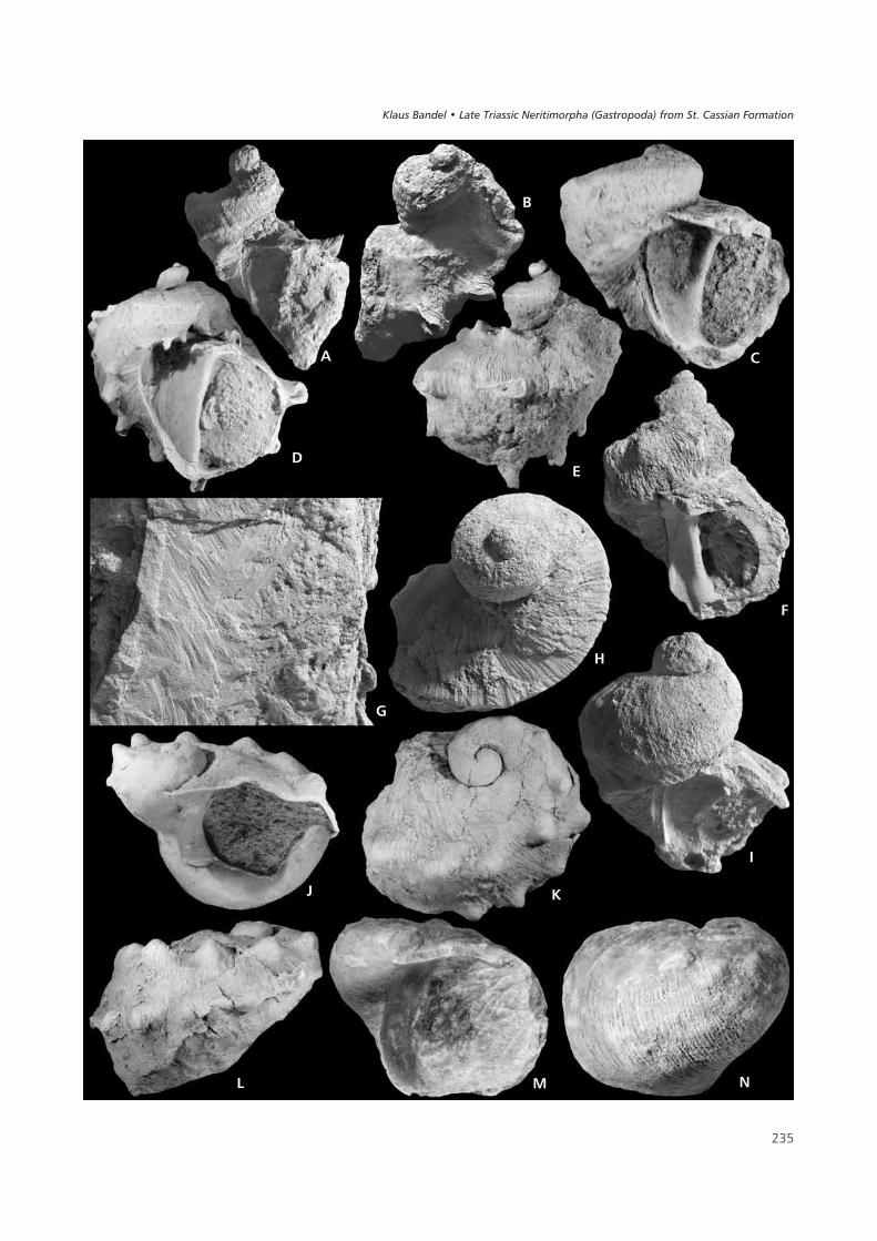
��
&
�
�
(
�
�
'
�
) *
���
���� ������ ��������� ���������� !�"�� �� ��#���$��%� ���&������

The body whorl may have a third spiral rib on the base.Shell composition is a thin outer calcitic layer and a thickinner crossed lamellar layer.
Differences. – Schwardtopsis münsteri differs from Del-phinulopsis binodosus by its narrow inner lip callus and thehigh spire in which the whorls do not detach from each ot-her during late stages of growth. The early juvenile shell ofDelphinulopsis binodosa is very similar in its elongate coi-led shape and the two spiral ribs of its ornament, but it de-velops a broad callus on the inner lip, develops two morespiral ribs later on, while in Schwardtopsis münsteri the in-ner lip is narrow from the beginning and remains that waythroughout growth, and differs additionally in having thenumber of ribs increasing only to three.
Name. – The genus is named for Annette Schwardt. She be-gan to assemble some of the data on the Neritimorpha ofthe St Cassian Formation and recognized that the genuscalled in her honor as distinct from Delphinulopsis.
Schwardtopsis münsteri (Klipstein, 1843)Figure 5F–I
Diagnosis. – Shell higher than wide with more than threewhorls up to 18 mm high. Whorls flattened on the sides andspiral ribs present on the corners to the shoulder and to thebase and on the base. Aperture ovoid in outline with a sim-ple thickened and grooved callus of the inner lip that coversthe umbilicus. From the umbilicus a furrow accompaniesthe inner lip and its margin is formed by basal spiral rib.
Description. – The conical shell is higher than wide and hasan ornament of two keels with tubercles, but it may alsocarry spines on the upper and lower corners of the whorls,which become hollow gutter-like in the last whorl. A row oftubercles or short spines on the base is covered by the succe-eding whorls. The shell is thick and when obtaining 2.5whorls the teleoconch measures about 4.5 mm wide by6 mm high. The largest shell known is about 18 mm high and15 mm wide with a final whorl with four rows of nodules ofwhich those in the second row are actually hollow spines.The whorl in the peripheral keel has 9–10 spines. The sidesare flattened and concave. A smaller basal spiral row sur-rounds the umbilical region, with the umbilicus closed bythe callus of the inner lip. Growth lines are straight and incli-ned in a posterior direction. The outer thin calcitic layer hasfine lamellar growth increments, which can be somewhatundulating. The bulk of the shell consists of aragonitic ma-terial with crossed lamellar structure. The aperture is relati-vely small and semicircular with a simple outer lip and aninner lip with a thickened margin and a smooth columellaredge. The inner walls of the shell are not dissolved.
Differences. – Schwardtopsis münsteri differs from Del-phinulopsis binodosa in its narrower inner lip that forms nowide septum, and the pointed high shell shape. A shellfrom the collection at NHMW was documented and deter-mined as Fossariopsis münsteri (Klipstein) by Laube(1869); despite being quite corroded, it displays the charac-ters of the species and genus. In Platychilina pustulosa theshell is lower and whorls more rounded.
Remarks. – Schwardtopsis münsteri was documented byZardini (1978, pl. 23, figs 8, 10) as Delphinulopsis bino-dosa. Yin & Yochelson (1983, fig. 3D) probably also had aspecimen of Schwardtopsis, but according to their descrip-tion it is closer to Delphinulopsis.
Subfamily Platychilininae n. subfam.
Type genus. – The subfamily is based on Platychilina, andincludes also Marmolatella. Both of these genera are re-cognized from the St Cassian Formation.
Diagnosis. – Shell diameter increases rapidly and orna-ment consists of fine collabral lamellae of the outer calciticlayer to which nodular spiral keels or spiral rows of nodesor hollow spines may be added. Shell walls are not dissol-ved in the interior of the protoconch or teleoconch. Proto-conch resembles that of the Cassianopsinae and Colubrel-lopsinae in size and shape. Operculum unknown. Inner sideof the inner lip is simple and straight without teeth or a gro-ove.
Remarks. – The protoconch of the Platychilininae is roun-ded and consists of several whorls and has a size of 0.5 mm,but its ornament is not well preserved. The shell of the te-leoconch consists of an outer calcitic layer and an inneraragonitic layer with crossed lamellar structure as is foundin most of the Neritimorpha of the St Cassian Formation.The shape of the inner lip, with a broad and simple callusband, places the subfamily near the Palaeonaricidae (seebelow), which has similar features. The shell shape may in-dicate that platychilininids are related to the Pseudorthony-chiidae Bandel & Frýda, 1999, which differs in havinga very characteristic protoconch and a thick outer calciticlayer in the teleoconch.
Genus Platychilina Koken, 1892
Type species. – The genus was based on a species from theRaibler beds of the Schlern Plateau, determined as Platy-chilina wöhrmanni Koken, 1892 and documented by Wenz(1938–1944, fig. 1005). However, as suggested as a possi-bility by Koken (1892), Platychilina wöhrmanni is closely
���
����������� ������ �������������
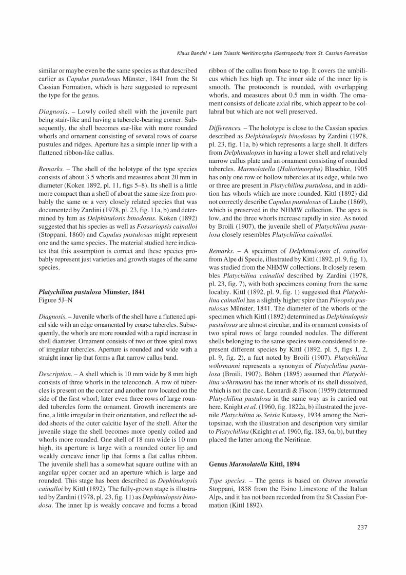
similar or maybe even be the same species as that describedearlier as Capulus pustulosus Münster, 1841 from the StCassian Formation, which is here suggested to representthe type for the genus.
Diagnosis. – Lowly coiled shell with the juvenile partbeing stair-like and having a tubercle-bearing corner. Sub-sequently, the shell becomes ear-like with more roundedwhorls and ornament consisting of several rows of coarsepustules and ridges. Aperture has a simple inner lip with aflattened ribbon-like callus.
Remarks. – The shell of the holotype of the type speciesconsists of about 3.5 whorls and measures about 20 mm indiameter (Koken 1892, pl. 11, figs 5–8). Its shell is a littlemore compact than a shell of about the same size from pro-bably the same or a very closely related species that wasdocumented by Zardini (1978, pl. 23, fig. 11a, b) and deter-mined by him as Delphinulosis binodosus. Koken (1892)suggested that his species as well as Fossariopsis cainalloi(Stoppani, 1860) and Capulus pustulosus might representone and the same species. The material studied here indica-tes that this assumption is correct and these species pro-bably represent just varieties and growth stages of the samespecies.
Platychilina pustulosa Münster, 1841Figure 5J–N
Diagnosis. – Juvenile whorls of the shell have a flattened api-cal side with an edge ornamented by coarse tubercles. Subse-quently, the whorls are more rounded with a rapid increase inshell diameter. Ornament consists of two or three spiral rowsof irregular tubercles. Aperture is rounded and wide with astraight inner lip that forms a flat narrow callus band.
Description. – A shell which is 10 mm wide by 8 mm highconsists of three whorls in the teleoconch. A row of tuber-cles is present on the corner and another row located on theside of the first whorl; later even three rows of large roun-ded tubercles form the ornament. Growth increments arefine, a little irregular in their orientation, and reflect the ad-ded sheets of the outer calcitic layer of the shell. After thejuvenile stage the shell becomes more openly coiled andwhorls more rounded. One shell of 18 mm wide is 10 mmhigh, its aperture is large with a rounded outer lip andweakly concave inner lip that forms a flat callus ribbon.The juvenile shell has a somewhat square outline with anangular upper corner and an aperture which is large androunded. This stage has been described as Dephinulopsiscainalloi by Kittl (1892). The fully-grown stage is illustra-ted by Zardini (1978, pl. 23, fig. 11) as Dephinulopsis bino-dosa. The inner lip is weakly concave and forms a broad
ribbon of the callus from base to top. It covers the umbili-cus which lies high up. The inner side of the inner lip issmooth. The protoconch is rounded, with overlappingwhorls, and measures about 0.5 mm in width. The orna-ment consists of delicate axial ribs, which appear to be col-labral but which are not well preserved.
Differences. – The holotype is close to the Cassian speciesdescribed as Delphinulopsis binodosus by Zardini (1978,pl. 23, fig. 11a, b) which represents a large shell. It differsfrom Delphinulopsis in having a lower shell and relativelynarrow callus plate and an ornament consisting of roundedtubercles. Marmolatella (Haliotimorpha) Blaschke, 1905has only one row of hollow tubercles at its edge, while twoor three are present in Platychilina pustulosa, and in addi-tion has whorls which are more rounded. Kittl (1892) didnot correctly describe Capulus pustulosus of Laube (1869),which is preserved in the NHMW collection. The apex islow, and the three whorls increase rapidly in size. As notedby Broili (1907), the juvenile shell of Platychilina pustu-losa closely resembles Platychilina cainalloi.
Remarks. – A specimen of Delphinulopsis cf. cainalloifrom Alpe di Specie, illustrated by Kittl (1892, pl. 9, fig. 1),was studied from the NHMW collections. It closely resem-bles Platychilina cainalloi described by Zardini (1978,pl. 23, fig. 7), with both specimens coming from the samelocality. Kittl (1892, pl. 9, fig. 1) suggested that Platychi-lina cainalloi has a slightly higher spire than Pileopsis pus-tulosus Münster, 1841. The diameter of the whorls of thespecimen which Kittl (1892) determined as Delphinulopsispustulosus are almost circular, and its ornament consists oftwo spiral rows of large rounded nodules. The differentshells belonging to the same species were considered to re-present different species by Kittl (1892, pl. 5, figs 1, 2,pl. 9, fig. 2), a fact noted by Broili (1907). Platychilinawöhrmanni represents a synonym of Platychilina pustu-losa (Broili, 1907). Böhm (1895) assumed that Platychi-lina wöhrmanni has the inner whorls of its shell dissolved,which is not the case. Leonardi & Fiscon (1959) determinedPlatychilina pustulosa in the same way as is carried outhere. Knight et al. (1960, fig. 1822a, b) illustrated the juve-nile Platychilina as Seisia Kutassy, 1934 among the Neri-topsinae, with the illustration and description very similarto Platychilina (Knight et al. 1960, fig. 183, 6a, b), but theyplaced the latter among the Neritinae.
Genus Marmolatella Kittl, 1894
Type species. – The genus is based on Ostrea stomatiaStoppani, 1858 from the Esino Limestone of the ItalianAlps, and it has not been recorded from the St Cassian For-mation (Kittl 1892).
���
���� ������ ��������� ���������� !�"�� �� ��#���$��%� ���&������

Diagnosis. – Shell ear-like in shape with a rounded to flatte-ned apex and large body whorl. Shell surface is smooth asidefrom increments of growth. Aperture is oval to round withthe callus of outer and inner lips connected and smooth.
Remarks. – Haliotimorpha Blaschke, 1905 was consideredto represent a subgenus of Marmolatella by Broili (1907,pl. 9, fig. 16–18). Knight et al. (1960, figs 180A, 1a, b, 181,9a, b) considered both to represent different genera of thesubfamily Naticopsinae, family Neritopsidae, even thoughboth are characterized by an ear-like shell that has its whorlscoiled to the side. Their juvenile shell is almost flat with itsapex low in the early whorls of the teleoconch, but later be-coming limpet-shaped. In the case of Haliotimorpha theperipheral side develops a row of nodules or open spines.Wenz (1938–1944, fig. 985) figured the apertural structure;the inner lip of the aperture is formed by a straight and wideribbon of callus. However, Haliotimorpha dieneri (Blasch-ke, 1905) from the Seiser Alp, which was clearly a large andlimpet-like species when fully-grown, quite evidently wasequivalent to Marmolatella (Marmolatella) Kittl, 1892 in itsearly ontogenetic shell. Neither growth stage has the charac-teristic depression on the inner lip as is at least partly charac-teristic to the family Neritopsidae in the definition that waspresented by Knight et al. (1960) for that taxon. They regard-ed Marmolatella as a subgenus of Naticopsis. This decisionthat was accepted by Jeffery et al. (1994) who described twospecies known only by teleoconch shape from the Carboni-ferous of Arkansas as belonginng to Marmolatella. However,this is unlikely since Naticopsis has a type species that livedin the Early Carboniferous and thus it may not be very clo-sely related to the Triassic species of Marmolatella that li-ved around 100 million years later.
Marmolatella fenestrata (Laube, 1869)Figure 6A–F
Diagnosis. – Horn-shaped shell with low spire. Ornamentconsists of lamellar collabral ribs and fine spiral lines. Pro-
toconch rounded consisting of 2.5 whorls with a distinctsuture and sinuous margin on the outer lip of the larvalshell.
Remarks. – The species was originally described as Capu-lus fenestratus Laube, 1869 and later determined as Nati-cella anomala Kittl, 1892 from the St Cassian Formation(Kittl 1892, pl. 9, fig. 7).
Description. – The shell shape resembles that of the bi-valve Gryphaea, with a low openly coiled whorl. Onewhorl of the teleoconch is about 11 mm wide and 7 mmhigh. Ornament consists of growth lamellae of the outer,calcitic layer of the shell, transected by very fine spiral li-nes producing a fenestrate pattern. The aperture has sim-ple, semi-circular shape and is very wide. The inner lip isalmost straight and extended to form a flattened simpleshelf of the callus. It covers the umbilical region. The pro-toconch is tightly coiled, consists of nearly 2.5 whorls thatform a helical spire. The suture of the larval whorls is dis-tinct, but the ornament of the whorls is not preservedin detail. The margin of the outer lip of the aperture of thefully-grown larval shell appears to have a wide lobe anda marginal low saddle. Shell composition consists of a so-lid but thin outer calcitic layer into which the densegrowth increments as well as the fine spiral lines are im-printed and a thick inner aragonitic layer composed ofcrossed lamellar structure with a smooth contact to theouter layer.
Remarks. – The shell is lowly coiled and has a depressedapical area, but is not sinistral as assumed by Kittl (1892) inthe case of Capulus(?) fenestratus Laube, 1869 or Nati-cella(?) anomala Kittl, 1892 (Kittl 1892, pl. 9, figs 7, 8).The holotype of Naticella anomala Kittl, 1892 fromNHMW belongs to this species. The same species has beenillustrated by Zardini (1978, pl. 24, fig. 2a–c) and was de-termined by him as Delphinulopis pustulosa. A poorly pre-served small individual of Marmolatella fenestrata was il-lustrated by Kittl (1892, pl. 9, fig. 8). He placed two quite
���
��#����/% A – apical view of Marmolatella fenestrata (Laube, 1869). Shell 4.5 mm wide (BSPG 2007 XVI 32). • B – apical view of Marmolatellafenestrata (Laube, 1869). Shell about 2 mm wide. Fig. 6C is a detailed view (BSPG 2007 XVI 33). • C – the protoconch of Marmolatella fenestrata(Laube, 1869) with rounded whorls and a simple margin. Width of protoconch about 0.5 mm. • D – apertural view of Marmolatella fenestrata (Laube,1869). Shell about 6 mm high (BSPG 2007 XVI 34). • E – somewhat recrystallized shell of Marmolatella fenestrata (Laube, 1869) broken to show therelatively thin outer calcitic layer (upper part) and thick crossed lamellar layer below. The shell is about 0.2 mm thick. • F – apertural viewof Palaeonarica concentrica (Münster, 1841). Shell about 4 mm wide (BSPG 2007 XVI 35). • G – apical view of Palaeonarica concentrica (Münster,1841). Shell about 4 mm wide. • H – juvenile shell of Palaeonarica pyrulaeformis (Klipstein, 1843) which is 4 mm in height (BSPG 2007 XVI 36).• I – the inner lip of the aperture of Palaeonarica pyrulaeformis (Klipstein, 1843) is simple and evenly smooth. The shell is about 10 mm high (BSPG2007 XVI 37). • J – shell structure of Palaeonarica pyrulaeformis (Klipstein, 1843) with the calcitic layer continuous and thin (upper part) underlainby the thick crossed lamellar layer. The visible shell is about 0.1 mm thick. • K – Palaeonarica pyrulaeformis (Klipstein, 1843) seen from the side.Shell about 5 mm wide. (BSPG 2007 XVI 38). • L – apical view of Palaeonarica pyrulaeformis (Klipstein, 1843) showing the protoconch withrounded whorls. The shell is about 1.8 mm wide (BSPG 2007 XVI 39). • M – specimen of Palaeonarica pyrulaeformis (Klipstein, 1843) showingsmooth inner lip. Shell about 10 mm wide (BSPG 2007 XVI 40).
����������� ������ �������������
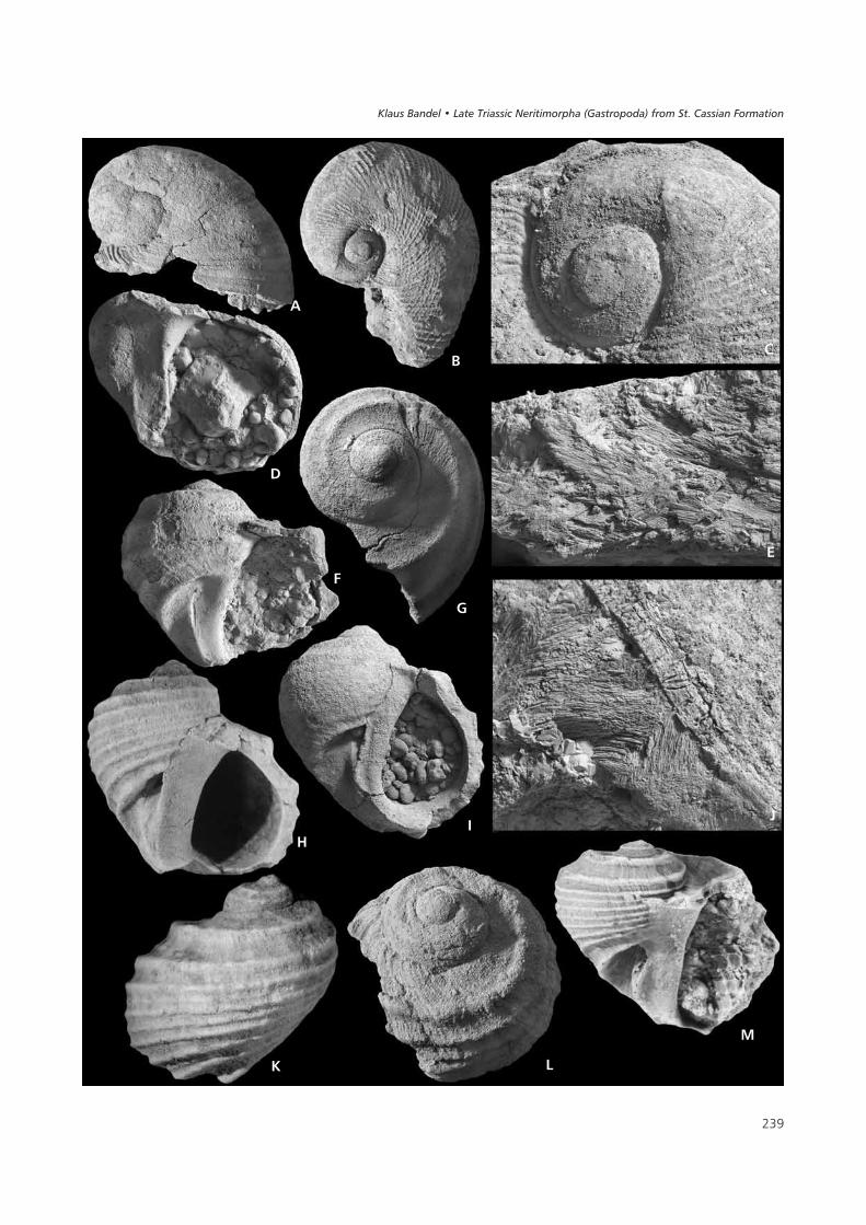
���
(�
�
�
&
)
�
'�
* �
�
���� ������ ��������� ���������� !�"�� �� ��#���$��%� ���&������

different species into his Naticella anomala, one that isclearly the same as that described by Laube (1869) and theother which may represent an individual of Cortinella Ban-del, 1988 (Kittl (1892, pl. 7, fig. 28), with a small planispi-ral shell and strong axial ribs. It had been placed with Bro-chidium by Kittl (1892) and was later, when its shellbecame better known, interpreted as belonging to its owngenus, Cortinella Bandel, 1988, and when its protoconchbecame known into an own family of the Neritimorpha(Bandel 2000), the Cortinellidae.
Not fully-grown shells of Marmolatella dieneri as de-scribed by Broili (1907, pl. 9, figs 16–18) would probablynot differ from Marmolatella fenestrata, since the charac-teristic row of hollow spines preserved in the individualsfrom Seiser Alp occur only on shells which are larger thanthe largest Marmolatella fenestrata that is known from theSt Cassian Formation.
Family Palaeonaricidae n. fam.
Type genus. – The family is based on the genus Palaeona-rica Kittl, 1892 from the St Cassian Formation.
Diagnosis. –Shell shape is Nerita-like and consists of threewhorls in the teleoconch. Ornament consists predominantlyof spiral ribs. Aperture is semicircular with a straight innerlip connected to a smooth ribbon-like callus and a roundedouter lip. Protoconch is globular with convolute whorls andmeasures about 0.5 mm in diameter. The shell is thick wal-led with a thin outer calcitic layer and a thick inner aragoni-tic layer with crossed lamellar structure. The inner whorls ofthe shell are not dissolved.
Differences. – The Nerita-like shape distinguishes thisnew family from the Platychilinae which have similar fea-tures of their aperture. The straight and smooth inner lipof the aperture distinguishes it from the Neritopsidaewhich has a groove and tubercles. The ornament ofthe spiral ribs distinguishes it from the Scalaneritinidaein which the axial ribs are characteristic. They also havea convex inner lip of the aperture, in distinction to the stra-ight inner lip of the Palaeonaricidae. The operculumis very solid and almost smooth with two scars on its in-ner side. It could be fitted into the aperture of Palaeona-rica.
Remarks. – Yin & Yochelson (1983) suggested that Fossa-riopsis differs from Palaeonarica in having only two keels,the upper of which forms the periphery and has a scale-likeshape in its tubercles. The shell of Palaeonarica is lowerand less stair-like, with a less prominent apical corner andmore spiral ribs or keels, but also differs in the shape of theinner lip.
Genus Palaeonarica Kittl, 1892
Type species. – The genus is based on Palaeonarica pyru-laeformis, which has more spiral ribs than Palaeonaricaconcentrica. Both species of Palaeonarica are similar toeach other in regard to size and shape.
Diagnosis. – Shell relatively large, as wide as high, ofovoid shape with a stair-like spire. Ornament consists ofspiral ribs. Aperture oval, a little inclined and the inner liphas a narrow smooth callus and a simple, smooth and we-akly concave inner edge.
Remarks. – Two species of Palaeonarica from the St Cas-sian Formation belong here. These species were originallydescribed as Naticella pyrulaeformis Klipstein, 1843 andNaticella concentrica Münster, 1841 and clearly they areclosely related to each other. Kittl (1892) discussed the re-lation of Palaeonarica to other gastropods and came to theconclusion that it more closely resembles Nerita and theNeritidae than Fossarus, as had been assumed by Laube(1869).
Palaeonarica pyrulaeformis (Klipstein, 1843)Figure 6H–M
Diagnosis. – The genus diagnosis also applies to the spe-cies. Whorls are ornamented by many spiral ribs. Umbili-cus mostly open with a deep umbilical groove.
Remarks. – The species illustrated by Zardini (1978, pl. 18,figs 4, 5) is quite characteristic with its spiral ornament andbasal corner at the umbilical groove.
Description. – The teleoconch consists of about three who-rls and is up to 23 mm in width and 20 mm high. It has athick wall and a spherical to pear-shaped form withstair-like whorls which rapidly increase in diameter. Thenarrow flat of the concave apical flank ends in a keel, but itis only the next keel that forms the periphery. Between thefirst two keels there is a wide space, while several morekeels follow in close distance to each other below on thesides. The last spiral rib is broader than the others and sur-rounds the umbilicus. Growth lines are strong and straight.The ornament is somewhat variable, but usually there are10 to 14 spiral ribs. The aperture has an oblique semicircu-lar shape with a rounded outer lip and an inner lip that ex-tends in a smooth callus pad and has a weakly concave in-ner margin. It is wider in its upper part and is wall-like nextto the umbilicus that usually remains open, but in somespecimens is also completely covered. The shell has a solidthin outer calcitic layer that is underlain by a thick aragoni-tic layer with crossed lamellar structure.
���
����������� ������ �������������
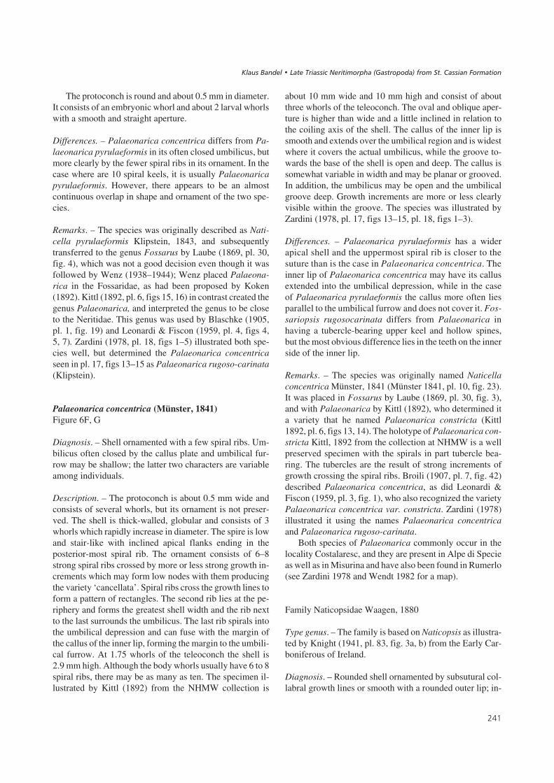
The protoconch is round and about 0.5 mm in diameter.It consists of an embryonic whorl and about 2 larval whorlswith a smooth and straight aperture.
Differences. – Palaeonarica concentrica differs from Pa-laeonarica pyrulaeformis in its often closed umbilicus, butmore clearly by the fewer spiral ribs in its ornament. In thecase where are 10 spiral keels, it is usually Palaeonaricapyrulaeformis. However, there appears to be an almostcontinuous overlap in shape and ornament of the two spe-cies.
Remarks. – The species was originally described as Nati-cella pyrulaeformis Klipstein, 1843, and subsequentlytransferred to the genus Fossarus by Laube (1869, pl. 30,fig. 4), which was not a good decision even though it wasfollowed by Wenz (1938–1944); Wenz placed Palaeona-rica in the Fossaridae, as had been proposed by Koken(1892). Kittl (1892, pl. 6, figs 15, 16) in contrast created thegenus Palaeonarica, and interpreted the genus to be closeto the Neritidae. This genus was used by Blaschke (1905,pl. 1, fig. 19) and Leonardi & Fiscon (1959, pl. 4, figs 4,5, 7). Zardini (1978, pl. 18, figs 1–5) illustrated both spe-cies well, but determined the Palaeonarica concentricaseen in pl. 17, figs 13–15 as Palaeonarica rugoso-carinata(Klipstein).
Palaeonarica concentrica (Münster, 1841)Figure 6F, G
Diagnosis. – Shell ornamented with a few spiral ribs. Um-bilicus often closed by the callus plate and umbilical fur-row may be shallow; the latter two characters are variableamong individuals.
Description. – The protoconch is about 0.5 mm wide andconsists of several whorls, but its ornament is not preser-ved. The shell is thick-walled, globular and consists of 3whorls which rapidly increase in diameter. The spire is lowand stair-like with inclined apical flanks ending in theposterior-most spiral rib. The ornament consists of 6–8strong spiral ribs crossed by more or less strong growth in-crements which may form low nodes with them producingthe variety ‘cancellata’. Spiral ribs cross the growth lines toform a pattern of rectangles. The second rib lies at the pe-riphery and forms the greatest shell width and the rib nextto the last surrounds the umbilicus. The last rib spirals intothe umbilical depression and can fuse with the margin ofthe callus of the inner lip, forming the margin to the umbili-cal furrow. At 1.75 whorls of the teleoconch the shell is2.9 mm high. Although the body whorls usually have 6 to 8spiral ribs, there may be as many as ten. The specimen il-lustrated by Kittl (1892) from the NHMW collection is
about 10 mm wide and 10 mm high and consist of aboutthree whorls of the teleoconch. The oval and oblique aper-ture is higher than wide and a little inclined in relation tothe coiling axis of the shell. The callus of the inner lip issmooth and extends over the umbilical region and is widestwhere it covers the actual umbilicus, while the groove to-wards the base of the shell is open and deep. The callus issomewhat variable in width and may be planar or grooved.In addition, the umbilicus may be open and the umbilicalgroove deep. Growth increments are more or less clearlyvisible within the groove. The species was illustrated byZardini (1978, pl. 17, figs 13–15, pl. 18, figs 1–3).
Differences. – Palaeonarica pyrulaeformis has a widerapical shell and the uppermost spiral rib is closer to thesuture than is the case in Palaeonarica concentrica. Theinner lip of Palaeonarica concentrica may have its callusextended into the umbilical depression, while in the caseof Palaeonarica pyrulaeformis the callus more often liesparallel to the umbilical furrow and does not cover it. Fos-sariopsis rugosocarinata differs from Palaeonarica inhaving a tubercle-bearing upper keel and hollow spines,but the most obvious difference lies in the teeth on the innerside of the inner lip.
Remarks. – The species was originally named Naticellaconcentrica Münster, 1841 (Münster 1841, pl. 10, fig. 23).It was placed in Fossarus by Laube (1869, pl. 30, fig. 3),and with Palaeonarica by Kittl (1892), who determined ita variety that he named Palaeonarica constricta (Kittl1892, pl. 6, figs 13, 14). The holotype of Palaeonarica con-stricta Kittl, 1892 from the collection at NHMW is a wellpreserved specimen with the spirals in part tubercle bea-ring. The tubercles are the result of strong increments ofgrowth crossing the spiral ribs. Broili (1907, pl. 7, fig. 42)described Palaeonarica concentrica, as did Leonardi &Fiscon (1959, pl. 3, fig. 1), who also recognized the varietyPalaeonarica concentrica var. constricta. Zardini (1978)illustrated it using the names Palaeonarica concentricaand Palaeonarica rugoso-carinata.
Both species of Palaeonarica commonly occur in thelocality Costalaresc, and they are present in Alpe di Specieas well as in Misurina and have also been found in Rumerlo(see Zardini 1978 and Wendt 1982 for a map).
Family Naticopsidae Waagen, 1880
Type genus. – The family is based on Naticopsis as illustra-ted by Knight (1941, pl. 83, fig. 3a, b) from the Early Car-boniferous of Ireland.
Diagnosis. – Rounded shell ornamented by subsutural col-labral growth lines or smooth with a rounded outer lip; in-
���
���� ������ ��������� ���������� !�"�� �� ��#���$��%� ���&������

ner lip thickened. Whorls rounded and separated from eachother by deep sutures. Shells may be relatively large. Proto-conch consists of more than two whorls, is about or largerthan 0.5 mm. Where known, ornament consists of sinuouscollabral ribs on its larval whorls which reflect the presenceof a deep median sinus in the outer lip.
Remarks. – The thickening of the inner lip is typical of thegenus that is central to the family (Knight 1941, pl. 83,fig. 3a, b), as well as the ornament of the larval shell (Ban-del 2002, figs 143–147).
Genus Naticopsis M’Coy, 1844
Type species. – The type of the genus is Naticopsis phillip-sii M’Coy, 1844 from the Lower Carboniferous of Ireland(Knight 1941; Wenz 1938–1944, fig. 976).
Diagnosis. – Shell large (up to 60 mm high and wide), withmore than three whorls bearing fine collabral ornament.Whorls smoothly rounded; adult shell consists primarily ofthe body whorl. Sutures deep. Base simple without umbili-cus. Inner lip concave covering the former shell wall in athick callus layer. Protoconch and shell structure of typespecies unknown.
Remarks. – In the case of Naticopsis ventrica (Norwood &Pratten, 1855) from the Late Carboniferous of Texas theprotoconch is known. According to Kues & Batten (2001,figs 12, 1–9), the shell of this species reaches a relativelylarge size (almost 30 mm) but they did not study the proto-conch. The early whorls of the teleoconch are ornamentedby collabral small sharp ribs, which are not developed onlater whorls. Its shape closely matches that of the genotypeof Naticopsis. A protoconch that was considered to belongto that species (Bandel 2002, figs 143–147) was found toconsist of a smooth embryonic whorl of 0.15 mm in diame-ter, succeeded by 1.5 whorls of the larval shell with an orna-ment of sinuous axial lines. The lines reflect the shape of theouter lip that has a deep median sinus and a narrow basallobe. The teleoconch has straight growth lines. The larvalshell measures about 0.6 mm in width and height. Similarprotoconch ornament was found by Herholz (1992) amongmicrogastropods from the Late Carboniferous of Germany.
Among the Neritimorpha of the St Cassian Formation,two new subfamilies, the Ampezzonaticopsinae and theHologyrinae, are here included with the Naticopsidae.
Subfamily Ampezzonaticopsinae n. subfam.
Type genus. – The subfamily is based on Ampezzonaticop-sis n. gen.
Diagnosis. – Low conical small shell with pointed spireand evenly coiled whorls having deep sutures. Larval whorlsof the protoconch conspicuously ornamented by sinuouscollabral axial ribs which reflect a lobed outer lip. Apertureof the teleoconch rounded with a concave callus of the in-ner lip accompanied by an umbilical groove around theopen umbilicus. Shell has a continuous calcitic outer layerand a thicker, internal aragonitic layer of crossed lamellarstructure.
Genera included. – Ampezzonaticopsis n. gen. and Corti-naticopsis n. gen.
Differences. – The larval shell of Hologyra has sinuous ribson the upper and lower side of its whorls ending in a me-dian ribbon, which is not present in Cortinaticopsis n. gen.However, in the case of Ampezzonaticopsis n. gen., the me-dian zone has an ornament of rows of tubercles connectedto some median spiral lines. The larval shell of Cortinati-copsis n. gen. comes closest in shape and ornament to thatof the Pennsylvanian Naticopsis, as illustrated by Bandel(2002). The teleoconch of the Ampezzonaticopsinae has arelatively thin shell compared with that of the Hologyrinae.In addition, another difference is the shape of the umbilicalgroove in the two species of the Ampezzonaticopsinae de-scribed here, which is not partly filled with a ridge as in thecase of Hologyra.
Genus Ampezzonaticopsis n. gen.
Type species. – The genus is based on Natica sublineataMünster, 1841 from the St Cassian Formation.
Diagnosis. – Small shell consisting of rounded whorls setoff from each other by deep sutures in the teleoconch thathas more than three whorls. The last whorl makes up atleast two thirds of spire height. Outer calcitic shell layerhas a lamellar pattern and easily peels off exposing thesmooth surface of the inner aragonitic crossed lamellarlayer. Aperture rounded with the callus of the inner lip bea-ring a groove and an inner margin that is concave. Base ofthe shell opens to a narrow, rounded umbilicus. Ornamentof the larval shell consists of sinuous and collabral rows oftubercles with a median whorl having spirals composed offine tubercles.
Name. – The species that serves as the type to the new genusresembles the Paleozoic genus Naticopsis and comes fromregion of Ampezzo in the Italian Dolomites. Hence, thename Ampezzonaticopsis, which is a combination of both.
Remarks. – The type was determined as Naticella subline-ata by Kittl (1892, pl. 7, figs 41, 42) and is well illustrated
���
����������� ������ �������������
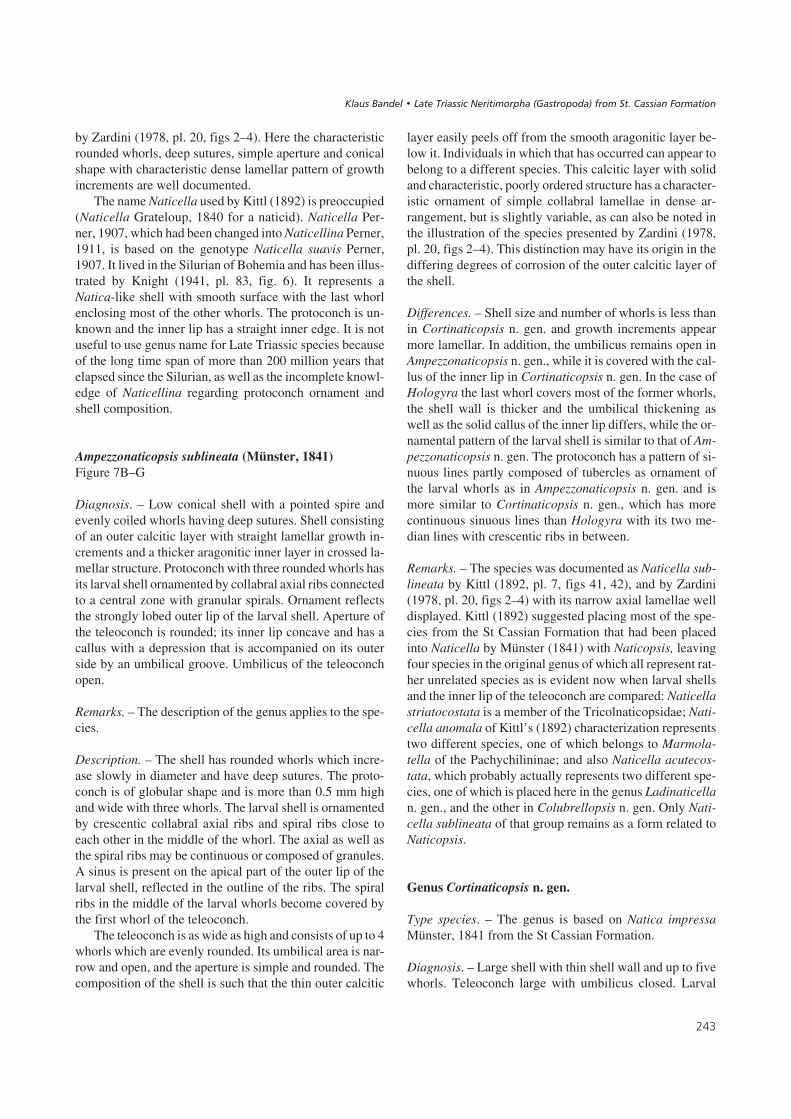
by Zardini (1978, pl. 20, figs 2–4). Here the characteristicrounded whorls, deep sutures, simple aperture and conicalshape with characteristic dense lamellar pattern of growthincrements are well documented.
The name Naticella used by Kittl (1892) is preoccupied(Naticella Grateloup, 1840 for a naticid). Naticella Per-ner, 1907, which had been changed into Naticellina Perner,1911, is based on the genotype Naticella suavis Perner,1907. It lived in the Silurian of Bohemia and has been illus-trated by Knight (1941, pl. 83, fig. 6). It represents aNatica-like shell with smooth surface with the last whorlenclosing most of the other whorls. The protoconch is un-known and the inner lip has a straight inner edge. It is notuseful to use genus name for Late Triassic species becauseof the long time span of more than 200 million years thatelapsed since the Silurian, as well as the incomplete knowl-edge of Naticellina regarding protoconch ornament andshell composition.
Ampezzonaticopsis sublineata (Münster, 1841)Figure 7B–G
Diagnosis. – Low conical shell with a pointed spire andevenly coiled whorls having deep sutures. Shell consistingof an outer calcitic layer with straight lamellar growth in-crements and a thicker aragonitic inner layer in crossed la-mellar structure. Protoconch with three rounded whorls hasits larval shell ornamented by collabral axial ribs connectedto a central zone with granular spirals. Ornament reflectsthe strongly lobed outer lip of the larval shell. Aperture ofthe teleoconch is rounded; its inner lip concave and has acallus with a depression that is accompanied on its outerside by an umbilical groove. Umbilicus of the teleoconchopen.
Remarks. – The description of the genus applies to the spe-cies.
Description. – The shell has rounded whorls which incre-ase slowly in diameter and have deep sutures. The proto-conch is of globular shape and is more than 0.5 mm highand wide with three whorls. The larval shell is ornamentedby crescentic collabral axial ribs and spiral ribs close toeach other in the middle of the whorl. The axial as well asthe spiral ribs may be continuous or composed of granules.A sinus is present on the apical part of the outer lip of thelarval shell, reflected in the outline of the ribs. The spiralribs in the middle of the larval whorls become covered bythe first whorl of the teleoconch.
The teleoconch is as wide as high and consists of up to 4whorls which are evenly rounded. Its umbilical area is nar-row and open, and the aperture is simple and rounded. Thecomposition of the shell is such that the thin outer calcitic
layer easily peels off from the smooth aragonitic layer be-low it. Individuals in which that has occurred can appear tobelong to a different species. This calcitic layer with solidand characteristic, poorly ordered structure has a character-istic ornament of simple collabral lamellae in dense ar-rangement, but is slightly variable, as can also be noted inthe illustration of the species presented by Zardini (1978,pl. 20, figs 2–4). This distinction may have its origin in thediffering degrees of corrosion of the outer calcitic layer ofthe shell.
Differences. – Shell size and number of whorls is less thanin Cortinaticopsis n. gen. and growth increments appearmore lamellar. In addition, the umbilicus remains open inAmpezzonaticopsis n. gen., while it is covered with the cal-lus of the inner lip in Cortinaticopsis n. gen. In the case ofHologyra the last whorl covers most of the former whorls,the shell wall is thicker and the umbilical thickening aswell as the solid callus of the inner lip differs, while the or-namental pattern of the larval shell is similar to that of Am-pezzonaticopsis n. gen. The protoconch has a pattern of si-nuous lines partly composed of tubercles as ornament ofthe larval whorls as in Ampezzonaticopsis n. gen. and ismore similar to Cortinaticopsis n. gen., which has morecontinuous sinuous lines than Hologyra with its two me-dian lines with crescentic ribs in between.
Remarks. – The species was documented as Naticella sub-lineata by Kittl (1892, pl. 7, figs 41, 42), and by Zardini(1978, pl. 20, figs 2–4) with its narrow axial lamellae welldisplayed. Kittl (1892) suggested placing most of the spe-cies from the St Cassian Formation that had been placedinto Naticella by Münster (1841) with Naticopsis, leavingfour species in the original genus of which all represent rat-her unrelated species as is evident now when larval shellsand the inner lip of the teleoconch are compared: Naticellastriatocostata is a member of the Tricolnaticopsidae; Nati-cella anomala of Kittl’s (1892) characterization representstwo different species, one of which belongs to Marmola-tella of the Pachychilininae; and also Naticella acutecos-tata, which probably actually represents two different spe-cies, one of which is placed here in the genus Ladinaticellan. gen., and the other in Colubrellopsis n. gen. Only Nati-cella sublineata of that group remains as a form related toNaticopsis.
Genus Cortinaticopsis n. gen.
Type species. – The genus is based on Natica impressaMünster, 1841 from the St Cassian Formation.
Diagnosis. – Large shell with thin shell wall and up to fivewhorls. Teleoconch large with umbilicus closed. Larval
���
���� ������ ��������� ���������� !�"�� �� ��#���$��%� ���&������

shell has an ornament of sinuous axial ribs with a characte-ristic strong median lobe and sinus. Continuous and thinouter calcitic layer and inner aragonitic layer with crossedlamellar structure.
Differences. – The teleoconch closely resembles that ofAmpezzonaticopsis but attains larger size and has a closedumbilicus.
Name. – The genus name is a combination of Cortina (afterthe Cortina d’Ampezzo) and Naticopsis.
Cortinaticopsis impressa (Münster, 1841)Figure 7A, H–L
Diagnosis. – Shell with regular narrow growth incrementsstrongly imprinted in the outer calcitic layer and having anaperture with a rounded inner lip covered by a narrow cal-lus ribbon with a groove in its middle. Callus covers theconcave umbilical area. Protoconch consists of three whorlsand teleoconch of up to five whorls and larval whorls orna-mented by crescentic axial ribs with a narrow, strong me-dian sinus.
Description. – The shell may be 8 mm wide and 7 mm highwith about three whorls in the teleoconch, in the case of thespecimen determined by Kittl (1892) and studied from theNHMW collection. The shells of large individuals haveusually been crushed by compaction, and they may reach asize of up to 20 mm in size. Whorls are rounded and havecollabral growth increments with a short distance fromeach other, sometimes somewhat irregular. The aperture issemicircular with a straight inner lip and rounded outer lip.Callus of the inner lip consists of a ribbon that in its apicalportion is a little wider than it is further towards the baseand bears a shallow rounded groove in its middle. Next tothe callus lies a wide umbilical furrow that starts in the na-rrow umbilicus, which is covered by the apical portion ofthe callus. The thin callus of the inner lip covers the con-cave umbilical area and fully-grown shells consist of up to5 whorls in the teleoconch.
The ornament of the protoconch is characterized by
collabral ribs on the larval shell that reflect the shape of thelobe and sinus on the outer lip. The protoconch consists ofabout three rounded whorls and is about 0.6 mm high andwide. The succeeding teleoconch has straight incrementsof growth. The species occurs commonly at localityCampo, where the shells are often more or less deformed.
Remarks. – The shell illustrated by Kittl (1892, pl. 7,figs 16, 17) resembles that illustrated by Zardini (1978,pl. 19, fig. 21). Natica berwerthi Kittl, 1892 is also describedas thin and with a closed umbilicus. The large shell of aspecies determined as Solarioconulus nudus (Münster,1841) and illustrated by Zardini (1978, pl. 11, fig. 11) hasrounded whorls bearing the original color pattern of zigzaglines, deep sutures, a wide base and an umbilicus closed bythe callus of the inner lip. Specimens from Misurina andAlpe di Specie also probably also belong to this species ha-ving relatively large and thin-walled shells that are usuallydeformed and crushed.
Subfamily Hologyrinae n. subfam.
Type genus. – The subfamily is based on the genus Holo-gyra Koken, 1892 as redefined from the St Cassian Forma-tion.
Diagnosis. – Shell has rounded whorls with a low spireand large final whorl. Inner walls preserved and outer wallsare thick. Protoconch has a larval shell with an ornamentof collabral ribs on the lower and upper side, interruptedin the middle of the whorl by two distinct spiral lines,which have curving ribs between them forming a chevron.Callus of the inner lip of the teleoconch lies next to acolumn-like ridge composed of growth increments; thisarticulated ridge extends from the umbilical furrow to thebase, ending before reaching the margin of the outer lip.Operculum calcareous and with angular inner side bea-ring two rounded attachment scars and a rounded outerside; the latter has an inner triangular portion with straightincrements of growth and a semicircular outer part withrounded concentric growth lines, the nucleus lying in thepoint of the triangular part.
���
��#����0% A – slightly deformed specimen of Cortinaticopsis impressa (Münster, 1841). Shell about 11 mm wide (BSPG 2007 XVI 44).• B – protoconch of Ampezzonaticopsis sublineata (Münster, 1841) with shell shown about 0.7 mm high. • C – larval shell of Ampezzonaticopsissublineata (Münster, 1841) with ornament and about 0.5 mm high (BSPG 2007 XVI 41). • D – protoconch of Ampezzonaticopsis sublineata (Münster,1841) in early teleoconch, about 0.8 mm high (BSPG 2007 XVI 42). • E – apical view of the protoconch of Ampezzonaticopsis sublineata (Münster, 1841)of the same specimen as in Fig. 7D. • F – apical view of juvenile shell of Ampezzonaticopsis sublineata (Münster, 1841). Shell about 0.7 mm wide (BSPG2007 XVI 43). • G – Ampezzonaticopsis sublineata (Münster, 1841). The same shell (about 0.7 mm high) as in Fig. 7F, but in apical view. • H – juvenileshell of Cortinaticopsis impressa (Münster, 1841). Shell almost 3 mm high (BSPG 2007 XVI 45). • I – the same shell of Cortinaticopsis impressa(Münster, 1841) as in Fig. 7H seen from above. Shell is 2.8 mm wide. • J – Cortinaticopsis impressa (Münster, 1841). Shell about 1.5 mm wide (BSPG2007 XVI 46). • K – protoconch of Cortinaticopsis impressa (Münster, 1841) with the initial portion of the teleoconch about 1.2 mm high (BSPG 2007XVI 47). • L – larval shell of Cortinaticopsis impressa (Münster, 1841) in a juvenile specimen. Shell about 0.8 mm high (BSPG 2007 XVI 48).
����������� ������ �������������
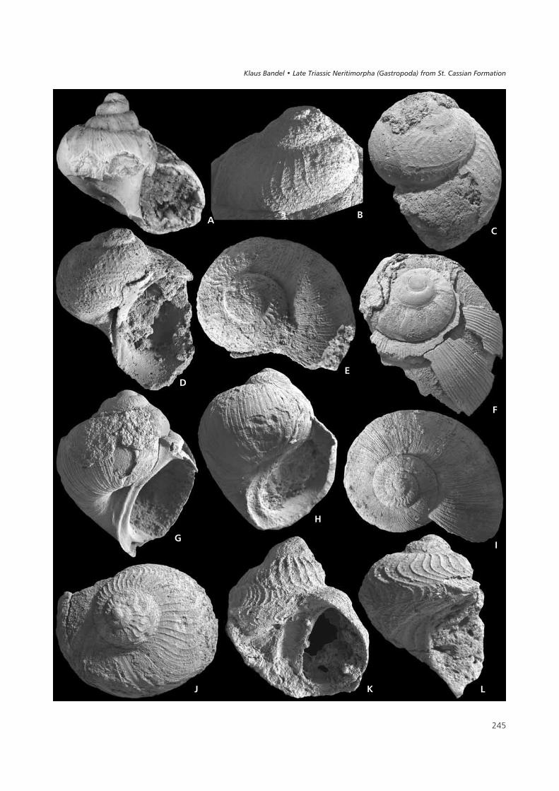
��
(
�
�
&
�
�
'
�
) * �
���� ������ ��������� ���������� !�"�� �� ��#���$��%� ���&������
Differences. – The protoconch has a characteristic orna-ment on the larval shell and a ridge in the umbilical grooveof the teleoconch, which distinguishes the Hologyrinaefrom all other Neritimorpha of the St Cassian Formationwith globular shells. The larval shell of the Ampezzonati-copsinae also has sinuous axial ribs, but the shape of the te-leoconch differs in its relatively higher spire, a less thickshell wall, concave umbilical furrow, and deeper sutures.The chevron in the middle of the larval whorls is foundonly in Hologyra. The Fedaiellidae with similar, solidrounded shells have a lowly rounded protoconch that re-sembles that of the Cassianopsinae, in addition to the angu-lar groove on their inner lip of the teleoconch. The Neritari-idae with Neritaria mandelslohi also has individuals with arelatively large rounded shell, but the lowly rounded proto-conch also has low axial ribs. The shell has its inner whorlsdissolved.
Remarks. – Zardini (1978, pl. 21, figs 5–7) illustrated thetwo characteristic muscle attachment scars. The operculumpreserved in Hologyra cassiana has the same general fea-tures as that known as Hypodema Koninck, 1853, which isconnected to a Naticopsis from the Carboniferous (Gordon& Yochelson 1982). It consists of a shield-shaped triangleat the base and on top of it a rounded part with concentricgrowth lines with their center present in the point of the tri-angle (Knight 1941, pl. 83, fig. 5); Knight suggested thatthe operculum belongs to Naticopsis, which occurs in thesame Lower Carboniferous beds at Visé in Belgium. In ad-dition, Kittl (1892) compared the operculum of the Carbo-niferous Naticopsis that was described by Koninck (1853)from Lower Carboniferous deposits with that of Hologyracassiana from the St Cassian Formation.
Genus Hologyra Koken, 1892
Type species. – The genus is based on Natica cassianaWissmann, 1841 from the St Cassian Formation. The choiceof a different type as originally suggested by Koken (1892)is discussed below.
Diagnosis. – Globular shell with a short spire and straightincrements of growth. Umbilical furrow in juvenile shellsis bordered by an inner lip with a sharp outer keel andin it a ridge composed of growth increments; ridge inmature shells can be covered by the thick callus of the in-ner lip. During shell growth teleoconch morphologychanges from highly rounded juvenile shell to broadlyrounded adult shell. Protoconch characteristized by twomedian spiral lines and axial ribs between forming a rib-bon of crescentic lines with the same spacing as is foundin the sinuous axial ribs on whorls above and below.Vertical aperture of the teleoconch can be sealed with acalcareous operculum that has a triangular base next tothe inner lip and an outer side bearing concentric growthlines.
Differences. – Kittl (1892) characterized this genus and no-ted that the pillar in the umbilical furrow may be more orless callus covered. Thus, Naticopsis cassiana merges withNaticopsis expansa Laube, 1869, which represent differentgrowth stages of Hologyra and also reflect different de-grees of corrosion of the shell. The pillar in the umbilicalfurrow distinguishes it from Fedaiella, and the latter genusin addition has a groove bordered by teeth on the inside ofits inner lip. Among the Neritimorpha from the St CassianFormation, only Hologyra has an umbilical ridge withgrowth increments.
Hologyra cassiana (Wissmann, 1841)Figure 8A–M
Diagnosis. – Rounded shell quite large with inner whorlsnot dissolved (checked by cutting shells). Solid callus plateof the inner lip connects to or may cover the roundedplug-like ridge that fills the inner side of the umbilical fur-row; the column is small in juvenile shells, well developedwith growth increments in larger shells and may be coveredby the callus plate in fully-grown shells of older individu-als. Operculum has a triangular basal part and outer portionwith rounded growth lines.
���
��#����1% All figures (A–M) illustrate Hologyra cassiana (Wissmann, 1841). • A – operculum is present with its nucleus in the outer half. The shell isabout 2 mm wide (BSPG 2007 XVI 49). • B – operculum of an older individual with a shell about 5 mm wide (BSPG 2007 XVI 50). • C – the large tri-angular basal part of the operculum is almost smooth, while the outer part is rounded and concentric (BSPG 2007 XVI 51). • D – the callus of the innerlip is accompanied by a ridge in the umbilical groove next to it. Shell about 6 mm wide (BSPG 2007 XVI 52). • E – juvenile shell with the first whorl ofthe teleoconch still with indistinct umbilical ridge and the visible larval ornament. Shell 1.8 mm high (BSPG 2007 XVI 53). • F – older shell with callusof the inner lip thickened and covering most of the umbilical groove and its ridge. Shell about 9 mm in diameter (BSPG 2007 XVI 54). • G – the umbili-cal ridge (funiculus) and the groove next to it with strong growth increments. Shell about 5 mm high (BSPG 2007 XVI 55). • H – the larval shell with itscharacteristic ornament of collabral ribs and two median lines with pattern of lunate ribs between them. The juvenile teleoconch is about 1.8 mm high(BSPG 2007 XVI 56). • I – detailed view of the larval ornament, as in Fig. 8H. • J – protoconch connected to the teleoconch with the characteristic pat-tern of the genus. The shell is 1.8 mm wide. • K – a shell with 3 mm in height and with the calcitic layer with lamellae and the bulk crossed lamellar inthe thick wall (BSPG 2007 XVI 57). • L – juvenile shell as in Fig. 8K seen from the apex. • M – the protoconch of about 0.5 mm in its teleoconch, de-tailed view of Fig. 8L.
����������� ������ �������������

���
( �
�
&�
�
'
�
)
* � �
���� ������ ��������� ���������� !�"�� �� ��#���$��%� ���&������

Description. – The solid teleoconch is of globular shape,about as wide as high and consists of about four whorls. Itmay be up to 25 mm in width and height. A shell with 2.75whorls of the teleoconch is about 4 mm wide and less than4 mm high, with a spire less than 0.5 mm high. The bodywhorl covers most of the former whorls. Ornament consistsof thread-like growth increments which are straight in earlywhorls, and later develops a slight curvature. The evenlyrounded outer lip ends with an anterior notch. It forms agroove between the outer lip and the anterior part of the in-ner lip. The aperture has a semicircular shape and obliqueorientation to the axis of coiling of the shell. The inner lipforms a flat callus that ends next to the umbilical groovewhich has the ridge of the so-called “funiculus” (columel-lar ridge – see below) in it (Kittl, 1892). This ridge has acharacteristic pattern of step-like increments of growth.Due to the change in callus dimension during growth of theshell this umbilical ridge may be more or less covered by it.This results in the description of a number of supposed spe-cies centered around this species which differ by callusshape and are just varieties. Ornamental patterns of darkspiral zigzag ribbons may be preserved on the shell (Kittl1892, pl. 7, fig. 16; Tichy 1980, pl. 3, fig. 9).
The protoconch is about 0.5 mm wide and high and inpart covered by the first whorl of the teleoconch, exposingthe ornament of its larval shell only up to the apical spiralline in the middle of the whorl. The embryonic whorl isabout 0.1 mm wide and is succeeded by about 1.75 larvalwhorls with an ornament of about 40 collabral ribs on onewhorl. Axial ribs have a curving shape reflecting the aper-ture and on the last half whorl have a spiral rib just abovethe suture and a second spiral rib which is covered by thesucceeding whorl. Short axial ribs form a chevron-like rib-bon between the spiral ribs.
The operculum is constructed of calcitic material andhas a darker color than the shell. Its semicircular shapematches that of the aperture. Its inner side reflects the con-cave curvature of the inner lip. With the exception of thegranular, inner marginal rounded area, the outer side of theoperculum has a fine pattern of growth increments. The nu-cleus lies eccentrically near the middle but towards theouter lip. From its growth increments can be followed theoutline of the outer lip, but it ends in a triangular inner zoneor continues here as weakly developed straight growth in-crements. The inner side of the operculum is well illus-trated by Zardini (1978, pl. 21, figs 5–7). Here a medianridge is accompanied on both sides by grooves with a con-centric pattern. Its outer margin is smooth and thickened.
Differences. – Hologyra cassiana is distinguished from theother species of the “Naticopsis” group by the presence ofan articulated column in the umbilical groove. This charac-ter also distinguishes it from Neritaria mandelslohi, whichalso has a strong tooth on the inner side of the inner lip near
the base of its aperture. Both species attain large size, buthave very different shaped opercula. In addtion, Neritariamandelslohi dissolves its inner shell walls. The operculumof Hologyra cassiana is distinguished from that of the ot-her species by having a more marginal position of the nuc-leus and concentric furrows on its outer side.
Remarks. – Natica cassiana of Wissmann (1841 inMünster 1841, pl. 10, fig. 3) was also documentedby Laube (1869) who, in addition, described probablythe same species also as Natica expansa Laube, 1869and Natica althusii Klipstein, 1843. Kittl (1892, pl. 7,figs 18–21, pl. 9, figs 9, 10) called it Naticopsis cassiana,but also determined a Naticopsis expansa. He suggested arelationship with Naticopsis impressa (Münster, 1841),and Naticopsis involuta Kittl, 1892, which have no pillar inthe columellar furrow. He distinguished a whole group ofspecies to comprise the Naticopsis cassiana- like species ofthe St Cassian fauna. Kittl (1892) recognized the relation-ship with Naticopsis due to an operculum that had beendescribed by Koken (1889) from Naticopsis deshayesiKlipstein, 1843. This latter species also belongs here sinceit has the columellar ridge (funiculus). In Koken (1892) asimilar shell was called Hologyra alpina Koken, 1892 andHologyra carinata Koken, 1892. Böhm (1895) used Holo-gyra alpina, and created Hologyra kokeni Böhm, 1895.The species from Seiser Alp was determined as Neritariacassiana by Blaschke (1905, pl. 20, fig. 11) and also byBroili (1907, pl. 9, figs 8, 12, 15). Leonardi & Fiscon(1959, pl. 3, fig. 3) called it Hologyra expansa, as did Zar-dini (1978, pl. 19, figs 6, 10–12, 23, 24; 1985, pl. 8, fig. 3).Zardini (1978, pl. 21, figs 5, 6, 10) also described its oper-culum. Similar individuals from the Muschelkalk werecalled Hologyra noetlinigi Picard, 1903 and Hologyra bey-erichi Picard, 1903 and of these Hologyra noetlingi wasconfirmed by Gründel (1980, pl. 1, figs 9, 10). Knight et al.(1960, fig. 181, 4a, b) suggested Hologyra to be a synonymof Dicosmos Canavari, 1890.
Family Tricolnaticopsidae n. fam.
Type genus. – Tricolnaticopsis n. gen. The family is basedon Tricolnaticopsis striatulus (Münster, 1841) from the StCassian Formation.
Diagnosis. – Smooth shell is higher than wide. Callus ofthe inner lip extends into the umbilical area and is convexon its margin to the aperture; basal part of the callus accom-panied by a narrow furrow with a sharp outer margin. Thinouter calcitic layer underlain by a thick uniform aragoniticlayer in crossed lamellar structure. Aperture shaped to ac-commodate a bean-shaped operculum, calcitic with twomuscle attachment scars on its inner side and no inner pro-
���
����������� ������ �������������

jection, with a rounded thickened margin; nucleus lies atthe inner basal margin and is visible on the outer side.Operculum grows by concentric increments of uneventhickness.
Remarks. – The type species Tricolnaticopsis striatulus(Münster, 1841) was documented as Natica pseudospirataby Zardini (1978) from the St Cassian Formation. Two spe-cies of Pachyomphalus are considered to belong here, dueto the shape of their aperture into which a similarbean-shaped operculum can be fitted. Some species whichresemble the modern vetigastropod Tricolia in having asmooth shell and preserved inner whorls are here included,such as the slender Natica pseudospirata with roundedwhorls, here described as Tricolnaticopsis striatulus. Pa-chyomphalus uhligi with less rounded whorls as describedby Kittl (1892) and the shorter Pachyomphalus angustus asdescribed by Laube (1869) are also similarly characterized.The apertural rim of Rinaldopsis ampezziensis resemblesthat in Pachyomphalus and may indicate a relationship tothe Tricolnaticopsidae.
Differences. – The genus Vernelia Böhm, 1895 is based onVernelia excelsa (Hauer) and has similar shell shape, but isof much larger size (Wenz 1938–1944, fig. 892). Rinaldo-psis as illustrated in Zardini (1978, pl. 30, figs 13, 14,pl. 31, figs 3–5) has a tricoliform shell shape, but has aninner lip that forms a wide callus. It differs also by itsmore irregular growth, while the shape of the apertureshows similarities with that of the Tricolnaticopsidae.
Genus Tricolnaticopsis n. gen.
Type species. – The type is Tricolnaticopsis striatulus fromthe St Cassian Formation, locality Seelandalpe (Alpe diSpecie), as illustrated by Zardini (1978, pl. 20, figs 17, 19,pl. 21, figs 2–4).
Diagnosis. – Shell higher than wide. Rounded whorls com-pose the shell with the final whorl higher than the spire.Protoconch consists of three rounded whorls with a straightmargin in the larval shell. Shell composition with a thincalcitic outer layer and a thick inner aragonitic layer. Colorpattern consisting of rounded dots arranged in rows or zig-zag lines can be preserved. Inner lip of the oval aperture isconvex and forms a callus pad that covers the umbilicalarea.
Genus name. – The name is derived from the Tricolia-likeshape of this member of the Neritimorpha which showscharacters of Naticopsis group as documented by Kittl(1892). As such, the name represents a combination of bothtaxonomic names.
Tricolnaticopsis striatulus (Münster, 1841)Figures 9A, B, N, 10A
Diagnosis. – Smooth shell resembles that of a Tricolia andhas a continuous thin calcitic surface that bears coloreddots. Aperture is rounded and the last whorl is higher thanthe spire. Callus of the inner lip has a convex bulge into thespace of the aperture and forms a callus pad that covers theumbilicus. Rounded whorls of the teleoconch with deep su-tures. Protoconch conical and its whorls rounded.
Description. – The shell consists of 4 whorls in the teleo-conch and is about 7 to 8 mm high and about 6 mm widewith an apical angle of about 50–60°. Whorls are rounded,smooth, and separated by deep sutures. The outermost layerof the shell is a very thin but continuous layer of calcite,which commonly preserves the original color pattern ofdots that are commonly arranged in zigzag lines orientedaxially. This pattern varies strongly among the few indivi-duals that were studied. The inner aragonitic layer is thickand consists predominantly of crossed lamellar structure.The orientation of the main lamellae is parallel to the outerlip. The callus of the inner lip is simple and smooth. It co-vers the umbilicus and forms a rounded pad and expands tothe inner margin of the aperture and forms a rounded lobe.The inner walls of the shell are not dissolved.
The protoconch consists of more than two roundedwhorls and is about 0.5 mm wide and high. The ornamentof its larval whorls is not preserved.
Differences. – This species was well illustrated and descri-bed by Zardini (1978, pl. 20, figs 17, 18, pl. 21, figs 2–4)and was determined by him as Natica pseudospirata. Someof the individuals described as Natica argus Kittl, 1892 byKittl (1892, pl. 7, figs 44–46) might belong here. The shapeand size of Tricolnaticopsis striatulus resembles that ofPachyomphalus uhligi, but differs in having a relativelyhigher spire, deeper sutures, a more regular callus of the in-ner lip, an ornament of dots and smoother and more evenlycurved outer shell surface.
Remarks. – Natica argus Kittl, 1892 as illustrated by Kittl(1892, pl. 7, figs 44–46) appears to represent the speciesthat was illustrated by Zardini (1978) and determined asNatica pseudospirata. Kittl (1892) described his Natica ar-gus as connected to a protoconch with whorls very tight intheir coiling mode. This is not so in Tricolnaticopsis stria-tulus, which has a protoconch of the ordinary globular androunded shape as usual among the Neritimorpha of St Cas-sian. Natica pseudospirata Orbigny, 1849 was characteri-zed by Kittl (1892, pl. 7, fig. 43) as having a wider shell andless imprinted sutures, as in his Natica argus, which hascolored dots that are typical for Tricolnaticopsis striatulusas documented by Zardini (1978, pl. 21, fig. 2).
���
���� ������ ��������� ���������� !�"�� �� ��#���$��%� ���&������
Genus Pachyomphalus Böhm, 1895
Type species. – The genus is based on Pachyomphalus con-cinnus Böhm, 1895 from the Marmolata Limestone.
Diagnosis. – Conical egg-shaped shell with a pointed apexabout 6 mm high and rounded whorls with impressed sutu-res. Growth lines straight and body whorl occupies abouttwo-thirds shell height. Aperture egg-shaped forming anapical angle. Umbilical area covered by callus of the innerlip. Outer lip rounded and inner lip slightly convex andsmooth.
Differences. – Vernelia Böhm, 1895 is based on Natica ex-celsa Hauer from the Esino Limestone and it has apointed-egg-shaped shell similar to that of Pachyompha-lus, but is much larger (Wenz 1938–1944, fig. 982). Itsaperture is oval with a flat callus of the inner lip that isslightly concave and smooth on its inner side. Vernelia dif-fers from both Pachyomphalus and Tricolnaticopsis n. gen.in its concave inner lip.
Pachyomphalus angustus (Münster, 1841)Figure 9G–I, M, O
Diagnosis. – Shell wall thick and a little higher than wide.Outer shell wall consists of a thin calcitic layer and an innershell wall that is aragonitic with crossed lamellar structure.Margin of the aperture is thick with the inner lip accompa-nied by a marginal groove; inner lip forms a median lobethat covers the umbilicus and produces a convex shape onthe inner side towards the aperture. Inner walls of the shellnot dissolved. Protoconch rounded consisting of threewhorls which overlap with their margins.
Description. – The protoconch is about 0.4 mm wide withembryonic whorl succeeded by about two larval whorlswhich overlap onto each other. Their ornament is not pre-
served. The shell of the teleoconch is thick. Its shape is ob-liquely egg-like with rounded whorls and weak sutures,with the three whorls of the teleoconch being about 3 mmhigh. The body whorl covers most of the first teleoconchwhorl and only the protoconch extends over it in an apicaldirection. Ornament consists of growth lines with a lowlobe below the suture and later on with a straight outline.The collabral lines form a pattern of fine regular groovesseparated from each other by slightly wider smooth zones.The aperture is slightly inclined in relation to the shell axisand a little higher than wide; it is relatively small due to thethick callus of the inner lip and the convexity present in themiddle of the edge. The outer lip is rounded, thickened, andsmooth. In basal position the callus of the inner lip is ac-companied by an outer groove that has a marginal rim. Inmedian position the callus covers the umbilicus and conti-nues to the apical corner of the inner lip. The callus endsbefore it reaches the outer lip. With two whorls of the teleo-conch the shell is about 2 mm wide and 2.5 mm high, andthe fully-grown shell consists of more than three whorls ofthe teleoconch and is about 5 mm high and 4 mm wide.
The thin outer calcitic layer of the shell has a well de-veloped “ice flower” pattern formed by thin crystallitesof calcite spread out with general direction of their apicestoward the aperture. Shell walls are composed predomi-nantly of a thick aragonitic layer in crossed lamellar struc-ture. In cases where the color pattern is preserved, it con-sists of small irregular dots of light color on a darkbackground.
No operculum has been found in situ, but an operculumthat would fit into the aperture according to shape and size isthick, with an oval, bean-like shape and having a convexouter surface. The side that lies next to the inner lip forms alow sinus. The eccentric nucleus lies near the outer edge andis the center to which concentric lines with different thick-ness are added. The inner side of the operculum is occupiedby two pits separated by a broad ridge between them whichis developed on the inner side. The inner surface is thus di-vided into two unequal fields (Zardini 1978, pl. 21, fig. 12).
� �
��#����2% A – apical view of Tricolnaticopsis striatulus (Münster, 1841) with the same shell as seen in side view in Fig. 9B (BSPG 2007 XVI 58).• B – side view of Tricolnaticopsis striatulus (Münster, 1841). Same shell as that showing shell structure in Fig. 9N. The shell is about 8 mm high.• C – fully-grown Pachyomphalus uhligi (Kittl, 1892) is about 9 mm high (BSPG 2007 XVI 59). • D – the same shell as in Fig. 9C, Pachyomphalus uhligi(Kittl, 1892). • E – a more juvenile shell of Pachyomphalus uhligi (Kittl, 1892) with the callus less expanded than in C. The shell is about 8 mm high(BSPG 2007 XVI 60). • F – Pachyomphalus uhligi (Kittl, 1892) as in Fig. 9E with a shell about 6 mm high. View from behind. • G – the shell ofPachyomphalus angustus (Münster, 1841). Shell about 3 mm high, its apex is shown in Fig. 9I (BSPG 2007 XVI 61). • H – Pachyomphalus angustus(Münster, 1841) with bulging inner lip and shell as in Fig. 9O. The shell is about 2.5 mm high (BSPG 2007 XVI 62). • I – the apex of the shell ofPachyomphalus angustus (Münster, 1841) shown in Fig. 9G with a rounded protoconch and with about three whorls. • J – Rinaldopsis ampezzana n. sp.has strong and irregular increments of growth. The shell is a little inclined and was crushed, with diameter about 5 mm (BSPG 2007 XVI 63). • K – slightlycorroded Rinaldopsis ampezzana n. sp. showing the wide callus of the inner lip. The shell is about 11 mm high (BSPG 2007 XVI 64). • L – Rinaldopsisampezzana n. sp. as in Fig. 9K seen from behind. • M – apex of Pachyomphalus angustus as in Fig. 9I with the larval shell ending with a lobed outer lip andprotoconch with three whorls about 0.4 mm wide. • N – shell of Tricolnaticopsis striatulus (Münster, 1841) having a very thin calcitic outer layer with theincrements of growth forming lamellae, and a thick inner crossed lamellar layer organized with the direction of its lamellae parallel to increments ofgrowth on the margin of the outer lip. The shell is about 0.3 mm thick. • O – this shell of Pachyomphalus angustus (Münster, 1841) has a thin continuousouter calcitic layer and the crossed lamellar layer below forming the bulk. The shell is about 0.3 mm thick.
����������� ������ �������������

� �
( � �
&
�� '
� )*
�
� � 3
���� ������ ��������� ���������� !�"�� �� ��#���$��%� ���&������
Differences. – The shell in Pachyomphalus uhligi and Tri-colnaticopsis striatulus is higher and a little more elongatethan that of Pachyomphalus angustus. Diener (1926) sug-gested that Pachyomphalus angustus, which he determinedas Naticopsis kinkelini represents a member of Hologyra,but this is not confirmed. Pachyomphalus angustus differsfrom Hologyra cassiana by having a quite different shapeof its callus and the umbilical groove and also by a higherand more pointed shell. The operculum that fits into theaperture of Pachyomphalus differs from that of other spe-cies from St Cassian by its elongate narrow bean-likeshape.
Remarks. – This species was originally described as Naticaangusta Münster, 1841, and illustrated again by Laube(1869) with the individual present in the collection atNHMW. It was later described as Naticopsis kinkeliniKittl, 1892 (Kittl 1892, pl. 7, fig. 25) and the same authoralso described and illustrated a Natica angusta based on aspecimen that appears to represent juvenile shells of hisNaticopsis kinkelini. A typical Pachyomphalus angustuswas illustrated as Naticopsis kinkelini by Zardini (1978,pl. 20, fig. 1).
Pachyomphalus uhligi (Kittl, 1892)Figure 9C–F
Diagnosis. – Egg-shaped shell has a smooth surface andwell-expressed, but not deep sutures. Body whorl aboutfour times higher than the spire. Callus of the inner lipforms a thickened elongate ribbon that covers the umbilicalregion and is accompanied on its basal portion by a narrowumbilical groove. Aperture oval with a convex inner lip.
Description. – The teleoconch consists of at least four who-rls and the shell is about 10 mm high and 7 mm wide. It iscovered by a thin but continuous outer calcitic layer and athick inner aragonitic crossed lamellar layer. Here the lar-gest lamellae are oriented parallel to the apertural margin.In case where preserved color patterns exist, they consist offine brown zigzag lines in an axial orientation. The aper-ture is oval and relatively small. The inner lip is connected
to the callus that covers the umbilical region. The callus issmooth and forms a ribbon that in old individuals has anelongate callus pad. The inner lip has a slightly convexshape. The outer lip is rounded with its margin at the basecontinuous with a shallow groove next to the base of thecallus. The outer lip is narrower in fully-grown specimensthan it is in juveniles.
Differences. – Tricolnaticopsis striatulus has deeper sutu-res, a relatively larger spire, a similar callus and outer shelllayer, a different ornament of colored dots and a smootherouter surface. Pachyomphalus angustus (= kinkelini) is re-latively shorter. A ribbon basal of the inner lip callus as inPachyomphalus uhligi is not mentioned in the diagnosis ofVernelia (Wenz 1938–1944), and the shell of the latter is atleast double the size of the former.
Remarks. – The specimen studied by Kittl (1892) was illus-trated from the collection of NHMW. This shell is about9 mm high and 6 mm wide with a Bithynia-like shape andthe spire that is less than one fourth of the body whorl inheight. Pachyomphalus uhligi had been suggested as Pha-sianella uhligi by Klipstein (in manuscript according toKittl 1892) but was published as Naticopsis uhligi by Kittl(1892, pl. 7, fig. 26).
Genus Rinaldopsis n. gen.
Type species. – The genus is based on Rinaldopsis ampez-ziensis that is illustrated by Zardini (1978, pl. 31, fig. 3a, b)and determined as Oonia subtortilis from Alpe di Specie,St Cassian Formation.
Diagnosis. – Egg-like shaped shell with a pointed spireand body whorl about four times larger than earlier whorls.Aperture has a flat shelf-like structure on the inner lip en-compassing about half of its size; shelf has a narrow andraised rim on the inner lip and extends into the narrow um-bilicus.
Name. – The type to the genus was well documented by Ri-naldo Zardini (1978) and is named in his honor.
� �
��#����$4% A – juvenile shell of Tricolnaticopsis striatulus (Münster, 1841) with evenly rounded whorls and thick callus plate. Shell about 2 mm high(BSPG 2007 XVI 65). • B – juvenile shell of Rinaldopsis ampezziensis n. sp. with a 1.8 mm high shell and conical umbilicus (BSPG 2007 XVI 66). • C – ju-venile shell of Laubopsis haudecarinata (Münster, 1841) with open umbilicus and about 1.5 mm in height (BSPG 2007 XVI 67). • D – Laubopsishaudecarinata (Münster, 1841) with rounded whorls and open umbilicus. Shell is 15 mm high (BSPG 2007 XVI 68). • E – Ladinaticella münsteriana(Orbigny, 1849) with a 5 mm high shell (BSPG 2007 XVI 69). • F – apical view of Ladinaticella münsteriana (Orbigny, 1849), same as in Fig. 10E.• G – protoconch of Ladinaticella münsteriana (Orbigny, 1849) with fine axial lines on the larval shell. Diameter about 0.5 mm. • H – juvenile shell ofLadinaticella costata (Münster, 1841). Shell about 1.4 mm high (BSPG 2007 XVI 70). • I – Ladinaticella costata (Münster, 1841). The same shell as inFig. 10H in apical view with a rounded, smooth protoconch. • J – Ladinaticella costata (Münster, 1841) with a 2.5 mm high shell (BSPG 2007 XVI 71).• K – Ladinaticella costata (Münster, 1841). The same shell as in Fig. 10J. • L – Ladinaticella striatocostata (Münster, 1841). Shell 10 mm high (BSPG 2007XVI 73). • M – Ladinaticella striatocostata (Münster, 1841). Shell 10 mm high. • N – Ladinaticella striatocostata (Münster, 1841). Shell 10 mm high.
����������� ������ �������������
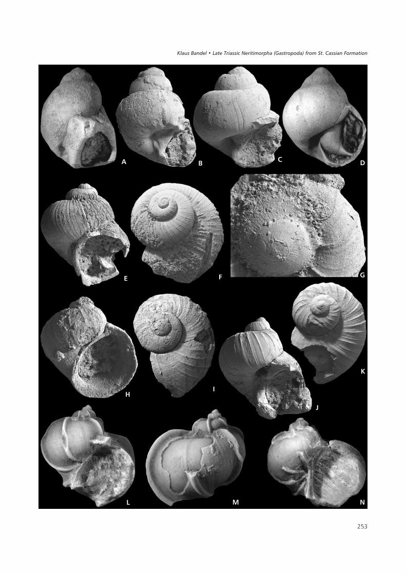
� �
(�
�
& � �
'�
*
)
� � �
���� ������ ��������� ���������� !�"�� �� ��#���$��%� ���&������
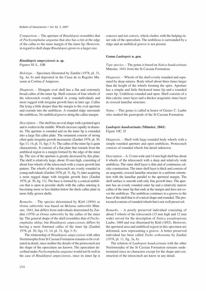
Comparison. – The aperture of Rinaldopsis resembles thatof Pachyomphalus angustus that also has a rim at the edgeof the callus to the inner margin of the inner lip. However,in regard to shell shape Rinaldopsis grows to a larger size.
Rinaldopsis ampezziensis n. sp.Figures 9J–L, 10B
Holotype. – Specimen illustrated by Zardini (1978, pl. 31,fig. 4a, b) and deposited in the Ciasa de ra Regoles Mu-seum at Cortina d’Ampezzo.
Diagnosis. – Elongate oval shell has a flat and extremelybroad callus of the inner lip. Shell consists of four whorls ofthe teleoconch evenly rounded in young individuals andmore rugged with irregular growth lines in later age. Callusflat lying a little deeper than the margin to the oval apertureand extends into the umbilicus. A rounded ridge surroundsthe umbilicus. No umbilical groove along the callus margin.
Description. – The shell has an oval shape with a pointed apexand is widest in the middle. Whorls increase rapidly in diame-ter. The aperture is rounded and on the inner lip is extendedinto a large flat callus plate. The ornament consists of strongoften quite irregular growth increments (Zardini 1978, pl. 30,figs 13, 14, pl. 31, figs 3–5). The callus of the inner lip is quitecharacteristic. It consists of a flat plate that extends from theumbilical region to a margin formed by the edge of the innerlip. The size of the aperture is greatly decreased by this plate.The shell is relatively large, about 10 mm high, consisting ofabout four whorls of the teleoconch with a coarse growth linepattern. The whorls of the teleoconch are evenly rounded inyoung individuals (Zardini 1978, pl. 31, fig. 5), later acquiringa more rugged shape with irregular growth lines (Zardini1978, pl. 30, fig. 13). The base is formed by a conical umbili-cus that is open in juvenile shells with the callus entering it,becoming more or less hidden below the thick callus plate inmore fully-grown shells.
Remarks. – The species determined by Kittl (1894) asOonia subtortilis was based on Melania subtortilis Mün-ster, 1841, but differs from individuals determined by Zar-dini (1978) as Oonia subtortilis by the callus of the innerlip. The general shape of the shell resembles that of Pachy-omphalus uhligi, but Rinaldopsis ampezziensis differs byhaving a more flattened callus of the inner lip (Zardini1978, pl. 30, figs 13, 14, pl. 31, figs 3–5).
The relationship of Rinaldopsis ampezziensis with otherNeritimorpha from St Cassian Formation remains to be eval-uated in detail, since neither the details of the protoconch northe shape of the operculum are known. The operculum de-scribed under Pachyomphalus angustus would not fit well inthe case of Rinaldopsis ampezziensis, since its inner lip is
concave and not convex, which clashes with the bulging in-ner side of the operculum. The umbilicus is surrounded by aridge and an umbilical groove is not present.
Genus Laubopsis n. gen.
Type species. – The genus is based on Natica haudcarinataMünster, 1841 from the St Cassian Formation.
Diagnosis. – Whorls of the shell evenly rounded and sepa-rated by deep sutures. Body whorl about three times largerthan the height of the whorls forming the spire. Aperturehas a simple and little thickened inner lip and a roundedouter lip. Umbilicus rounded and open. Shell consists of athin calcitic outer layer and a thicker aragonitic inner layerin crossed lamellar structure.
Name. – This genus is called in honor of Gustav C. Laubewho studied the gastropods of the St Cassian Formation.
Laubopsis haudcarinata (Münster, 1841)Figure 10C, D
Diagnosis. – Shell with large rounded body whorls with asimple rounded aperture and open umbilicus. Protoconchconsists of rounded whorls but detail unknown.
Description. – A 12 mm wide and 14 mm high shell has about6 whorls of the teleoconch with a deep and relatively wideumbilicus. The outer shell layer is thin and of continuous cal-citic construction. The inner shell layer is thick and consists ofan aragonitic crossed lamellar structure in a uniform orienta-tion with the lamellae parallel to the apertural margin. Theshell surface is smooth with only fine growth lines. The aper-ture has an evenly rounded outer lip and a relatively narrowcallus of the inner lip that ends at the margin and does not co-ver the umbilicus. The umbilicus continues in a groove to thebase of the shell that is of conical shape and rounded. The pro-toconch consists of rounded whorls but is not well preserved.
Remarks. – A poorly preserved individual consisting ofabout 5 whorls of the teleoconch (15 mm high and 12 mmwide) served for the description of Natica pseudospirataLaube, 1869 and was illustrated by Kittl (1892). However,the apertural area and umbilical region in this specimen aredeformed, now representing a groove. A better preservedindividual has been called Turbo orthostoma by Zardini(1978, pl. 11, fig. 3a, b).
The relation of Laubopsis haudcarinata with the otherNeritimorpha of the St Cassian Formation remains unde-termined since no characters except for the shape and con-struction of the teleoconch are know in any detail.
� �
����������� ������ �������������

Family Scalaneritinidae n. fam.
Type genus. – The family is based on Scalaneritina n. gen.from the St Cassian Formation, and includes also the twospecies of Ladinaticella n. gen. and possibly also Lance-dellopsis inaequiplicata.
Diagnosis. – Shell has an ornament of strong lamellaraxial ribs and a simple straight or rounded aperture with-out grooves or teeth. Shell composition consists of a thinouter calcitic layer and a thick inner aragonitic layer withcrossed lamellar structure. Inner whorls of the shell notdissolved. Protoconch consists of rounded whorls withoutornament on its larval shell.
Remarks. – The species contained within the Scalaneritini-dae have been previously placed within the Epitoniidae(Scalariidae, genus Scalaria), Naticidae (genus Naticella),genus Naticopsis and Trochidae (genus Eucycloscala); seeMünster (1841), Laube (1869), Kittl (1892), Bandel (1993).
Genus Scalaneritina n. gen.
Type species. – The genus is based on the high-spired Sca-laneritina triadica (Kittl, 1892; as Scalaria) from the StCassian Formation, Alpe di Specie.
Diagnosis. – Conical shell has rounded whorls ornamentedby few straight, lamellar axial ribs with a fine backgroundof collabral lamellae between. Inner walls of the shell notdissolved and shell wall has an outer calcitic layer. Proto-conch of conical shape and consists of few rounded whorlswith impressed sutures.
Differences. – Scalaneritina is more conical and has a higherprotoconch than is present in Ladinaticella.
Name. – The shell of Scalaneritina triadica resembles Epi-tonium Röding, 1789 (= Scalaria Lamarck), while proto-conch, shell construction and the shape of the inner lipplace the species here contained into the Neritimorpha.The shell structure and general shape resembles that of spe-cies found among modern Neritina. Scalaneritina is a freecombination of generic names Scalaria and Neritina.
Scalaneritina triadica (Kittl, 1892)Figure 11H–M
Diagnosis. – Elongate conical egg-shaped shell with wi-dely spaced axial ribs and a simple aperture. Protoconchhas a conical shape with rounded whorls. Thin calcareousoperculum apparently of semicircular shape.
Description. – The holotype preserved in the NHMW col-lection retains the remnants of a thin calcareous opercu-lum, but the latter is only preserved fragmentarily. The pro-toconch is succeeded by more than three whorls of theteleoconch. The shell is higher than wide, and turreted. Theteleoconch has an ornament of lamellar axial ribs of which5–7 are present on each whorl. They represent former sta-ges of interrupted shell growth (varices). The outer lip isrounded and the inner lip almost straight. The callus of theinner lip narrows in the apical direction and widens to co-ver the umbilicus and continues as a flat ribbon to the base.In the umbilical region the ribs of the shell end without anumbilical groove being present. An operculum of calcare-ous, probably calcitic composition is preserved in one indi-vidual (NHMW) that appears to have some kind of projec-tion on its apical side and a smooth surface on the largerbasal side. The shell is about 6 mm high and slightly less inwidth.
Remarks. – The species is relatively common at the localityAlpe di Specie (Seelandalpe). The species was interpretedto represent Scalaria triadica by Kittl (1892, pl. 8, figs 34,35), and also by Broili (1907, pl. 10, fig. 8) and to belong tothe Scalariidae Lamarck, 1812 (= Epitoniidae Berry,1910). It was later considered to represent a member of theArchaeogastropoda (Eucycloscala Cossmann, 1895).The species was documented by Zardini (1978, pl. 17,figs 8–10) as Eucycloscala triadica. Bandel (1993) recog-nized that it is not related to Eucycloscala, since neither theprotoconch is similar nor is the shell structure nacreous, asis the case of true species of Eucycloscala. The holotype ofScalaria triadica from Alpe di Specie was studied from thecollection at NHMW and clearly represents this species.
Kittl (1892) suggested a relationship with Naticellastriatocostata. The species placed here in Ladinaticellacostata differs from Scalaneritina triadica in having anopen umbilicus, having fewer axial ribs and a lower, rela-tively broader shell.
Genus Ladinaticella n. gen.
Type species. – The genus is based on Ladinaticellamünsteriana with a rounded shell in which the whorls havesimple, straight, broad axial ribs with many fine ones inbetween. Also included are the species Ladinaticella stria-tocostata with a similar rounded shell, but an ornament ofwidely spaced axial ribs and a pointed apex, and Ladinati-cella costata with a similar shape of the shell as in Ladina-ticella striatocostata, but having an ornament more likethat of Ladinaticella münsteriana.
Diagnosis. – Shell about as high as wide consisting of threerounded whorls of the teleoconch with a straight axial orna-
�
���� ������ ��������� ���������� !�"�� �� ��#���$��%� ���&������
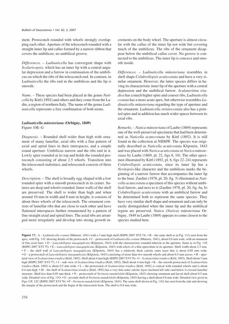
ment. Protoconch rounded with whorls strongly overlap-ping each other. Aperture of the teleoconch rounded with astraight inner lip and callus formed by a narrow ribbon thatcovers the umbilicus; no umbilical groove.
Differences. – Ladinaticella has convergent shape withScalaricopsis, which has an inner lip with a central angu-lar depression and a furrow in continuation of the umbili-cus on which the ribs of the teleoconch end. In contrast, inLadinaticella the ribs end in the umbilicus and the lip issmooth.
Name. – These species had been placed in the genus Nati-cella by Kittl (1892) and others and they come from the La-din, a region of northern Italy. The name of the genus Ladi-naticella represents a free combination of both terms.
Ladinaticella münsteriana (Orbigny, 1849)Figure 10E–G
Diagnosis. – Rounded shell wider than high with orna-ment of many lamellar, axial ribs with a fine pattern ofaxial and spiral lines in their interspaces, and a simpleround aperture. Umbilicus narrow and the ribs end in it.Lowly spire rounded at its top and holds the rounded pro-toconch consisting of about 2.5 whorls. Transition intothe teleoconch indistinct, and teleoconch consists of threewhorls.
Description. – The shell is broadly egg-shaped with a lowrounded spire with a smooth protoconch in its center. Su-tures are deep and whorls rounded. Inner walls of the shellare preserved. The shell is wider than high and whenaround 10 mm in width and 8 mm in height, it consists ofabout three whorls of the teleoconch. The ornament con-sists of lamellar ribs that are close to each other and haveflattened interspaces further ornamented by a pattern offine straight axial and spiral lines. The axial ribs are arran-ged more irregularly and develop into strong growth in-
crements on the body whorl. The aperture is almost circu-lar with the callus of the inner lip not wide but coveringmuch of the umbilicus. The ribs of the ornament disap-pear below the umbilical callus cover. No groove is con-nected to the umbilicus. The inner lip is concave and smo-oth inside.
Differences. – Ladinaticella münsteriana resembles inshell shape Colubrellopsis acuticostata and have a very si-milar ornament. However, the latter species differs in ha-ving its characteristic inner lip of the aperture with a centraldepression and the umbilical furrow. Scalaneritina tria-dica has a much higher spire and coarser ribs, Ladinaticellacostata has a more acute apex, but otherwise resembles La-dinaticella münsteriana regarding the type of aperture andthe ornament. Ladinaticella striatocostata also has a poin-ted spire and in addition has much wider spaces between itsaxial ribs.
Remarks. – Natica münsteriana of Laube (1869) representsone of the well-preserved specimens that had been determi-ned as Naticella acutecostata by Kittl (1892). It is stillfound in the collection at NHMW. The species was origi-nally described as Naticella acuticostata Klipstein, 1843and was placed with Narica acuticostata or Narica münste-riana by Laube (1869, pl. 22, figs 8, 10). The other speci-men illustrated by Kittl (1892, pl. 6, figs 22–24) representsColubrellopsis acuticostata, since its inner lip has aNeritopsis-like character and the umbilicus marks the be-ginning of a narrow furrow that accompanies the inner lipto the base. Zardini (1978, pl. 20, fig. 5) illustrated as Nati-cella acutecostata a specimen of this species without umbi-lical furrow, and next to it (Zardini 1978, pl. 20, fig. 6a, b)Colubrellopsis acuticostata with an umbilical furrow andhe determined both to represent the same species. Theyhave very similar shell shape and ornament and can only beeasily distinguished when the inner lip and the umbilicalregion are preserved. Natica (Narica) münsteriana Or-bigny, 1849 in Laube (1869) appears to come closest to thespecies studied here.
� �
��#����$$% A – Ladinaticella costata (Münster, 1841) with a 3 mm high shell (BSPG 2007 XVI 74). • B – the same shell as in Fig. 11A seen from theapex, with Fig. 11C showing details of the protoconch. • C – protoconch of Ladinaticella costata (Münster, 1841), about 0.6 mm wide, with an ornamentof fine axial lines. • D – Lancedellopsis inaequiplicata (Klipstein, 1843) with the characteristic rounded tubercle in the aperture. Same as in Fig. 11E(BSPG 2007 XVI 75). • E – Lancedellopsis inaequiplicata (Klipstein, 1843) with relicts of a thin operculum in its aperture. Shell width about 2.5 mm.• F – the shell wall of Lancedellopsis inaequiplicata (Klipstein, 1843) has a relatively thick calcitic outer layer that is about 0.05 mm wide.• G – a protoconch of Lancedellopsis inaequiplicata (Klipstein, 1843) consisting of more than two smooth whorls and about 0.5 mm across. • H – aper-tural view of Scalaneritina triadica (Kittl, 1892). Shell about 4 mm high (BSPG 2007 XVI 76). • I – Scalaneritina triadica (Kittl, 1892). Shell about 5 mmhigh (BSPG 2007 XVI 77). • J – side view of Scalaneritina triadica (Kittl, 1892). Shell about 4 mm high. • K – the smooth protoconch of Scalaneritinatriadica (Kittl, 1892) is about 0.5 mm wide. • L – the protoconch of Scalaneritina triadica (Kittl, 1892) is conical with rounded whorls and is about0.4 mm high. • M – the shell of Scalaneritina triadica (Kittl, 1892) has a very thin outer calcitic layer (inclined left side) and below it crossed lamellarstructure. Shell less than 0.05 mm thick. • N – protoconch of Neritaria mandelslohi (Klipstein, 1843) showing ornament and larval shell about 0.5 mmwide. Detailed view of Fig. 11O. • O – juvenile shell of Neritaria mandelslohi (Klipstein, 1843) having a shell about 0.8 mm wide. Detailed view given inFigs 11P, 12C (BSPG 2007 XVI 78). • P – Neritaria mandelslohi (Klipstein, 1843). The same shell shown in Fig. 11O, but seen from the side and showingthe margin of the protoconch and the begin of the teleoconch from. The shell is 0.8 mm wide.
����������� ������ �������������
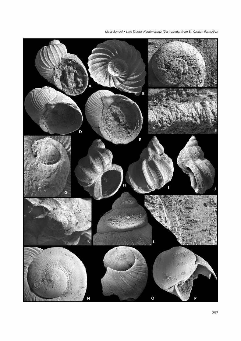
� �
(
�
&
�
�
�
'� )
* � �
� 3 5
���� ������ ��������� ���������� !�"�� �� ��#���$��%� ���&������
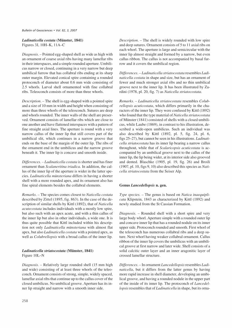
Ladinaticella costata (Münster, 1841)Figures 3J, 10H–K, 11A–C
Diagnosis. – Pointed egg-shaped shell as wide as high withan ornament of coarse axial ribs having many lamellar ribsin their interspaces, and a simple rounded aperture. Umbili-cus narrow or closed, continuing in a very narrow but deepumbilical furrow that has collabral ribs ending at its sharpouter margin. Elevated conical spire containing a roundedprotoconch of diameter about 0.6 mm wide consisting of2.5 whorls. Larval shell ornamented with fine collabralribs. Teleoconch consists of more than three whorls.
Description. – The shell is egg-shaped with a pointed spireand a size of 10 mm in width and height when consisting ofmore than three whorls of the teleoconch. Sutures are deepand whorls rounded. The inner walls of the shell are preser-ved. Ornament consists of lamellar ribs which are close toone another and have flattened interspaces with a pattern offine straight axial lines. The aperture is round with a verynarrow callus of the inner lip that still covers part of theumbilical slit, which continues in a narrow groove thatends on the base of the margin of the outer lip. The ribs ofthe ornament end in the umbilicus and the narrow groovebeneath it. The inner lip is concave and smooth inside.
Differences. – Ladinaticella costata is shorter and has finerornament than Scalaneritina triadica. In addition, the cal-lus of the inner lip of the aperture is wider in the latter spe-cies. Ladinaticella münsteriana differs in having a shortershell with a more rounded apex, and its ornament also hasfine spiral elements besides the collabral elements.
Remarks. – The species comes closest to Naticella costatadescribed by Zittel (1895, fig. 863). In the case of the de-scription of similar shells by Kittl (1892), that of Naticellaacutecostata includes individuals with a mostly low spire,but also such with an apex acute, and with a thin callus ofthe inner lip but also in other individuals, a wide one. It isthus quite possible that Kittl included within his descrip-tion not only Ladinaticella münsteriana with almost flatapex, but also Ladinaticella costata with a pointed apex, aswell as Colubrellopsis with a broad callus of the inner lip.
Ladinaticella striatocostata (Münster, 1841)Figure 10L–N
Diagnosis. – Relatively large rounded shell (15 mm highand wide) consisting of at least three whorls of the teleo-conch. Ornament consists of strong, simple, widely spaced,lamellar axial ribs that continue up to the callus cover of theclosed umbilicus. No umbilical groove. Aperture has its in-ner lip straight and narrow with a smooth inner side.
Description. – The shell is widely rounded with low spireand deep sutures. Ornament consists of 5 to 11 axial ribs oneach whorl. The aperture is large and semicircular with theinner lip almost straight and formed by a narrow, but evencallus ribbon. The callus is not accompanied by basal fur-row and it covers the umbilical region.
Differences. – Ladinaticella striatocostata resembles Ladi-naticella costata in shape and size, but has an ornament offewer and much stronger axial ribs and no thin umbilicalgroove next to the inner lip. It has been illustrated by Za-rdini (1978, pl. 20, fig. 7) as Naticella striatocostata.
Remarks. – Ladinaticella striatocostata resembles Colub-rellopsis acuticostata, which differs primarily in the cha-racters of the inner lip. They were confused by Kittl (1892)who found that the type material of Naticella striatocostataof Münster (1841) consisted of shells with a closed umbili-cus, while Laube (1869), in contrast to his illustration, de-scribed a wide-open umbilicus. Such an individual wasalso described by Kittl (1892, pl. 5, fig. 24, pl. 6,figs 25–27), but cannot be seen in his illustrations. Ladinati-cella striatocostata has its inner lip bearing a narrow callusthroughout, while that of Scalaricopsis acuticostata is ac-companied by an umbilical groove next to the callus of theinner lip, the lip being wider, at its interior side also groovedand dented. Blaschke (1905, pl. 19, fig. 26) and Broili(1907, pl. 10, figs 9, 10) also described this species as Nati-cella striatocostata from the Seiser Alp.
Genus Lancedellopsis n. gen.
Type species. – The genus is based on Natica inaequipli-cata Klipstein, 1843 as characterized by Kittl (1892) andnewly studied from the St Cassian Formation.
Diagnosis. – Rounded shell with a short spire and verylarge body whorl. Aperture simple with a rounded outer lipand concave inner lip that has a rounded nodule on its innerupper side. Protoconch rounded and smooth. First whorl ofthe teleoconch has numerous collabral ribs and a deep su-ture. Next whorl having weaker collabral ornament. Callusribbon of the inner lip covers the umbilicus with an umbili-cal groove at first narrow and later wide. Shell consists of asolid calcitic outer layer and an inner aragonitic layer ofcrossed lamellar structure.
Differences. – In ornament Lancedellopsis resembles Ladi-naticella, but it differs from the latter genus by havingmore rapid increase in shell diameter, developing an umbi-lical groove, and having a rounded nodule in the upper partof the inside of its inner lip. The protoconch of Lancedel-lopsis resembles that of Ladinaticella in shape, but its orna-
� �
����������� ������ �������������

ment is not well enough preserved for good comparison. Itmay be that Lancedellopsis is not a member of the Scalane-ritinidae, but represents another branch of the Cycloneriti-morpha.
Name. – In honor of Rolando Lancedelli from Cortinad’Ampezzo who collected many of the gastropods thathave been described by Rinaldo Zardini.
Lancedellopsis inaequiplicata (Klipstein, 1843)Figure 11D–G
Diagnosis. – Shell having a globular protoconch of 0.4 mmwith about two whorls. Whorls of the teleoconch rapidlyincrease in width until the shell reaches 10 mm in heightand width and consists of 2.5 whorls. Teleoconch ornamentof collabral ribs, which within the second whorl may disap-pear. Aperture has the inner lip with a narrow callus ribbonthat has a wider apical part with a rounded tubercle on itsinner side. Umbilicus closed continuing in a groove. Oper-culum thin and of semicircular shape. Shell composed of acontinuous and relatively thick calcitic outer layer with alayer of crossed lamellar structure beneath.
Description. – The shell is rounded and has a wide obliqueaperture with a straight inner lip and an evenly rounded outerlip. The globular protoconch is not well preserved but ap-pears to have been smooth. The first whorl of the teleo-conch has an ornament of more than 40 simple collabralribs that continue from the suture to the edge of the umbili-cal groove or the callus cover of the umbilicus in about thesame composition. In the case of the original individualstudied by Kittl (1892), present in the NHMW collection,the ornament on the second whorl of the teleoconch beco-mes weaker until only increments of growth remain. With2.5 whorls of the teleoconch the shell is about 10 mm wideand high. The aperture has the inner lip expanded to form acallus ribbon that is narrow except in its apical part, whereit covers the umbilical region. It is accompanied by an um-bilical groove that increases in width with shell growth. Inthe fully-grown shell the groove shows growth increments,in a shell with only 1.2 whorls of the teleoconch it is verynarrow. The aperture has a rounded thickening on the up-per part of the inner lip. A partially crushed operculum ispreserved, and is thin and similar to that of Ruganeritaria.However, in contrast to the latter genus its composition inregards to growth line patterns is not preserved.
Remarks. – Kittl (1892) compared Lancedellopsis inaequ-iplicata (as Naticopsis) with Colubrellopsis acuticostata(as Naticella acutecostata) and noted their resemblance.Kittl (1892) suggested that they could be best distinguishedfrom each other by the ornament up to the body whorl. In
Colubrellopsis it is supposedly present and ornament isweaker on the body whorl in Lancedellopsis. This diffe-rence can no longer help, since such a change in ornamentis also noted here in the case of Colubrellopsis acuticos-tata, where some individuals also have an almost smoothbody whorl. The only solid difference between these spe-cies is in regard to the shape of the inner side of the innerlip, which was not noted by Kittl, and also not by Broili(1907). The inner lip has the character of Neritopsis in thecase of Colubrellopsis, and is even and smooth in its lowerportion and possesses a characteristic rounded nodule in itsupper portion in the case of Lancedellopsis.
Superfamily Neritoidea Rafinesque, 1815
Diagnosis. – Inner walls of the whorls of the shell are dissol-ved during larval life and in most cases later. Protoconchwhorls tightly coiled, convolute and composed of aragonite.Shell of the teleoconch has an outer calcitic layer and an in-ner layer composed of aragonitic crossed lamellar structure.
Remarks. – The central group of the Neritoidea is the Re-cent Neritidae still alive today. The most ancient knowngenera of the superfamily are represented by the Neritarii-dae from the St Cassian Formation.
Differences. – Among the living marine species with ashell, the only genus that does not belong into this superfa-mily and has the inner whorls preserved is Neritopsis of theNeritopsoidea.
Family Neritariidae Wenz, 1938
Diagnosis. – Globular shell composed of a very thin outercalcitic layer and a thick uniform inner aragonitic layer incrossed lamellar structure. Shell margin simple and suturesdistinct. Aperture rounded with simple outer lip and innerlip with a bulging callus. Inner lip callus not continuous tothe base, but ends in a depression with its outer margin a ribthat connects to the base of the outer lip. Inner whorls of theshell dissolved. Protoconch globular with convolute who-rls. Ornament of larval shell consists of rounded axial ribsand sometimes also fine spiral ribs.
Remarks. – The family is based on genus Neritaria Koken,1892 from the St Cassian Formation. The family also inclu-des Ruganeritaria and Dentineritaria forming the Neritari-inae and Oncochilus and Cassianpisulina composing theOncochilinae, and perhaps also Trachyneritaria of the Tra-chyneritariinae.
Description. – The larval shell of Neritariidae is of globularshape with distinct sutures, all whorls are visible and bear
� �
���� ������ ��������� ���������� !�"�� �� ��#���$��%� ���&������
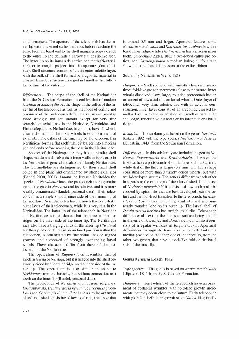
axial ornament. The aperture of the teleoconch has the in-ner lip with thickened callus that ends before reaching thebase. From its basal end to the shell margin a ridge extendsto the outer lip and delimits a narrow flat or slit-like area.The inner lip on its inner side carries one tooth (Neritarii-nae), or its margin projects into the aperture (Oncochili-nae). Shell structure consists of a thin outer calcitic layer,with the bulk of the shell formed by aragonitic material incrossed lamellar structure arranged in lamellae that followthe outline of the outer lip.
Differences. – The shape of the shell of the Neritariidaefrom the St Cassian Formation resembles that of modernNeritina or Smaragdia but the shape of the callus of the in-ner lip of the teleoconch as well as the mode of coiling andornament of the protoconch differ. Larval whorls overlapmore strongly and are smooth except for very finescratch-like axial lines in the Neritidae, Neritinidae andPhenacolepadidae. Neritariidae, in contrast, have all whorlsclearly distinct and the larval whorls have an ornament ofaxial ribs. The callus of the inner lip of the teleoconch inNeritinidae forms a flat shelf, while it bulges into a medianpad and ends before reaching the base in the Neritariidae.
Species of the Naticopsidae may have a similar shellshape, but do not dissolve their inner walls as is the case inthe Neritoidea in general and also their family Neritariidae.The Cortinellidae are distinguished by their small shellcoiled in one plane and ornamented by strong axial ribs(Bandel 2000, 2001). Among the Jurassic Neritoidea thespecies of Neridomus have the protoconch more globularthan is the case in Neritaria and its relatives and it is moreweakly ornamented (Bandel, personal data). Their teleo-conch has a simple smooth inner side of their inner lip ofthe aperture. Neritidae often have a much thicker calciticouter layer of their teleoconch, while it is very thin in theNeritariidae. The inner lip of the teleoconch in Neritidaeand Neritinidae is often dented, but there are no teeth orridges on the inner side of the inner lip. The Neritiliidaemay also have a bulging callus of the inner lip (Pisulina)but their protoconch lies in an inclined position within theteleoconch, is ornamented by fine spiral lines or alignedgrooves and composed of strongly overlapping larvalwhorls. These characters differ from those of the pro-toconch of the Neritariidae.
The operculum of Ruganeritaria resembles that ofmodern Nerita or Neritina, but it is hinged into the shell ob-viously aided by a tooth or ridge on the inner side of the in-ner lip. The operculum is also similar in shape toNeridomus from the Jurassic, but without connection to atooth on the inner lip (Bandel, personal data).
The protoconch of Neritaria mandelslohi, Ruganeri-taria subovata, Dentineritaria neritina, Oncochilus globu-losus and Cassianpisulina bullata have a similar ornamentof its larval shell consisting of low axial ribs, and a size that
is around 0.5 mm and larger. Apertural features uniteNeritaria mandelslohi and Runganeritaria subovata with abasal inner ridge, while Dentineritaria has a median innertooth, Oncochilus Zittel, 1882 a two-lobed callus projec-tion, and Cassianpisulina a median bulge; all four taxashow indistinct basal depression of the callus ribbon.
Subfamily Neritariinae Wenz, 1938
Diagnosis. – Shell rounded with smooth whorls and some-times fold-like growth increments close to the suture. Innerwhorls dissolved. Low, large, rounded protoconch has anornament of low axial ribs on larval whorls. Outer layer ofteleoconch very thin, calcitic, and with an acicular con-struction. Inner layer consists of an aragonitic crossed la-mellar layer with the orientation of lamellae parallel toshell edge. Inner lip with a tooth on its inner side or a basalridge.
Remarks. – The subfamily is based on the genus NeritariaKoken, 1892 with the type species Neritaria mandelslohi(Klipstein, 1843) from the St Cassian Formation.
Differences. – In this subfamily are included the genera Ne-ritaria, Ruganeritaria and Dentineritaria, of which thefirst two have a protoconch of similar size of about 0.5 mm,while that of the third is larger (0.8 mm) and has a shapeconsisting of more than 3 tightly coiled whorls, but withwell-developed sutures. The genera differ from each otherin regards to the ornament of their larval shell. In the caseof Neritaria mandelslohi it consists of low collabral ribscrossed by spiral ribs that are best developed near the su-ture and the indistinct transition to the teleoconch. Rugane-ritaria subovata has undulating axial ribs and a promi-nently rounded lobe on its outer lip. The larval shell ofDentineritaria neritina has straight axial ribs. Teleoconchdifferences also exist in the outer shell surface, being smoothin the case of Neritaria and Dentineritaria, while it con-sists of irregular wrinkles in Ruganeritaria. Aperturaldifferences distinguish Dentineritaria with its tooth in amedian position on the inner side of the inner lip, from theother two genera that have a tooth-like fold on the basalside of the inner lip.
Genus Neritaria Koken, 1892
Type species. – The genus is based on Natica mandelslohiKlipstein, 1843 from the St Cassian Formation.
Diagnosis. – First whorls of the teleoconch have an orna-ment of collabral wrinkles with fold-like growth incre-ments that may occur close to the suture. Early teleoconchwith globular shell; later growth stage Natica-like; finally
���
����������� ������ �������������

ovoid in shape. Whorls smooth; body whorl abandoning re-gular coiling mode, twisting off and then having an inclinedsuture. Protoconch large and globular with larval whorls ha-ving a pattern of rounded axial ribs and spiral lines develo-ped especially near the suture. Final part of larval shell alsodeviates from the coiling mode and has an inclined suture.Outer calcitic layer of teleoconch very thin. Inner lip formsa callus pad that covers the umbilical region; inner sidewith fold near the base that runs slightly inclined almostparallel to the inner lip.
Differences. – Only Ruganeritaria has a similar inner foldon its inner lip, but is quite distinct from Neritaria in ha-ving a folded shell surface, smaller size, and ornament onthe larval shell.
Remarks. – According to Wenz (1938–1944, fig. 1002), theshell is Neritina-like. The genus is based on Natica plicati-lis Klipstein, 1843 = Natica mandelslohi, which closely re-sembles Neritaria similis Koken, 1892 from the Upper Tri-assic Raibler Formation, Schlern-Plateau, Alps. Koken(1892) based his genus especially on Neritaria mandel-slohi when he described Neritaria similis. Because of bet-ter preservation of specimens, Neritaria mandelslohi ishere proposed to represent the type of the genus.
Kittl (1892) utilized Neritaria mandelslohi as the char-acteristic species to a group of St Cassian Natica. In thegroup he united species that have a callus pad of their innerlip which covers the umbilicus. The species of the groupdiffer somewhat in the height of the spire, presence or ab-sence of ornamental folds, as well as the width of the callusof the inner lip. Kittl (1892) also included into the groupNatica angusta Münster, 1841, and Natica plicatilisKlipstein, 1843, which are not at all related to Neritaria,but represent relatives to Neritopsis, with the characteristicfeatures of the inner lip of the aperture of that group(Fedaiella). Knight et al. (1960, fig. 183, 1) stated thatNeritaria is a synonym to Protonerita Kittl, 1894 and char-acterized the shell as having a protuberance in the middleof its callus of the inner lip, but smooth inner side of the in-ner lip. The latter author suggested that the genus be basedon Neritaria candida Kittl, 1894.
Neritaria mandelslohi (Klipstein, 1843)Figures 11N–P, 12A–H
Diagnosis. – Juvenile shell globular, becoming helicoid andelongate ovoid shape when fully-grown. Spire short and poin-ted with axial folds. Callus of inner lip with a thickened me-dian pad beneath which the columellar lip has a flat area withan outer ridge. Inner side with a fold near the base. Operculumgenerally Nerita-like. Protoconch measures about 0.5 mmand has an ornament of axial and spiral ribs.
Remarks. – Both the thickened median pad of the callusand beneath it the flat area with an outer ridge of the colu-mellar lip are the same as in Ruganeritaria.
Description. – The shell has a rounded shape with a charac-teristic ornamental pattern of folds in axial orientation. Thelatter begin at the suture and decrease in strength or disap-pear further down on the whorls. With 1.7 whorls the teleo-conch is globular in shape and measures 2.6 mm in heightand 2.7 mm in width. Sutures are indistinct and the orna-ment consists of collabral elements that may be indistinctor quite distinct axial ribs. Shell shape is similar up to twowhorls of the teleoconch, but in the third whorl the diame-ter increases more rapidly and the shape of the shell beco-mes more elliptical. The shell surface is smooth in thefourth (final) whorl of the teleoconch. Coiling mode devia-tes from the former whorls so that the suture is a little incli-ned. Maximum shell size is around 10 mm high and 8 to9 mm wide. Shells of that size belong to fully-grown adultspecimens as is indicated by the inclined suture and the cal-lus pad of the inner lip, which is very strong and thick atthat stage, more so than in earlier stages of growth. Colorpattern of the shell is commonly preserved. It is quite vari-able with zigzag axial lines of dark and light color and spi-ral lines such as four narrow, brown ribbons. Some indivi-duals have rows colored dots, others a uniform colorationof brown.
The aperture has a straight corner of the inner lip andan evenly rounded outer lip. Its orientation is oblique. Theedge of the outer lip continues in a narrow rim into theumbilical area, and ends under the cover of the callus ofthe inner lip. The area between this rim and the basal partof the inner lip is flattened. The callus band expandsabove the umbilical region into a rounded pad. An inter-nal fold lies next to the base on the inner side of the innerlip; the hinge of the operculum fitted in the groove be-tween this tooth-like ridge and the inner lip but the ridge isnot continuous into the shell interior. The operculum re-sembles that of Ruganeritaria in shape, and is thin with asmooth surface. Only fragmentary preservation wasnoted, though.
The protoconch is up to 0.6 mm wide and consists ofmore than three whorls. It is partly covered by theteleoconch. The embryonic whorl is connected to morethan two larval whorls which expand rapidly and are coiledin convolute manner. They are ornamented by undulatinglow axial ribs and spiral ribs. Both form a reticulate patternthat is best developed next to the suture. The spiral ribs areat first weak, later on the final whorl spreading out in afan-like fashion. Sutures are not impressed but still distinct.The margin of the larval shell is sinuous and not thickened.Its transition into the teleoconch is quite indistinct. It isconnected with the change of sinuous growth lines tostraight increments of growth.
���
���� ������ ��������� ���������� !�"�� �� ��#���$��%� ���&������

Differences. – Neritaria mandelslohi is larger than Ruga-neritaria but closely resembles it regarding the apertureand operculum. The irregular folded surface of Ruganeri-taria and its more rounded shape when fully-grown distin-guishes the genus. The tooth on the inner lip of the apertureis stronger and in more basal position than that of Dentine-ritaria neritina. Neritaria mandelslohi has its protoconchwith a fine reticulate ornament and a row of nodes near thesuture. This distinguishes it from the axial ornament ofDentineritaria as well as the undulating pattern of Rugane-ritaria but even more so from living Neritina and Nerita,which have their protoconch with a smooth surface and or-nament consisting of only very fine scratch-like straightaxial lines. In addition, the whorls of their larval shell morestrong overlap each other and their sutures are indistinct(Bandel 2001).
Remarks. – Laube (1869) discussed Natica mandelslohiKlipstein, 1843, characterizing it by the axial folds of itsornament. Its size of about 9 mm in height and possessionof about 4 whorls in the teleoconch fits with the observati-ons on the species. However, Laube (1869) did not note theinner fold on the base of the inner lip, which is characteris-tic to the species. In addition, Natica cassiana of Laube(1869, pl. 21, fig. 10) may belong to that species, as well asNeritaria mandelslohi of Kittl (1894) from the MarmolataLimestone. All of these were not described in sufficient de-tail especially in regards to the internal basal fold that is ac-tually characteristic to the species. This fold is also not seenin the specimens that were illustrated and described byKittl (1892, pl. 7, figs 31, 33) as Natica mandelslohi fromthe St Cassian Formation. Zardini (1978, pl. 19, figs 1–5)determined it as Neritaria mandelslohi (Klipstein), but alsoas Naticopsis cassiana (Wissmann) (Zardini 1978, pl. 19,fig. 15), Naticopsis limneiformis (Laube) (Zardini 1978,pl. 19, fig. 16), and Neritaria plicatilis (Klipstein) (Zardini1978, pl. 19, fig. 22), all without mentioning the basal foldin the shell. But this tooth is well seen in the illustration ofZardini (1978, pl. 19, figs 5, 15; 1985, pl. 9, fig. 3). Shell
shape and ornament is obviously very similar to Neritaria(Neritaria) plicatilis Klipstein, as illustrated by Wenz(1938–1944, fig. 1002), but that species was also characte-rized without details concerning its aperture. In the case ofthe Peruvian representatives of the genus Neritaria descri-bed by Haas (1953) from the Pucará Group, the color pat-tern is preserved as well as the little knob on the inner sideof the inner lip. This was confirmed by Bandel (1994, pl. 5,fig. 15) in the case of Neritaria distincta Haas, 1953.
Genus Ruganeritaria n. gen.
Type species. – The genus is based on Ruganeritaria subo-vata (Münster, 1841) from the St Cassian Formation,which is very common in the fauna from the locality Alpedi Specie.
Diagnosis. – Globular shell consists of 3 whorls of the tele-oconch. Protoconch is of about 0.5 mm in size and has itslarval shell ornamented by rounded and undulating axialribs. Outer calcitic layer of teleoconch very thin. Below theouter layer shell material originally contained much orga-nic material, deeper down more solidly mineralized. Innershell walls dissolved. Operculum concentric with hinge fit-ted into a groove formed by a basal tooth and the inner lip.Outer surface of teleoconch can be wrinkled. Callus of in-ner lip characterized by a rounded pad in a median positionabove the umbilicus.
Name. – This Neritaria-like member of the Neritoidea hascharacteristic wrinkles, = rugae in Latin, and the name is acombination of both terms.
Differences. – Ruganeritaria resembles Neritaria in seve-ral ways, but is very distinct in its wrinkled shell, whichprobably formed during diagenesis. The outer shell layers,especially of the outermost aragonitic part, held much or-ganic shell material. When it was destroyed or decomposed
���
��#����$+% A – juvenile shell of Neritaria mandelslohi (Klipstein, 1843) with callus pad and basal tooth on the inner side of the inner lip in the aperture.Shell 4 mm wide (BSPG 2007 XVI 79). • B – shell of Neritaria mandelslohi (Klipstein, 1843) with characteristic inner lip, and basal tooth-like ridge.3 mm wide shell (BSPG 2007 XVI 80). • C – ornamented larval shell of Neritaria mandelslohi (Klipstein, 1843), detailed view of Fig. 11O, P. • D – inter-nal walls of Neritaria mandelslohi (Klipstein, 1843) are totally dissolved in the apical shell part, here fractured (BSPG 2007 XVI 81). • E – the outercalcitic shell layer forms a continuous sheet, with the crossed lamellar layer below displaying the arrangement of the lamellae parallel to the aperture.• F – juvenile shell of Neritaria mandelslohi (Klipstein, 1843) with its callus very similar to Ruganeritaria, but differing having a smooth shell. Shellabout 2 m high (BSPG 2007 XVI 82). • G – fractured shell of Ruganeritaria subovata (Münster, 1841) showing dissolved inner whorls. Shell 4 mm wide(BSPG 2007 XVI 83). • H – shell of Neritaria mandelslohi about 0.08 mm thick and having a very thin outer calcitic layer, (upper side) and well organizedcrossed lamellar layer below. • I – protoconch of Ruganeritaria subovata (Münster, 1841) about 0.5 mm wide, ornamented by collabral ribs and having itsmargin fractured before the teleoconch formed (BSPG 2007 XVI 84). • J – wrinkled shell of Ruganeritaria subovata (Münster, 1841) with operculum stillin place. Shell 5 mm wide (BSPG 2007 XVI 85). • K – shell of Ruganeritaria subovata with an extremely thin outer calcitic layer (upper part) and crossedlamellar structure below. The shell is about 0.04 mm thick. • L – operculum in the aperture of Ruganeritaria subovata (Münster, 1841) with a nucleus inthe lower left corner (BSPG 2007 XVI 86). • M – shell of Ruganeritaria subovata (Münster, 1841) with wrinkles and basal tooth in the aperture. Shellabout 2 mm high (BSPG 2007 XVI 87). • N – characteristic wrinkled surface of Ruganeritaria subovata (Münster, 1841). Shell about 2.8 mm wide(BSPG 2007 XVI 88).
����������� ������ �������������

���
( �
&
�
�
�'
� )
*
� � �
���� ������ ��������� ���������� !�"�� �� ��#���$��%� ���&������

during earliest diagenesis, probably by microbial decay,the very thin calcitic layer on the shell surface collapsed lo-cally during compaction of the sediment and formed wrin-kles but these are not present in every individual shell. Theconstruction of the operculum is like Recent Nerita or Ne-ritina, but in contrast to these the hinge arrangement is dif-ferent with a groove formed by a basal tooth and the innerlip.
Ruganeritaria subovata (Münster, 1841)Figures 12G, I–N, 13K, L
Diagnosis. – Outer layer of shell transformed by folds ofdifferent orientation with layer below consisting of solidaragonitic crossed lamellar deposits. Rounded shell has aprotoconch of 3 whorls with larval whorls having fine un-dulating axial ribs. Teleoconch consists of about 3 whorlseither smooth or having collabral and irregular wrinkles.Inner lip granular with thick swelling in middle part thatextends onto a thickened pad covering the umbilicus. Cal-lus of fully-grown shells does not cover basal part of shell;here a ridge connects to outer lip. Inner walls of shell havebeen dissolved. Protoconch of more than three whorls withlarval whorls ornamented by low undulating axial ribs andapertures with sinuous outline.
Description. – The oblique egg-shaped shell has thick wa-lls with a very thin outer calcitic layer and massive aragoni-tic inner layer in crossed lamellar structure. A shell that isabout 3.4 mm high and 3.3 mm wide has about 1.25 whorlsof the teleoconch. With three whorls of the teleoconch theshell measures 6 mm in height and width and isfully-grown. The body whorls encompass almost the entireshell height and the sutures are indistinct. The final whorlhas a more inclined suture. The pattern of growth lines mayform shallow folds and an ornament of collabral folds orribs. Folds of irregular orientation are usually also present.The aperture is large, oblique and semicircular in outline.Its outer lip is acute and thickened within. The inner lipforms a thick broad callus with a rounded pad near the um-bilical region and a depression below it. The margin of thecallus is formed by a thin keel that runs in a curve to thebase. The callus surface is granular and its margin to theaperture is straight. A solid tooth lies near the base on theinner lip.
The protoconch is about 0.6 mm in diameter and partlycovered by the first whorl of the teleoconch. The embry-onic shell is succeeded by about two larval whorls. The fi-nal whorl declines a little. Ornament consists of lowrounded axial ribs with an undulating somewhat granularpattern. There are about 30 axial ribs on a whorl with an in-clined direction in collabral arrangement. The aperturalmargin has a wide lobe near the middle of the outer lip.
The operculum was preserved in place in several indi-viduals, but usually crushed, since it is relatively thin. It fitswith its straight inner margin to the straight edge of the innerlip. Its center of growth lies close to the basal inner lip. Fromthe spiral nucleus growth lines go to the outer, rounded mar-gin in a fan-like manner showing that growth was along theinner lip and faster towards the apex than towards the umbi-licus. This growth is quite like that in the operculum of mod-ern Neritina. The inner edge of the operculum is hinged intothe groove formed by the tooth on the inner lip and the basalmargin of that lip. A tooth on the inner side of the operculumcannot be made out but may be present.
Differences. – The whorls of Ruganeritaria subovata aremore convolute and lower than Neritaria mandelslohi andDentineritaria neritina. The inner lip callus of Ruganerita-ria is wider than that of Dentineritaria neritina and Nerita-ria mandelslohi resembling in shape that of the latter spe-cies. The shell size of Ruganeritaria is smaller than that ofNeritaria and larger than that of Dentineritaria.
The protoconch of Ruganeritaria in size and ornamentresembles that of Neritaria mandelslohi and has similaraxial but no spiral ornament, while the larval shell ofDentineritaria is larger and has straight axial ornament.The larval ornament of the protoconch of Cassianpisulinais quite similar to that of Ruganeritaria.
Remarks. – Laube (1869, pl. 21, fig. 8) noted a wrinkledsurface in the species he had determined as Natica subo-vata Münster, 1841, a feature that is characteristic to manyindividuals of this species. However, his size data do not fitin all aspects; in 1869 Laube noted up to 9 mm in diameter,a size that would only fit with that of Neritaria mandel-slohi. Zardini (1978, pl. 19, figs 8, 9) determined indivi-duals of this species recognizable by their characteristicshell folds as Hologyra expansa, others as Naticopsis lim-neiformis Laube (Zardini, 1978, pl. 19, fig. 16), and othersagain as Naticopsis cassiana (Zardini 1978, pl. 19, fig. 19)and as Naticopsis impressa (Zardini 1978, pl. 19, fig. 20).
Genus Dentineritaria n. gen.
Type species. – Natica neritina Münster, 1841.
Diagnosis. – Small shell (about 6 mm wide and 5 mm high)with a characteristic tooth on inner side of inner lip in me-dian position. Aperture large and whorls increase rapidly inwidth. Larval shell with ornament of axial ribs and consis-ting of three whorls reaching around 0.8 mm.
Name. – This Neritaria-like member of the Neritoidea hasa characteristic tooth, = dentis, “of a tooth” genitive in La-tin, combined to give Dentineritaria.
���
����������� ������ �������������

Remarks. – Shells shaped like Smaragdia which lives onsea-grass. Natica neritina Münster, 1841, according toLaube (1869) represents the smallest of the group of spe-cies from St Cassian. In the original description by Münster(1841) as well as the subsequent one by Laube (1869),this species could be fitted but Laube’s illustration doesnot agree with it. Kittl (1892) determined it as Natica neri-tina, and the originals to his illustrations present in theNHMW collection in part have the characteristic mediantooth on the inner side of their lip. This small central toothwas not described by Laube (1869) and is not found amongthe species described and illustrated by Zardini (1978).Since it occurs in the material that was in part collected andanalyzed by Rinaldo Zardini it was most probably not re-cognized.
Dentineritaria neritina (Münster, 1841)Figure 13A–H
Diagnosis. – Ovoid shell with broad callus of inner lip witha groove on its basal end. Inner side of the inner lip carries amedian tooth. Larval shell of large protoconch has an orna-ment of low rounded axial ribs. Teleoconch smooth.
Description. – Shell size is up to 6 mm high and 5 mm widewith only 2.5 whorls of the teleoconch. Whorls are smoothand increase rapidly in width. With one whorl of the teleo-conch the shell is about 2.3 mm in width and 2.4 mm inheight and has globular shape. The aperture is large, a littleinclined, and almost semicircular in outline. The outer liphas an acute edge and is a little thickened on its inside. Thecallus of the inner lip forms a thickened ribbon that coversthe umbilicus and connects with the outer lip on the apicalside and ends before it reaches the basal side. Here a finesharp keel connects with the edge of the outer lip at the baseand differentiates a narrow flat area next to the basal outerlip. The surface of the callus is granular. In the middle ofthe inner lip and toward the interior of the aperture a smalland distinct pointed tooth is developed. The inner walls ofthe shell have been dissolved.
The protoconch is 0.8 mm in diameter. Not all of it isvisible in the apex when the first whorl of the teleoconchsurrounds it. The embryonic shell is succeeded by 2.25 lar-val whorls that have an ornament of delicate axial ribs,which have a sinuous arrangement. 40 to 50 such ribs arepresent on the last whorl and the apertural margin is simpleand straight.
Differences. – The small number of whorls of Dentinerita-ria neritina distinguishes it from Neritaria mandelslohi aswell as from Ruganeritaria subovata both of which alsohave a tooth on their inner lip. However, their teeth are notin a median position as is the case in Dentineritaria neri-
tina. The latter has a larger protoconch, a higher spire and anarrower inner lip than present in Ruganeritaria subovataand Neritaria mandelslohi. The teleoconch of Ruganerita-ria commonly bears wrinkles, which are not found in Den-tineritaria neritina.
Remarks. – The genus Cryptonerita Kittl, 1895 with aNatica-like shell having a smooth inner lip is based onCryptonerita elliptica as illustrated by Wenz (1938–1944,fig. 1004). This is not the small Dentineritaria from the StCassian Formation, but resembles Natica berwerthi Kittl,1892, which is larger than Dentineritaria neritina. Kittl(1892) established Neritina imitans for a species from theSt Cassian Formation, which has the shape of Dentinerita-ria, but differs in having the callus of its inner lip characte-rized as wide with a groove. No tooth on its inner side wasnoted. Study of the type of the species preserved in the col-lection at NHMW shows that it actually represents a mem-ber of the Oncochilinae with a projection of the margin ofthe inner lip into the aperture. Kittl (1892) discussed thepossibility of Neritina imitans belonging to the modern Ne-ritina, without coming to a final conclusion.
Subfamily Oncochilinae n. subfam.
Type genus. – The subfamily is based on the genus Onco-chilus Zittel, 1882 from the Late Triassic.
Diagnosis. – Rounded shell with an ornament only ofgrowth increments and a characteristic swelling on the in-ner lip, which in the case of Oncochilus consists of a doubletooth, and in that of Cassianpisulina is a non-symmetricmassive projection of callus of the inner lip, which ends be-fore reaching the base. The groove formed below it has asan outer margin the continuation of the outer lip. Proto-conch resembles that of the Neritariinae and has an axialornament on its larval whorls.
Differences. – Here included are Oncochilus as well asCassianpisulina from the St Cassian Formation. Oncochi-lus also has species in the Jurassic (Zittel 1873, 1895). Theteleoconch of Cassianpisulina resembles modern Pisulinabut differs from it in regard to the shape and ornament ofthe larval shell.
Genus Oncochilus Zittel, 1882
Diagnosis. – Rounded smooth shell with inner lip bearingtwo characteristic rounded tubercles in central part.
Type species. – According to Wenz (1938–1944, fig. 1010)the genus is based on Natica globulosa Klipstein, 1843,
��
���� ������ ��������� ���������� !�"�� �� ��#���$��%� ���&������

which is the same species as Natica subovata Münster,1841 (Kittl, 1892) from the St Cassian Formation, ItalianAlps.
Oncochilus subovatus (Münster, 1841)Figure 14C, E–G
Diagnosis. – Globular shell with four whorls in teleoconchand short spire. Two projections of callus plate of inner lipextend into apertural opening. Callus of inner lip forms athickened pad over umbilical area; narrow umbilical fur-row below which the outer margin fuses with edge of outerlip. Protoconch globular and about 0.5 mm in diameter.
Description. – The round shell with short spire and largebody whorl has distinct sutures. In the last whorl of thefully-grown shell the suture is inclined. Growth lines havea curved outline. The outer calcitic layer may preserve acolor pattern of zigzag lines and dots. A fully-grown shellwith four whorls of the teleoconch may be up to 9 mm highand slightly less in width, but may also be about 8 mm highand 6 mm wide. Whorls are smooth. The semicircular aper-ture has a simple rounded outer lip, and a wide callus of theinner lip that expands into two strong folds on its inneredge. A thickened median pad with rounded outer marginforms a rounded bulging pad across the umbilical area. Itdoes not expand to the apical and umbilical sides but co-vers only the middle of the shell on the inner lip and con-nects with the two projections into the median space of theaperture. On the basal side a ridge connects to the outermargin of the outer lip and an indistinct umbilical groove isdeveloped here.
The protoconch is globular and consists of about 2.5whorls of which only about 0.3 mm are visible at the top ofthe teleoconch, with much of it is covered. Larval shell or-nament is not preserved but the size was at least 0.5 mm inwidth and height, as is seen in early juvenile shells.
Comparison. – A species of this genus from the Late Juras-
sic described by Zittel (1895) as Oncochilus chromaticushas a less bulging callus pad (Zittel 1873), and a more in-distinct umbilical groove, but otherwise is very similar toOncochilus subovatus.
Remarks. – Natica globulosa was described as Deshayesiaglobulosa by Laube (1869, pl. 22, fig. 11) and placed withOncochilus by Kittl (1892, pl. 6, figs 19–21) as in Zittel(1895, fig. 867, but here based on a Jurassic species). Kittl(1892) thought that the original of Natica subovata ofMünster (1841) represents Oncochilus globulosus as des-cribed by Klipstein (1843). One of the original specimensof the illustration of Kittl (1892) is present in the NHMWcollection and represents a fully-grown individual of thisspecies, described by him as Oncochilus globulosus Laube.The two strong folds on its edge are documented inLaube’s figure, which has also been reproduced by Koken(1897), Wenz (1938–1944, fig. 1010), and Knight et al.(1960). The species was called Neritodomus (Sphaerochi-lus) globulosus by Cossmann (1898). Zardini (1978, pl. 18,figs 9–13) illustrated individuals with one tooth on the edgeof the inner lip as Oncochilus globulosus, but they actuallyrepresent Cassianpisulina. However, in his description ofthese specimens Zardini (1978) stated that there are two te-eth projecting from the callus pad. In Zardini (1980, pl. 3,fig. 14) the two teeth on the inner lip are present and alsothe more rounded shell shape is evident, features characte-ristic of Oncochilus subovatus that also commonly has pre-served color patterns. Knight et al. (1960, fig. 213, 5) illus-trated an Oncochilus as it is in the species described here.They suggested placing it either with the Neritinae or withthe Subilitacea.
Genus Cassianpisulina n. gen.
Type species. – The genus is based on Cassianpisulina bul-lata from the St Cassian Formation.
Diagnosis. – Rounded shell with thin outer calcitic layer
���
��#����$,% A – larval shell of Dentineritaria neritina (Münster, 1841) with ornament of straight regular axial ribs, detailed view to Fig. 13B.• B – protoconch of Dentineritaria neritina (Münster, 1841) with collabral ornament, detailed view given in Fig. 13A. The shell is about 1.2 mm wide(BSPG 2007 XVI 89). • C – juvenile shell of Dentineritaria neritina (Münster, 1841) with tooth on the inner lip, same shell as in Fig. 13A, B. • D – the cal-lus of the inner lip of Dentineritaria neritina (Münster, 1841) has a granular surface in this detailed view to Fig. 13F. Picture about 0.7 mm high. • E – lar-val shell of Dentineritaria neritina (Münster, 1841) seen from the side with the transition into teleoconch. Shell width about 1.2 mm (BSPG 2007 XVI90). • F – shell of Dentineritaria neritina (Münster, 1841) is about 4 mm high. The tooth on the inner side of the aperture is seen in the middle of the innerlip (BSPG 2007 XVI 91). • G – structure of the shell of Dentineritaria neritina (Münster, 1841) with the calcitic layer (right side) underlain by crossedlamellar structure. Shell section about 0.05 mm wide. • H – shell of Dentineritaria neritina (Münster, 1841) about 4 mm high and exposing the tooth onthe inner lip (BSPG 2007 XVI 92). • I – shell of Cassianpisulina bullata (Koken, 1897) in apical view. Shell about 4 mm wide (detailed view given inFig. 13J) (BSPG 2007 XVI 93). • J – protoconch of Cassianpisulina bullata (Koken, 1897) showing ornament of larval shell, detailed view to Fig. 13I.• K – protoconch of Ruganeritaria subovata (Münster, 1841) in juvenile shell about 0.6 mm in width. • L – protoconch of Ruganeritaria subovata(Münster, 1841) having a sinuous margin on its larval shell. Shell about 0.6 mm wide (BSPG 2007 XVI 94). • M – juvenile shell of Cassianpisulinabullata (Koken, 1897) with a shell about 1.5 mm high (BSPG 2007 XVI 95). • N – Cassianpisulina bullata (Koken, 1897) with an about 2 mm high shell(BSPG 2007 XVI 96). • O – Cassianpisulina bullata (Koken, 1897) with a 2 mm wide shell and hollow interior (BSPG 2007 XVI 97).
����������� ������ �������������
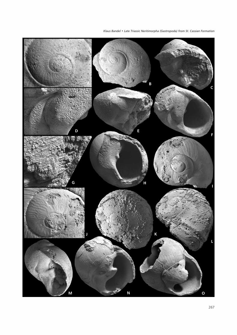
���
(�
� &�
� '�
) *
�
� � 3
���� ������ ��������� ���������� !�"�� �� ��#���$��%� ���&������
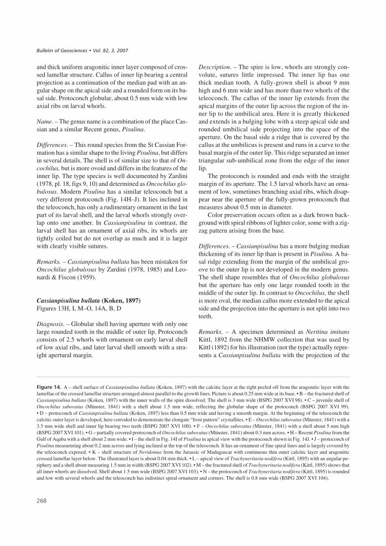
and thick uniform aragonitic inner layer composed of cros-sed lamellar structure. Callus of inner lip bearing a centralprojection as a continuation of the median pad with an an-gular shape on the apical side and a rounded form on its ba-sal side. Protoconch globular, about 0.5 mm wide with lowaxial ribs on larval whorls.
Name. – The genus name is a combination of the place Cas-sian and a similar Recent genus, Pisulina.
Differences. – This round species from the St Cassian For-mation has a similar shape to the living Pisulina, but differsin several details. The shell is of similar size to that of On-cochilus, but is more ovoid and differs in the features of theinner lip. The type species is well documented by Zardini(1978, pl. 18, figs 9, 10) and determined as Oncochilus glo-bulosus. Modern Pisulina has a similar teleoconch but avery different protoconch (Fig. 14H–J). It lies inclined inthe teleoconch, has only a rudimentary ornament in the lastpart of its larval shell, and the larval whorls strongly over-lap onto one another. In Cassianpisulina in contrast, thelarval shell has an ornament of axial ribs, its whorls aretightly coiled but do not overlap as much and it is largerwith clearly visible sutures.
Remarks. – Cassianpisulina bullata has been mistaken forOncochilus globulosus by Zardini (1978, 1985) and Leo-nardi & Fiscon (1959).
Cassianpisulina bullata (Koken, 1897)Figures 13H, I, M–O, 14A, B, D
Diagnosis. – Globular shell having aperture with only onelarge rounded tooth in the middle of outer lip. Protoconchconsists of 2.5 whorls with ornament on early larval shellof low axial ribs, and later larval shell smooth with a stra-ight apertural margin.
Description. – The spire is low, whorls are strongly con-volute, sutures little impressed. The inner lip has onethick median tooth. A fully-grown shell is about 9 mmhigh and 6 mm wide and has more than two whorls of theteleoconch. The callus of the inner lip extends from theapical margins of the outer lip across the region of the in-ner lip to the umbilical area. Here it is greatly thickenedand extends in a bulging lobe with a steep apical side androunded umbilical side projecting into the space of theaperture. On the basal side a ridge that is covered by thecallus at the umbilicus is present and runs in a curve to thebasal margin of the outer lip. This ridge separated an innertriangular sub-umbilical zone from the edge of the innerlip.
The protoconch is rounded and ends with the straightmargin of its aperture. The 1.5 larval whorls have an orna-ment of low, sometimes branching axial ribs, which disap-pear near the aperture of the fully-grown protoconch thatmeasures about 0.5 mm in diameter.
Color preservation occurs often as a dark brown back-ground with spiral ribbons of lighter color, some with a zig-zag pattern arising from the base.
Differences. – Cassianpisulina has a more bulging medianthickening of its inner lip than is present in Pisulina. A ba-sal ridge extending from the margin of the umbilical gro-ove to the outer lip is not developed in the modern genus.The shell shape resembles that of Oncochilus globulosusbut the aperture has only one large rounded tooth in themiddle of the outer lip. In contrast to Oncochilus, the shellis more oval, the median callus more extended to the apicalside and the projection into the aperture is not split into twoteeth.
Remarks. – A specimen determined as Neritina imitansKittl, 1892 from the NHMW collection that was used byKittl (1892) for his illustration (not the type) actually repre-sents a Cassianpisulina bullata with the projection of the
���
��#����$-% A – shell surface of Cassianpisulina bullata (Koken, 1897) with the calcitic layer at the right peeled off from the aragonitic layer with thelamellae of the crossed lamellar structure arranged almost parallel to the growth lines. Picture is about 0.25 mm wide at its base. • B – the fractured shell ofCassianpisulina bullata (Koken, 1897) with the inner walls of the spire dissolved. The shell is 3 mm wide (BSPG 2007 XVI 98). • C – juvenile shell ofOncochilus subovatus (Münster, 1841) with a shell about 1.5 mm wide, reflecting the globular shape of the protoconch (BSPG 2007 XVI 99).• D – protoconch of Cassianpisulina bullata (Koken, 1897) less than 0.5 mm wide and having a smooth margin. At the beginning of the teleoconch thecalcitic outer layer is developed, here corroded to demonstrate the elongate “frost pattern” crystallites. • E – Oncochilus subovatus (Münster, 1841) with a3.5 mm wide shell and inner lip bearing two teeth (BSPG 2007 XVI 100). • F – Oncochilus subovatus (Münster, 1841) with a shell about 5 mm high(BSPG 2007 XVI 101). • G – partially covered protoconch of Oncochilus subovatus (Münster, 1841) about 0.3 mm across. • H – Recent Pisulina from theGulf of Aqaba with a shell about 2 mm wide. • I – the shell in Fig. 14I of Pisulina in apical view with the protoconch shown in Fig. 14J. • J – protoconch ofPisulina measururing about 0.2 mm across and lying inclined at the top of the teleoconch. It has an ornament of fine spiral lines and is largely covered bythe teleoconch exposed. • K – shell structure of Neridomus from the Jurassic of Madagascar with continuous thin outer calcitic layer and aragoniticcrossed lamellar layer below. The illustrated layer is about 0.04 mm thick. • L – apical view of Trachyneritaria nodifera (Kittl, 1895) with an angular pe-riphery and a shell about measuring 1.5 mm in width (BSPG 2007 XVI 102). • M – the fractured shell of Trachyneritaria nodifera (Kittl, 1895) shows thatall inner whorls are dissolved. Shell about 1.5 mm wide (BSPG 2007 XVI 103). • N – the protoconch of Trachyneritaria nodifera (Kittl, 1895) is roundedand low with several whorls and the teleoconch has indistinct spiral ornament and corners. The shell is 0.8 mm wide (BSPG 2007 XVI 104).
����������� ������ �������������
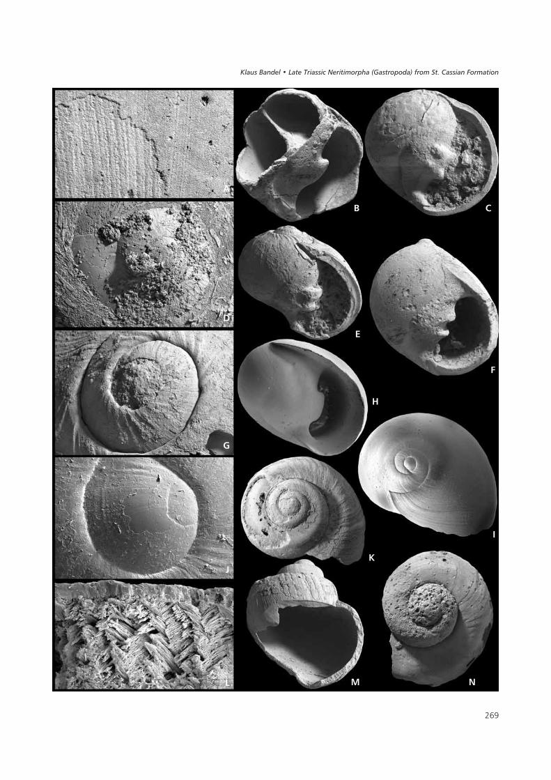
���
( �
�
&
�
�
'
�
)
�
*
� �
���� ������ ��������� ���������� !�"�� �� ��#���$��%� ���&������

inner lip. However, in his description of the Neritina imi-tans Kittl (1892, pl. 8, figs 1, 2) documents nothing of thatmedian projection, so the species remains in doubt.
Subfamily Trachyneritariinae n. subfam.
Type genus. – The subfamily is based on Trachyneritaria n.gen. from the St Cassian Formation, Alpe di Specie.
Diagnosis. – Teleoconch of angular shape with flattenedapical ramp, rounded corner to the side and a few more orless distinct rounded spiral ribs. Inner whorls of the shelltotally dissolved. Inner lip thickened and its inner edgeconvex. Callus forms a pad that ends near the base with anindistinct groove; its outer margin connected to the outerlip. Protoconch globular measuring about 0.3 mm. Shellstructure is crossed lamellar aragonitic with a very thin outercalcitic layer.
Differences. – Members of the Trachyneritariinae have theinner whorls of their shell totally dissolved, as is the case ofthe Neritariidae in general but they differ in the more angu-lar shape of their teleoconch, the simple convex bulge oftheir inner lip, and the relatively small globular proto-conch.
Remarks. – Existing information is limited for this groupsince protoconch ornament is unknown. The holotype tothe genus Trachynerita Kittl, 1894 is a large shell of 30 mmin diameter, whereas the specimens studied here and refer-red to Trachyneritaria are small with a size range of 2 to3 mm when fully-grown. Trachynerita as characterized byKnight et al. (1960, fig. 183, 16a, b) is a very different ge-nus to the Trachyneritaria described below.
Genus Trachyneritaria n. gen.
Type species. – The genus is based on Trachynerita nodi-fera Kittl, 1894, originally from the Marmolata Limestone,Italian Dolomites, and as newly defined here is based on in-dividuals from Alpe di Specie near Cortina d’Ampezzo(map in Zardini 1978, Wendt 1982, fig. 1).
Diagnosis. – Teleoconch with angular shoulder. Innerwhorls of shell dissolved. Globular protoconch consistingof several (about three) rounded whorls.
Name. – The name of the genus represents a combinationof the genus name Trachynerita (which has a similar shapebut larger shell size) and Neritaria to which the describedspecies is related. The small species is named with a com-bination of both: Trachyneritaria n. gen.
Trachyneritaria nodifera (Kittl, 1894)Figure 14L–N
Diagnosis. – Small shell reaching 3 mm in size consistingof about four whorls in teleoconch. Inner whorls totallydissolved. Rounded protoconch consisting of a few (aboutthree) convolute whorls and measuring about 0.3 mm.Whorls of teleoconch with rounded corner on convex sidesand rounded corner to the base. Additional spiral ornamentmay be present or absent. Aperture large with simple outerlip. Callus of inner lip forms rounded low pad that coversthe umbilical area and ends before reaching the margins ofthe outer lip on apical and basal sides. Margin of outer lipcontinues onto the margin of a weak depression on the baseending in the median callus pad of the inner lip.
Description. – The shell with 2.5 whorls of the teleoconchis only a little more than 2 mm high and a little less widewith a blunt spire. A size of about 3 mm in width and heightis attained in fully-grown shells containing four whorls ofthe teleoconch. The end of shell growth is indicated by thepresence of dense growth increments in the final part of thebody whorl. The protoconch of a little more than 0.3 mm indiameter has a simple apertural margin, and consists of se-veral whorls with the ornament of the larval shell not pre-served. First whorl of the teleoconch has a step-like shape.Whorl sides are weakly convex and end in a rounded corneron the convex base. A spiral rib may indicate the widestpart of the whorl and where crossed by growth lines mayhave low tubercles. The sutures are deep. Collabral growthlines are present and may be strong. Additional spiral ribsmay appear and disappear again in later whorls. The aper-ture is oval in shape and about twice as high as wide. Theouter lip is simple with the apical corner expressed. The in-ner lip widens in the middle and covers the umbilical re-gion with a callus. The inner side of the inner lip is weaklyconvex.
Differences. – Trachyneritaria nodifera may have nodularanterior and posterior edges and in general shape resemblesTrachynerita. Its protoconch is smaller (about 0.3 mm indiameter) than that of the other Neritariidae of the fauna ofthe St Cassian Formation.
Remarks. – The genus Trachynerita Kittl, 1894 is based onTurbo quadrata Stoppani, 1858 = Trachynerita foronensisKittl, 1894 from the Marmolata limestone, Italian Alps(Kittl 1894, pl. 3, figs 9–12; Böhm 1895, pl. 10, fig. 4).Turbo quadrata Stoppani, 1858 was originally describedfrom Esino and Kittl (1899, pl. 2, figs 1, 2) also recognizedfrom that locality a Trachynerita fornonensis Kittl, 1895.Nützel & Senowbari-Daryan (1999) described a speciesdetermined as Trachynerita quadrata from the Late Trias-sic of Iran that may even be up to 40 mm in size. This spe-
���
����������� ������ �������������

cies placed in the Neritidae by the authors is not sufficientlywell preserved to determine shell structure, protoconchshape or presence or absence of internal walls.
The type species of the genus Trachynerita has a muchlarger shell than the type to Trachyneritaria n. gen. as heredescribed based on Trachyneritaria nodifera (30 mm incontrast to 2–3 mm). The latter has been introduced asTrachynerita nodifera Kittl, 1894 from the Marmolatalimestone (Kittl 1894, pl. 3, figs 15, 16), and was describedagain by Böhm (1895, pl. 10, figs 1, 2) and mentioned byCossmann (1925, pl. 9, fig. 44). It does not resembleTrachynerita as described by Knight et al. (1960, fig. 183,16a, b), which has a strong spiral row of tubercles and isbased on a different type species (Trachynerita depressaHoernes, 1855). In the case of Trachynerita, color patternsof dark zigzag ribbons were noted by Kittl (1894). Zardini(1985, pl. 3, fig. 9a) illustrated Trachynerita quadrata anddetermined it as Naticopsis zitteli. Also Trachyneritatambosolensis Haas, 1953 from the Late Triassic of Peru(Haas 1953, pl. 11, figs 18, 19, 24) is similar. It has therounded aperture with a simple inner lip that covers theumbilical region with a thick callus (Bandel 1994). Fromthe St Cassian Formation, Trachynerita quadrata was de-termined by Leonardi & Fiscon (1959, pl. 4, fig. 15), andalso illustrated by Tichy (1980, pl. 4, fig. 18). Zardini(1978, pl. 10, fig. 16a–d) documented a shell resembling itin general shape as Turbo subcarinatus (Münster, 1841). Itis up to 10 mm high and has color patterns preserved.The shell of Trachynerita quadrata (Stoppani, 1858) hasan apical angle and otherwise is of rounded outline (Wenz1938–1944, fig. 1007). It resembles Trachynerita altoni(Kittl, 1892) that is present with its holotype in the NHMWcollection and had originally been described as Naticopsisaltoni by Kittl (1892, pl. 7, fig. 14). Its shell is of roundedoutline with a short spire forming the shoulders and whorlsand distinguished from each other by groove-like sutures.The body whorl is large and has a simple large aperturewith a well-developed inner lip callus. Its shell is largerthan that of Trachyneritaria nodifera and nothing is knownabout its internal composition.
�6�7���#����
Originally, Annette Schwardt (Hamburg) began this study sup-ported by financial aid from a DFG project to revise theNeritimorpha from the Triassic St Cassian Formation in the vicin-ity of Cortina d’Ampezzo in the Italian Alps, but discontinued herwork by end of 1994. She borrowed some originals from theNaturhistorisches Museum at Vienna (NHMW) from the studiesof Laube (1869) and Kittl (1892) that have been included in thisstudy. More recent material has been collected by the author(aided by his students) during several trips in the region of Cortinad’Ampezzo with localities pointed out by the late RinaldoZardini. Zardini and Rolando Lancedelli had collected in the area
over many years and also provided some of the specimens thatwere studied here. Robert Welti and Ivonne Milker helped in doc-umenting the material with the scanning electronic microscope.Photographs of specimens were made and plates assembled byEva Vinx. Marlies Becker helped in handling the manuscript dur-ing its production. I also thank Robert B. Blodgett and AlexanderNützel for their thoughtful reviews of the manuscript and SusanTurner for revising the English.
8���������
BANDEL, K. 1982. Morphologie und Bildung der frühontogene-tischen Gehäuse bei conchiferen Mollusken. Facies 7, 1–198.
BANDEL, K. 1988. Repräsentieren die Euomphaloidea einenatürliche Einheit der Gastropoden? Mitteilungen aus demGeologisch-Paläontologischen Institut der Universität Ham-burg 67, 1–33.
BANDEL, K. 1990. Shell structure of the Gastropoda excluding theArchaeogastropoda, 117–134. In CARTER, J.G. (ed.) SkeletalBiomineralization: Patterns Processes and EvolutionaryTrends. Volume I. Van Nostrand Reinolds, New York.
BANDEL, K. 1993. Trochomorpha aus der triassischen St. CassianFormation (Gastropoda, Dolomiten). Annales des Naturhisto-rischen Museums in Wien 95, 1–99.
BANDEL, K. 1994. Comparison of Upper Triassic and Lower Ju-rassic gastropods from the Peruvian Andes (Pucará Group)and the Alps (Cassian Formation). Palaeontographica 233A,127–160.
BANDEL, K. 1997. Higher classification and pattern of evolutionof the Gastropoda. A synthesis of biological and paleon-tological data. Courier Forschungsinstitut Senckenberg 201,57–81.
BANDEL, K. 2000. The new family Cortinellidae (Gastropoda,Mollusca) connected to a review of the evolutionary history ofthe subclass Neritimorpha. Neues Jahrbuch für Geologie undPaläontologie, Abhandlungen 217(1), 111–129.
BANDEL, K. 2001. The history of Theodoxus and Neritina con-nected with description and systematic evaluation of relatedNeritimorpha (Gastropoda). Mitteilungen aus dem Geolo-gisch-Paläontologischen Institut der Universität Hamburg85, 65–164.
BANDEL, K. 2002. Reevaluation and classification of Carbonifer-ous and Permian Gastropoda belonging to the Caenogastro-poda and their relation. Mitteilungen aus dem Geologisch-Paläontologischen Institut der Universität Hamburg 86,81–188.
BANDEL, K. & FRÝDA, J. 1999. Notes on the evolution and higherclassification of the subclass Neritimorpha (Gastropoda) withthe description of some new taxa. Geologica et Palaeon-tologica 33, 219–235.
BANDEL, K. & HEIDELBERGER, E. 2001. The new family Nerrhe-
���
���� ������ ��������� ���������� !�"�� �� ��#���$��%� ���&������
nidae (Neritimorpha, Gastropoda) from the Givetian of Ger-many. Neues Jahrbuch für Geologie und Paläontologie,Monatshefte 2001(12), 705–718.
BANDEL, K. & KIEL, S. 2003. Relationships of Cretaceous Neriti-morpha (Gastropoda, Mollusca), with the description of sevennew species. Bulletin of the Czech Geological Survey 7, 53–65.
BIGGELAAR, J.A.M. VAN DEN & HASZPRUNAR, G. 1996. Cleavagepatterns and mesentoblast formation in the Gastropoda: anevolutionary perspective. Evolution 50, 1520–1540.
BLASCHKE, F. 1905. Die Gastropodenfauna der Pachycardien-tuffe der Seiseralpe in Südtirol nebst einem Nachtrag zurGastropodenfauna der roten Raibler Schichten vom Schlern-plateau. Beiträge zur Paläontologie und Geologie Öster-raich-Ungarns und des Orients 17, 161–222.
BLENDINGER, W. & BLENDINGER, E. 1989. Windward-leewardeffects on Triassic carbonate bank margins in the Dolomites,northern Italy. Sedimentary Geology 64, 143–166.
BLODGETT, R.B., FRÝDA, J. & STANLEY, G.D. 2001. Dephinulo-psidae, a new neritopsoidean family from the Upper Triassic(upper Carnian to lower Norian) of the Wallowa terrane,northeastern Oregon. Journal of Czech Geological Society 46,307–318.
BÖHM, J. 1895. Die Gastropoden des Marmolatakalkes. Palaeon-tographica 42, 211–308.
BOGGILD, O.B. 1930. The shell structure of the molluscs. DetKongelige Danske Videnskabernes Selkabs Skrifter, Natur-videnskabelig og Mathematisk Afdeling 9, 231–326.
BOUCHET, P. & ROCROI, J.-P. 2005. Classification and nomen-clator of gastropod families. Malacologia 47, 1–397.
BOURNE, G.C. 1908. Contributions to the morphology of thegroup Neritacea of aspidobranch gastropods. Part I. The Neri-tidae. Proceedings of the Zoological Society of London 1908,810–887.
BRÖSAMLEN, R. 1909. Beitrag zur Kenntnis der Gastropoden desschwäbischen Jura. Palaeontographica 56, 177–321.
BROILI, F. 1907. Die Fauna der Pachycardientuffe der Seiser Alp.Scaphopoden und Gastropoden. Palaeontographica 54,69–138.
COSSMANN, M. 1898. Referat Koken: Die Gastropoden der Triasvon Hallstadt. Revue critique de paléozoologie 2, 148–150.
COSSMANN, M. 1925. Essais de Paléontologie Comparée.Treizième Livraison. 345 pp. Les Presses Universitaires deFrance, Paris.
DIENER, C. 1926. Fossilium Catalogus I: Animalia, Pars 34 –Glossophora triadica. 242 pp. W. Junk, Berlin.
FRETTER, V. & GRAHAM, A. 1962. British prosobranch molluscs,their functional anatomy and ecology. 755 pp. Ray Society,London.
FRÝDA, J. 2000. Some Givetian (late Middle Devonian) gastro-pods from the Paffrath area (Bergisches Land, Germany).Memoirs of the Queensland Museum 45, 359–374.
FRÝDA, J. & BANDEL, K. 1997. New Early Devonian gastropodsfrom the Plectonotus (Boucotonotus)–PalaeozygopleuraCommunity in the Prague Basin (Bohemia). Mitteilungen ausdem Geologisch-Paläontologischen Institut der UniversitätHamburg 80, 1–57.
FRÝDA, J., BLODGETT, R.B. & STANLEY, G.D. 2003. New neri-topsoidean gastropods (Neritimorpha) from the Late Triassic(Late Carnian–Early Norian) of the Wallowa Terrane, north-eastern Oregon. Mitteilungen aus dem Geologisch-Paläon-tologischen Institut der Universität Hamburg 87, 55–72.
FÜRSICH, F.T. & WENDT, J. 1977. Biostratinomy and palaeoecol-ogy of the Cassian Formation (Triassic) of the southern Alps.Palaeogeography, Palaeoclimatology, Palaeoecology 22,257–323.
GOLIKOV, A.N. & STAROBOGATOV, Y.I. 1975. Systematics ofprosobranch gastropods. Malacologia 15(1), 185–232.
GORDON, M. & YOCHELSON, E.L. 1982. A Naticopsis operculumfound in situ (Gastropoda: Mississippian). Journal of Paleon-tology 56, 260–265.
GRÜNDEL, J. 1980. Zur Gastropodenfauna des Unteren Muschel-kalkes im Raume Halle-Unstruttal (DDR). FreibergerForschungshefte C348, 41–59.
HAAS, O. 1953. The Late Triassic gastropods from Central Peru.Bulletin of the American Museum of Natural History 101,9–328.
HASZPRUNAR, G. 1988. On the origin and evolution of the majorgastropod groups, with special reference to the Streptoneura.Journal of Molluscan Studies 54, 367–441.
HEIDELBERGER, D. 2001. Mitteldevonische (Givetische) Gastro-poden (Mollusca) aus der Lahnmulde (südliches RheinischesSchiefergebirge). Geologische Abhandlungen Hessen 106,1–291.
HEIDELBERGER, D. & BANDEL, K. 1999. Micromorph Gastropodafrom the Middle Devonian (Givetian) limestone of the Soe-tenich Syncline (Eifel). Mitteilungen aus dem Geologisch-Pa-läontologischen Institut der Universität Hamburg 83,129–162.
HERHOLZ, M. 1992. Mikromorphe Gastropoden aus demrheinisch-westfälischen Steinkohlerevier (Oberkarbon).Neues Jahrbuch für Geologie und Paläontologie, Monatshefte1992, 242–256.
HOLTHUIS, B.V. 1995. Evolution between marine and freshwaterhabitats: a case study of the gastropod Neritopsina. 286 pp.Doctoral dissertation, University of Washington.
JAGT, J.W.M. & JANSSEN, A.W. 1988. Jouannetia (Bivalvia,Pholadidae) and Neritopsis (Gastropoda, Neritopsidae), twomollusks from the Danian (Paleocene) of the Maastricht area(SE Netherlands and NE Belgium). Mededelinden Werkgroepvoor Tertiare en Kwartaire Geologie 25, 163–174.
JEFFERY, D.L., HOARE, R.D., MAPES, R.H. & BROWN, C.L. 1994.Gastropods (Mollusca) from the Imo Formation (Mississip-
���
����������� ������ �������������
pian, Chesterian) of north-central Arkansas. Journal of Pale-ontology 68, 58–79.
KAIM, A. & SZTAJNER, P. 2005. The opercula of neritopsid gastro-pods and their phylogenetic importance. Journal of MolluscanStudies 71, 211–219.
KANO, Y., CHIBA, S. & KASE, T. 2002. Major adaptive radiationin neritopsine gastropods estimated from 28S rRNA se-quences and fossile records. Proceeding of the Royal Society2699, 2457–2465.
KANO, Y. & KASE, T. 2000. Taxonomic revision of Pisulina(Gastropoda: Neritopsina) from submarine caves in the tropi-cal Indo-Pacific. Paleontological Research 4, 107–129.
KITTL, E. 1892. Die Gastropoden der Schichten von St. Cassiander südalpinen Trias. Part 2. Annalen des kaiserlich-könig-lichen naturhistorischen Hofmuseums 7, 35–97.
KITTL, E. 1894. Die triadischen Gastropoden der Marmolata undverwandter Fundstellen in den weissen Riffkalken Südtirols.Jahrbuch der Kaiserlich-königlichen geologischen Reichs-anstalt 44 (1894), 99–182.
KITTL, E. 1899. Die Gastropoden der Esinokalke, nebst einer Re-vision der Gastropoden der Marmolatakalke. Annalen deskaiserlich-königlichen naturhistorischen Hofmuseums 14,1–237.
KITTL, E. 1901. Bakonyi Triasz-Gastropodák. 54 pp. Budapest.KLIPSTEIN, A.V. 1843. Beiträge zur geologischen Kenntnis der
östlichen Alpen. 311 pp. Georg Friedrich Geyer, Giessen.KNIGHT, J.B. 1941. Paleozoic gastropod genotypes. Bulletin of
the Geological Society of America, Special Paper 32,1–510.
KNIGHT, J.B., COX, L.R., KEEN, A.M., BATTEN, R.L., YOCHEL-
SON, E.L. & ROBERTSON, R. 1960. Part I, Mollusca. System-atic descriptions, I169–I351. In MOORE, R.C. (ed.) Treatise onInvertebrate Paleontology. University of Kansas Press, Law-rence.
KONINCK, L. de 1853. Notes sur le genre Hypodema. Mémoires dela Societé Royale des Sciences de Liége 8, 140–144.
KOKEN, E. 1889. Über die Entwicklung der Gastropoden vomCambrium bis zur Trias. Neues Jahrbuch für Mineralogie,Geologie und Paläontologie, suppl. vol. 6, 305–484.
KOKEN, E. 1892. Über die Gastropoden der rothen Schlern-schichten nebst Bemerkungen über Verbreitung und Herkunfteinige triassischer Gattungen. Neues Jahrbuch für Minera-logie 1892(2), 25–36.
KOKEN, E. 1897. Die Gastropoden der Trias von Hallstatt. Ab-handlungen der Kaiserlich- königlichen geologischen Reichs-anstalt in Wien 17(4), 1–112.
KUES, B.S. & BATTEN, R.L. 2001. Middle Pennsylvanian gastro-pods from the Flechado Formation, North-Central New Mex-ico. Journal of Paleontology, Memoir 54, Supplement 1(75),1–95.
LAUBE, G.C. 1869. Die Fauna der Schichten von St. Cassian.
Kaiserliche Akademie der Wissenschaften, Denkschrift 30,1–48.
LEONARDI, P. & FISCON, F. 1959. La fauna Cassiana di Cortinad’Ampezzo. 3. Gasteropodi. Memorie degli Istituti di Geo-logia e Mineralogia dell’Università di Padova 21, 1–103.
MOJSISOVICS, E. VON 1879. Die Dolomit-Riffe von Südtirol undVenetien. Beiträge zur Bildungsgeschichte der Alpen. 552 pp.Hölder, Wien.
MÜNSTER, G.G. VON 1841. Beschreibung und Abbildung der inden Kalkmergelschichten von St. Cassian gefundenen Ver-steinerungen, 25–152. In WISSMANN, H.L. & MÜNSTER, G.G.Beiträge zur Geologie und Petrefacten-Kunde des südöst-lichen Tirol’s vorzüglich der Schichten von St. Cassian 4.Buchner, Bayreuth.
NÜTZEL, A. & ERWIN, D.H. 2004. Late Triassic (Late Norian)gastropods from the Wallowa Terrane (Idaho, USA). Palä-ontologische Zeitschrift 78, 361–416.
NÜTZEL, A. & SENOWBARI-DARYAN, B. 1999. Gastropods fromthe Later Triassic (Norian-Rhaetian) Nayband Formation ofcentral Iran. Beringeria 23, 93–132.
PONDER, W.F. & LINDBERG, D. 1997. Towards a phylogeny ofgastropod molluscs: an analysis using morphological charac-ters. Zoological Journal of the Linnean Society 119, 83–265.
ROBERTSON, R. 1971. Scanning electron microscopy of plank-tonic larval marine gastropod shells. The Veliger 14(1), 1–12.
SASAKI, T. 1998. Comparative anatomy and phylogeny of the Re-cent Archaeogastropoda (Mollusca: Gastropoda). The Univer-sity Museum, The University of Tokyo, Bulletin 38, 1–223.
SASAKI, T. 2001. Macro- and microstructure of shell and oper-culum in two recent gastropod species, Nerita (Theliostyla)albicilla and Cinnalepeta pulchella (Neritopsina Neritoidea).Palaeontological Research 5, 21–31.
SCOTT, B.J. & KENNY, R. 1998. Superfamily Neritoidea, 694–702.In BEESLEY, P.L., ROSS, G.J.B. & WELLS, A. (eds) Mollusca,The Southern Synthesis. Fauna of Australia 5, Part B. Com-monwealth Scientific and Industrial Research Organization,Canberra.
SUZUKI, S., TOGO, Y. & UOZUMI, S. 1991. Exosceletal adapta-tions for life environment in neritacean gastropods, 397–401.In SUGA, S. & NAKAHARA, H. (eds) Biological Systems.Springer Verlag, Tokyo.
THIELE, J. 1929–1931. Handbuch der systematischen Weichtier-kunde. Gustav Fischer Verlag, Jena.
TICHY, G. 1980. Über die Erhaltung von Farben und Farbmusternan traissischen Gastropoden-Gehäusen (Colour retention inTriassic gastropods). Verhandlungen der Geologischen Bun-desanstalt A 1980(2), 175–217.
THOMPSON, F.G. 1980. Proserpinoid land snails and their rela-tionships within the Archaeogastropoda. Malacologia 20,1–33.
TROSCHEL, F.H. 1856. Das Gebiss der Schnecken zur Beg-
���
���� ������ ��������� ���������� !�"�� �� ��#���$��%� ���&������

ründung einer natürlichen Classification 1. 252 pp. Nico-laische Verlagsbuchhandlung, Berlin.
TROSCHEL, F.H. & THIELE, J. 1865–1893. Das Gebiss derSchnecken zur Begründung einer natürlichen Classification 2.409 pp. Nicolaische Verlagsbuchhandlung, Berlin.
URLICHS, M. 1974. Zur Stratigraphie und Ammonitenfauna derCassianer Schichten von Cassian (Dolomiten/ Italien).Schriftenreihe der Erdwissenschaftlicher Kommission 2,207–222.
URLICHS, M. 1994. Trachyceras Laube, 1869 (Ammonoidea) ausdem Unterkarn (Obertrias) der Dolomiten (Italien). Stutt-garter Beiträge zur Naturkunde, Serie B 217, 1–55.
WENDT, J. 1982. The Cassian patch reefs (Lower Carnian, South-ern Alps). Facies 6, 185–202.
WENZ, W. 1938–1944. Gastropoda, Teil I. 1639 pp. In SCHINDE-
WOLF, O.H. (ed.) Handbuch der Paläozoologie, Bd. 6.Gebrüder Borntraeger, Berlin.
WILSON, B.R. 1993. Australian marine shells. Prosobranch gas-tropods. Part 1 (Prosobranch gastropods). 408 pp. OdysseyPublishing, Kallaroo, Western Australia.
YIN, H.-F. & YOCHELSON, E.L. 1983. Middle Triassic Gastro-poda from Qingyan, Ghizhou Province, China: Euomphalaceaand Loxonematacea. Journal of Paleontology 57, 1098–1127.
ZARDINI, R. 1978. Fossili Cassiani (Trias Mediosuperiore)Atlanti dei Gasteropodi della Formazione di S. CassianoRaccolti della Regione Dolomitica Atorno a Cortinad’Ampezzo. 58 pp. Cortina d’Ampezzo.
ZARDINI, R. 1980. Fossili Cassiani (Trias Mediosuperiore)Atlanti dei Gasteropodi della Formazione di S. CassianoRaccolti della Regione Dolomitica Atorno a Cortinad’Ampezzo. 14 pp. Cortina d’Ampezzo.
ZARDINI, R. 1985. Fossili Cassiani (Trias Mediosuperiore)Atlanti dei Gasteropodi della Formazione di S. CassianoRaccolti della Regione Dolomitica Atorno a Cortinad’Ampezzo. 16 pp. Cortina d’Ampezzo.
ZITTEL, K.A. 1873. Die Gastropoden der Stramberger Schichten.Paläontologische Mittheilungen aus dem Museum des könig-lich Bayrerischen Staates 2, 311–491.
ZITTEL, K.A. 1895. Grundzüge der Palaeontologie. 971 pp.R. Oldenbourg, München and Leipzig.
���
����������� ������ �������������