Embed Size (px)
Citation preview


OCCASIONAL PAPERS
OF THE
CALIFORNIA ACADEMY OF SCIENCES
No . 128 , 78 pages , 31 figures , 1 table June 22 , 1978
Cranial Nerves of a Percoid Fish , Polycentrus schomburgkii
(Fam ily Nandidae) , a Contribution to the Morpho logy and Classification
of the Order Perciformes
By
Warren C . Freihofer
Department of Ichthyo logy , California A cademy of Sciences ,
Go lden Ga te Park , San F rancisco , Ca lifornia 941 18
SAN FRANC ISCO
PUBLISHED BY THE ACADEMY
COMMITTEE ON PUBL ICATIONS
Laurence C . B inford , Cha irman
Tom-io Iwamoto , Edito r
Paul H . Arnaud , Jr .
Wi ll iam N . Eschmeyer
George E . L i ndsay
The Cal iforn ia Academy of Sc ience s
Golden Gate Park
San Franc i sco , Cal ifornia 94 1 18
PRINTED IN THE UNITED STATES OF AMERICABY ALLEN PRESS INC . . LAWRENCE , KANSAS
TABLE OF CONTENTS
ABSTRACT
INTRODUCT ION
METHODS
FAMILY NANDIDAE
NAMES OF CRAN IAL NERVES
NERVE COMPONENTS
DESCRIPT IONS OF NERVES
Rad ix Profundu s
Fifth and Seventh C ran ial Nerve Root s , Trunks , and Rami
Ro o ts and gang lia of nervus trig eminus
Communis ro o t and g enicula te gang lion
Do rsal la teralis roo t offacial nerveGasserian gang lion of nervus trig eminusRoo ts and gang lion of nervus fac ia lisRamus communicans of nervus trig em inus
Truncu s Supraorb i tal i s
Ramus ophtha lm icus sup erfic ia lis trig eminus and r . op hth . supf.fac ia lisTruncu s I nfraorb i tal i s
Ramus bucca lis fac ia lisRamus maxillaris trig em inus
Ramus mandibularis trigeminus
Ramus op ercularis trig eminus
Innerva tio n of cheek muscle
Ramulus mandibularis cutaneus trig em inus
Ramulus mandibularis externus trig em inus
Ramulus mandibularis internus trigeminus
Ramus Ot i cu s
Ramus Palat i nu s
Tru ncu s Hyomandibularis
Ramus op ercularis p rofundus fac ialisRamus op ercularis sup erficia lis fac ialisRamus hyo ideus
Ramus mandibularis fac ia lisRamus bucca lis accessorius fac ialisRamus mandibularis externus fac ialis and ramus
mandibularis internus facialisRamus Lateral i s Acces soriu s
The o rbito -
p ec to ra l branch (RLA - OP)The parie to
- do rsa l branch (RLA -PD)I nnervat ion Of t he G i l l Arches
Nervu s GlOSSOpharyngeus
Nervu s Vagus I
Intracranial do rsal vaga l ramus
Rami cutanei dorsales vag i
Ramus op ercularis vag i
Ramus sup ra temp ora lis vag i
F irs t vaga l branchia l trunk
Second vaga l branchia l trunk
Third vaga l branchia l trunk
Fourth vagal branchia l trunk
Summary of G i l l - Arch Musc le I nnervat ion
Lateral L i ne Nerves Of the Trunk
Nervus L i nae Late ral i s
D ISCUSSION
Free Cephal i c Lateral i s Organs
Comparison with Perca fluv iatilus
Compa rison with MenidiaComparison with cyp rinids
Compariso n with eso co ids
Comp arison with Amia
Cephal i c Lateral i s Canal s and Neu romas t s
I nnervat ion of Upper Jaw
Cheek Muscl e I nnervat ion
Correct Name for Ramus Lateral i s Acce s soriu s
Trunk Late ral L i ne Nerves
An External - I nternal Latera l L i ne Canal Nerve Re lat ionsh ip
Rad ix Profund'
us
A S tretch Receptor Nerve to Base of Max i l lary Tendon
S impl ify i ng Cran ial Nerve Stud ie s
SUMMARY
ACKNOWLEDGMENTS
L ITERATURE C ITED
ABBREVIATIONS FOR FIGURESILLU STRATION S
ABSTRACT
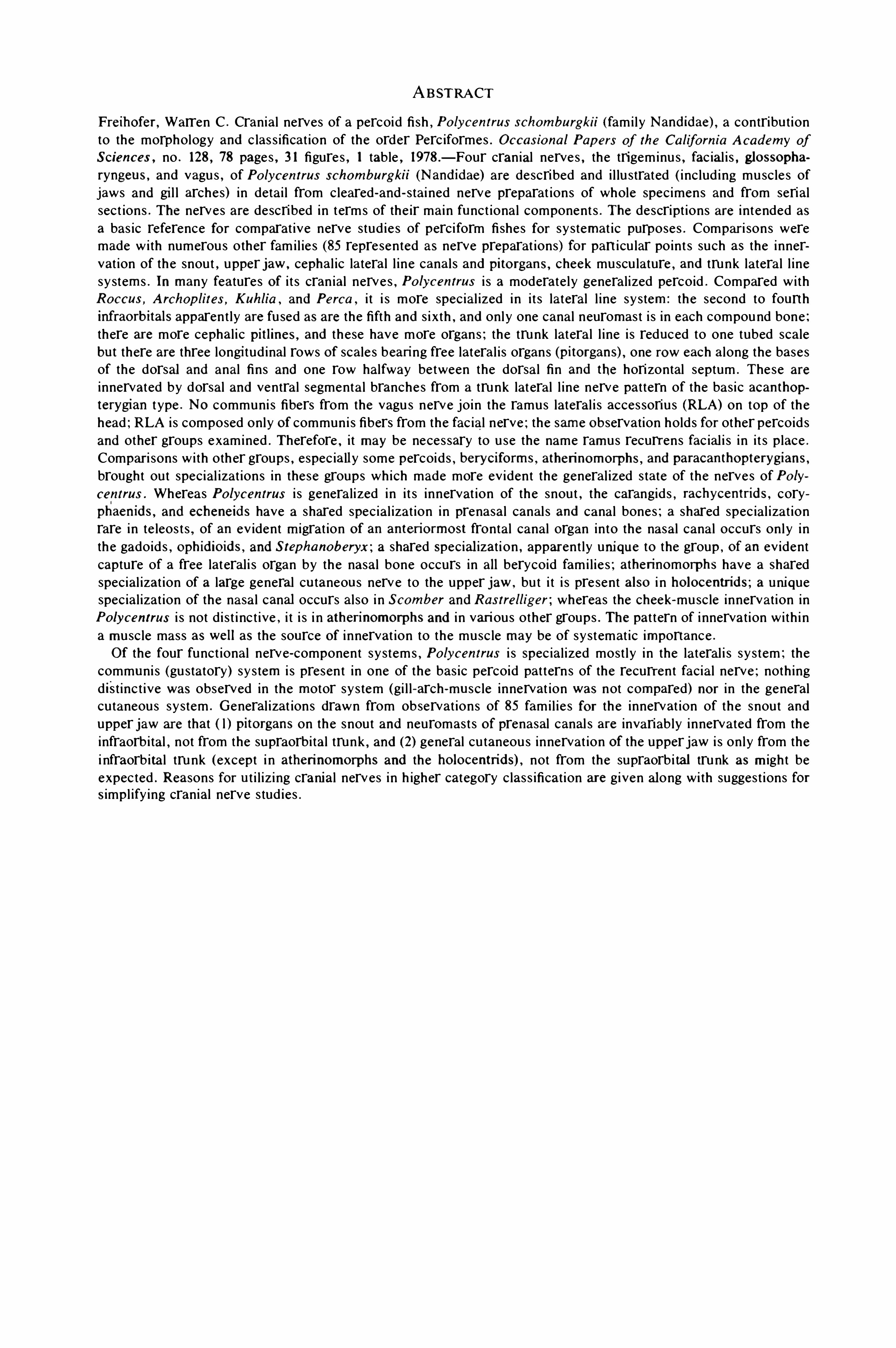
Freihofer, Warren C . Cranial nerves of a percoid fi sh ,Po lycentrus schomburgkii (fami ly Nandidae) , a contribut ion
to the morphology and c las s ificat ion of the order Perc iformes . Occas io na l Pap ers of the Ca lifornia Academy ofSciences , no . 128 , 78 pages , 3 1 figure s , 1 table , 1978 — Four cranial nerves , the trigeminu s , fac ial i s , glossopharyngeus , and vagu s , of Po lycentrus schomburgkii (Nandidae) are descri bed and i l lu strated ( i nc lud ing musc le s ofjaws and gi l l arches) i n detai l from cleared - and- stai ned nerve preparat ions of whole Spec imens and from serialsect ions . The nerves are descri bed in terms of the ir mai n funct ional component s . The descript ions are i ntended asa bas ic reference for comparat ive nerve stud ie s of perciform fi shes for sy stemat ic purposes . Compari sons weremade wi th numerou s other fami l ie s (85 repre sented as nerve preparat ions) for part i cu lar point s such as the innervat ion of the snout , upper jaw , cephal ic lateral l i ne canal s and pito rgans , cheek muscu lature , and trunk lateral l i nesy stems . In many features Of i t s cran ial nerves , Po lycentrus i s a moderate l y general i zed percoid . Compared wi thRoccus , Archop lites , Kuhlia , and Perca , i t i s more spec ial ized in i t s lateral l i ne sy stem : the second to fourthinfraorbitals apparen t ly are fu sed as are the fifth and s i xth , and onl y one canal neuromas t i s in each compound bone :there are more cephal i c pitlines , and these have more organs ; the trunk lateral l i ne i s reduced to one tubed scalebut there are three longi tud inal rows Of scale s beari ng free lateral i s organs (pitorgans) , one row each along the basesof the dorsal and anal fins and one row halfway between the dorsa l fin and the hori zontal septum . These are
i nnervated by dorsal and ventral segmental branches from a trunk latera l l i ne nerve pat tern of the bas ic acanthopterygian t ype . No communi s fibers from the vagu s nerve join the ramus lateral i s acces sori u s (RLA) on top of thehead ; RLA i s composed only Of commun i s fibers from the fac ial nerve ; the same Observation holds for other percoid sand other groups examined . Therefore , i t may be neces sary to u se the name ramus recurrens fac ial i s i n i t s p lace .
Compari sons wi th other groups , espec ia lly some percoids , beryciforms , atherinomorphs , and paracanthopterygians ,
brought out special izat ions in these groups wh ich made more ev ident the general ized s tate of the nerves of Po lycentras . Whereas Po lycentrus i s genera l i zed in i t s innervat ion of the snout , the carangids , rachycentrids , coryphaenids , and echeneids have a shared spec ial izat ion in prenasal canal s and canal bones ; a shared spec ial i zat ionrare in te leos t s , of an ev ident m igrat ion Of an anteriormost frontal canal organ into the nasal canal occurs on l y i nthe gadoid s , Ophidio ids , and Stephanoberyx ; a shared Special izat i on , apparent ly unique to the group , Of an ev identcapture of a free lateral i s organ by the nasa l bone occurs in all berycoid fami l ie s ; atherinomorphs have a Sharedspecial izat ion of a large general cutaneous nerve to the upper jaw , bu t i t i s pre sent also i n ho locentn
'
ds ; a uniquespec ial izat ion of the nasa l canal occurs al so i n Scomber and Ras trelliger ; whereas the cheek—musc le innervat ion inPo lycentrus i s not d i s t i nc t ive , i t i s i n atherinomorphs and i n vari ous other groups . The pat tern of i nnervat ion wi th i na musc le mass as we ll as the source Of i nnervat ion to the musc le may be of sy stemat ic importance .
Of the four funct ional nerve- component sy stems , Po lycentrus i s Spec ial i zed most l y i n the lateral i s sy s tem ; thecommuni s (gu statory) sy s tem i s presen t in one of the bas ic percoid pat terns of the recurrent facial nerve ; noth ingd is t inc t ive was observed in the motor sy s tem (gi l l - arch -musc le innervat ion was not compared) nor i n the generalcu taneous sy stem . General i zat ions drawn from Observat ions of 85 fami l ie s for the innervat ion of the snout andupper jaw are that ( l ) pitorgans on the snout and neuromas t s Of prenasal canal s are invariably innervated from theinfraorbi tal , not from the supraorbi tal trunk , and (2) general cu taneou s innervat ion Of the upper jaw i s on ly from thei nfraorbital trunk (except i n atheri nomorphs and the ho locentrids) , not from the supraorbi tal trunk as m igh t beexpec ted . Reasons for ut i l i z i ng cranial nerves in h igher category c las s ificat ion are given along wi th sugges t ions forS impl ify ing cranial nerve s tud ies .


l er preparat ion s ( see under Methods sec t ion
for u se) may las t 15 to 20 years and no doubt
wi l l las t longer . The reference col l ect ion,once
as sembled and growing , can be u sed for con
duct ing su rvey s Of promi s i ng nerve complexe s .
Several ne rve complexes were surveyed dur
i ng the t ime th i s de scri pt i ve account OfPo lycen
trus was i n progres s . The resu l t s are i n variou s
s tage s of complet ion and are planned for later
publ icat ion . The stud ie s embrace compari sons
rangi ng from Am ia up to callionymids . S i h l er
nerve preparat ion s represent i ng up to 90 fami l ie s
were u sed . Two Of the nerve complexe s in
vo l v ed t h e i n ne rvat io n Of lat e ra l l i n e cana l
bones ; a th i rd was a large general cu taneou s
nerve Of the supraorb i tal t ru nk ; a fou rth ln
volved pat tern s Of i nnervat ion to the cheek mus
c le mas s ; a fifth concerned new pat tern s of a
gu s tatory nerve , the recurrent fac ial ; and the
s i x th was on t runk lateral l i ne nerve pat te rn s .
Each Of these stud i e s revealed nerve charac
t ers of prom i s i ng sy stemat ic importance . A few
are apparent ly convergent in some groups . Two
at t ribute s Of nerve s gi v ing them sy stemat i c pO
tential were Observed i n these s tud ie s . One i s
that nerves fol low the i r end organ s ( tas te bud s ,lat eral l ine canal organs , and muscle s) . The Oth
er i s t hat , in general , each nerve i nnervat i ng a
lat eral l i ne canal neuromas t has a certai n mor
pho logical i n tegri ty connected wi th that bone
and organ . NO other nerve to a canal neu romas t
may enter through the substance Of that bone
and supply a canal organ there . Thi s Observat i on
might be s tated as a ru le . AS wi th all ru l e s , they
are alway s ( i t s eems) occas i onal l y broken , but
there are alway s good reason s for the i r be i ng
broken ; the except ion s to the ru le , if the except ion s are val id , Shou ld prove the ru le . Important
for the sy s temat i s t u s i ng nerves i s the fact t hat
t he except i on s re su l t in new Spec ial i zat i on s
bei ng formed . These may become Characte rs
usefu l to t he sy stemat i s t . If nerves alway s fol
lowed the ru le s,there would be fewer important
sy s temat i c characters .
Some example s Of what nerves do that are
i mportant to sy stemat i s t s come from study Of
t he nerves and neu romas t s Of the canal bones .
No t on ly may the total number Of canal o rgan s
be S ign ificant for a ma i n branch Of the cephal i ccanal sy s tem ,
but the changes in the number Of
organ s in each canal bone may be espec ial l y im
portant . These changes i nvolve how canal or
gan s i n a bone may i ncrease or decrease i n num
OCCASIONAL PAPERS O
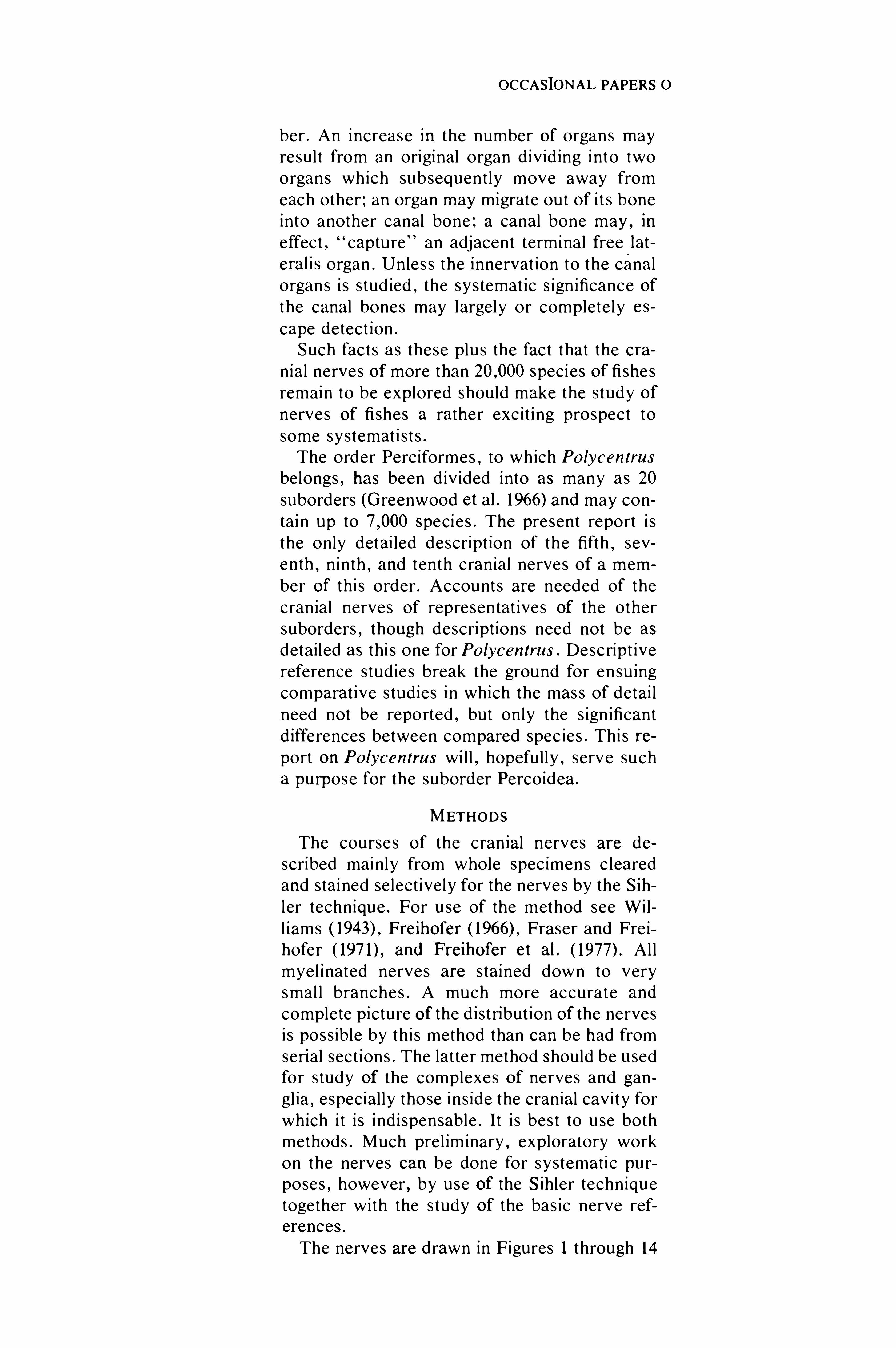
ber . An increas e in the number Of organ s mayre sul t from an origi nal o rgan d iv id ing i nto two
organ s wh ich subsequent ly move away from
each other ; an organ may migrate ou t Of i t s bone
i nto another canal bone ; a canal bone may ,in
effect,captu re
#
an adjacent termi nal free lat
eralis organ . Un le s s the innervat ion to the canal
organs i s stud ied ,the sy s temat ic S ign ificance Of
the canal bones may largel y or complete l y e s
cape detect ion .
Such fact s as these plu s the fact t hat the cra
nial nerves Of more than Speci es Of fi she s
remai n to be explored Should make the s tudy Of
nerves Of fi shes a rather exc i t i ng prospect to
some sy stemat i s t s .
The order Perc iformes , to wh ich Po lycentrus
belongs , has been d iv ided i nto as many as 20
suborders (Greenwood et al . 1966) and may con
tai n up to Speci e s . The present report i s
the only detai led descript ion of the fifth , sev
enth , ninth , and tenth cran ial ne rves of a mem
ber Of th i s order . Account s are needed of the
cran ial nerves Of representat i ve s Of the other
suborders , though descript ions need not be as
detai l ed as th i s one for Po lycentrus . Desc ript i ve
reference s tud ie s break the ground for en su ing
comparat i ve s tud ies i n which the mas s of detai l
need not be repo rted , but on ly the s ign ificant
d ifferences between compared spec ie s . Thi s re
port on Po lycentrus wi l l , hopefu l l y , serve su ch
a pu rpose for the suborder Percoidea .
METHODS
The course s of the cran ial nerves are de
scribed mai n l y from whole Spec imen s c leared
and stai ned se lect i ve l y for the nerves by the Sih
l er technique . For u se of the method see Wi l
l iams Freihofer Fraser and Fre i
hofer and Freihofer et al . Al l
mye l i nated nerves are s ta i ned down to very
smal l branche s . A much more accurat e and
complete picture Of the d i s t ribut ion Of the nerves
i s poss ibl e by th i s method than can be had from
serial sect ions . The lat ter method Should be used
for s tudy Of the complexes Of nerves and gan
gl ia , especial l y those i n s ide the cranial cav i ty for
wh ich i t i s i nd i spensable . I t i s bes t to u se both
methods . Much pre l im inary , exploratory work
on the nerves can be done for sy stemat i c pu r
poses , however , by use Of the S i h ler techn ique
together wi th the s tudy Of the bas ic nerve ref
erences .
The nerves are drawn in Figures 1 through 14

OMBURGKII
and Val enc iennes ( 1828) descri bed the c ran ial
ne rves ofPercafluvia ti/is from d i s sect ion s . Al l i s
( 1903) carefu l l y d e sc ri bed the nerve s of the
mackere l , Scombe r scombe r , but not complete ly
nor in much detai l . Maheshwari ( 1965 ) descri bed
the cranial nerves of the Spiny eel , Mas ta cem
be lus a rma tus , and Saxena ( l969a) d id l i kewi se
for the nandid,Nandus nandus ,
but both of
these authors u sed on ly d i s sect ion methods and
omi tted detai l s in the i r descript ions,d im in i sh i ng
the sy s temat ic u sefu lnes s of the i r work .
The mos t u sefu l references for th i s study were
the papers by Herri ck ( 1899 , 1900 ,e spe
c ially hi s monograph ( 1899) on the cran ial nerve s
of Men idia (fami l y Atheri n idae) . His Menidia
paper pioneered the anal y s i s Of nerves of fi she s
accord i ng to the majo r t ypes Of funct ional nerve
fibers they carry . The nerve componen t s , as
Herrick cal l ed the funct ional types‘
Of nerve fi
bers ( see sect ion on nerve component s below) ,are t he bes t bas i s for anal yz ing , ident ify i ng , and
unders tand ing the nerves . Other papers found
usefu l becau se they were SO carefu l l y and thor
ough ly done were those by Al l i s ( 1897 , 1903 ,
19 10) on the bowfin , Am ia ca lva , the mackere l ,Scombe r scomber ,
and on Sco rpaena ; Norri s
( 1925 ) on t he ge ne ra A c ip ens er,Po lyo do n ,
Am ia ,and Lep iso s teus ; Norri s and Hughe s
( 1920) on the spi ny dogfish , Squa lus acan thias ;
Pancratz ( 1930) on the toadfish , Op sanus tau ;
Manigk ( 1934) on Phoxinus ; and , las t l y , the most
recent study by Ray ( 1950) on Lampanyc tus .
Si nce 1960 t he fol lowi ng authors publ i shed
general de script i ve account s on some or all Of
the cranial ne rve s of fi shes : Fre ihofer ( 1963 ,
1970 , G i lmore Gupta Ma
heshwari Mithel ( 1964a , 1964b) ; Nara
wane Saxena ( 1966 ,1967 , l969a , 1969b) ;
Saxena and Ras togi Spri nger and Fre i
hofer Vash i sht and Ubero i In all
these s tud ie s except my own ,in which serial
s ec t ion s and/or Si h l er whole - nerve preparat ion s
were u sed,d i s sect i on Of preserved spec imen s
was t he on l y technique employed .
FAM ILY NAND IDAE
Po lycen trus schomburgkii Mil ller and Tros
che l, 1848 ,
i s a small Spiny - rayed fi sh l i v i ng in
fresh water i n northeas tern South Ameri ca and
Tri n idad . I t be longs to the famil y Nandidae ,
wh ich i s one of about 70 fami l ie s of the suborder
Percoidea ,probably the leas t spec ial i zed Of t he
20 recogn i zed suborders Of t he orde r Pe rc i

OCCASIONAL PAPERS OF THE CALIFORNIA ACADEMY OF SCIENCES , NO . 128
formes (Greenwood et al . wi th perhaps
spec ie s .
Po lycentrus i s Special i zed for s tal k i ng i t s prey .
I t s bare ly percept ib le swimming movement s and
co lorat ion make i t look rather l i ke a dead , float
i ng leaf when approaching i t s prey fi sh . After an
i nve s t igat i v e forward movemen t , i n s t ead Of
turn ing around and swimming away , Po lycen
trus u sual l y sw ims backward s and downwards ,which i t can do for some d i s tance unobtru s i ve l y .
The lateral l i ne Shows Special izat ion seemi ngl y
for these way s Of swimming . The large , pro tru
s ib le jaws are adapted for suddenl y engu lfing the
prey . L i em ( 1970) has done a comparat i ve func
tional anatomical s tudy Of the feed ing mecha
ni sm of the Nandidae . In the res t Of i t s mor
pho logy ,except for the i ncrease in number Of
dorsal and anal fin sp i ne s , Po lycentrus appears
to be a general i zed percoid .
The Nandidae are of Spec ial zoogeograph i cal
i nteres t . There are two monotyp ic genera (Afronandus and Po lycen trops is ) i n Africa , two
monotyp ic genera (Po lycentrus and Mo no c ir
rhus ) in South America , and one genu s , the leas t
Specia l i zed (Nandus ) , with 2 spec ie s in I nd ia and
Southeas t As ia . The ancestors Of the Afr ican
and South Ameri can genera were ev ident l y l i v
i ng i n the area of separat ion between Africa and
South America when these cont i nent s drifted
apart .
NAMES OF CRA N IAL NERVES
There are actua l l y e l even pa i r s of c ran ial
nerves in fi shes,not ten . The nervu s terminal i s ,
which has the number zero ,was not d i scovered
(Pi nkus 1894) unt i l long after the other ten pa i rs
had been numbered . The present term inology
for the cranial nerve s may not be the best that
cou ld be dev i sed , but i t i s so wel l es tabl i s hed i n
the l i terature of vertebrate s that i t can hard l y be
uprooted .
Cran ial
Nerve
Number Name
0 nervus terminal i s
I nervu s Olfactoriu s (Olfactory)I I nervus Opt icus (Opt i c)I I I nervus ocu lomotorius (ocu lomotor)IV nervu s trochl eari s ( t rochlear)V nervus trigeminus ( trigemi nal )VI nervus abducens (abducent)VI I nervus facial i s (facial)
Name
nervu s acu s ti cus (aud i tory ,
acousficaDnervus glos sopharyngeus
(glos sopharyngeal)nervus vagus (vagal)
NERVE COMPONENTS
Herrick ( 1897 , 1899 , 1903) used a Si mple func
tional anal y s i s in describ ing the cranial nerves
OfMenidia . His sy s tem i s fol lowed here for the
cranial nerves OfPo lycentrus .
Each root of a nerve i s con s idered to be com
posed of one funct ional componen t . C ran ia l
nerves I (O lfact ion) , I I (Vi s ion) , 111 (motor) , IV
(motor) , VI (motor) , and VI I I (acoustico later
al i s) are very S impl e in that each has character
istically only one mai n funct ional component
whi ch i s e i ther sensory or motor . The other cra
n ial nerves are more complex , contai n i ng one or
more sensory and one motor root . A sen sory
root i s e i ther lateral i s (acoustico lateralis) , com
mun i s,or general cu taneous in i t s funct ional
component . These components are defined be
l ow . Cran ial nerve V ( trigeminu s) has two root s :
one motor and one general cu taneous ; nerve VI I
(fac ial i s) has four root s : two are lat eral i s , one i s
communi s , and one i s motor ; nerve IX (glos
sopharyngeus) has two root s in most fi shes : one
motor and one commun i s ; nerve X ( the vagu s
together wi th the late ral l i ne nerve of the t runk)has four root s : one motor , one lateral i s , one
communi s , and a smal l general cu taneou s root .
The cran ial ne rve root s are easy to fol low in
the i r courses from the bra i n out to the firs t cra
n ial nerve gangl ia ,but at t hi s point i n s ide the
cranium where mos t Of the cranial gangl ia are
l ocated , nerves V ,VI I
, and X begin to form an
astomo sing complexes , one Of which i s the tri
gemi no - fac ial i s complex . Thi s complex forms
i ntracran ial l y near the c losely spaced trigemi nal
and fac ial foramina Of the proot ic bone . Four
sen sory root s and the i r g angl ia and two motor
root s are more or le s s i nt imate l y compacted . All
neces sary fiber i nte rchanges are made there .
Three ma i n nerve trunks ari se from the trigem
i no - facial i s complex : the t runcus supraorbi tal i s ,t runcu s i nfraorbi tal i s , and t runcu s hyomandib
u lan’
s, as wel l as the palat i ne nerve .
The cran ial nerves of Po lycentrus are ana
OMBURGKII
c le s of the jaws and gi ll arche s and are car
ried in cran ial ne rves V,VI I
,IX
, and X .
Thi s component in the present paper i s
u sual l y s imply refe rred to as motor , S i nce
on ly the jaw and gi l l—arch musc le s are d i s
cu ssed .
DESCRIPT IONS OF NERVES
Rad ix Profundu s
C lo se ly as soc iated at the brai n w i th the root
of the fifth , or tri geminal , nerve , but separat e
from i t , i s t he rad ix profundu s (Figs . 14 and
The rad ix profundu s suppl ie s v i sceral sen sory
i nnervat ion to mu sc le s which move the len s and
change the d iameter of the i r i s . Thi s nerve d i
v i des i nto two d ivergi ng rami , the ramus c i l iari s
longu s and the ramus c i l iari s brev i s (cal led the
rad ix longu s up to the poi nt Of i t s contact wi th
the c i l iary gangl ion) . Both rami rece i ve major
fibers from the fifth,or trigem inal , sympathet ic
gangl ion and separate l y enter the rear Of the eyebal l .
In one spec imen ofPo cen trus , the rad ix pro
fundu s has i t s root o rigi n in t he medu l la at the
dorsal surface of the base of the trigemi nal root .
Both the profundu s and the trigemi nal root s
emerge from the m id lateral wal l of the anterior
end of the medul la , ventral to the posterior end s
of the Opt ic lobe s and a l i t t l e poste rior to the
root Of t he nervu s troch leari s . The rad ix profun
du s remai n s separat e from the trigeminal root
for the res t of i t s i n t racran ial cou rse . In another
Spec i men t he rad i x p rofu ndu s was app l i e d
c lo se ly to the t rigeminal root for some d i s tance
from the brai n , after whi ch the two root s sepa
rated .
En rou te to i t s cran ial exi t , the rad ix profun
du s l ie s fai r l y c lo se to the nervu s troch leari s ,both nerves u sual l y be ing on the dorsal surface
of a large b lood ve s se l , wi th the profundu s me
d ial and a l i t t l e ventral to the t roch leari s , and
both root s bei ng med ia] to the root s and gangl ia
of the t ri gemi nal and fac ial nerves . No t far from
i t s c ran ial e x i t , the rad ix profundu s bears o n i t s
dorsal su rface a gangl ion of about two dozen cel l
bod ie s (Fig . Short ly beyond i t s gangl ion , t he
profundu s,now cal l ed the truncus c i l iari s pro
fundu s,i s cros sed dorsal l y by the i n trac ran ial
parietodorsal branch (RLA -PD) of the ramus lat
eralis acces sorius (RLA ) . Short l y beyond thi s
point,the truncu s c i l iari s profundu s enters the
trigemi nal foramen ,pas s i ng th rough i t at the dor
so lateral corner of the foramen . The t runcu s i s

OCCASIONAL PAPERS OF THE CALIFORNIA ACADEMY OF SCIENCES , NO . 128
pres sed up agai n st t he wal l of the foramen by
the large dorsal fiber mas s of the truncu s su
prao rbitalis . The t ru ncu s c i l iar i s profu nd u s
moves down to l i e vent ral to the large horizon
tal l y e l l ip t i cal fiber mas s of the fifth- seventh
complex that i s pres sed up agai n s t the proot ic
roof of the depres s ion ( the trigemino fac ialis fos
sa of Al l i s) in wh ich the external part of the fifth
seventh complex l i es . The truncus c i l iari s pro
fundus d iv ides i nto two part s . The lateral d iv i
s ion i s the ramus c i l iaris longu s , and the med ial
d i v i s ion i s the ramus c i l iar i s brev i s . The ci l iari s
longu s rece ives a S i zeable bundle of fibers from
the t rigemi nal sympathet i c gangl ion l y ing be low
i t . The c i l iari s brev i s passe s ventral l y and then
med ial l y through or pas t t he t ri geminal s ympa
thet i c gangl ion,apparent ly rece i v i ng sympathet
ic fibers from i t . On the left s ide Of the Spec imen ,
t ri geminal sympathet i c nerve fibers c learly cou ld
be seen enteri ng the c i l iar i s brev i s , but not on
the righ t S ide (Fig . Leav i ng the tri gemina l
sympathe t i c gangl ion , the c i l iari s brev i s , which
at th i s po int can be termed the rad i x longa ad
gangl ion c i l iare or rad ix longa , pas se s med ial l y
over to the vent rolateral su rface of the ocu lo
motor nerve,which l i e s on top of a large b lood
ves se l or S i nus c lose to the proot i c wal l Of the
cranium . Some gangl ion cel l s form at the poi nt
of contact between the ocu lomotor and the rad ix
longa (Fig . The rad ix longa course s for a
short d i s tance wi th the ocu lomotor nerve, and
the two separate and each pas se s ventral l y down
Oppos i t e S ides Of a large b lood ve s se l , the rad ix
longa pas s i ng down the lateral S id e and the oculomo to r down the med ial S ide . About two
th i rd s of the way down the lateral S ide and after
the format ion Of a few more gangl ion ce l l s in i t ,the rad ix longa pas se s med ial l y around the large
b lood ves se l and jo i n s t he ci l iary gangl ion lo
cated on the ocu lomotor nerve . The ramus c i l
lari s brev i s d i rect l y depart s from the c i l iary gan
gl ion and pas se s anterodo rsally and latera l l y
over to the rear Of the eyebal l i n company wi th
an Ophthalm ic b lood ves se l and penetrate s the
sc lera close to the opt ic nerve and vein . I n s ide
the sclera the c i l iari s brevi s pas se s ventral l y on
the S ide of a large ve i n or S i nu s reach ing the
Iateroventra l edge of the ret i na where the re
t ractor lent i s musc le at taches to the base of the
i ri s (Fig . The c i l iari s brev i s send s a branch
anteri orly and another posteri orly along the periphery of the iri s , each branch ramify i ng over
the ventral su rface of the i ri s .
The rema i nder of the c i l iari s longu s i s de
sc ri bed next . At the point where i t has rece i ved
fibers from the trigeminal sympathet ic gangl ion,
t he c i l iar i s longu s Short l y l eaves the prox im i ty
of the fifth- seventh complex and pas se s anter
oventrally through the fibrous coat i ng separat i ng
the proot i c foramina] area from the orb i tal cav
i ty , enters the orbi ta l cav i ty in company wi th an
Ophthalm i c arte ry , pas se s lateral l y over to the
dorsal rear of the eyebal l , and enters the eyebal l
about two - th i rd s of the way middorsally above
the entrance of the Opt i c nerve (Figs . 14 and
The course and re lat ionsh ips Of the rad ix pro
fundu s are es sent ial l y the same on both S i de s of
the sect ioned spec imen .
Fifth and Seventh Cranial Nerve Root s ,Trunks , and Rami
The trigemino - fac ial i s complex i s the resu l t Of
Six root s of the fifth and seventh cran ial ne rves
(each nerve root carry ing fibers of one funct i onal
component) coming together , formi ng gangl ia if
they are sensory root s , exchanging fibers , and
then leav ing agai n w i th nerve component s re
combi ned i n to the t hree ma i n ne rve t runk s
wh ich i nnervat e t he ante ri or half Of the head .
These three nerve trunks are the truncu s su
praorbitalis ,truncu s i nfraorbi tal i s , and t runcu s
hyomandibularis . The t runcu s Supraorb i ta l i s
course s anteriorl y from the rear Of the orb i tal
cavi ty dorsal to the eye and i nnervate s the top
of the head and the snout (Fig . 3) but not , except
rare ly ,the upper jaw . The i nfraorb i tal t runk
pas se s anteriorly from the rear of the orbi tal
cav i ty along the vent ral edge of the orb i t and
branches i nto three mai n rami : 1) the ramus
maxi l lar i s t rigem inu s to the upper jaw ; 2) the
ramus buccal i s fac ial i s to the lateral l i ne organs
as soc iated wi th the i nfraorbi tal canal and to the
adjacent Ski n ; and 3) the ramus mand ibulari s
t rigeminus to the musc le s and ski n Of the cheek
and lower jaw (Figs . 1 , 6 The t runcus hyo
mand ibu lari s fac ial i s l eaves the cran ium a l i t t l e
further posteriorl y than do the fi rs t two trunks
and pas se s ventral l y along or through the hyO
mand ibu lar and preopercu lar bones and d iv ides
i nto two ma i n rami , the ramus mand ibulari s fa
c ialis and the ramus hyoideus (Figs . 2 and
The ramus mand ibulari s i nnervate s the preoper
cular area ,the anteri or part of the opercu lar
area , and the lower jaw . The ramus hyo ideus
pas ses down the hyoid arch i nnervat i ng the Ski n
and muscle s Of the gi l l membrane and branchio

OMBURGKII
bers from the dorsa l root of the 7th c ran ia l
nerve , the i nfraorb i ta l t runk recei ves a large
bund le of general cu taneous fibers from the Gas
serian gangl ion for the sk i n . I t al so rece i ves the
whole of the motor root of the 5 th cranial n erve .
Both Of these t runks,the truncu s supraorbi tal i s
and t ru ncu s i nfraorb i tal i s , l eave the cran i um
through the trigemi nal foramen on the ou ter face
of t he proot ic bone and then d iverge on the rear
wal l of the orb i tal cav i ty,one t runk pas s i ng dor
soanteriorly above , the other ventroanteriorly
be low , the orb i t .
The t runcu s hyomandibularis i n Po lycentrus
i s formed by the whol e Of the motor root of the
7th cranial nerve , plu s a large bund le of com
muni s fibers from the genicu late gangl ion of the
7th cran ial nerve , and by the whole of the ven
t ral lateral i s root Of t he 7th nerve . The truncu s
hyomandibularis u sual l y con ta i n s a S i zeab l e
bund le of general cu taneous fibers from the Gas‘
Serian gangl ion . These fibers reach the truncu s
hyomandibularis as t he ramus commun i can s n .
t ri gemin i ad n . facialem . Thi s ramus pas se s po s
teriorly in a chamber or lateral pas sageway that
l i e s i n the wal l of the proot ic between t he tri
gemi nal and fac ial foramina . The ramus com
municans tu rn s lat e ral l y beyond the proot i c
chamber and jo i n s the truncu s hyomandibularis
as th i s trunk enters i t s pas sageway in the hyo
mand ibu lar bone .
No t all Of the communi s fibers of the genicu
late gangl ion Of the 7th cranial nerve go i nto the
three trunks Of the 5 th and 7th cran ial nerve s .
Some gen i cu late fibers form the ramus palat i nu s ,wh ich pas se s ou t of the cranium into the myo
dome and t hen along the med ial edge Of the pal
ate next to the parasphenoid bone , i nnervat i ng
tas te bud s and the mucosa Of t he palate and up
per jaw . I n many fi shes there are s t i l l othe r gus
tatory branches ari s i ng from the genicu late gan
gl ion . These are con s idered together under the
t erms ramus lat eral i s acces sorius ( ramus recu r
ren s facial i s) . The branches Of this ramus course
back from the head onto the trunk .
AS wi l l be seen in t he detai l ed descript ion s for
Po lycentrus ,both the ramus mand ibular i s t rige
mi nu s Of the truncus i nfraorbi tal i s and the ramus
mand ibulari s fac ial i s Of the t runcu s hyomandi
bularis cours e onto the lower jaw and out to i t s
t i p.Al though the t rigeminal (fifth) and fac ial
( seven th) nerves paral l e l each other in t he i r
courses on the lower jaw , each nerve se rve s a
d ifferent funct ion there . Where these rami reach

OCCASIONAL PAPERS OF THE CALIFORNIA ACADEMY OF SCIENCES , NO . 128
the lower jaw i n Po lycentrus and in t el eo s t s in
general , the ramus mand ibu lari s trigeminu s car
ri es onl y motor fibers for musc les and general
cu taneous fibers for the Sk in Of the lower jaw ,
whi le the ramus mand ibu lari s fac ial i s carri e s on
ly lateral i s fibers for free lateral i s organ s and
canal neuromas t s , and commun i s fibers for the
tas te bud s and mucosa of the lower jaw .
Ro o ts and gang lia of nervus trig em inus
The S i ngle sensory root and s i ngl e motor root
Of the nervus trigeminu s (V) are indistinguish
ably bound together as they ar i se from the m id
lateral wal l of the medu l la Oblongata d i rect l y
posterior to the root of the ocu lomotor nerve,
anterior and ventral to the fac ial root s, and pos
terior to the lateral emergence Of the nervus
t rochleari s (Fig . The rad ix profundus l i e s on
the dorsal surface Of the trigeminal root as t hese
two roots l eave the brai n . They soon separat e
but remai n rat her c lose for most Of the i r i n tra
cranial courses . The trigemi nal root remai n s um
connected to the root s and gangl ia of the fac ial i s .
When c lose to i t s foramen,the compound motor
and sensory trigemi nal root l i e s dorsal to the
gen icu lat e gangl ion Of the fac ial nerve and medioventral to the dorsal lateral i s gangl ion Of the
fac ial nerve . The Gas se rian gangl ion of the tri
geminal ne rve forms ou t s ide the tr igem ina l foramen where the final i n terchanges between the
t rigemi nal and fac ial nerves occur to form the
t runcu s supraorb i tal i s and truncu s infraorbi tal i s .
The communis ro o t and g enicula te gang lion
The commun i s root Of the fac ial nerve carri e s
gu statory fibers to tas te buds in the mouth andto terminal buds on the body , and communi s
fibers to the mucosa Of the buccal cav i ty . Dorsal
to the fac ial foramen ,the communi s root em
larges i nto a round,dense mas s Of
'
large and
smal l gangl ion ic cel l s,the gen iculat e gangl ion
(Fig . From the gen icu late gangl ion ari se two
ventral l y d i rected,large fiber bundle s ; one i s the
anterior ramus palat i nu s which i nnervate s tas te
buds on the palate and upper jaw , and the other
i s the communi s bund le which join s the truncus
hyomandibularis as t h i s nerve pas ses through
the facial foramen .
Several fiber bund les leave a dorsoanterior
extens ion Of the gen icu late gangl ion . One Of
these i s a small bundle , the pari eto - dorsal
branch of the ramus lateral i s acces sori u s (Fig .
which pas se s lateral l y around the ventral
surface Of the trigeminal root and then dorsal l y
up the s ide Of a blood ves sel where i t i s j oi ned
by a s im i lar bund le from the med ial Side of the
genicu late gangl ion . The enlarged nerve thu s
formed Short l y comes i nto contact wi th the intracranial gangl i on of the rad ix profundu s and
t hen pas se s dorsal l y up to the pari etal bone,
through which - it pas se s . The further course of
the parieto - dorsal branch Of the ramus lateral i s
acces soriu s (RLA) , i s desc ribed in another sec
t ion . There i s no fiber exchange between the
pari eto - dorsal branch Of the RLA and the rad ix
profundu s . The remai nder Of the dorsoanterior
part of the genicu lat e gangl ion gi ves ri s e to a
fiber mas s whi ch pas se s through the trigeminal
foramen , l y ing , as i t doe s SO ,on the late ral su r
face of a mas s of trigemi nal fibers . AS th i s com
muni s bund le goes out the trigeminal foramen ,
i t detache s fibers that course wi th the ramus
ot icus . These fibers of the gen icu late gangl ion
cons t i tu te the orbi to - pectoral branch (Fig . 3 1 ,
RLA -OP) Of the RLA . General cutaneous fibers
leave the Gas serian gangl ion ou ts ide the trigem
i nal foramen and al so joi n the ramus ot i cu s . On
ser ial sect ions no fiber bund le from the gen ion
lat e gangl ion was observed to pas s i nto the in
fraorbital t runk , but on a d i s sect ion a fiber bun
dle from the gen icu late gangl ion appears to enter
the i nfraorb i tal t runk , and another apparent l y
enters the supraorbi tal t runk , but i t cou ld not be
defini te ly determined that they do .
In summary,the fiber bund le s l eaving the ge
niculate gangl ion and joi n ing variou s part s of the
t rigemi no - fac ia l i s complex are as fol lows : ( 1) a
large bund le of fibers that forms the ramus pal
atinus ; (2) a large bund le to the truncu s hyomandibularis ; (3) a dorsal l y d i rected intracra
n ial branch ( the pari eto - dorsal branch Of the
RLA) to the dorsal fin ; (4) a Si zeable bund l e that
forms the orbi to - pectoral branch Of the RLA to
the pectoral , pe lv ic , and anal fins ; (5) a S i zeable
bundle , probably to the infraorbi tal t ru nk , and
a smal l bund le , probabl y to the supraorb i tal
t runk . Herrick ( 1899 : 35 1) s tate s that i nMen idia
such fibers i n smal l numbers enter the supraor
bi ta l t runk .
Do rsal la teralis ro o t of the facia l nerve
The dorsal lateral i s root forms a rather large
gangl ion of from smal l to large - s ized , compact l y
grouped cel l bod ie s . On i t s course to i t s gangl ion
(Fig . the dorsal lateral i s root l ie s o n the
med ial S ide of the i n ternal ear and i s lateral and

OMBURGKII
Ro o ts and gang lia of the ne rvus fac ia lis
The nervus fac ia l i s ari se s by four root s which
are fused basal l y gi v i ng the appearance of one
root (Fig . A short d i s tance from the medulla , t he basa l roo t separate s i n to t hree part s
wh ich , however , represent fou r fac ial root s , the
motor fac ial and t he vent ral lat eral i s fac ial root s
bei ng SO close together as to appear as one root
(Fig . The fac ial root s emerge from the me
dul la d irect l y dorsal and a l i t t l e anterior to the
anteriormo st aud i tory root . The fac ial root s areas fol lows , start i ng dorsal l y : the dorsal lat eral i s
root ; t he commun i s root , which enlarges ante
riorly i nto i t s gen icu late gangl ion ; the motor
root ; and , most ven tral , the vent ral late ral i s
root , wh ich ari se s d i rec t l y ventral or med ia] to
the anteriormo st aud i tory root . A Short d i s tance
from the bra i n , th i s ven tral lat eral i s root comes
to l i e on the vent ral su rface of the motor root .
The s e two roo t s be c ome i nd i s t i ngu i s hab l y
bound together . More anteriorl y they leave the
cran ium as the t runcu s hyomandibularis th rough
the fac ial fo ramen in the proot ic bone . Before
jo i n ing the motor root , the vent ral lateral i s root
rece ives a ramus wh ich i s connected to the las t
two aud itory root s . An intracran ial connect i on
between the aud i tory and fac ial nerves i s appar
ent ly common i n fi shes,being found in Men idia
(Herrick Lampanyc tus (Ray and
Scomber (Al l i s A l l the se fac ial i s root s
near the i r poi nt s Of origi n l i e over the posterior
end of the lobu s inferi ori s of the i r Sid e . All the
root s run anteroventral l y to the trigem ino - fac i
al i s foramina ,where the ir re lat ion sh ips become
complex i n the format ion of the supraorb i tal , in
fraorbital , and hyomand ibu lar t runks .
A few gangl ion ce l l s appear on the med ial S i de
of the vent ral lateral i s root a Short d i s tance afte r
the ramus from the aud i tory roo ts jo ins i t s late ral
su rface (Fig . A l i t t le further d i s tal l y the ven
tral lateral i s and motor root s joi n and many gan
glionic ce l l bod ie s appear on the lateral surface
of the compound root, but they do not form as
dense and compact a mas s as occurs in the ge
niculate or Gas serian gangl ia . The gangl ion ic
cel l bod ie s whi ch are most abundant on the ven
t ral half Of the jo i ned motor and lat eral i s root s
(Fig . 3 1) are part Of the ventral lateral i s root .
Beyond the gangl ion the compound trunk of
motor fac ial i s and vent ra l late ral i s root s pas se s
med ial to t he int racran ial flange of the proot i c ,which separates the fac ia l and t rigemi nal fora

10 OCCASIONAL PAPERS OF THE CALIFORNIA ACADEMY OF SCIENCES , NO . 128
mina , rece i ves commun i s fibers from the gen iculate gangl ion , and , as the t runcus hyomandi
bularis , pas se s out the fac ial foramen .
Ramus communicans of the nervus trig em inus
The ramus commun icans of the nervu s trigemi
nus i s large in Po lycentrus and forms from the
posterior end Of the part Of the Gas serian gan
gl ion that extend s i nto the proot i c chamber (Fig .
The ramus communicans run s posteriorl y
in t h i s chamber , emerges c lose to the fac ial fo
ramen ,but rema i n s separated by large b lood
ves se l s from the t runcu s hyomandibularis as the
lat ter t runcu s leaves the fac ial foramen . The
truncus hyomandibularis and i t s ramu s com
municans from the trigeminal nerve pu rsue sep
arat e course s wh ich converge lateral l y and ven
trally at the med ial S ide of the hyomand ibu lar.
bone , which they enter together and cours e i n
a pas sageway i n thi s bone , the ramus commun
icans l y ing on the dorsal su rface Of the t runcu s
as the two go into the hyomand ibular . The fibers
of the ramus communicans Short l y become a
part Of the truncus and cannot further be fol
lowed separate l y on t he serial sect ion s . Al l of
the ramus communi cans was Observed on a Sih
l e r preparat ion OfLiparis pulchellus (L i paridae)to leave the proximi ty Of the truncu s hyoman
dibularis , not hav i ng become at tached to that
t runcus , and to const i tute almost the ent i re ra
mus hyoideu s . In Menidia (Herrick 1899 ; fig . 3)
all of the ramus commun icans enters the ramus
hyo ideus , none apparent ly enteri ng the other
ma i n d iv i s ion Of the truncu s hyomandibularis ,
t hat i s , the ramus mand ibu lari s fac ial i s . In Po ly
centras some general cutaneou s fibers cont i nue
in the lat ter nerve al so .
Truncu s Supraorb i tal i s
The dorsal lateral i s and communi s root s of the
fac ial nerve together wi th the general cu taneou s
root of the t rigeminal nerve cont ribute to the
format ion at the t rigeminal foramen Of the fol
lowing nerves that con st i tute , Or are as soc iated
wi th , the truncu s supraorbital i s : ( 1) the ramus
ot icu s (general cu taneous and dorsal lateral i s
components) ; (2) the orb i to - pectoral b ranch of
the ramus lateral i s acces sori u s (commun i s fibers
from genicu late gangl ion) ; and (3) the t runcu s
supraorb i tal i s (dorsal lateral i s and general cu
taneous fibers and pos s ib ly some communi s fi
bers) . The supraorb i tal t runk separates i n to i t s
two mai n rami,the ramus ophthalm icu s super
fic ialiS trigeminu s (general cu taneou s fibers ) andt he ramus oph tha lm i cu s superfic ialis fac ia l i s
( lat eral i s fibers) , Short l y beyond the frontal commissure of the supraorb ital late ral i s cana l .
Ramus ophtha lm icus sup erfic ia lis trig em inus
and ramus ophtha lmicus superfic ia lis fac ia lis
The firs t branch of the truncu s Supraorbi tal i s
i s t he ramus oti cu s (Fig . 3 ,ROT) . I t i s consid
ered separate l y i n the next sect ion . The second
branch Of the truncu s , SORB 2,suppl i e s ski n
and certa i n scal e pocket s beari ng free late ral i s
organs on the head dorsal to the orb i tal r im
(Figs . 1 , 3 , and 5) and i s as s igned ,therefore
,to
the ramus ophthalm icus superfic ialis fac ial i s , al
though it carri e s some general cutaneou s fibers .
I t detaches from the truncu s a Short d i s tance
after the orb i to - pectoral branch Of the ramus lat
eralis acce s soriu s (RLA -OP) leaves from al ong
s ide the truncu s . Branch SORB 2 runs dorsal l y
a Short way and pene t rat e s t he a l i s phenoi d
th rough a re lat i ve ly large foramen . On other
Spec imens the foramen for SORB 2 was occa
sionally i n the Sphenot i c bone . Enteri ng the cra
n ial cav i ty , SORB 2 ri se s dorsal l y along the in
ner surface Of the frontal bone . En rou te i t i s
cros sed by SORB 3 (Fig . 5) from the truncus
supraorb ital i s . SORB 3 pas se s through the fron
tal bone , and afte r c ros s i ng SORB 2 ,it jo ins wi th
the pari e to—dorsal b ranch Of the ramus lateral i s
acces soriu s (RLA -PD) . SORB 3 was absen t on
the other s ide of the Specimen . Near the cran ial
roof, SORB 2 detaches several m inute nerves
wh ich appear to i nnervat e , in part , the meni nges
of the bra i n in the region dorsal and anterior to
the anterior semici rcu lar canal . Media] to the
supraorb i tal canal , but at about i t s dorsa lmost
leve l , SORB 2 d iv ides i nto SORB 2a and SORB
2b (Fig . The sma l l er , SORB 2a , pas s es
through the midmedial roof Of the cran ium , turn s
anteriorl y , run s over the su rface of the horizon
tal myoseptum between the dorsal and ventral
c ranial roof muscu latu re , curves lat eral l y over
to the Skin , tu rn s posteriorly underneath i t , andi nnervate s free late ral i s organ s on one Of the
scal e pocket s . I ntracranial l y SORB 2a gives Off
a t i ny twig wh ich appears to supply the men
i nges .
At the poi nt Of origi n Of SORB 23 , a larger
branch , SORB 2b , run s int racran ial l y dorsoan
teriorly and soon penetrates the frontal bone
med ia] to the frontal sen sory canal . I t curves
over the external su rface Of the canal and pas se s

12 OCCASIONAL PAPERS OF THE CALIFORNIAACADEMY OF SCIENCES , NO . 128
the supraorbi tal canal . The two organ s l i e c lo se
together poste rior to the frontal commi s sure of
the supraorbi tal canal . The branch to each canal
organ detaches a s lender ramus , which i s prob
ably lateral i s or perhaps both lat eral i s and gen
eral cu taneou s in funct ion . Each penetrate s the
fron tal bone and i nnervate s t i s sue near the o r
bi tal rim .
The truncu s supraorbi tal i s cont i nues anterior
ly and a l i t t le lateral l y , l y i ng up aga i n s t the roof
of the orbi tal cav i ty underneath the fron tal ca
nal , and gives Off two branches (Fig . 3) of un
even S i ze , SORB 5 and SORB 6 , which toge ther
cons t i tu te most of the ramus ophthalmi cu s tri
gemi nu s and are general cu taneou s i n funct ion .
The much sma l le r branch , SORB 5 , curve s lat
eroanteriorly clo se to the edge of the orb i tal r im
where two smal l tw igs detach , one of wh ich was
los t i n t he Ski n connect ing the orb i tal ri m to the
eyeball ; the other i s d i s tributed to the orbi tal
su rface borderi ng the rim . The res t Of SORB 5
(Fig . now labeled COR 2 ,cont inues in Sk in
d irect ly lateral to the orbi tal r im , unt i l in the
anterodorsal quadrant of the orb i t i t tu rns to
wards the center Of t he eye , giv i ng branche s to
Ski n connecti ng the eye and orb i tal rim ; t he res t
of COR 2 , which i s the larger part , goe s to the
conjunct i va and cornea . SORB 6 , which i s the
major port ion of the ramus ophthalm icu s super
ficialis trigemi nu s ( r . Oph . sup . and t he re
mainder Of the truncu s , which i s equ ival en t to
the ramus ophthalm icu s superficialis fac ial i s ( r .
oph . sup . fac . ; see Fig . d iverge rather Sharpl y
from each other , then , after some d i s tance , approach each other and cont inue to the anterior
end Of the orb i tal roof where they leave together
and pas s ou t onto the snout beneath the nasal
bone . The r . Oph . sup . t ri . course s acros s the
ventral surface of the frontal bone i n the orbi tal
cav ity and t he r . Oph . sup . fac . courses in a bony
canal beneath or to the S ide Of the frontal canal .
Short ly before leav ing the orbi tal r im , SORB
6 detaches SORB 6a , wh ich pas se s dorsoante
riorly up the med ial s ide Of the nasal canal and
i nnervate s Skin overly i ng the junct ion s Of t he
anterior end of the frontal canal and the po ste
rior end Of the nasal canal . The res t Of SORB 6
cont inues ante ri orly a l i t t l e below the ventral
S ide of the nasal canal . Two—th i rd s Of the wayalong the nasal canal and at the vent ro lateral
S i de Of i t , SORB 6 gi ves Off several branche s .
The fi rs t Of these i s a smal l branch , SORB 6b ,
which pas se s dorsal l y and i nnervate s Ski n on the
med ial S ide Of the nasal canal . I t l ie s d i rec t l y i n
front of the term inal branch ing of the lateral i s
branch SORB 1 1a from the mai n t ru ncu s su
praorbitalis . The next branch , SORB 6c ,larger
than the SORB 6b , pas se s around the med ial
S ide Of the nasal bone , rise s to the dorsal l eve l
of th i s bone , and i nnervates Skin med ial to and
a l i t t l e beh ind the anterior end of the nasal bone .
The remai nder (SORB 6d) of SORB 6 pas se s to
Skin in front Of, and lateral to ,the anterior narial
open ing . Here i t b ifu rcate s , the dorsal fork pas s
i ng med ial l y around the front end of the nasal
bone to i nnervat e Sk in beh ind the head Of the
max i l la , the other fork pas s ing anteroven tral l y
towards the dorsal half of the Shaft of the max
i l la , where i t i nnervate s Ski n in front of the an
terior nar ial open i ng and below the nasal bone .
Branches SORB 6b , 6c , and 6d are es sen t ial l y
the same on both S ide s Of the spec imen . There
i s no i nd icat ion that these branches i nnervat e
free lateral i s organs in the i r Vi c i n i ty . The nu
merous free lat eral i s organs located near the na
rial and nasal canal open ings (Fig . 19) are most
ly , i f not comple te l y , i nnervat ed by fac ial i s
fibers of the i nfraorb i tal t ru nk (Fig . Further
s tudy i s needed of the areas Of the front end of
the snout and nasal and nar ial openi ngs where
the t runcu s Supraorb i tal i s and t runcu s infraor
bitalis meet .
The res t of the t runcu s Supraorbi tal i s , after
detach ing SORB 6 , i s the ramus Ophthalm i cu s
superficialis fac ial i s , which carri e s most ly lat
eralis fibers but apparentl y al so some general
cutaneou s fibers . The ramus , cours i ng i n a bony
pas sageway in the frontal bone , curves to l i e
med ial to the fronta l canal and gives Off branch
SORB 7 t hat course s along the orbi tal roof.
SORB 7 Short ly detaches branch SORB 7a t hat
enters t he frontal bone , cu rves med ial l y and dor
sal l y around the frontal canal , cont i nues cours
i ng anteriorly along the med ial S ide Of the canal
unt i l , a Short d i s tance beh ind the poste rior end
of the nasal bone , i t ri se s to the Ski n over the
frontal canal and i nnervates i t . I t cou ld not be
determined if thi s branch i s general cu taneous
or lateral i s or both . The res t of SORB 7 course s
anteriorly al ong the lateral s ide of the frontal
cana l and d iv ides i nto four d ivergi ng , anteriorly
d i rected branches ; two Of these emerge on the
dorsal surface Of the head , supply i ng Skin in
front Of the anterior pore Of the frontal canal ,one branch pas s i ng anteriorly acros s the floor Of
the pore . Another Of the branches cont i nue s

OMBURGKII 13
bound together for some d i s tance after l eav ing
the area Of t he tri geminal foramen .
Ramus bucca lis fac ia lis
D i v i s ion 10 1 , the ramus buccal i s fac ial i s , car
ri e s mai n ly lateral i s fibers i nnervat i ng the canal
organ s Of t he i nfraorbi tal s erie s of bone s,th e se
lateral i s fibers hav ing come from the dorsal lateralis root of the fac ial nerve . InPo lycentrus the
ramus buccal i s a l so carries a general cu taneou s
component , the fibers of which come from the
Gas serian gangl ion Of the t rigeminal nerve . They
supply the ski n Of the cheek area and preoper
cular region s as wel l as Ski n on or near the infraorbital bones . The lateral i s fibers of the ramus
buccal i s suppl y four canal neuromas t s enc lo sed
wi th in the lachrymal canal as wel l as a neuro
mas t enc lo sed in the canal of each of the second
and th i rd bones of the i nfraorbi tal se ri e s (Figs .
1 and The fourth infraorbi tal , that on the
Sphenot ic bone , has i t s neu romas t i nnervated by
a branch of the ramus oti cu s, a nerve whi ch i s
no t cons idered by Herri ck ( 1899) to belong to
e i ther the supraorb i tal or infraorb i tal t runks bu t
to l i e in between the two . Free lat eral i s organ s
are d i s tributed along the lower edge of the sec
ond and th i rd i nfraorbi tal bone s , along the po s
terior one - fifth of the lachrymal , and on the
membrane of the second pore Of the lachrymal
(Fig . The ramus buccal i s fac ial i s i nnervat e s
these naked lat eral i s organs . The ramus buccal i s
al so apparent ly i nnervate s free lateral i s organ s
med ial o r dorsal to the anteri or and poste rio r
end s Of the nasal bone and a patch Of the se or
gan s that l i e s i n Ski n lateral and ventral to the
anteri or and posterior nar ial Open ings (Figs . 1 ,
3 , and A patch Of free lateral i s o rgan s in
nervated by the ramus buccal i s al so occurs on
the anterior end Of the max i l la , ventral to the
ante ri or nost ri l . Some branches to canal neuro
mas t s detach a branch that innervates free lat
eralis organ s near an adjacent canal po re . An
example i s t he neu romast branch for the second
neuromast and second pore of the lachrymal ca
nal (Fig . 1 , IO ld2) .
The ramu s buccal i s , d iv i s ion 10 ] Of the t run
cu s i nfraorbi tal i s , forms from the dorsal lateral i s
root Of the fac ial ne rve and moves through the
dorsolateral corner Of the t rigeminal foramen
and t hrough the trigemino - fac ial i s complex ou t
s ide the foramen . A fai rly large cont ribut ion Of
general cu taneou s fibers from the Gas se rian gan
gl ion jo i n s i t as t he ramus buccal i s pas se s ven
14 OCCASIONAL PAPERS OF THE CALIFORNIAACADEMY OF SCIENCES , NO . 128
trally down the lat eral s ide of the trigemino - fa
cialis complex and late ral to t he re s t of the
t runcu s i nfraorbi tal i s .
The fi rs t branch i s IO l a (Fig . 1) to the lateral i s
organ in the fourth i nfraorbi tal bone ( the S i x th
organ Of the i nfraorbi tal canal) . The ramu s buc
cal i s short ly d iv ides , the larger part rema i n i ng
as the ramus buccal i s , the smal l er part , IO lb ,in
turn d iv ides i nto four branches , all being appar
ent ly the general cu taneou s fibers going to sk i n
of the cheek area . The fi rs t Of these i s branch 1h
(Fig . which pas se s under the posterodorsal
end Of the second i nfraorb ital bone and acros s
the cheek underneath Ski n near the sen sory ca
nal Of the preoperc le . Branch 1h tu rn s Sharpl y
vent ral ly and i nnervate s Skin i n front Of and o n
top Of the preoperc le about midlength Of the ca
nal . Branch 1g pas se s d i rect ly poste riorly and
cros ses the preopercular canal , i nnervat i ng Ski n .
Branch If i s short . I t pas se s under the second
infraorbi tal and ends i n Skin partway acros s the
ch e ek t oward s t h e preope rc l e . The fou r t h
branch , le ,after contact ing a th i ck branch Of
10 2b from the ramus mand ibu lari s trigemi nu s
pas se s under the second infraorbi tal and
ext end s almost in a st raight l ine over to the pre
operc le,where i t breaks up in Sk in overl y ing the
canal Of t h i s bone . Branch IOZb of the ramus
mand ibulari s t rigeminu s i s ment ioned here be
cause it Shou ld be part Of the ramus buccal i s
al though i t detache s from 10 2 , the r . mand .
trigeminu s . No compari son of thi s branch was
made on other Spec imens . Branch IO2b also
pas ses under the second infraorbi tal bone about
m idway along the length Of th i s bone after hav
i ng been in contact wi th branch l e Of the ramus
bu c ca l i s . B ranc h IOZb d i v i d e s i n t o c h e e k
branches 1c and 1d , both Of which pas s vent ro
posteriorly over the cheek,supply i ng apparent l y
general cu taneou s i nnervat ion to Ski n over to
and on top Of the preopercu lar canal .
The ramu s bucca l i s next d e tache s branch
IO lc ,which Short ly d i v ides i nto IO lc l (Figs . 1
and which Suppl ie s the lateral i s organ located
hal fway along the length Of the second infrao r
bi tal , and IO l c2 , which i n turn gives Off two
branches . The first branch , COR6 , i nnervate s
conjunct iva and cornea Of the posterovent ral
part Of t he eyebal l and ski n connect i ng the eye
bal l and the second infraorb i tal bone . The res t
of IO lc2 pas s es under the second suborbi tal ,emerges at the ventral edge Of th i s bone , ri se s
to ski n, and d iv ides i nto branches 3b and 3c .
B ranch 3b pas se s anteriorly , i nnervat i ng a series
Of fou r or five free lateral i s organ s i n a row . I t
anastomoses wi th branch 3d . Branch 30 turn s
pos te riorl y and i nnervate s a row Of four or fivelateral i s organs al ong the vent ra l edge of the an
terior half Of the second suborbi tal bone . The
res t Of the ramus buccal i s cont i nues anteriorly
over the floor Of the orb i tal cav i ty med ial to the
i nfraorbi tal bones and gives Off branch IO ld ,
which pas ses anteriorl y giv ing Off branche s to
the suborbi tal bones and sk i n of the cheek . The
firs t branch of Io ld (not labeled i n Fig . 1) l eaves
IO ld near the ante ri or end Of the second infraor
bi tal and d iv ides i nto three branche s . Branches
1a and 1b pas s ventral l y down the cheek ,i nner
vating Sk in be low the eye and anterior to the
preopercu lar canal . Branches 3d and 3e both
supply a row of free lateral i s organ s ly ing at the
vent ral edge Of the anterior end of the second
i nfraorb i tal bone and about three or four organs
extend ing onto the ventral edge Of the posterior
end of the lachrymal (Fig . Branch 3c i s not
cont i nuous wi th branch 3f, al though they are
Shown cont inuous i n Fig . 1 . Branch 3d anasto
moses poste riorl y wi th 3b . The las t branch of
the ramus buccal i s to supply free lateral i s organs
in the i nfraorbi ta l row i s branch 3f, which in
nervates fou r organ s in a row anteri or to those
i nnervated by 3c . The four organs l i e a l i t t l e
above the vent ral edge of the lachrymal bone
wel l posterior to the canal Of th i s bone . The next
branch , IO ld2 , de tachi ng almost at the same
poi nt as branch 3f, pas se s anteriorl y some d i s
tance and i nnervates the second lateral i s organ
in t he lachrymal as wel l as the membrane over
the second lachrymal canal pore . The branch
innervat i ng the pore membrane detache s from
10 2d2 i n s id e t he lachrymal cana l . The next
branch Of #O ld i s 3g (Fig . which pas se s to
the exterior of the lachrymal bone and anteriorly
some d i stance , reaching the membrane over the
thi rd pore of the lachrymal . Branch 3g i nner
vate s the patch of free late ral i s organs on th i s
membrane . Here i s another example Of a later
al i s branch to a canal organ detaching al so a
branch to free lat era l i s organs on the membraneof the adjacent cana l pore . The las t two branch
es Of IO ld each i nnervate a canal neuromas t lo
cated near the thi rd lachrymal pore and i t s mem
brane .
After detach ing IO ld , the ramus buccal i s con
tinues anteriorl y some d i s tance and d iv ides i nto
IO l e and IO lf about halfway al ong the lachry
OMBURGKII 15
Branch IOlfb (Fig . 1) cont i nues for a short
d i s tance anterodorsally and med ia] to the lach
rymal before d iv id i ng i nto two branche s . One
branch pas se s anterolateral l y to i nnervate Sk i n
overl y i ng the dorsal edge of the head Of the max
i l la and Sk i n ventra l to the anteri or narial open
i ng where there are free lateral i s organ s (Fig .
The other branch pas se s dorsal l y up the
med ial Si de of the nasa l canal and end s i n Ski n
med ial and dorsal to the anterior end of the nasal
canal . There i s a patch Of free lateral i s organ s
here , which th i s branch ev ident ly suppl i e s .
Ramus maxillaris trig eminus
The ramus max i l lari s t rigeminu s of the t ru n
cu s i nfraorb i tal i s has two mai n branche s , the
ramulu s max i l lari s superi o ri s to the snou t and
symphy seal area of the upper jaw and the ram
u lu s max i l lari s i nferiori s to the upper jaw . I n
Po lycentrus , at l eas t in t he Spec imen on whi ch
the desc ript ion s are based , the supe rior ramulu s
i s not a separate nerve but courses , to whatever
extent i t i s present , wi th the ramus buccal i s fa
cialis . The inferior ramulu s (10 3 ; Fig . 1) to the
upper jaw i s moderate l y developed . I t i n ner
vate s the Ski n Of the premaxi l la and max i l la . The
fo l lowing descri pt ion covers onl y the i nferior
ramulu s .
The ramulu s max i l lari s i nferiori s t rigemi nu s
leaves the floor of the orbi tal cav i ty about half
way acros s it and pas se s anterolat eral l y and
vent ral l y of the palat i ne bone , then acros s the
lateral s ide Of the palat i ne but med ia l to t he lach
rymal , giv i ng off as i t does two smal l branche s
(not shown i n Fig . 1) which anastomose wi th
branch IO lf Of the ramus buccal i s facial i s . The
next branch ,IO3a , l eaves the inferior ramulu s
max i l lari s and send s a branch to Skin on the
lachryma l , dorsal to the second and th i rd pores
Of t he lachrymal canal . The remai nder of the
IO3a (Figs . 1 and 16) i s branch COR 4, which
cont i nues onto the anterovent ral quadran t Of the
eye,i nnervat i ng Ski n on the edge of the eye and
then the conjunct i va and cornea .
The mai n part Of the i nferior ramulus (10 3)cont i nue s anteriorl y late ral to the palat i ne and
cros ses the med ial s i de of the al veo lar Shaft of
the max i l la a l i t t l e ventral to the curved proces s
of the maxi l la in which the premax i l lary ascend
ing proce s s rides . As the i nferior ramu lu s (10 3)l eaves the max i l la ,
i t rece ives the mai n part of
the palat i ne nerve . The ramulus courses paral l e l
and vent ral to the ascend ing proce s s Of t he pre
16 OCCASIONAL PAPERS OF THE CALIFORNIA ACADEMY OF SCIENCES , NO . 128
maxi l la ,l y i ng between membranes Of the upper
jaw (Fig . The external membrane i s the Sk in
and the i nterna l membrane i s the mucosa] l i n i ng
of the mouth,both having become mod ified in to
a s t retchable , th i n , tough pai r Of membrane s .
Before reach ing the premax i l la ,branch IO3b i s
detached med ial to the lachrymal as 10 3 pas se s
beyond the palat i ne (Fig . Branch IO3b al so
run s between the jaw membranes , pas se s lat eral
to the A I tendon which fasten s the cheek mu s
cle s to the max i l la , and runs c lose to the dorsal
edge Of the max i l la towards i t s d i s tal end . Two
th ird s along the maxi l la ,IO3b cros ses onto the
lateral surface of th i s bone and Short l y d i v i des
i nto three branches . A Smal l branch ru ns an
terodo rsally i n to the jaw membrane anterior to
the maxi l la ,another end s i n Skin over the Shaft
near the d i s tal end Of the '
maxilla , and the ma i n
part Of IO3b cont inues to the d i s ta l end Of t he
max i l la , cours i ng along the ventral edge of the
l igamentou s connect ion between the d i s tal end s
Of t he maxi l la and premaxi l la . Branch IO3b ram
ifies in folds Of Ski n i n th i s region . After pas s i ng
beyond the max i l la on i t s way to the premax i l la ,
10 3 rece i ves , as ment ioned ,the anterior end of
t he pa lat i ne nerve . The juncture of t he two
nerves vari e s somewhat on d ifferent Specimen s
and on the two Si de s on the same Specimen . On
one S id e , the palat i ne nerve cros ses IO3 but then
cont inues alongs ide i t , the two nerves remai n i ng
separate for some d i s tance before joi n i ng . On
t he other s ide,the two nerves rema i n separate
for some d i s tance , except for a smal l branch ,
unt i l after 10 3 bifurcat es at two - th i rd s Of the
way toward the shaft Of the premaxi l lary bone .
The pa lat i ne nerve then jo ins the dorsal fork ,
10 30 (Fig . The dorsal fork and the pa lat i ne
nerve cont i nue as one nerve toward s the base
of the ascend ing proce s s,giv i ng Off twigs at right
angles , which run i n the thi n membrane of the
upper jaw and i nnervate i t . Jus t before IO3c
pas se s med ial to t he Shaft Of the premax i l la , i t
b reak s up i n to two large and s evera l sma l l
nerves . The larges t branch IO3c 1 , pas se s on to
the med ial s ide Of the premaxi l la , penetrate s the
premax i l la at the edge of the alveolar region , and
emerges on i t s dorsa l (externa l) su rface and d i
v ides , one branch goi ng toward s the symphy s i s
of the upper jaw and out onto the l ip . The res t
Of IO3c ] i nnervate s teeth and l ip t i s sue adjacent
to the symphys i s . Branch IO3c ] i s the ma i n in
nervat ion to the lip t i s sue i n front Of the sym
phys i s . The fai r- s ized remai nder of IO3c i nner
vat es the teeth and adjacent t i s sue Of the res t Of
the premaxi l lary . I t send s a smal l branch to the
premaxi l lary oral flap and a smal l branch to the
premaxi l lary membrane (nei ther branches Shown
in Fig . 1) and t hen pas ses acros s the med ial su r
face of the Shaft Of the premaxi l lary unt i l i t
reaches the al veo lar region , where i t tu rn s d i s
tal l y and runs - ih the gum t i s sue between the
midd le rows of teeth , supply i ng the teeth and
gums out to the end of the tooth region .
The posterior fork,IO3d , formed from the
bifurcat ion of 10 3 , S lant s anteroventral l y acros s
the jaw membrane and reaches the lateral su r
face Of the premaxi l la about one - th i rd Of the wayalong i t s Shaft , giv i ng off en route two branches
which supply the area Of the l i p d i s tal to that
suppl ied by the dorsoanterior fork . The res t Of
t he po st er i o r fork pas se s in Sk in d iagona l l y
acros s the premax i l la , giv ing Off branche s to the
upper l ip ; leaving the premaxi l la one—th i rd Of the
way from i t s d i s ta l end , i t i nnervates the pos
terior end Of the l ip . Branch IO3d must carry
predominant l y general cutaneous fibers . Of the
three mai n branches Of the compound 10 3 and
palat i ne nerves , the branch to the al veolar re
gion i s t he smal l e s t .
The conspicuous part Of the superior maxil
lary ramulu s , the branch to the symphy seal re
gion Of the upper jaw that i s apparent l y genera l l y
present in many te leost s , i s absent in Po lycen
trus . The res t Of the superior ramu lus that in
nervates the ski n of the anterior end of the snout
no doubt course s as' part s Of branches IO lfa and
IO lfb Of the ramus buccal i s .
Ramus mandibularis trig em inus
The ramu s mand ibu lar i s t r igem i nu s ( I0 2)courses for some d i s tance bound to the re s t Of
t he truncu s i nfraorbi tal i s . I t leaves the t runcus
and turns ventral l y . Medial to t he second ih
fraorbital bone , i t de taches i t s fi rs t branch ,IO2a ,
the ramus opercularis t rigem inu s (Fig . 1) and
cont i nues toward s the cheek muscles and the
lower jaw .
Ramus op ercularis trig em inus
The ramu s opercularis t r i gem i nu s , l 0 2a ,
pas se s pos teriorly toward s the hyomand ibu lar ,curves dorsal l y ,
cros ses the anterior edge of th i s
bone a l i t t le ventral to i t s anterior art i cu lat i ng
head,cont i nue s pos teriorly c lose to the lateral
su rface of the hyomand ibu lar and d iv ides en
route i nto a dorsa l and a ventral branch . The
ventral branch breaks up to supply the levator

OMBURGKII 17
and Short ly d i v ides , the posteri or branch sup
ply i ng the more post erolateral part of A , , theante ri or branch going to the more vent rolatera l
part of the anterior ha l f of A 1 .
The fifth branch , 10 2e ,qu i te large , l eaves the
ramus mand ibu lari s t rigeminu s at t he same po in tas does 10 2d and pas se s ventropo steriorly (Fig .
The fi rst branche s to come off l0 2e ante
rio rly form a rather den se ramificat ion on t he
mos t med ial S id e of the mu scle mas s . These
branche s l ie med ial to the ma i n t runk of the ra
mus mand ibulari s t rigeminu s (10 2) and appear
to supply mai n l y t he mu scl e fibers wh ich i n se rt
by way Of t he prearticular tendon (TA3 ; see Fig .
Branches from the poster ior s ide Of l0 2e
supply the more lateral and ventral fibers Of t he
posterior part Of A I in th i s region , fibers whi ch
i n sert on the ventral end of the max i l lary t endon
(TA l ; see Fig . Most Of the res t Of lo2e
pas se s anterovent ral l y toward s the tendon con
necting t he mental i s (Aw) d iv i s ion of the adduc
tor mand ibu lae , suppl y i ng en route the ven
troanterio r fibers of thi s mu scle , that i s , t hose
wh ich mai n l y con st i tu te A 2 . Some of the more
dorsolat e ral mu sc le fibers wh ich l0 2e i nner
vate s i n sert on the vent ral end Of t he max i l lary
tendon . After gi v i ng off 10 2d and l0 2e , the ma i n
t ru nk of 10 2 ,the ramus mand ibu lari s trigemin
u s , cont i nue s anteroventral l y , pas s i ng between
the body of the cheek muscle mas s al ong a rath
er c lear- cu t separat ion between what has been
des ignat ed as d iv i s i on s A l and A2 Of t he jaw
muscle s . AS the mai n ramus reache s the anterior
edge of the quadrate a l i t t l e above i t s art i cu lat i ng
head where 10 2 i s covered lat eral l y by fibers Of
A2 , the nerve forms three large subd iv i s ion s .
The mos t vent ral of these , the ramulu s mand ib
ularis cu taneu s t rigemi nu s (10 2f; see Fig .
pas se s d i rect l y anterior to the art icu lat ing head
Of the quadrat e onto the lateral surface of the
posterior end of the art icu lar bone .
Ramulus mandibularis cutaneus trig eminus
The firs t branch detached en route , 10 2f1 ,pas s e s med ial l y around the anterior edge of the
quadrat e . I t end s i n a dense , l ocal i zed ramifi
cat ion Of branche s (Fig . 10) i n the t end inou s
mucosa which l ie s on the i n s ide Of the mouth
d i rec tl y i n front of the ante ri or edge Of the quad
rate near the art i cu lat ion head Of the bone . Thi s
tend inous mucosa fan s ou t toward s the vent ral
end Of the max i l lary tendon . Branch 10 2fl mayi nnervat e st retch receptors located in the jaw

18 OCCASIONAL PAPERS OF THE CALIFORNIA ACADEMY OF SCIENCES , NO . 128
membrane s i n t h i s reg ion . Ju s t before 10 2f
reaches the quadrat e , three branches of 10 2f are
given off. One of these , 10 2f2 , run s dorsal l y be
neath the Ski n coveri ng the ventral area Of the
cheek (Fig . 6 ; on ly the stub of th i s branch i s
Shown) and wh ich fas ten s onto t he art i cu lar
bone . One twig i nnervate s th i s ski n . A more dor
sal twig i nnervate s the Skin area d irect l y poste
r ior to the ventral end Of the maxi l lary tendon .
A second of the three branches , 10 2f3 (Fig .
6 ; s tump on ly Of branch Shown) , runs beneath
Ski n posteriorl y ,gives Off a smal l twig to Skin
ly i ng over the art i cu lat i ng head Of the quadrate ,and then cont i nue s dorsal l y a Short d i s tance ,paral l e l to the ventral edge of the quadrate and
b ifurcat es . One of the two forks extends poste
riorly to the ventra l end Of the max i l lary tendon
and end s i n very fine branches in the Sk i n near
t he area suppl ied by 10 2f1 . The other,larger
fork S lant s po steroventrally over the preoper
cular canal and ends i n fine branches in sk i n
d i rec t l y dorsal to t he ventral open i ng Of t he pre
opercu lar canal . I t i nnervates Skin c lose to that
i nnervat ed by a branch Of the ramus mandibu
lar i s fac ial i s of the t ru ncu s hyomandibularis ,
TH I6 , but TH I6 (Fig . 2) has termi nal Sprays
typi cal of nerves supply i ng free lat eral i s organ s
i n t he Sk i n , whereas for the sma l l e s t d i s ta l
branches of the larger fork Of 10 2f3 , the t ermi
nat ions as V i ewed on S i h ler Spec imens are very
de l icate , wide l y forked twigs and apparent l y are
of the general cu taneou s component .The las t Of t he three branches
,10 2f4 (Fig .
pas se s a nte riorly over the lateral S id e Of the pos
terior end of the art icu lar , gives Off a smal l ven
t ral branch to sk i n , t hen S lant s dorsa l l y and
Short l y b ifurcat e s , wi th fork IO2f4b pas s i ng
c lose to the dorsal edge of the art icu lar . A Short
d i s tance up the art i cu lar , IO2f4b give s Off a
smal l branch that en ters the fold Of bone con
stituting the dorsal edge Of the art i cu lar . Thi s
nerve pas se s to the end Of the dorsal edge and
onto the ventra l surface Of the cart i laginou s pad
between the posterior end of the alveo lar pro
ces s Of the dentary and t he anterior end of the
dorsal edge of the art i cu lar . I t breaks up i n to
t i ny branches i n th i s region . The re s t of 10 2f4bcont i nues Up the dorsal art icular proce s s , de
taches a branch which pas se s onto the cartilaginous pad , and then cont inues in a curved courseanteriorly along the ventral edge Of the al veolar
proce s s of t he den tary ,gradual l y becom ing
smaller . I t i s apparent l y a cutaneous nerve . A
second branch , which was given Off at the same
t ime as th i s branch from 10 2f4 , al so pas se s to
the cart i lagi nou s pad where i t cou ld no t be fol
lowed . The th i rd branch Of 10 2f4b curves anterodorsally al ong the lat eral su rface of the
broad anteri or art iculat ing shaft Of the art i cu lar
bone , becomes th i nner as i t approaches the posterior edge of the body Of the dentary , and d i sappears between the lateral wal l Of the dentary
and the anterior shaft Of the art i cu lar .
The o t he r fork of t h e gen e ra l c u taneou s
branch Of 10 2f4 , branch 10 2f4a (Fig . extend s
anteri orl y under ski n above the art i cu lar seg
ment of the mand ibu lar lateral l ine canal , gi v i ng
Off occas ional branche s whi ch run downward i n
the Skin over t he canal . I t becomes progres s i ve
ly Smal l er un ti l i t d i sappears i n Sk in . Branch
10 2f4 i s apparent ly all general cu taneous .
Short ly after 10 2f pas ses onto the art i cu lar,
i t i s jo i ned by TH I6 , a branch from the ramus
mand ibu lar i s fac ial i s Of the tru ncu s hyomandibularis . Branch TH I6 cou ld be carry i ng e i ther
lateral i s or cutaneous fibers or both . Branch10 2f from the r . mand ibu lar i s t rigeminu s wou ld
be carry i ng general cu taneou s fibers bu t no t mo
tor fibers . S i nce 10 2f and TH I6 d i s t ribute to the
same areas and 10 2f carri e s on ly or most l y gen
eral cu taneou s i nnervat ion , i t i s no t l i ke l y thatTH I6 i s carry i ng th i s component . More l i ke l y
,
s ince i t detaches from the r . mand ibu lar i s ex ter
nus (Fig . 2) which carri e s lateral i s fibers mai n l yor only , TH I6 mus t carry lateral i s fibers , whichi t suppl i es to free lateral i s organs Of t he art i cu larand preopercu lar areas . The compound nerve
(TH I6 l 0 2f) , after a Short d i s tance , detaches
IO2f5 (Figs . 6 and which pas se s postero
ventral l y d i rect l y ventral to the art i cu lat i ng head
of the quadrate . Branch 10 2f5 Short ly send s asmal l branch wh ich contributes to the i nnerva
t ion of the Skin over the posterior Open ing of the
latera l l i ne canal in the art i cu lar bone . Branch
IO2f5 pas ses med ial to the lateral l i ne canal and
comes out under Ski n cove ri ng the posterior pro
ces s Of t he art i cu lar and angu lar bones . He re i t
breaks up into severa l branches . Branch 10 2f5a
turns ante riorly beneath Ski n at the postero
ventral surface Of the art icu lar , where i t disappears as fine branche s ; the other branche s come
Off t he rema i nder of 10 2f5 . Branch 10 2f5b (Fig .
6) d iv ides into an anterior branch,which pas se s
i n ski n Of the art i cu lar ventral to i t s sensory ca
nal , and i n to a long , th i n posterior branch which
run s posteri orl y i n the l igament connect i ng the
20 OCCASIONAL PAPERS OF THE CALIFORNIA ACADEMY OF SCIENCES , NO . 128
TH l9a l (Fig . detaches from TH 19a Short ly
after the lat ter l eaves TH 19 and run s forward to
an area a l i t t l e pos terior and lateral to t he sym
phys i s,where i t end s in Ski n . Short ly before i t
breaks up ,TH 19a 1 i s j o ined by a long an terior
branch of TH 19b l (Fig . Branches Of TH 19
Should be carry i ng commun i s fibers for tas te
buds and mucosa .
After detach ing l0 2h ] , the remai nd er Of the
ramulu s mand ibu laris externu s trigeminus (10 2h)
(Fig . 8) course s anteriorl y to near the end of
the dentary and detaches a fa i rl y stout branch ,
10 2i , at the curve Of the lower jaw . Branch 10 2i
goes to the antero lat eral corner of the dentary ,
near t he symphy s i s,and probably carri es most l y
general cu taneou s fibers . I t pas se s through t he
dentary d irec t ly above the attachment Of the la
bial cart i lage of the l ower l ip and d i rect ly d i v ides
i nto th ree large and several smal l b ranc hes
whi ch supply the lower l ip . The larges t of the se
branches fol lows the cu rve of the labial cart i lage
posteriorly , giv ing Off branches to the po st erior
half Of the l ip . One of these branches cu rves
posteriorly acros s the dorsal su rface Of t he l ip
and runs in Ski n Of the l i p above the teeth of the
dentary . Branches pas s lateral l y and anteriorly
supply ing the anterior half Of the l ip . After de
tach i ng 10 2i , t he res t Of 10 2h cont i nues ante
riorly as 10 2j (Fig . which pas se s under the
teeth ,has a connect ion wi th TH 19b 1 , and , near
the symphys i s,comes close to and probab ly
jo i n s wi th the res t of TH 19b ,which carrie s com
mun i s fibers from the r . mand ibu lari s i n ternus
fac ial i s .
Ramulus mandibularis inte rnus trigem inus
Branch l0 2g (Figs . 7 and 10) Of the r . man
dibularis t rigemi nu s (10 2) remai n s to be de
sc ribed . The res t of the r . mand ibu lari s fac ial i s
(branches TH I9 through TH25) are desc ri bed i n
another sec t ion along wi th the res t Of the r . man
dibularis fac ial i s and t he truncus hyomandibu
lari s . The ramulus mand ibulari s i n ternu s trigem
i nu s (10 2g) i nnervate s the protractor hyoid ei
(gen iohyoideus) , the intermandibularis musc l e s ,and the Skin Of the anterior end of the lower jaw
i n the area Of these mu scle s . I t carrie s a lateral i s
contribut ion from the r . mand ibulari s ex ternu s
fac ial i s , which i nnervate s free lateral i s organs
between the anterior end s Of the dentary bones .
Ju st before the mai n t runk Of the r . mand ibu lari s
t r igem inu s (10 2) reache s t he dorsa l s i d e Of
Meckel’s cart i lage , about halfway along the ar
Ramus Ot icus
The ramus ot i cus forms intracranial l y from a
larger bund le Of fibers from the dorsal lateral i s
root and a smal l er bund le from the ventral lat
eralis root , both bei ng roots of the fac ial nerve .
ticular bone , branch l 0 2g i s d etached . 10 2g
pas se s ventral l y around the cart i lage and comes
to l i e on the med ial surface of the r . mand ibulari s
fac ial i s , run s wi th i t for some d i s tance on the
ventral surface Of Meckel’
s cart i lage , l eaves the
r . mand ibulari s fac ial i s , and cont i nues dorso
anteri orly med ial to the cart i lage . After branch
TH I9 i s detached (Fig . 10) from the r . mand ib
ularis fac ial i s ( see sect ion on the t runcu s hyo
mand ibu lari s for descript ion of TH I9) , the re
mainder Of the r . mand ibu lari s facial i s ( lateral i s
fibers from the ramulus mand ibu lari s ex te rnu s
fac ial i s) plu s the trigemi nal branch 10 2g (general
cu taneou s and motor fibers) cont i nue anteriorlymed ial to Mecke l
’s cart i lage . The fac ial and t ri
geminal part s Of th i s nerve cannot be separated .
En route to the mu sc le s and sk in of the floor of
the lower jaw , branches TH20 to TH24 are de
tached . These branches mai n l y con st i tute the
remai nder of the r . mand ibu lari s ex ternu s fac i
al i s , al though some cutaneous i nnervat ion Of the
trigeminus course s wi th them . Only branches
l0 2g2 and 10 2g2a (Fig . which i nnervate the
protractor hyoide i and t he intermandibularis
musc les , respecti vel y , and overly i ng sk i n are la
be led as deri v i ng from l0 2g Of the r . mandibu
lari s t rigemi nus . Branch l0 2g2 pas se s antero
med ial l y (Fig . 8) and detaches 10 2g2a , which
cont i nues anteriorl y and i s almost cont iguou s at
t he m idvent ral l i ne wi th a S im ilar branch from
the Oppos i te Si de , the two together i nnervat i ng
the intermandibularis musc le (Figs . 8 , 9 , and
The remai nder Of l0 2g2 enters the protractor
hyoide i musc le,course s posteriorly in i t near the
med ial edge of the mu sc le , and send s lateral
branches i nto the mu scle . Near the po in t Of d i
vergence Of t he two part s Of the prot ractor
hyo ide i whe re t he be l l y Of each mu s c l e i s
c ros sed by a myoseptum , t he rema i nd er Of
branch l0 2g2 of each S ide meet . There appears
to be some cros s i ng of the nerve fibers Of each
s ide over to the Oppos i te Side as branch 10 2g2
cont i nues posteriorl y i n the protractor hyo idei ,10 2g2 final l y overlapping , as far as cou ld be de
termined ; the anterior extent of the ramus hyo ideus pas ses anteriorly from the hyohyo ideus
muscle .
FREIHOFER : CRANIAL NERVES OF POL YCENTRUS SCHOMBURGKII
The two bund le s jo i n , pas s out t he late ral s id e
of the t rigemi nal foramen , and rece i ve general
cu taneou s fibers from the Gas serian gangl ion .
The compound nerve pas ses laterodo rsally over
the surface of the orbi tal cav i ty and i nto a fo
ramen i n the Sphenot ic bone . En route to th i s
foramen , a branch i s detached which i nnervat e s
the next - to - las t lat eral l i ne canal organ Of the
i nfraorb i tal s eri e s , organ 6 . I n Figu re 1 th i s
branch i s labeled 10 la and i s Shown as detach ing
from the ramus buccal i s fac ial i s . U sua l l y th i s
branch i s as soc iated wi th the ramus ot i cu s (Fre i
hofer 1963 : fig . A cutaneou s branch (not
shown i n Fig . 1) extend s from IO la underneath
the i nfraorb i tal bone and ramifies i n sk i n po s
terior to th i s bone . Upon emergence from the
Sphenot i c bone , t he ramus oti cus (Fig . 5 ) l i e s
underneath the las t i nfraorbi tal bone . I t d etaches
a lat eral i s branch i nnervat i ng the Si ngle lateral i s
organ i n the las t i nfraorbi tal , which l ie s over the
Sphenot i c , and a l so de tache s a branch ( no t
shown in Fig . 1) wh ich pas se s from under t h i s
last i nfraorb i tal . I t i nnervate s sk i n overl y i ng the
d i lator operculi musc le . The res t of the ramus
Ot icu s , al ong wi th the orb i to - pectoral branch Of
t h e ramu s lat e ra l i s acc e s so r i u s (RLA - OP) ,which jo i n s i t i n the porou s pas sageway through
the Sphenot i c , enters a bony pas sageway at t he
lateral s ide Of the pterot i c canal , sh ift s med ial l y ,
and enters t he pterot i c canal , where i t i n nervate s
two lateral l i ne canal organs l y i ng c lo se together .
The orb i to - pectoral branch of the ramus late ral i s
acces soriu s cont i nues on and i s described in a
later sect ion .
Ramus Palat i nu s
The ramus palat i nu s carrie s commun i s fibers
for the mucosa of the palate and i t s tas te buds ,and for tas t e buds of the upper jaw . The ramus
leaves the gen icu late gangl ion Of the fac ial ne rve
and pas se s above the anterior end Of the fac ial
foramen . I ntracran ial l y the ramus pa lat i nu s apparently d iv ide s i nto an anteri or and a poste rior
ramus , th i s d i v i s ion not being d i scern ib le on s e
rial sect ion s Or on S i h l er preparat ion s . I t may be
that both rami ari se separate l y from the gen i culate gangl ion . Two rami are iden t ifiabl e ou t s ide
the cranium . The anterior ramus i s the ma i n one .
The posterior ramus leaves the cran ium wi th the
t runcu s hyomandibularis as branch TH ] (Fig . 4)to the pseudobranch area . The anterior pa lat i ne
ramus drops sharp ly ventral l y and a l i t t l e anteriorly , pas s i ng through i t s foramen in the proot i c
2 1
bone , and enters the prooti c chamber connecting the t rigem inal and fac ial foramina . I t d i rect l y
enters the wal l of the proot ic and pas se s antero
ventral l y nex t to the lateral proot i c wa l l Of the
po sterior myodome chamber . I t course s i n a
bony space out s id e that Of the myodome . Leav
i ng the wal l Of the proot i c , i t pas se s through the
parasphenoid rather c lo se to the anterior edge
of the proot i c wi ng Of th i s bone . I t cont i nues
next to the top edge Of t he paraspheno id ,grad
ually sh ift s ventral l y , and final l y comes to l i ealongs ide the parasphenoid d i rect ly above the
origi n of the adductor arcu s palat i n i mu sc l e . The
fi rs t branch (not i l lu s t rat ed ) pas se s anterodor
sal l y in the palatal mucosa ,cont i nue s halfway
anteri orly acros s the adductor arcu s palat i n i ,and cu rv e s lat e ra l l y . I t s e nd s s e v e ra l l o ng
branches posteriorl y that i nnervat e the mucosa
Of the posteri or area of the palate . On the ot her
s ide Of the Specimen , a branch was given Off
from t he r i gh t an te r io r pa lat i ne ramu s t hat
cou rse s near the m idd le Of the parasphenoid
bone for about half the length Of th i s bone , and
i n nervate s mucosa . The next branch , P2 , i s gi v
en off a l i t t l e before the mai n ramus pas se s an
terio rly beyond the end of the palatal roof (Fig .
Branch P2 pas s e s lat eral l y reach ing the dor
sal su rface of the palat i ne bone near i t s anterior
end . I t detaches en rou te a branch that course s
s t rai gh t posteriorl y some d i s tance , i nnervat i ng
the muco sa Of the pa late . The res t of P2 b ifu r
cate s at the palat i ne bone . The anteri or fork
gives Off several twigs ; some innervate anterior
palat i ne teeth . One branch cont i nues anteriorly
acros s the vent ral surface of the pa lat i ne l i ga
ment connect i ng the palat i ne and prevomer and
i nnervate s mucosa in th i s area . Another branch
from P2 pas se s anteriorly acros s the vent ral su r
face Of t he art i cu lat ion pad Of the max i l lary wi th
the prevomer and i nnervat e s muco sa in th i s
area . Another branch serve s the prevomer area .
The posteri or,larger fork Of P2 course s par
al l e l and med ial to t he palat i ne teeth and gi ves
Off a smal l nerve at t he posterior extent Of t he se
t ee t h,t he ne rve pas s i ng an te ri o rl y beneat h
t ee th . The re st Of the posteri or fork cont i nues
onto the ectopterygoid bone , where branches
s u pp l y muco sa and t e e t h . N ume rou s s ma l l
branches occur in t he mucosa med ia l to t he ah
terio r end of the palat i ne bone and dorsal to the
palat i ne - prevomer l igament . The res t Of the ah
terio r ramu s pa lat i nu s , aft e r de tach i ng P2,
cou rses anteriorl y ,pas ses beneath the proce s s

22 OCCASIONAL PAPERS OF THE CALIFORNIA ACADEMY OF SCIENCES , NO . 128
of the max i l lary in which the premaxi l lary spi ne
moves , and cont i nues paral l e l to the ascend i ng
proces s of the premax i l la and the ramus maxil
lari s trigeminu s (10 3 ; Fig . The anterior pal
at i ne ramus final l y jo in s the dorsal fork of the
ramus max i l lari s t ri geminus (Fig .
On the other s ide of the spec imen , the anterior
ramus palat i nu s jo in s the maxi l lary ramus (10 3)proximal to the spl i t t i ng of 10 2 i n to pri mary
branches to the a l veo lar region of the premaxillary ramus .
Truncu s Hyomandibularis
A ll of the nerve fibers and component s of the
t runcu s hyomandibularis are de ri ved from the
root s of the fac ial nerve except the general cu
taneous fibers which come from the t rigeminal
nerve Via the ramus commun icans and al so some
sympathet i c fibers from the fac ial sympathe t i c
ganghon .
There are four mai n branches of the truncu s
hyomandibularis . These are ( 1) the ramus oper
cularis profundus ; (2) the ramus opercularis su
perficialis fac ial i s ; (3) the ramus hyo ideu s ; and
(4) the ramus mand ibu lari s fac ial i s , wh ich has
two mai n d i v i s ion s : the ramus mand ibu lari s fa
cialis externu s and t he r . mand . fac . i n ternus .
The ramus opercularis profundus fac ia l i s car
rie s v i sceral motor component fibers and i nner
vate s the mu scle s of the pa lat e and those mov ing
the hyomand ibu lar bone and operc le , musc les
whose ori gi n i s med ial to these bones .
The ramus Operculari s superficralIS fac ial i s
carries lateral i s and general cu taneou s fibers . In
Po lycentrus i t i nnervate s the dorsalmost two ca
nal organs of the mand ibulo - preopercu lar canal
and free lateral i s organ s on certai n scal e s : one
scal e near t he dorsal end of the preoperc le ; ah
other on the operc le near the operculO- hyoman
dibular art i cu lat ion ; and a th i rd o n t he ante rior
end of the Side of the suboperc le .
The ramus hyoideus fac ial i s fol lows a course
mai n l y along the ventra l edge of the hyo id arch ,
end ing a l i t t l e beyond the anterior end of the
arch . In Po lycen trus i t contai n s all or nearl y all
of the general cutaneous fibers that jo in the t runcu s hyomandibularis from the Gas se rian gan
gl ion of the trigemi nal nerve Via the ramu s com
municans . I t al so carrie s v i sceral motor fibersfor the branch iostega l mu sc le s . The ramus hyo ideus apparent ly carri e s no late ral i s or com
muni s fibers . Almost all of the ramus hyoideus
i nnervat e s the hyoid arch , a sma l l part of i t apparently cont i nu i ng i n to the posterior end Of the
protractor hyoidei mu sc le , where i t appears to
anas tomose wi th a poste riorl y cours ing branch
of the ramus mand ibu lari s trigeminus wh ich innervates most of the lat ter musc le .
The ramus mand ibu lari s fac ial i s conta i n s lateralis and commun i s fibers , but apparent l y none
or few general cutaneous fibers and no motor
fibers . I t has three mai n branches . One of these
i s a latera l i s branch , the ramus buccal i s acces
sorius (TH 10 ; Fig . which in Po lycen trus ih
nervates free lateral i s organs around some pores
Of the preopercu lar canal , a vert ical row Of free
late ral i s organs on the check in front of the pre
opercu lar canal , and free lateral i s organ s al ong
the th i rd and fourt h i nfraorb i tal bones . Another
i s t he ramu s mand ibu lari s fac ia l i s ex t e rnu s ,which i s a lat eral i s branch that separate s from
the rema i nder of the r . mand ibulari s fac ia l i s at
the symplec t ic bone , the remai nder of the r .
mand ibu laris fac ial i s at th i s poin t be i ng the r .
mand ibu lari s fac ial i s i nte rnu s . These two rami
rejoi n beyond the symplect ic but rema i n more
or le s s d i s t i nc t en t i t i e s , componentwi se , but
such d i s t i nctnes s i s not evident external l y . The
ramu s mand i bu lar i s fac ia l i s ext e rn u s i s s o
named becau se i t course s c loser to the external
su rface of the lower jaw , where i t suppl ie s lat
eralis organs in the mand ibu lar canal and free
lateral i s organ s i n ski n on the lower jaw . The
las t of the three branches i s t he ramus mand ib
ularis fac ial i s i n ternu s,which course s separate l y
from the r . mand ibulari s fac ial i s externu s in the
region of the symplect i c,rejoi n ing the r . man
dibularis facial i s externus , in most but not all
fi shes , med ial and anterior to the symplec t ic . I t
carri e s communi s fibers for tas te buds located
ma i n l y at t he ante ri or end of the lower jaw , and
those on the lower l i p and gum area of the teeth .
In Po lycen trus there are apparent ly few or no
cutaneous fibers i n the r . mand ibulari s fac ial i s
beyond the preoperc le . Herrick ( 1899) found a
S im i lar cond i t ion for the atherinid Menidia .
In t he labe l ing of Figures 2 and 8— 13 , all of
the branches of the t runcu s hyomandibularis are
numbered con secut i ve ly , and given the prefixTH , and occas ional l y a name where one i s ava i l
able .
In Po lycen trus the ventral lateral i s and motor
root s of the fac ia l nerve pas s through the fac ial
foramen as one combined t runk , the truncu s hy

OMBURGKII 23
hyomandibularis . The lateral of the two d iv i
s ion s of TH3 pas se s through the midd l e of the
levator opercularis muscl e , send ing branche s ihto th i s mu scle , except for two smal l branche s
wh ich joi n wi th branche s of the vagu s cours i ng
down the med ial su rface of the operc le . One Of
these vagal branches apparent ly suppl ies t he an
terior part , and the other the posterior part , of
the uppermost port ion Of the hyoide i adductores
muscle . I t has not been pos s ib l e to verify th i s
Observat i on . The lateral d iv i s ion Of TH3 c ros s
e s , at ri ght angle s , three branches of the vagu s
and cont i nue s i nto the body of the levator Oper
cularis muscle , where i t breaks up in to a dorsal
and a vent ral branch of about equal s i ze and a
midd le , smal l branch . Al l run posteri orly in t he
mu scl e .
No other branche s l eave the truncu s before i t
pas se s through i t s foramen into the hyomandib
u lar bone . As i t enters th i s bone , the ramus com
municans from the Gas serian gangl ion jo i n s the
tr . hyomandibularis on i t s poste rior surface (Fig .
2 ; r com V) .
Short ly after enter i ng i t s canal i n t he hyoman
dibular and tu rn i ng ventral l y , the tr . hyoman
dibularis detaches the ramus opercularis super
ficialis fac ial i s (Fig . 2 ; TH4) .
Ramus op ercularis sup erficia lis facia lis
The ramus opercularis superficialis fac ial i s
(Fig . 2 ; TH4) d i rec t ly Suppl ie s the twe lfth , or
las t , late ral l i ne organ of the mand ibu lo - pre
opercu lar canal . The res t of the opercu lar fac ial
ramus send s a branch wh ich pas se s d i rect l y pos
teriorly and ,j u st in front Of the Opercl e and a
l i t t l e be low the operculo- hyomand ibu lar jo i nt ,
d iv ides i nto TH4aa and TH4bb . The vent ral l y
d i rected branch TH4bb pas se s onto the lateral
su rface of the operc le a l i t t l e below the art i cu lar
head ,enters a foramen near the lead ing edge Of
the operc le,emerge s on i t s lateral su rface , and
short ly detaches branch TH4bb 1 and ,a short
d i s tance further , branch TH4bb2 , which soon
rej oi n and supply the pocket Of a large scal e .
About 15 free lateral i s organs were Observed on
a scal e i n t h i s area (Fig . The res t of TH4bb
pas se s ventral l y ,cros se s the anteroven t ral co r
ner of the operc le , cont i nues onto the suboper
cle a l i t t l e posterior to the i nteroperc le , and ih
nervates free lateral i s organ s in the ski n of th i s
region . There are 15 such organs at th i s locat ion
(Fig .Branch TH4aa pas se s dorsal l y , late ra l
24 OCCASIONAL PAPERS OF THE CALIFORN IA ACADEMY OF SCIENCES , NO . 128
to the hyomandibulo - opercular art i cu lar head ,
and ends in Skin a l i t t l e below the poin t where
the preopercu lar , pterot ic , and Supratemporal
cana l s open . The terminal tw igs Of th i s branch
appear the same as t hose which supply free lat
eralis organs in the Ski n el s ewhere . There are six
or more free late ral i s o rgans i n th i s area (Fig .
Branch TH4aa apparent l y suppl ie s these
organs .
Branch THS leaves the hyomand ibu lar , pen
etrates the preopercu lar lateral l i ne canal , and
after a Short d i s tance , i nnervate s late ral l i ne ca
nal organ number 1 1 of the mand ibulo -
preoper
on lar canal .
A Short d i s tance further'
TH6 , a th i n branch ,
detaches and pas se s posteriorl y onto the ante
rior edge of the surface of the operc le that art i c
ulates with the preoperc le (Fig . Branch TH6
courses down the art i cu lat i ng edge , send i ng a
branch lateral l y onto the opercle , where i t appears to end by several t i ny branches in a sca l e
pocket dorsal to the pocket suppl ied by TH4bb ] .
I t does no t appear to i nnervate free late ral i s o r
gan s nor are there any at thi s locat ion on s i x
whole , formal i n - preserved spec imens examined .
A t iny twig i s sen t from TH6 i nto sk i n over the
l i gament which connects the preoperc le to the
i nteroperc le . A smal l cu taneou s branch ,TH7
(Fig . was present on one s ide of the Spec imen
bu t not t he other .
About three - fou rth s of the way toward the
art icu lat ion of the i nterhyal wi th the hyoman
dibular and symplect i c bones , the t runcu s hyomandibularis detaches THS, which Supp l i e s
lateral i s organ number 10 Of the mand ibu lo - pre
opercu lar canal . The truncu s next d i rec t l y gi ve s
Off the S i zeable ramus hyoideu s (TH9) , which
pas se s down near the posteri or edge of the hyo id
arch and detaches several cutaneous branches
to ski n over the anteroventral end s of the Oper
c le and suboperc le , and the ventral hal f of the
i nteroperc le as wel l as branches to the hyoidei
mu sc le s and to the branch ios tegal membrane .
The res t of the truncus short ly d iv ides i nto the
rami mand ibulari s fac ia l i s externus and i n ternus .
Ramus hyo ideus
The ramus hyoideus (TH9 : Fig . 2) cont i nues
v en t ra l l y t oward s t he i n t e rhya l , but befo re
reaching thi s bone i t l eaves the hyomand ibu lar ,turn s po steroventrally paral le l i ng the int erhyal
and gives Off en route a thi n branch , TH9A
(Figs . 2 and Branch TH9A cros ses the d i sta l
end of the i nterhyal and pas se s onto the pos
teromedial su rface Of t he ep i hya l , whe re i t
breaks up i n the mucou s l i n i ng Of the mouth .
The ma i n part Of the ramus hyoideu s (TH9)pas ses around the posteri or edge of the i nter
hya l - epi hyal art i cu lat i on and detache s TH9B
(Fig . which pas se s a short d i s tance to a point
above the posterodorsal end of the i nteroperc le
where i t d iv ides i nto TH9BO and TH9B 1 (Fig .
Branch TH9B 1 goes through the broad l iga
ment connect i ng the i nteroperc le to the Shaft of
the hyomand ibular and short ly d iv ides i nto fou r
branche s , TH9B 1a th rough TH9B ld (Fig .
t reated toge t h er , and i n to branch TH9B 1e .
Branch TH9B 1a send s long thi n branche s i nner
vating Ski n coveri ng the joi nt between the i nte r
opercle and operc le . Branch TH9B lb course s
up the anterior edge of the operc le and anasto
moses wi th a de scend ing branch of the vagu s
and wi th TH6 . These branche s apparent l y sup
ply general cutaneous i nnervat ion overl y i ng the
anteri or edge of the opercle . Branch TH9B 1c
pas se s to sk i n coveri ng the jo i n t between the
suboperc le and the operc le , o ne sma l l b ranch
end i ng i n th i s joi n t . Branch TH9B 1d cou rses
posteriorl y above and somewhat paral l e l to the
ventral end of the Operc le ; another branch Of
TH9B 1d pas se s over the joi nt and cu rves pos
terio rly along the vent ral edge of the operc le for
a short d i s tance , anas tomoses wi th a branch of
TH9B02 , and detaches severa l twigs which pas s
down over the suboperc le a short d i s tance ,in
nervating Skin . The rema i nder of TH9B 1 , after
detach ing branches TH9B la through TH9B ld ,
i s TH9B l e ,which pas se s al ong the med ial s i de
of the dorsal edge of the in teroperc le , cont i nues
anterovent ral l y , and turn i ng horizontal l y , pas se s
med ial to the preoperc le . About midway acros s
the i nteropercle, TH9B 1e curves vent ral l y , con
t inues anteroventral l y acros s the med ial su rface
of the in teropercle,detach ing as i t does at l east
five branches wh ich emerge from under Ski n on
the late ra l surface of the interoperc le below the
preoperc le . These branches suppl y the ski n of
ten scal e pocket s on the exposed lat era l surface
of the i nteropercle . No free lat eral i s organ s were
observed on any of these scal e s on the Six whole
Specimen s examined . The anteriormo st terminal
branch of TH9B 1e anas tomoses wi th a branch
from 10 2f5b (Fig . a general cu taneou s branch
from the r . mand ibu lari s trigeminu s .
FREIHOFER : CRANIAL NERVES OF POLYCENTRUS SCHOMBURGKII
The detai l s Of the se branches Of TH9B d iffer
on the two s ides of the spec imen , but the same
areas are se rved by s im i lar branches .
B ranch TH9B0 ari se s at t he same poi n t as
TH9B ] . I t pas se s ventro lateral l y onto the po s
teromedial surface of the i nteroperc le where i t
b ifurcate s . The pos teroventral fork TH9BOZ ,
send s a branch to the anterolat eral su rface Of
the suboperc l e where i t i nnervat e s sk i n al ong
the ventroanterior half of the med ia l su rface of
the suboperc le . I t doe s not supply free laterali s
organs . Another branch of TH9B02 goes to Ski n
covering the art i cu lat i ng surfaces between the
suboperc le and i n teroperc le and to Ski n o n t he
anteromed ial su rface of the suboperc l e . B ranch
TH9B03 runs anteri orl y on the medial Si de of
the i nteroperc le and cros ses the cent ral area of
th i s bone,where i t i s j oi ned by a branch from
TH9C of the ramus hyoideus (Fig . The com
bined nerve Short l y b ifurcate s ; fork TH9B04
cont i nues toward s the ventral edge of the i nt er
operc le,supply ing sk i n med ial l y in th i s region .
I t cont i nue s anteriorl y near the vent ral edge Of
the preopercle and i n to ski n on the ventromed ial
surface Of t he po st erior end of the lower jaw at
t he junct ion of the i nte ropercl e , angu lar , and
art i cu lar bones . The other fork . TH9B05 , run s
ventral l y c lo se to the ventral edge of the i n te r
ope rc l e,t u rn s an t e r i o rl y and dor sa l l y , and
course s beyond the i nte roperc le onto the fold of
sk i n connect i ng the vent ral edge of the lower
jaw and the protractor hyoide i mu sc le s , whi ch
form,i n part , the floor of the mouth . The branch
i nnervate s th i s sk i n and send s a branch toward s
the angu lo - i nteropercu lar jo i nt .
B ranc h e s TH9B 1 and TH9B 0 appare n t l y
shou ld be as s igned to the general cu taneous
component . The med ia l Si de Of t he i nteroperc le
i s suppl i ed ma i n l y by branch TH9B03 , whereas
t he lateral s i de Of the i nteroperc le i s suppl i ed
most ly by TH9B I3 .
Branch TH9C (Figs . 13 and which leaves
the ramus hyoideu s (TH9) beyond TH9B ,in
nervates the port ion of the hyo ide i adducto res
musc le connect i ng the S i x th branch io stegal rayto the Operc le (Fig . Immed iate l y after ari s
i ng , TH9C detaches a th i n branch , TH9C ] (Fig .
wh ich pas se s to the med ial Si de of the in
teropercle ,where i n part i t anastomose s wi th a
branch of TH9B0 and then cont i nues as a smal l
nerve wh ich run s forward to the art i cu lat i ng
head of the s i x th branch ios tegal ray and then
25
Ramus mandibula ris fac ia lis
The truncu s hyomandibularis (Figs . 2 and
after detach ing TH8 and the ramus hyoideu s
(TH9) , i s refe rred to at th i s poi nt as the ramus
mand ibulari s fac ial i s . I t course s away from the
s ide of the preopercular canal and toward s the
po i nt where the i nterhyal art icu lat e s wi th the
along th i s ray i nnervat i ng apparent ly mu sc l e and
Ski n in thi s area . The res t Of TH9C Suppl i e s the
dorsalmost port ion Of the hyo ide i adducto res
musc l e , as al ready ment ioned . A few branche s
of TH9C cros s onto the med ial S ide Of the s ixth
ray and i n to the mu scle Spann ing the s i x th and
fifth ray s . A few twigs i nnervate the branch io
stegal membrane Spann ing the d i s tal end s of the
fifth and fourth ray s .
The remai nder of the ramus hyoideu s (Figs .
2 and 13) cont i nues ante riorly al ong the vent ral
edge of the ep ihyal and acros s the med ial Si d e
of the S i x t h branch io s tegal ray , gi v ing Off en
route a very thi n , l ong nerve (not shown ) which
run s posteri orl y in the fi rst (dorsalmo st) segment
of the hyoide i adductores musc le . The t i s sue
was broken i n t h i s region , but the nerve appar
ent l y rejo i n s the mai n branch Of TH9C . As the
ramus hyoideu s cros se s the Si x th ray , another
nerve,TH9D (Fig . i s gi ven Off which pas se s
posteriorl y acros s the Si xth , fifth , and fou rth
ray s , decreas i ng i n s i ze as i t suppl ie s the mu scle
and ski n connecti ng the se ray s . AS i t cro s ses the
fou rth branch iostegal ray (counti ng t he anterior
most ray fi rs t) , the ramus hyoideu s d i v i des i n to
TH9E and TH9F . B ranch TH9B pas se s around
the anterior edge of the th i rd branch iostegal rayand i n to t he po ste ri or end Of the prot rac tor
hyoide i (gen iohyoideu s) muscl e , which i t i nner
vate s , and then anas tomoses wi th the po sterior
end of branch 10 2g2 of the ramus mand ibu lari s
t rigem i nu s suppl y i ng the major part of the pro
tractor hyoide i mu sc le . A branch (no t illustrat
ed ) Of TH9 in t he prot ractor hyo ide i area run s
back to the base s of the branchio stegals and ap
pears to i nnervat e the t endon s and musc le fibers
of the hyohyo idei abductores (H AB ; Fig .
Branch TH9f suppl i e s the anteriormo st segment
of the hyoide i abductores (H AB ) and the mu scle
and Skin connect i ng the firs t and second ray s .
Smal l twigs are detached from TH9 as i t c ro s se s
the fifth to second ray s , which i nnervate t he
mu sc le S l i p s Of the hyoide i adductores that ori
ginate on the ventral edge Of the ceratohyal .
26 OCCASIONAL PAPERS OF THE CALIFORNIA ACADEMY OF SCIENCES , NO . 128
symplect i c,pas s i ng a l i t t l e anterior to that po i n t .
Short ly i t detache s TH 10 , the ramus buccal i s
acces sori u s fac ial i s .
Ramus bucca lis accesso rius facia lis
The ramus buccal i s acces soriu s (Fig . 2, TH 10)
ri se s through the adductor mand ibu lae mu sc le
to t h e Sk i n ,whe re i t b i fu rcat e s ; one fo rk ,
(TH IOA ; Fig . tu rn s dorsal l y and courses be
neath Ski n up to the second suborb i tal bone
where,near t he po s ter ior end of th i s bone ,
TH 10A innervates a seri es of free lateral i s o r
gans that cont i nue s posteriorl y near the ventral
edge of the th i rd suborbi tal (Figs . 2 and The
other fork,TH 10B , runs ventroanteriorly , par
alleling the preopercu lar canal , and Short ly gi ve s
off branch TH IOB I , a fai rl y large nerve wh i ch
pas se s anterodorsally a short d i s tance and d iv i des
i nto a dorsal and a ventral branch wh ich together
course Vert ical l y supply i ng a s erie s of free lat
eralis organs in the sk i n of two large scal e pock
et s . A thi rd scal e pocke t in the seri e s i s supp l i ed
by TH 10B2 ,wh ich ari se s more vent ral l y from
TH 10B . The free lateral i s organs on the se three
scal e s form a vert i cal row (PCL ; Fig . Duri ng
i t s course down the preoperc le , TH IOB has a
smal l connect ion wi th the r . mand ibu lari s fac i
al i s , cros ses the ante ri or arm of the preopercu lar
cana l , and de tache s TH IOB3 and TH 10B4 .
Branch TH 10B4 i nnervat es free lateral i s organ s
i n the membrane over the pore of the anter ior
e nd of t h e p reope rcu lar cana l , and b ranch
TH IOB3 i nnervates a cluster Of free lateral i s o r
gans at t he second preopercu lar canal pore .
Branch TH 10 , therefore , suppl i e s apparent l y
on ly free lateral i s organs . These free lateral i s
organs i ncl ude tho se around the second pore of
the preopercu lar canal p lus a Vert i cal row of
about 20 organs extend ing dorsal l y up the cheek
area (Fig . 2) and some free lat eral i s organ s ad
jacent to the junct ion of the second and t h i rd
suborb i ta l bo n e s . B ranch TH 10 s hou l d b e
termed the ramus buccal i s acces soriu s . I t maybe a remnant of the ramus canal i s lateral i s sy s
tem (Freihofer A Sim i lar nerve i s present
i n other percoid s .
Ramus mandibula ris externus fac ia lis and ra
mus mandibularis in te rnus fac ia lis
After giv ing Off TH IO,the res t of the ramus
mand ibu lari s fac ial i s pas ses d i rect ly anterior to
t he prox imal end Of the i nterhyal , at which point
i t detache s TH l l (Fig . which i nnervate s the
fou rt h canal organ of the preopercle (count ing
from anteriorl y) , the n inth organ of the mand ib
u lo - preopercu lar canal . The r . mand ibulari s fac ialis next c rosses the med ial surface of the
prox imal end of the symplec t ic and d iv id es i n to
two part s , the r . mand ibulari s i n ternus fac ial i s
and the r . mand ibu laris externu s fac ial i s , t he internus port i on cont i nu i ng anterovent ral l y d i
rectly i n front of the symplect ic and the externu s
port ion pas s i ng down the posterior Side of the
symplect i c . The r . mand ibu lari s i nternu s ap
proaches the externu s beyond the d i s tal end of
the symplect i c bone , t he two rejo in ing on the
med ial surface of the art icular head of the quad
rate . Halfway down the quadrate , the r . man
dibularis i n t e rnu s se nd s a l ong ,t h i n nerve
,
TH 12a , dorsal l y up the i nner wal l of the mouth
to the mucosa coveri ng the anterior end Of the
adductor arcu s palat i n i , where the nerve cou ld
not be fol lowed . I t presumably i nnervate s tas te
bud s , commun i s fibers bei ng the main ,i f not
so le , component in the r . mand ibu lari s i n ternus .
Further along i t s cou rse , not far from where i t
rejo i ns the r . mand ibulari s externu s,two more
thi n branches leave t he r . mand ibu lari s i n ternu s
(TH I2) . One branch (TH 12b,not i l l u st rated) i s
soon joined by another twig al so detached from
TH 12 . Branch TH 12b pas se s anteriorl y on the
med ial su rface of the quadrate to near the an
terior edge Of th i s bone and then tu rn s dorsal l y
onto the pterygo id bone and pas se s among the
smal l pat ch Of pterygoid teeth,where i t appears
to anas tomose wi th a branch Of t he palat i ne
nerve . Branch TH l 2c (no t i l l u s trated) , which
ari se s at the same point as TH 12b ,pas se s ven
trally and t hen lateral l y around the po sterior endof the angu lar and the art icu lar bones anas
tomo ses wi th the posterior end of 10 2f
(branch 10 2f5d) . B ranche s v e ry S i m i lar t o
TH 12a and TH 12B were observed on S i h l er
preparat ion s of several other perco id fi shes . On
the oppos i te s ide of the spec imen, a branch cor
respond ing to TH 12a detaches more proxi mal l y
al ong the r . mand ibulari s i n ternu s (TH I2) and
goes to the same area of Skin in the roof Of the
mouth .
Some d i s tance beyond t he detachmen t of
TH 10 , branch TH I3 l eaves the r . mand ibu lari s
fac ial i s at the point of i t s d iv i s ion i nto the i n te r
nal and external fac ial rami and pas se s toward s
the preopercu lar canal and bifurcates at the edge
of the canal (Fig . TH 13a send s Off one branch
which end s i n the externa l sk i n near the fourth

28 OCCASIONAL PAPERS OF THE CALIFORNIAACADEMY OF SCIENCES , NO . 128
TH20c , runs anteriorl y on the external surface
of the dentary canal almost up to t he thi rd pore
of the dentary , where i t b ifu rcates ; the med ial
fork pas se s around the canal and send s one
branch anteriorly and another poste riorl y . The
anterior of these two branches cou ld not be fol
lowed, but the posteri or one emerges i n sk i n on
the vent ral surface of the dentary and run s back
med ial to the canal . The lateral fork l i kewi se
runs posteriorl y beneath ski n but lat eral to the
sensory canal . Al l the se branches coming Off of
TH20 appear to be long to the general cu taneou s
sy stem . Apparent l y no free lateral i s organ s oc
cur in the sk i n over the dentary canal where
these branches term inat e .
At the point of origi n of TH2O, the res t Of the
compound r . mand ibu lari s fac ial i s p lu s l0 2g d i
v ides in to two mai n branches , TH I9 and TH2 I
(Fig . Branch TH I9 apparent l y carri e s com
muni s fibers from the r . mand . i n ternu s fac ial i s
t hat suppl y the tas te buds on the lower l ip , labial
cart i lage , and gum area of the teeth as wel l as
other branches to the floor of the anterior end
of the mouth . I t s branches are described afte r
those of TH21 .
Branch TH2 1 (Fig . 10) conta i n s the V i sceral
motor and general cutaneous fibers carr ied by
l0 2g from the r . mand ibu lari s trigem inu s as wel l
as lat eral i s fibers from the r . mand ibu lari s ex
ternus fac ial i s for the large patch of free lateral i s
organs near the t ip Of the lower jaw . I t probably
carri e s few or no commun i s fibers . A number Of
branches , TH22 to TH25 , ar i se from TH2 1 .
Branch TH22 , contai n i ng mai n l y t he lat eral i s
port ion and some general cutaneou s fibers of
TH2 1 , cont i nues forward along the med ial S ide
of the lateral l i ne canal of the dentary and de
taches TH22a to the second canal neuromas t Of
t he dentary not far from the symphys i s of the
lower jaw (Figs . 8 The rema i nder of TH22 ,
after detach i ng TH22a ,cont i nues anteri orl y i n
Skin over the ventral surface of the mand i bu lar
canal . Halfway toward s the second pore Of t h i s
canal , the remai nder of TH22 bifurcate s (ne i ther
fork i l lu st rat ed) , one fork pas s ing lateral l y and
one med ial l y around the canal , both emerging
onthe ventral surface of the dentary . The med ial
fork i nnervate s Skin med ial to the second pore
of the canal . The lateral fork send s a branch
ante riorly and one poste riorly,i nnervat i ng Sk in
lateral to the canal .
Branch TH23 , the next most med ial branchari s i ng from TH2 I , courses some d i s tance an
teriorly in mucosa coveri ng the dorsal surface
of the dentary , penet rate s the bone , enters the
dentary canal , and i nnervate s the fi rs t canal neuromast Of the dentary . Branch TH24 pas se s an
teriorly unti l i t reaches the med ial edge of the
dentary , where i t detaches two thi n branche s ,one of wh ich ,
TH24a (Figs . 8 and pas se s up
the i nner s ide of the dentary and i nnervate s sk i n
near the dorsal edge of the first pore at the ah
terior end of the mand ibular canal . The other
th i n branch (TH24b ; Fig . 9) pas se s up the ex
t ernal surface of the dentary med ial to the an
terior end of the canal and i nnervates sk i n at the
fi rs t canal pore . Branch TH24b pas se s between
the med ial edge Of th e dentary and the ante rior
tendons of the protractor hyoidei (gen iohyo id
eu s) muscle s , reach ing Skin on the external sur
face Of the dentary near the symphy s i s of the
lower jaw be low the intermandibularis muscle ,where i t curves toward the midvent ral l i ne de
tach ing smal l b ranches to sk in between the an
terior end s of the rami of the lower jaw . I t t hen
tu rn s poste riorly i n sk i n over the med ial edge of
the protractor hyoide i and i nnervate s Ski n . The
branches ofTH24 going to ski n over the anterior
end Of the dentary canal may be i nnervat i ng
some free lateral i s organs . More l i ke ly , all the
numerou s free lateral i s organ s on the ch in be
tween the two den tary rami are suppl i ed by
TH2S,but th i s point cou ld not be defin i te l y de
term ined .
Branch TH25 ,the next most med ia l of the
branches ari s i ng from TH2I , pas se s anterome
dially and cont i nues between the dentary bone
and p ro t rac t o r h yo i d e i mu sc l e of i t s S i d e ,
emerges beneath the sk i n med ia] to the anterior
tendon Of th i s musc le , and end s in branches
whi ch appear to i nnervate numerou s free lat er
al i s organs i n th i s area (Figs . 9 and Branch
10 2g2 , which corre spond s to branch 10 2g from
the r . mand ibulari s trigeminus , i nnervate s t he
intermandibularis and protractor hyoidei mu s
c le s and overl y ing Sk in . I t has al ready been de
sc ri bed under the branches Of the r . mandibu
lari s trigemi nu s .
Branch TH I9 of the r . mand ibu lari s fac ia l i s
rema i n s to be desc ri bed (Figs . 8 and I t prob
ably contai n s cutaneous fibers of branch 10 2gof the r . mand ibu lari s t rigeminu s , but apparent l y
mos t of the branches Of TH I9 are of the com
muni s component for taste buds on the l i ps and
t he i n s ide of the ante rior end of the lower jaw .
On S i h ler preparat ion s these communi s fibers
FREIHOFER : CRANIAL NERVES OF POLYCENTRUS SCHOMBURGKII
cou ld be seen to deri ve from branches of the r .
mand ibu lari s fac ial i s i n te rnu s . B ranch TH 19
(Fig . 8) cont i nues anteri orl y on t he vent ra l sur
face of Mecke l’s cart i lage some d i s tance and
d i v i d e s i n t o b ran c h e s TH 19a and TH 19b .
Branch TH 19a i s short l y joi ned by branch 10 2h
conta i n ing apparent l y general cu taneou s fibers
from the r . mand ibulari s t rigeminus The
two branche s course together to the i nne r edge
of the al veolar region of the dentary , where they
d iv ide . An anterior branch ,TH 19a4 , enters t he
dentary beneat h the i nner rows of teeth and
course s anteriorly toward s the symphy s i s of the
lower jaw , supply i ng teeth and t i s sue su rrou nd
ing the teeth . Two pos terior branche s , TH 19a2
and TH 19a3 (Fig . cou rse along the i nner edge
of the al veolar region,branch TH 19a2 i n bone
under the t eeth and TH 19a3 next to the al veo lar
region but i n muco sa . The lat t er nerve break s
up into several paral l e l nerves , one of wh ich , a
l i t t l e poste rior to the teeth , drops a l i t t l e ven
trally and i nnervat e s an area Of the muco sa] lin
ing of the i n s ide of the lower jaw . A thi n nerve,
TH 19a ] (Fig . d etache s from TH 19a and
courses forward to an area a l i t t l e po ste rior and
late ral to the symphy s i s , where i t end s i n mu
cosa that may contai n tas te bud s . Short l y before
i t breaks up , TH 19a ] i s jo i ned by part of a l ong
anterior branch , TH 19b l (Fig . Branch TH 19b
depart s from TH 19a , cont i nues anteriorl y me
d ial t o Mecke l’s cart i lage , and d i v i d es i n to
TH 19b1 and TH 19b2 . TH 19b l pas se s anteri orly
up to the pos terior edge of the intermandibularis
musc le where i t cu rves med ial l y and connect s
to 10 2j and TH 19a ] by a Short branch to each
one . The res t OfTH 19b 1 cont i nues med ial l y i n to
the bone of the al veo lar region adjacent to the
symphy s i s and cou ld not be fol lowed . TH 19b2
cu rves med ial l y and detaches TH 19b3 , a t h i n ,
long nerve that arche s acros s the tendon of the
protractor hyo ide i mu sc le and run s c lo se to the
lateral S ide of t he po ster ior end of the tendon Of
the oppos i te s ide , where i t cou ld not be fol lowed
fu rther . A S im i lar nerve from the Opposi te s ide
comes to l i e near i t . These branches are appar
ent l y carry i ng commun i s fibers to the floor of
the anterior end of the buccal cav i ty . After de
tac h i ng TH 19b3 , t h e rema i nd e r of TH 19b2
pas se s posteriorl y as TH 19b4 and i nnervate s t he
mucosa of the buccal cav i ty . About ha lfway
a long i t s length TH 19b l detaches TH 19b la t hat
cu rve s po stero laterally i n the oral membrane of
the dentary gi v ing Off many fine branche s to i t .
29
Ramus Lateral i s Acces soriu s
The ramus lat eral i s acces soriu s (RLA ) , al so
referred to as the ramus recurren s fac ial i s o r
S impl y the recurrent fac ial nerve,has been de
scribed ih detai l for Po lycentrus and some other
percoid s in another paper (Fre ihofer Onl y
a summary of i t s course i s given here . The name
ramus late ral i s acces soriu s i s used i n the pre sen t
paper . The nerve i t se lf has noth i ng to do wi th
the lat eral l i ne innervat ion . I t i nnervate s tas teand termi nal bud s , both of whi ch apparent ly arebas i cal l y t he same ki nd Of sense organ ,
the lat te r
be ing located on the external body surface (Her
ri ck The nerve fibers of RLA be long tothe commun i s sy s tem of nerve component s .
The ramus lat eral i s acces sorius i n Po lycentrus
con s i s t s Of two mai n branches, an orb i to - pec
tora] branch (RLA -OP) t hat courses to the pec
toral , pel v i c , and anal fins, and a parieto - dorsa]
branch (RLA -PD) t hat courses to the dorsal fin ,
wi th some fibers apparent ly reach i ng the caudal
fin.Both the s e branche s ari s e i n t racran ia l l y
from the geni cu late gangl ion (Fig .
The o rbito -
p ec to ra l branch (RLA -OP)
The orb i to - pec tora] branch Of RLA ari se s
from a Si zeable bund le of fibers l eav i ng the anterior end of the gen i cu late gangl ion anterior to
the fac ial foramen . The bundle pas se s th rough
the t rigem i nal foramen and i nto and t hrough the
ex tracran ial t r igemi no - fac ial i s complex (Fre i
hofer 1963 : figs . 1 and then dorsolateral l y
over the posteri or wal l Of t he orb i tal cavi ty up
to and t hrough the Sphenot ic bone from which
i t emerge s to l ie underneath the las t i nfraorbi tal
bone . RLA -OP courses the overal l d i s tance i n
c lose prox im i t y or cont iguous wi th the ramus
Ot i cu s . From under the las t i nfraorb i tal bone , i t
pas se s al ongs ide the anterior end of t he pterot i c
cana l as far posteriorl y as i t s midlength , where
i t pas se s d iagonal l y u nder th i s canal over to ' i t s
med ia] s ide and then ri ses to the overl y i ng sk i n ,
at which point i t turn s posteri orly and pursue s
a shal l ow course beneath the Ski n , cont i nu i ng
e i ther under the vent ral end of the supratem
poral cana l or between th i s canal and t he po s
terior end of the pterot ic canal , then pas t t he
dorsa l rami of the ramus supratemporalis vagi
and t he ma i n branche s of the nervu s l i nae lat
eralis and onto the med ia] S ide of the Supra
cle i thrum . C ro s s i ng th i s bone , i t d rops Sharp l y ,
vent ral l y down the med ial s id e of the c le i thrum
onto the firs t po stcle ithrum , t hen alongs ide the
30 OCCASIONAL PAPERS OF THE CALIFORNIA ACADEMY OF SCIENCES , NO. 128
posterior edge of the second po stcle ithrum , and
cont i nues more or l e s s d iagonal l y under sk i n to
near the m idventral l i ne , reachi ng i t j u st po ste
rior to the anu s . From thi s point po steriorl y,i t
courses next to the base s of the anal sp i ne s, ap
parently supp ly i ng commun i s fiber s t o each
branch Of a segmental ventral Spi nal ramus that
en ters an anal Spine or ray .
In i t s cou rse pas t t he pectoral fin , RLA -OP
detaches branches wh ich i nnervate the pectoral
fin (for deta i l s see Fre ihofer A branch i s
detached to the pelv ic fin as RLA -OP pas se s th i s
fin .
The parieto- dorsa l branch (RLA—PD)
The parieto - dorsal b ranch of the ramus later
al i s acces sori u s arise s by two root s from the
large geni cu lat e gangl ion wh ich separate l y pas s
dorsal l y around the overl y i ng dorsal lat eral i s
root of the fac ial nerve and jo i n above i t (Fig .
RLA - PD pas se s lat eral l y over to the i n s ide
of the cran ial wal l,encounters the rad ix profum
du s and i t s gangl ion , or nearly does , and t hen
ri se s to the cran ial roof beneath the pari e tal
bone , where i t i s met by the intracran ial dorsal
vagal ramus , which was ident ified on serial s ec
t ion s as be longing to the rami cutanei dorsal e s
vagi . Thi s vagal ramus d id not actual l y contact
RLA—PD in two S i h l er preparat ion s examined
but was seen to rema i n separate from i t and to
pas s anteriorly a s hort d i s tance to i nnervat e
ski n . I t was somet imes Observed on S i h l er nerve
preparat ions of several k i nds of fi shes no t to
jo i n wi th RLA -PD nor course wi th the lat ter
nerve . I t seems un l i ke l y that t h i s vagal ramus
carri es communi s fibers as mai n ta i ned by Herri ck
After pas s i ng through the pari e tal bone , RLAPD cont inues lateral l y acros s the pari etal ri dge
and to the overly i ng sk i n and then po ste ri orly
toward s the middorsal l i ne and the dorsal fin . I thas s imi lar re lat ion s to branches of the dorsal
spi nal rami innervat i ng the fin sp ines , ray s , andmembrane that RLA -OP does for the anal fin .
None of the branches of the ramus supratem
poralis vagi fu se wi th RLA -PD as Herrick 1899)found in the atherinid Menidia .
I t cou ld not be determ ined by the methods
u sed in th i s s tudy whether or no t any taste buds.
were i nnervated by the two ma i n branches ofRLA between the ir c ranial ex i t and the fin s or
whether there are tas te bud s in t hese areas .
Innervat ion of the Gil] Arches
There are fou r filament- bearing gi l l arches i nPo lycentrus (Fig . 18 , CB l—CB4) and a fifth gi l l
arch whi ch bears no gi l l fi laments and has on ly
one bone support i ng i t , the fifth ceratob ranch ial
(Fig . 18 , CBS) . Each Of the firs t four arche s hastwo vert ical rows , a med ia] and a lateral
,of gi ll
fi laments and a lateral and med ia] row Of gi l l
rakers (Fig . 20 , LGR , MGR) .
Two pa i rs of cran ial nerves , the glo ssopharyngeus ( IX) and the vagus (X) , i nnervate the
five arches and the i r as soc iated mu scle s (Fig .
The second or th i rd gi l l arch exh ibi t s the more
complete pat tern of branch ing of a branch ial
nerve trunk to i t s gi l l arch . Typi cal l y a branchia]
nerve t runk d iv ides i n to two rami,a pretremat i c
and a po sttrematic . The pretremat ic ramu s of a
branchia] nerve trunk pas se s down i t s gi l l arch
ante rior to i t s gi l l s l i t ; the po sttrematic ramus
pas se s down the succeed ing gi l l arch l y ing pos
terior to the same gi l l s l i t . For the th i rd gi l l arch ,
i t s pretremat i c branch ial ramus (Fig . 19 ,V2PR)
i nnervate s the area of the med ia] row Of gi l l fil
ament s , and i t s po sttrematic ramus (Fig . 19 ,
V2PO) i nnervates the lateral row of gi l l fi laments
of the next poster ior gi l l arch . The glo ssopha
ryngeus nerve i nnervat i ng the fi rs t gi l l arch has
a po sttrematic ramus (Fig . 19 , GLR) pas s i ng
down the firs t arch , but the pret remat i c ramus
(G2) for the glo ssopharyngea] nerve course s ah
teriorly onto the palat e , where i t j oi n s t he pos
terior ramus palat i nu s . The med ia] s ide of the
fi rst gi l l arch and the whole second,th i rd
, and
fourt h gi l l arches are i nnervated by vagal b ran
ch ial t runks one , two ,and three
,each of wh ich
has a pretremat i c and po sttrematic ramus ( s ee
symbol s for each arch , Fig . After pas s i ng
onto a gi l l arch , po sttrematic rami 2 , 3 , and 4
each gives Off a smal l nerve (Shown d iagram
matically large i n Fig . 19) that pas se s down the
anterior face of a ceratobranch ial bone , cours ing
between the latera l and med ia] rows Of gi l l rak
ers,i nnervat i ng t hese rakers and the mucous
epi the l ium between them . I t i s referred to as the
gi l l raker ramus (G6 ,VlB , V2F) . I t apparent ly
does not i nnervate g i l l fi laments . The pretrem
at i c ramus of each branchial arch nerve trunk
al so has a pharyngeal ramus . Other branche s of
the pretremat i c or po sttrematic rami innervate
the#
variou s gi l l arch musc les and mucou s ep i
the l ium on the floor Of the gi l l arches . In Po ly

FREIHOFER : CRANIAL NERVES OF POLYCENTRUS SCHOMBURGKII
centras one gi l l arch mu sc le , the pro tractalis
pec toral i s , i s i nnervated by a branch that comes
from the ramus supratemporalis vagi (Fig . 19 ,
PRPB) . Branches to the muscle s of the gi l l arch
e s and gi l l fi lament s detach from the po sttrem
at i c , not from the pret remat ic rami .
Nervu s G l o s sopharyngeu s
The glos sopharyngeal , or n i nth pai r of c ran ial
nerves,ari se s by a s i ngle root on each s id e a
l i t t l e be low the mid lat eral exten t Of t he medu l la
and courses po steroventrally a rather short d i s
tance to i t s cranial e x i t i n the exocci p i ta l bone
at the posterodorsal end of the swel l i ng of theot i c bu l la (Figs . 26 , The glo ssopharyngea]
foramen l ie s d i rec t l y in front of the vagal foramen but separated from the lat te r . Sho rt l y be
yond i t s cran ial ex i t , the g lo s sopharyngeu s i s
jo i ned on i t s ventral surface by the sympathe t i c
t ru nk . The two course c lose ly bound together ahteriorly three - quarters of the way acros s the ot i c
bu l la at which po in t the petrosal gangl ion forms
on the glos sopharyngeu s . A l i t t l e before or afte r
the pet rosal gangl ion , some gangl ion ce l l s form
in what mus t be the sympathe t i c part of the com
pound nerve . The two nerves then separat e , t he
glos sopharyngeu s cont inu ing anteriorly . At the
an te ri or end of the ot i c bu l la i t detache s b ranch
G 1 (Fig . which pas se s anterolat eral l y and
i nnervate s the levator i n ternu s I I branch ia] mus
cle (Fig . 18 , LIZ) . On some S i hler nerve prepa
rat i o n s , t h e g l o s sop hary ngeu s i t s e l f pa s s e s
through th i s mu sc le as i t i nnervate s i t . The glos
sopharyngeus next detaches branch G2 , t he ra
mus pretrematicus 1X , wh ich pas se s anteri orl y
and then med ial l y . Upon reach ing the prox imal
head Of t he firs t , or su spen sory infrapharyngo
branch ia] bone , G2 meet s the poster ior palat i ne
ramus (PPR , Fig . 19; TH 1 , Fig . 4) from the trun
cu s hyomandibularis . The se two nerve s jo i n
form ing Jacobson’s anas tomos i s
,then course
ante riorly pas t the pseudobranch and i nnervat e
sk i n over the pos terior end of the pa lat e i n theVlClI
‘
llly -Of the pseudobranch , wh ich i s a h igh l y
vascu lari zed sacl i ke s tructure i n Po lycentrus .
The glo s sopharyngeu s , i n tu rn i ng late ra l l y ,
cros ses the anterolat eral surface Of the levator
i nternu s 11 (LIZ ; Fig . 18) and pas se s around the
med ia] and anteroventral su rface of the levator
externu s I (LE I) branch ia] muscle wh i ch i t ihnervates by branch G3 (Fig . The glo ssopharyngeus ( IX ; Fig . 19) cont inues onto the dorsal
Nervu s Vagu s
31
The funct ional components of the nervu s va
gu s,or tenth pa i r of cranial ne rve s are ( 1) Vis
ceromo to r and commun i s fibers contai ned in the
su rface of the firs t epibranch ia l bone (EPI ; Fig .
18) and lateral l y al ong th i s bone , l y i ng under the
base s of a lateral row of gi l l fi laments , th i s part
of the glo s sopharyngeu s be i ng the po sttrematic
branch ia l ramus (GLR) Of the fi rs t gi l l s l i t . I t
cont i nue s vent ral l y on to t he ceratobranc h ial
al ong i t s poste rolate ral s ide under the lat eral row
of gi l l fi laments . Halfway down the fi rs t ceratobranchial , the po sttrematic ramus (GLR) de
taches a smal l gi l l raker branch that pas se s ante rio rly a s ho r t d i s tanc e and wh i c h ,
upo n
reach ing the ante ri or face of the fi rs t g i l l arch ,i s jo i ned by a s im i lar gi l l raker branch (not la
bel ed on Fig . 19) from the pretremat i c firs t vagal
b ranch (not labeled) from the pret remat i c fi rs t
vagal b ranchia] ramus (VIPR) . The joi ned
branches (G6 ; Figs . 19 , 20) pas s ventral l y between the lat eral and med ial rows of gi l l rakers
innervat i ng them . As the po sttrematic ramus of
the glo s sopharyngeu s (GLR ; Fig . 19) proceed s
down the posterolateral s ide of the ceratobran
ch ial along the base s of the lateral row Of gi l l
fi lament s , a s eri e s Of very smal l branche s are
sequent ial l y detached , each Of whi ch pas se s
poste riorly and jo i n together forming a vert i cal
b ranch , G5 (Fig . B ranch G5 weave s i n and
out between the gi l l fi laments , apparent l y sup
ply i ng the muscle s Of t he fi lament s .
The po sttrematic of t he glo s sophary ngeu s
(GLR) detaches branch G7 (Fig . 19) near the
ventral end of the firs t ceratobranch ial ; G7 in
nervates the Obl iquu s ventral i s 1 (OV l ) musc le
(Fig . The remai nder of the po sttrematic
(GLR ,Fig . cont i nue s down the lateral face
of the firs t hypobranch ial bone and ventral l y
around i t s anterior end and Up med ial l y and an
teriorly to the s ide of the bas ihyal bone , along
wh ich i t course s anteriorly i nnervat i ng mucosa
at the s ide of the tongue lateral to t he bas i hyal .
En route pas t the fi rs t hypobranch ial , the glo s
sopharyngeus detaches branch G8 (Fig . 19) for
the rectu s ventral i s 1 musc le (RV l , Fig . A
branch i s gi ven Off for the mucosa on the floor
of the mouth dorsal to the firs t bas ibranch ial and
dorsal hypohyal . The las t branch given Off run s
po stero laterally on the dorsal su rface of the dor
sal hypohyal toward s the fi rs t ceratohya l .
32 OCCASIONAL PAPERS OF THE CALIFORN IA ACADEMY OF SCIENCES , NO . 128
pharyngobranchia] and i n te st i nal rami : (2) gen
eral cu taneous and pos s ib ly commun i s fibers i n
the rami cutanei dorsal e s vagi : genera] cuta
neous fibers in t he opercu lar ramus : lateral i s andgeneral cutaneou s fibers in the supratemporalramus ; and (3) v i sceromotor fibers in the card iac
ramus . The inte st i nal and card iac rami are not
con s idered further . The lateral l i ne nerve of the
t runk of the body i s con s idered separat el y from
the vagus . Herrick ( 1899) found the genera] cu
taneous component to be very smal l in the vagu s
of the atherinidMenidia , but he found the motor
component to be of cons iderable S i ze , wi th the
commun i s component by far the large s t . S im i lar
re lat i ve deve lopment s ex i st in the perco id Po lycen tras . The ce l l bod ie s for the genera] cuta
meou s component are l ocated in the jugu lar gan
gl ion of the vagal nerve ; nei ther thi s gangl ion
nor i t s root was d i s t i ngu i shable from the res t of
the gangl ion and common root of the vagal nerve
i n Po lycentrus .
Accord ing to Herri ck fibers Of t he
communi s sy stem are Of two k ind s : fibers that
i nnervat e tas te bud s and fibers that i nnervate
general Vi sceral and mucous surfaces by the
S imple st free nerve end ings . The lat te r wou ld be
comparable to the genera] cutaneous fibers Of
external body surfaces .
The root of the vagu s ari se s from the midlat
eral s ide of the medul la by four root let s on d i s
sected spec imen s , but t hese appear very c lo se
together on se rial l y sect ioned specimen s and are
not d i s t i ngu i shable . The vagal root pas se s a
Short d i s tance posteroventrally to i t s foramen in
t he exocc ip i tal bone , t he foramen for the glo s
sopharyngea] nerve ly i ng a short d i s tance ante
rior to that for the vagu s (Figs . 4 , 25 , 28
I nt racranial l y the nervus vagus i s separate from
the nervus l i nae lat eral i s (NLL) . The two nerves
pas s through the same foramen together , the
nervu s l i nae lateral i s a l i t t le anterior and med ia]
to the nervu s vagu s .
In trac ran ial do rsa l vaga l ramus
Two - th i rd s al ong i t s i ntracranial cou rse , the
nervu s vagus detaches a fai rl y large ramus . Thi sVagal ramus may be equ i valen t i n part to the
rami cutane i dorsal e s vagi of Menidia , which
rami Herrick ( 1899) found to ari se from the j ug
u lar gangl ion out s ide the cranium ,not i n s ide as
i n Po lycen trus . Part of the rami cutane i dorsa l e s
vagi of Po lycen trus does pas s ou t the cran ium
as i n Menidia through the vagal foramen .
The i ntracran ial vagal ramus in Po lycentruspas se s dorsal l y a short d i stance from the root Of
the nervu s vagus and detaches a branch about
one - th i rd i t s own s i ze , t he ext racranial vagal ramus , which pas se s toward s the vagal foramen
in the exoccipi tal bone where the ext rac ran ial
vagal ramus forms a gangl ion jus t before reach
ing the foramen . Thi s gangl ion i s the jugular gan
gl ion of the vagal nerve , t he gangl ion for general
cu taneous sen sory neu ron s . The ext rac ran ial
vagal ramus ( the rami cutane i dorsal e s vagi , in
part) pas s es through the dorsolateral co rne r Of
the vagal foramen between the nervus l i nae lat
eralis and the s l ight ly posteri or root Of t he ner
vus vagus . The ext racranial vagal ramus was
t raced on serial s ect ion s almost up to three s ize
able fiber bund le s ly i ng lateral to the nervu s l i n
ae lateral i s as th i s nerve leaves the cran ium . The
extracran ial vagal ramus could not be traced beyond th i s point . The ext racran ial vagal ramus
ev ident l y jo i n s wi th the three groups of fiberswh ich are ev identl y lateral i s , and together form
the ramus supratemporalis vagi . More gangl ion
ce l l s form i n t he suprat empora l ramus as i t
moves laterodorsally and anteri orly away from
the nervu s l i nae lat eral i s . The furt her course of
the ramus SUpratempo ralis vagi i s descri bed af
ter t he i ntracran ial vagal ramus .
One of the three fiber bund les forming t he ra
mus supratemporalis vagi , or part of one of the se
bund le s , mus t be Of motor fibers for the protractalis pectoral i s muscle . Another of the bun
d le s i s of lateralis fibers for the posttemporal and
suprat emporal canal s . The fiber bund le from the
i nt racran ia l vagal ramus must be carry i ng gen
eral cu taneou s fibers and pos s ib ly communi s fi
bers for tas te bud s , but the methods u sed i n the
present s tudy d id not perm i t defin i te determi
nat ion Of the presence or absence of commun i s
fibers in any of these rami .
Short ly after the i nt racranial dorsal vagal ra
mus detaches the ext racran ial ramus that ev i
dently jo ins the ramus supratempo ralis vagi , the
i ntracran ial d iv i s ion forms a Si zeable gangl ion .
The ramus then ri se s dorsoanteriorly and me
dially i n the cranial cavi ty up to the parietal
bone,through which i t pas ses c lose to the ex i t
of the ramus late ral i s acces soriu s (RLA - PD) but
med ia] to RLA and about on the same tran sverse
p lane wi th i t . Before reaching the parietal , the
intracran ial dorsal vagal ramus bifurcate s . Both
branches exi t through the parie ta l , pas s t ran s
versely acros s the parietal ridge , and i nnervate

FREIHOFER: CRANIAL NERVES OF POLYCENTRUS SCHOMBURGKII
s k i n on the s i de of the head dorsal to th i s ridge .
The in tracran ial vagal ramus ex i t i ng through the
parietal i s m i s s i ng i n Menidia (Herr ick I t
i s ab sent in t he atherinid A therinop s affin is
(Fre ihofer
I t seems most probable that th i s i n trac ran ial
vagal ramus i s no t carry i ng commun i s i nnerva
t ion for tas te buds , s ince RLA which doe s carry
such innervat ion pas se s through or c lo se to the
area on top of the head served by the i nt racran ial
vagal ramus . Nor wou ld i t l i ke ly be carry i ng lat
eralis fibers , s i nce the r . supratemporalis vagi ,wh ich carr i e s lat eral i s fibers , i nnervate s the ad
jacent area posteriorly , and SORB2 of t he su
praorbital trunk , al so carry i ng lat eral i s fibers ,i nnervat e s the adjacent area anteriorl y . If t he
foregoi ng s tat ement s are correc t and the i n tra
cranial vagal ramus carri e s ne i ther lateral i s no r
communi s fibers to the ski n , the ramus mu st be
carry ing general cu taneous fibers .
Ram i cutanei do rsa les vag i
The rami cutane i dorsal e s vagi compri se the
general cu taneou s bund le s of fibers cours i ng
with the nervu s vagu s (Herrick B ranch r .
supr . 2a in Po lycen trus (Fig . 5 ) i s apparent l y a
cu taneou s dorsal branch of the suprat emporal
vagal ramus and be longs to the rami cutane i dor
sal e s vagi sy s tem ,but i t i s desc ri bed in Po ly
cen tras unde r the ramus supratemporalis vag i .
The int racran ial dorsal vagal ramus described
above al so apparent l y belongs to the general cu
taneous sy s tem of branches compri s i ng the rami
cu tane i dorsal e s vagi . Although de scribed in
Po lycen trus as a separat e ramu s , t he ramus
opercularis vagi (Figs . 1 1— 12 , and 19) i s appar
ent ly genera] cu taneou s in funct ion and al so be
longs to the rami cu tane i dorsal e s vagi .
Ramus opercularis vag i
The ramus opercularis vagi i s as soc iated prox
imally wi th the ramus SUpratempo ralis vagi (Fig .
In Po lycen trus the ramus opercularis vagi
ari se s from a t h i n , e longate gangl ion l y i ng out
side’
the cran ium and beneath the nervu s l i nae
lateral i s as th i s nerve leave s the vagal foramen .
The cutaneou s root from which the gangl ion
came cou ld not be observed as a separat e ent i ty .
Herrick ( 1899) as s ign s the r . operculari s vagi to
the rami cu tane i dorsal e s vagi . AS seen o n ser ial
sect ion s , the ramus opercularis vagi i s a rather
i ndependent en t i t y , a l t hough i t l i e s in c l o se
prox im i ty to the basal part of both the nervus
l i nae late ral i s and the ramus supratempo ralis
33
vagi (Fig . The ramus opercularis vagi l eave s
the s ide Of t he ramus supratempo ralis vagi near
t he ori gi n Of the lat te r from the nervu s l i nae lat
eralis (Fig . 12) and detaches i t s fi rs t b ranch
(PRPE : Fig . which i s a v i sceromotor ne rve
i nnervat i ng t he nearby pro tractalis pectoral i s
mus c le ly ing dorsal to the fourt h gi l l arch . Thi s
motor branch i s not cons idered part Of, but on ly
as soc iated wi th , t he rami cu tanei dorsal e s vagi .
The temporal branch ( r . supr . 2a) Of the r . su
pratempo ralis vagi may come Off as part of the
ramus opercularis vagi (Fig . 5 ) or as a separate
branch from the ramu s supratempo ralis vagi ,d etach ing before the ramus opercularis vagi .
The ramu s ope rcularis vagi pas s e s do r sa l l y
above the adductor opercularis musc le , acros s
the t i p of thi s musc le , and down i t s lateral s id e
to the dorsal edge of the operc le , where i t pen
etrates t he Operc le a l i t t l e poste rior to the hyomandibulo - opercu lar art icu lat i on at about the
leve l of the hori zontal t h i cken i ng of the bone
right at the l i ne of i n sert i on of the adductor Oper
cu l i mu sc le . Here i t d iv ides i nto three branche s ,r . Op . 1—3 (Fig . B ranch r . Op . 1 appears to
go to the mucosa coveri ng t he dorsoposterior
s ide of the po steri or wal l of the las t gi l l s l i t .
B ranch r . Op . 2 pas se s to ski n dorsal to the oper
c le and give s Off branch r . op . 2a , wh ich comes
c lo se to the terminat ion s of branch TH4AA Of
the truncu s hyomandibularis in the area where
a large scal e bears a row of free lat eral i s organ s
(Fig . These lateral i s organ s are i nnervated by
TH4AA,not by a branch of r . Op . 2a . The res t
of r . op . 2a i nnervate s sk i n on the dorsoposteri or
lat eral s ide of the operc le (Fig . Branch r .
Op . 3 i nnervate s mos t Of t he med ial su rface of
t he operc le down to the subopercle (Fig .
where i t anas tomoses wi th dorsa l l y cours ing
branches of TH9C (from the ramus hyo ideu s)
t hat t ermi nate in t he branch io stegal membrane .
One dorsal branch Of r . Op . 3 pas se s po ste riorl y
and i nnervates the opercu lar membrane both
d o r sa l and ve n t ra l t o t he ope rcu lar Sp i n e .
B ranch r . Op . 3 i s t he on ly branch found to ex
tend be low the operc le any d i s tance . Where i t
doe s extend below the Opercle , i t enters t he
branch io stegal membrane and does not appear
to course on the subope rc le . The innervat ion to
the suboperc le i s , in part , by several fine branch
e s coming down from a branch of the ramus
opercularis vagi that extend s ho rizontal l y al ong
the l i ne of contact between the operc le and sub
operc le (Fig . 12) and ,in part , by fine branches

34 OCCASIONAL PAPERS OF THE CALIFORNIA ACADEMY OF SCIENCES , NO. 128
from the ramus hyoideu s coming onto i t at the
anterior end of the gi l l cover (Fig .
Ramus supra tempo ra lis vag i
The ramus supratempo ralis vagi (Fig . 5) con
s i s t s mai n l y of lateral i s fibers from the root of
the nervu s l i nae lateral i s and , to a smal l ex tent ,of general cu taneou s fibers . Most of the cuta
neous fibers are apparent l y carried i n branch r .
supr . 2a (described be low) and al so i n branch r .
supr . 1b (Fig . Communi s rami detaching from
the r . supratemporalis vagi and from the ante
riormo st branches of the nervu s l i nae lateral i s
were observed by Herr ick ( 1899 : fig . 3 ; a . r .
VI I I,2 , and 3) to jo i n the ramus late ral i s ao
cessorius (RLA) extracranially on the top of the
head and nape . No such commun i s rami were
ident ifiable in Po lycentrns . The component s for
the r . supratemporalis vagi in Po lycentrus are
apparent ly onl y lateral i s and general cu taneou s
( see al so under sect ion on rami cu tane i dorsal e s
vagi) . Herrick may be wrong in hi s i nte rpreta
t ion of the three rami in Menidia .
As the r . SUpratemporalis vagi and nervu s l i n
ae lateral i s move away from each another , a
gangl ion forms i n t he ventral part of the r . su
pratemporalis vagi , and the ramus short ly d i
v ides i nto two mai n branches : branch r . supr . 1 ,
carry ing ma i n ly lat era l i s fibers innervat i ng lat
eralis organs in the pos ttempora l and SUpratem
poral canal s , and free lateral i s organ s i n a ver
t i cal row of scal e s above the suprat emporal
canal , but carry i ng al so some cutaneou s fibers
for sk i n dorsal to the se canal s (Fig . and
branch r . supr . 2,carry i ng lateral i s fibers to free
lateral i s organ s i n a vert ical row anterior to the
suprat emporal canal (STL ; Fig . 17) as wel l as
some cu taneous fibers to ski n of the temporal
region (Fig .
Branch r . sum. 1 , after separat i ng from r .
supr . 2 , pas ses Straight lateral l y acros s the base
Of the cranium and sw ings dorsal l y med ia] to the
cranio - pectoral - girdle musc le , the l evator
pectoral i s , and cont i nues dorsal l y med ia] to the
supratemporal canal , where i t detache s branch
r . supr . 1a , which i nnervate s the lateral i s organ
i n#
the posttemporal canal . From branch r . supr .
1a , a branch pas se s ventral l y ( shown extend ing
horizontal l y i n Fig . 7) below the posttemporal
canal , where i t Short ly breaks up i n th i ck sk i n
i n front of the dorsal end of the supracle ithrum .
Also detached med ial l y of the supratemporal
canal i s r . supr . 1b . I t i nnervate s sk i n located
F irs t vagal bro nchia l trunk
The firs t vagal b ranchia] t runk , VI , for the
second gi l l arch detaches i t se lf from the pha
ryngo- i nte s t i na] t runk of the vagus (Fig . 19; see
symbol s for each branch ia] trunk) Short ly after
the lat ter leaves the cranium at the anterior end
of the head kidney . I t cont i nues anteri orly and
then anterolateral l y and ventral l y , pas s i ng me
d ial to the i nternal and external l evator branch ial
musc l es . Media] to these musc le s the fi rs t vagalbranchial t runk detaches VIA (Fig . which
i nnervat es the levator externu s 1] musc le (LE2 ;Fig . Before the detachment of VIA , a gan
gl ion forms on the fi rst branch ial t runk . A short
d i s tance further , i t detaches a second branch
(no t shown in Fig . 19) to the levator externu s I I
mu scle . Before pas s ing around the second le
vator i nternus branch ial musc l e , the fi rs t vagal
between the anterior , epiot ic arm of the post
temporal bone and the posterior end of the su
pratempo ral . The res t of r . supr . 1 detaches r .
supr . 1c and r . supr . 1d , each of wh ich i nne r
vate s a lateral i s canal organ in the supratemporal
canal . Branch r . supr . I then cont i nues in a dor
soanterior cu rve toward s the middorsal l i ne , ihnervating th ree scal e s , each of wh ich bears a
row of free lateral i s organs that together form a
cont i nuous , almost vert i cal l i ne of organ s (Fig .
Branch r . supr . 2a (Fig . 5) i s apparent ly a gen
eral cu taneous branch and be longs to the sy s tem
of branches that Herri ck ( 1899) cal l s the rami
cu tane i dorsal e s vagi . R . supr . 2a pas se s dor
sal l y med ial to the poste rior end Of t he pterot i c
canal and med ia] to the anteroventral end of the
suprat emporal canal where i t tu rn s anteri orly ,
cours i ng under and i nnervat i ng Ski n on the s ide
of the head dorsal to the pterot i c in the temporal
area of the head . The rema i nder of r . supr . 2
cont i nue s dorsallv and detaches another branch
that cou rse s toward s the t emporal area bu t
pas se s lateral to t he levator pectoral i s mu sc le
that extend s from the posterolateral corner of
the cranium back to the proximal end of the opistho tic arm Of the post temporal bone . B ranch
r . supr . 2 curves dorsopo steriorly and i nnervat e s
t he long vert i cal row (Fig . 17 , STL) of free lat
eralis organ s l y i ng ac ros s t hree con se cu t i v e
scal e s ant erior to the supratemporal canal . En
rou te r . supr . 2 c ro s se s t he o rb i to - pec tora]
branch of the ramus lateral i s acces soriu s and
run s a short d i s tance anteriorly along RLA -OP.
36 OCCASIONAL PAPERS OF THE CALIFORN IA ACADEMY OF SCIENCES , NO . 128
(LE4) branch ial mu sc le . At the Si te of the two
cro ss connect ion s , or i ndependent l y of them and
from the second pretremat i c branch ia] ramus ,branch V2C detache s and pas se s anteri orl y be
tween the pret remat ic and po sttrematic s econd
vagal rami . I t cont i nues paral l e l to the lat ter and
above the Obl iquu s dorsal i s I I I mu sc le (OED3) ,detaches a branch (not labe led on Fig . 19) t hat
i n nervat e s t h i s mu s c l e and anot he r branch ,
V2D,that pas ses anteriorly and i nnervat e s the
transversu s dorsal i s I I (TD2) branch ia] musc le .
The second po sttrematic vagal ramus (V2PO)cont i nues anteroventral l y med ia] to the large levator externu s IV branch ial musc le (LE4) and
d orsa l t o t h e Ob l i qu u s d o r sa l i s I I I mu s c l e
(OBD3) . Then i t pas se s onto the dorsal surface
of the thi rd ep ibranch ial and underneath the dor
sal end of the lat eropo ster ior row of gi l l fila
ments of the th i rd arch and cont i nues under
neat h the row of fi lament s to i t s end at the base
of the thi rd ceratobranch ial bone . A gi l l raker
branch (V2F) i s detached at the joi n t between
the second ep i and ceratobranchials and pas se s
around the arch to i t s anterior su rface where i t
meet s a more dorsal gi l l raker branch (V3E)from the pretremat i c ramus of the fourth vagal
branch ia] t runk . Branch V2F pas se s vent ral l y
between the two rows of gi l l rakers , i nnervat i ng
them and i n terven ing mucous ep i the l ium to the
end of the th i rd ceratobranch ial .
The rema i nder of the po sttrematic ramu s
(V2PO) of the second vagal branchia] t runk , af
ter gi v ing Off branch V2F ,cont i nues down the
th i rd ceratobranch ial at i t s lateroposterior s ide
beneath the base s Of the lateral gi l l fi lament s to
the end of the row . At the base of the th i rd cer
atobranchial , the remai nder Of the po sttrematic
ramus gives off branches innervat i ng four ven
tral branchial mu sc le s and then end s i n mucou s
ep i the l ium overly i ng the hypobranchial and ba
sibranchials of the thi rd arch . The four branches
are branch V2] to the thi rd Obl iquu s ventral i s
musc l e (OV3) ; branch V2H to the rectu s ventralis IV (RV4) musc le ; a large branch ,
V2G ,to
the rectu s commun i s (RCOM) musc le ; and a
branch (no t Shown in Fig . 19) to the rectu s ven
tralis V (RVS) musc l e .
Third vaga l branchial trunk
The th i rd vagal branch ia] trunk for the fourth
gi l l arch separates prox imal l y from the second
vagal branch ia] trunk ,forms a gangl ion , pas se s
ven t ral l y a short d i s tance , and detaches branch
V3A which drops ventropo sterio rly and i nner
vate s the ret ractor dorsal i s (RETD) musc le , en
tering th i s long muscle anteriorl y near i t s at
tachment to the th i rd infrapharyngobranchial
bone . Next , the th i rd vagal branch ia] t runk d i
v ides i nto the pretremat i c (V3PR) and po sttrem
at i c (V3PO) rami , which cont i nue close together
anteroventral l y toward s the fourth ep ibranch ial .
The pretremat i c ramus (V3PR) pas se s med ia] to
the levator externu s IV muscle (LE4) , cros se s
the dorsal su rface of the fourth and th i rd ep i
branchials , and detaches the pharyngeal ramus ,V3P. The pharyngeal ramus pas se s between the
two epibranchials and gi ves Off a med ia] branch
wh ich pas ses beneath the mucosa toward s the
th i rd pharyngeal bone , and a lateral b ranch ,
V3E ,which pas se s onto the anterior surface on
the thi rd epibranch ial and then down i t between
the two rows of gi l l rakers . The remai nder of
V3E jo i n s branch V2F at the top of the th i rd
ceratobranchial ; The pretremat i c ramus (V3PR)then cont i nues ventral l y down the th i rd epibran
ch ial and ceratobranch ial to the end of the lat te r
underneath the med ia] row Of gi l l fi lament s .
The po sttrematic ramus Of the th i rd vagal
branch ial ramus (V3PO) , after depart i ng from
the pretremat i c ramus , pas se s ventroanteriorlyand gives off a gi l l raker branch V3B which
courses over the prox imal end Of the fourth cer
atobranchial and onto i t s anterior su rface be
tween the two rows Of gi l l rakers for the anterior
th i rd of th i s bone . The res t of the po sttrematic
pas ses the joi nt between the fourth ep i and cer
atobranchials ,med ia] to the levator posterior
(LEP) musc le . I t then cros ses the dorso lat eral
surface Of the Obl iquu s poste ri or muscl e (OP)and at the top of the fourth arch detaches branch
3BC which i nnervat e s the adductor IV (AA4)branch ia] musc le . The po strematic then cou rses
down the fourth ceratobranch ial underneath the
lateral row Of gi l l fi laments , near the end of thi s
bone detach ing branch 3BD which pas se s me
dially and i nnervate s the t ransversu s vent ral i s
IV (TV4) muscle . The end Of the po sttrematic
branch innervate s the floor Of the pharyngeal
cav i ty at the base of the fourth gi l l arch . En
route down the fourth ceratobranchial , the post
t remat i c d etache s abou t s i x short g i l l raker
branches , each of wh ich i nnervates a seri e s of
gi l l rakers and adjacent mucous ep i the l ium on
the anterior surface .
FREIHOFER : CRAN IAL NERVES OF POLYCENTRUS SCHOMBURGKII 37
F ourth vaga l branchia l trunk
After departu re Of the fi rs t three vaga l bran
ch ial rami , the vagu s cons i s t s Of t he i nte s t i nal
and card iac rami and the fourth vagal b ranch ial
t ru n k . On l y t h e fi r s t t h ree e sophageal ram i
wh ich detach segmen tal l y are i l lust rated . The
i n te s t i nal and card iac rami we re not furt he r
s tud ied . The gangl ion for the fourth vagal bran
ch ia l trunk forms on t he dorsolate ral surface of
the th i rd gangl ion . N ear the anterior end of the
e sophagu s , t he fourth vagal t runk separat e s i nto
pretremat i c and po sttrematic rami , V4PR and
V4PO re spect i ve l y . The pretremat i c (V4PR) ra
mus pas s e s anteroventral l y toward s the pharynx
where, at t he junct ion of the fourth ceratobran
ch ial and fourth ep ibranch ial , i t detache s a smal l
branch (no t label ed on Fig . 19) which pas se s
med ial l y under the fou rth epibranch ial bone , in
nervating the dorsal extens ion of the med ia] row
of gi l l fi lament s . The res t of the fourth pretrem
at i c ramus cont i nues ventral l y abou t ha lfwaydown the fourt h ceratobranch ial beneat h the
med ia] row of gi l l rakers . Al though the gi l l S l i t
beh i nd the fou rth arch i s reduced to abou t half
Si ze , the med ia] row Of fi lament s extend s some
d i s tance further , but the innervat ion to i t was
not detected .
The fourth vagal po sttrematic ramus (V4PO)drops vent ral l y toward s the dorsal end of the
fifth cerat obranch ial bone , pas s i ng along the
posteri or su rface of the Obl iquu s posteri o r mu s
cl e (OP) , detach i ng a branch to th i s mu sc le ( the
upper branch Of V4B) , and another ( the lower
branch OfV4B) to the adductor V (AAS) musc l e .
Ju s t pas t the dorsa l head of the fifth ceratobran
ch ial , t he fourth po sttrematic vagal ramu s de
taches branch V4E , wh ich in tu rn d iv ide s , one
part i nnervat i ng the pharyngoclav icularis i n ter
nu s (PCI) and the other the pharyngo clav icularis
externu s mu sc le s (PCB) . Branch V4D detaches
at t he same po i nt as V4E and d iv i de s ; one
branch i nnervate s the t ransversu s vent ra l i s IV
mu scle (TV4) and another branch (not Shown)i nnervate s the floor of the pharynx d i rect l y pos
terio r to the fifth ceratobranch ial . The rema i nder
(V4C) of the fourth po sttrematic ramus i nner
vate s the teeth and mucous ep i the l i um of the
fifth ceratobranch ial .
Summary of G i l l - Arch Muscle I nnervat ion
Glo s sopharyngeal nerve innervate s :
( 1) Levator i nternu s 11 (LIZ ) by branch G 1
(2) Levator externus l (LE I) by branch G3
(3) Rectus vent ral i s 1 (RV I) by branch G8
Firs t vagal b ranch ia] ramus i nnervate s :
( l ) Levator i nte rnus 111 (LI3) by branch V IA
(2) Obl iquu s vent ra l i s I I (OV2) by branchVIC
Second vagal branch ia] t runk innervate s :
( l ) Levator posteri or (LEP) by branch V2A
(2) Levator externu s IV (LE4) by branch V2B
(3) Obl iquu s dorsal i s I I I (OBD3) by branchV2C
(4) Tran sversu s dorsal i s 11 (TD2) by branchV2D
(5 ) Obl i quu s vent ral i s I I I (OV3) by branchV2J
(6 ) Rec tu s commun i s (RCOM) by b ranchV2G
(7) Rec t u s ve n t ra l i s V (ne rve su pp l y no t
shown on Figu re 19)
(8) Rectus vent ral i s IV (RV4) by branch V2H
Thi rd vagal branch ia] t runk inne rvat e s :
( I) Ret ractor dorsal i s (RETD) by branch V3A
(2) Adductor IV (AA4) by branch 3BC
(3) Tran sversu s ventral i s IV (TV4) by branch
3BD
Fou rth vagal branch ia] t runk innervat e s :
( l ) Obl iquus poste rior (OP) by upper branch
of V4B
(2) Adductor V (AAS) by lower branch of
V4B
(3) Pharyngoclav icularis i n ternu s (PCI) by
branch V4E
(4) Pharyngoclav icularis externu s (PCB) by
branch V4E
(5 ) Tran sversu s vent ral i s V (TVS) by branch
V4D
Ramus opercularis vagi innervate s :
( 1) Pro trac talis pectoral i s (PRP) by branch
PRPB
Lateral L i ne Nerves of the Trunk
Herric k ( 1899) d id no t cons ider the nervus l i n
ae lateral i s to be part of the vagal nerve . He
t reated i t as a separate ent i ty morpholog ical l y ,
S i nce i t has a d ifferent o ri gin i n the medu l la oh
longata and i s phy s ical l y separate from the va
gu s nerve, al though i t has a connect ion wi th the
vagu s by general cu taneou s fiber bund le s . Some
of the latter jo in the ramus supratempo ralis vagi
from the vagu s . The origi n of the lat eral l i ne

38 OCCASIONAL PAPERS OF THE CALIFORNIAACADEMY OF SCIENCES , NO. 128
nerve i s from the acoustico - late ral i s cente r i n
the medul la near t hat of the aud itory nerve (Her
rick
The term i nology for the trunk late ra l l i n e
nerves fol lows Fre ihofer
The lat eral l i ne Of the trunk of most fi she s i s
u sual l y a l i near serie s of scal e s along the s ide of
the body , each scal e hav ing a bony tube or canal
Open ing lateral l y by a pore onto the su rface . A
relat i ve ly large lateral l i ne canal organ l i e s about
midway i n s ide the tube Of each scal e . I n Po lycen trus t here i s on l y one tubed lateral l i ne scal e .
I t i s the fi rs t scal e after the post tempora l canal .
Although there i s V i rtual l y no scal ed or mem
branous lateral l i ne canal on the trunk in Po lycentrus ,
t he lateral l i ne nerve on the t runk i s
wel l deve loped . The branches Of the lateral l i ne
nerve innervate Short vert i cal rows of smal l , free
late ral i s organ s located on certai n s cal e s , t he lat
eralis scal e s form ing longi tud inal rows as de
picted i n Figure 2 1 .
Nervu s L i nae Lateral i s
The nervu s l i nae lat eral i s (NLL) ari se s fromthe bra i n about halfway down the s ide of the
medul la Oblongata and a l i t t l e anteri or to the root
of the nervu s glo s sopharyngeus (Figs . 28 and
NLL courses posteriorly , almost hori zon
tal l y , close to the medu l la .
As NLL cont inues pos teri orly , i t comes to l ie
lateral to the emerging root of the nervu s vagu s
at the m idside Of the medul la . The two root s
proceed separat e l y laterOpOsterio rly to t he i r
common foramen in the exocc ip i tal bone at the
top of the bulge of the wal l of the ot i c bu l la , the
nervu s l i nae lateral i s ly i ng dorsal and lateral to
the nervu s vagu s (Fig . A few gangl ion cel l s
form on the dorsal su rface Of NLL before i t
pas se s out through i t s foramen . More gangl i on
cel l s form out s ide the cranium in the reces s be
tween the ot ic wal l of the bu l la and the lat eral
ho ri zontal wal l of the exoccipi tal . Then gangl ion
cel l s form a large swel l ing which cont inues for
some d i s tance as NLL pas se s po stero laterallyth rough the head kidney . Short l y before l eav i ng
the head kidney,the S i zeable ramus supratem
poralis vagi i s detached (Fig . pas s i ng dorsal l y
and a l i t t l e posteri orl y toward s the med ia l s i de
of the supratemporal canal . Thi s ramus i s de
scribed ih a preced ing sect ion .
Immed iate ly anterior and media] to the Supra
c le i thrum , the nervu s l inae lateral i s spl i t s i n to
two large rami , NLL ] and NLL2 (Fig . Ra
mus NLL2 , the dorsa l longi tud ina l co l l ector
lat eral l i ne nerve , pas se s dorsopo sterio rly me
d ial to the shoulder gi rd le and detaches two
branches en rou te . The larger Of these , NLL2a ,
then gives off a re lat i ve ly short branch which
can be des ignated the dorsal fin branch . I t innervates the vert i cal row of free late ral i s organ s
on a scal e pocket dorsal to the posteri or opening
of the pos ttemporal canal (Figs . 17 and The
res t of NLL2a curves anterodo rsally and then
posteriorl y , cours i ng to the anterior end of the
dorsal fin where i t i nnervates free lateral i s o r
gans on two scal e pocket s anterior to thi s fin
(Fig . The other branch,NLL2b ,
i nnervate s
the scale pocket d i rect l y posterior to the po st
temporal canal , which i s the on l y lateral l i ne
scal e on Po lycentrus .
The mai n part of NLL2 , the dorsal long i tu
d ina] col lector lateral l i ne nerve, arche s dorso
posteriorly beneath the sk i n to nearly halfway
to the dorsa l fin , where i t level s Off and cont i n
ues posteriorly gradual l y dropping vent ral l y to
ward s NLL I which i t final l y overl ies at the po s
terior end of the soft anal fin . En route , NLL2
detaches ten long ramul i which i nnervate free
lat eral i s organs on ten i nd i v idual scal e pocket s
along the base of the sp i nou s dorsal fin , and two
long ramul i which s im i larly innervate free lat
eralis organ s on two scal e pocket s near the base
of the soft dorsal fin (Fig . En route i t al so
g i ve s off t h i rt ee n s hort e r ramu l i,i n c l u d i ng
NLL2b , twe lve of whi ch i nnervat e free lat eral i s
o rgans on twelve scale pocket s that l i e hori zon
tal l y along a l i ne o ne - th i rd to one - fourth of the
way toward s the dorsal fin . Ten of these branch
e s occur on the anterior half of the body . The
th i rteenth ramulu s i nnervat e s free lat eral i s o r
gans on a scal e pocket a l i t t l e be low NLL2 , not
far from the caudal peduncle . The mai n t runk Of
NLL2 terminates by spl i t t i ng i nto two ramul i
which supply free latera l i s organs o n two suc
cessive scale pocket s ante rior to the caudal fin .
The branche s NLL ] and NLL2 do not rejoi n
posteriorly .
The other ramus , NLL ] , the horizonta l sep
tum lateral l i ne nerve of the nervus l i nae lat er
al i s , cont i nues s trai ght to the caudal fin . En
route i t detaches three commi s sura] or co l l ector
branches , NLL la ,NLl , and NLL Ic , which
jo i n and form the dorsal longi tud inal col l ector
lateral l i ne nerve , NLL2 . Branch NLl de
taches a long ramu lus which pas se s dorsal l y to
i nnervate free lateral i s organs on a scal e pocket

FREIHOFER : CRANIAL NERVES OF POLYCENTRUS SCHOMBURGKII 39
l y i ng d i rect ly vent ra l to the s i xth spi ne of the
dorsal fin . The commi s su ral branch NLL Ic de
taches a ramulu s that pas se s vent ral l y toward s
the anterior anal spi ne s where i t spl i t s i n to three
branches , each i nnervat i ng free lateral i s organ s
on a scal e pocket d irect l y dorsal to t he anal
sp i nes . The mai n t runk , NLL I , next gi ve s off
two more ramul i , each innervat i ng a scal e pock
et and i t s free organ s near t he base s of ana l
sp i nes . The penu l t i mat e ventral ramulu s d i v i des
near the anal fin and i n nervate s the free lat eral i s
organ s on three more sca l e pocket s along the
base of the fin . Las t l y , a ramulu s i s detached
which in tu rn d iv ides i nto three more b ranche s
wh i ch supply free lateral i s organ s on three scal e
pocket s al ong the bas e of the soft anal fin .
The remai nder of NLL I courses po ster iorl y
and b ifurcate s near t he base of the caudal fin .
The dorsa l fork run s along the base s Of the ray s
of the dorsal half Of the fin and detache s a large
ramu lu s whi ch cont i nue s in the membrane be
tween the fifth and S i x th cauda l ray s , i nnervat i ng
a long row of about 24 free lateral i s organ s . The
res t of the dorsal fork pas se s to the Sk in of the
dorsal surface of the caudal pedunc le where i t
i nnervate s , in succe s s ion , free lat eral i s organ s
on four smal l scal e pocket s , one of the se branch
e s i nnervat i ng a s cal e pocket at the base Of t he
fourth caudal ray . The vent ral fork cont inue s i n
the membrane between the e leventh and twe lfth
caudal ray s , i nnervat i ng a long l i ne of abou t 24
free lateral i s organs .
D ISCU SSION
Free Cephal ic Lateral i s Organ s
Free cephal i c lat era l i s organs (pitorgans) or
the i r deri vat i ve s are found in all the c las se s of
jawed fi she s . The pitorgans are often rat he r
c lose ly as soc iat ed wi th the cephal i c lateral i s ca
mal s . The organs may occur i n c lu sters or l i ne s
or i n both arrangement s on the same fish . I n
Po lycentrus they are i n c lu s ters at the anterior
end of the head and i ncreas i ngly arranged i n
l i nesmore post er iorl y .
Al though con s iderable informat i on ex i s t s on
pitlines ( see D i s l er 197 1 ; StensiO there i s
not much i nformat ion on these organ s for per
e i form fi she s . StensiO ( 1947) broadl y homolo
gi zes Six pitlines of the cheek and lower jaw i n
all clas se s of gnathostome fi shes and of amphib
ian s . The pu rpose here i s mere ly to compare the
pitlines ofPo lycentrus wi th those of some other
percoid s , wi th Menidia , and wi th a few lower
groups to obtai n an id ea of the degree of pitline
spec ial i zat ion i n Po lycentrus . Simple locat ional
names for variou s pitlines are u sed for couve
nience .
Compariso n with Perca fluv iatilus
Perca (D i s ler 197 1 : fig . 56) has four organ s
med ia] to the ante rior end of the nasal canal and
a longi tud inal row of seven or e ight organ s ven
tral to the anterior and poste rior narial open i ngs .
There are three organ s i n the locat ion of the an
terior pitline OfAm ia (Al l i s 1889 : p] . 42 ,fig . 49)
and s i x organs in the locat ion of the supratem
pora] row ( the poster ior pitline ofAmia ) . Las t l y ,
Perca has a vert i cal row of about s ix organ s and
an Opercu lar row Of about e ight organ s . These
pitlines i n Pe rca co rre spond to pitlines i n Po lycentrus hav ing s im i lar locat ion and i nnervat ion .
Po lycentrus has more organ s at these locat ion s .
The mai n d ifferences between Pe rca and Po ly
centrus are t hat Po lycentrus has ,i n add i t ion to
the above l i ne s , wel l - developed supraorb i tal , in
fraorbital , and mand ibu lar pitlines . Pe rca lacks
the se l i ne s and pre sumably every organ of each
l i ne . Perca i s a more general i zed perco id in i t s
swimming and feed ing hab i t s than i s Po lycen
trus . These ext ra cephal i c pitlines i n Po lycen
trus ,together wi th the development of pitlines
on the trunk of the body and the lack of late ral
l i ne scal e s , sugge s t t hat the add i t ional pitlines i n
Po lycentrus are Special izat ion s .
There are i nd icat ion s that the ance stors of
percoid s may have had a fu l ler development of
the pitline sy stem than do recent spec ie s . Nu
merous beryc ifo rms s u ch as me lamphae ids ,
trachichthyo ids , and stephanoberyco ids have
many free cephal i c lat eral i s organs . Percopsi
forms have the sy s tem very wel l deve loped . So
do myctopho ids and variou s o ther pro tacan
thopterygians ,not to ment ion o stariophysans .
Pitlines are wel l deve loped i n the percoid fami ly
Apogon idae , where i t s e laborat ion i s apparent ly
a Special izat ion rather than a re tai ned ance s tral
cond it ion . I n spect ion Of nandid genera for p i t
l i ne s shows that Nandus has the sy s tem leas t
deve loped, and the others (Afi
'
o nandus ,Mo no
c irrhus ,and Po lycen trop s is ) have i t we l l d eve l
oped . The l ines in nandids have lengthened , but
whether a l i ne can be complete ly los t (not a Sin
gl e free organ present where the l i ne shou ld be)
and be redeve loped i s uncertai n . Both Po lycen
trus and Apogon may be examples i n whi ch at
l eas t some l ine s redeve loped after hav i ng been

40 OCCASIONAL PAPERS OF THE CALIFORNIA ACADEMY OF SCIENCES , NO . 128
l o st . One would th i nk that as l ong as t he fac ial
and vagal late ral i s root s are present , any partic
u lar pitline cou ld be completel y lost and later
reformed . Deta i l ed comparat i ve stud ie s of pi t
organs are needed and Should prove most i n ter
es t ing .
Compa riso n with Menidia
Herrick describes for Menidia some twigs of
a branch (SO . 14 ; Herri ck 1899zp] . 2 , fig . 3) of
the r . ophthalm icu s superficialis fac ia l i s t hat
supply free lateral i s organ s (g , h , and i ) l y i ng
between the dorsal margi n of the anterior narial
open ing and the anterior end of the nasal canal .
These organ s correspond to a clu s ter of organ s
i n the same locat ion i n Po lycentrus , but the in
nervat ion i s d ifferent in Po lycen trus ,being from
the r . buccal i s fac ial i s of the i nfraorb i tal t runk .
If Herrick i s correct , Menidia s tand s alone
among te leos t s , as far as I can determine , i n i t s
i nnervat ion of these snout organs . Examinat ion
of S i h ler preparat ion s of representat i ve s of 85
famil ie s d id not Show any spec ie s wi th free lat
eralis organs i n th i s area innervated from the
supraorbi tal t runk . Herrick found other p i t o r
gans on the snout of Menidia to be innervated
from the infraorbi tal t runk . These inc lude three
organs (a , b , and c ; Herrick 1899zpl . 2 ,fig . 3)
l y i ng between the anterior and posterior nost ri l s
and three more organ s on top of the anterior end
of the snout med ia] to the ante ri or nost ri l , one
organ l y ing qu i te far med ial l y . These pito rgans
correspond in pos i t ion and i nnervat ion to s imi lar
c lu s ters of pitorgans in Po lycentrus .
InMen idia there are pito rgans l y ing be low the
orb i t i n the area of the mi s s i ng part of the in
fraorbital canal t hat are i nnervat ed by the r . buc
cal i s fac ial i s . They presumably repre sent the
mod ified canal neuromas t s from the mi s s i ng ca
nal segment . In Po lycentrus there i s an infrao r
bi tal row contai n ing many more pitorgans t han
in Menidia which are i nnervated by the r . buc
cal i s , but i n Po lycentrus the canal and canal
bones are st i l l pre sent , al though there i s on ly
one canal neuromas t i n the fused second , th i rd ,
and fourth i nfraorb i tal bones . The i ntere s t i ng
quest ion i s , Do these pitorgans along the edge
of these infraorbitals represent mod ified canal
neu romast s or do they represent a prol iferat ion
of pitorgans that were never canal neuromas t s
bu t whi ch formed or prol iferated as the cana l
neuromast s d i sappeared# The impl icat ion by
other authors seems to be that they pre sumably
are mod ified canal neuromast s .
Menidia al so has one pito rgan at the do rsoanterio r corne r Of the Opercl e and four at the ventroanterio r corner , much as i n Po lycen trus (Fig .
2 , branches TH4AA for the operc le , and TH9B02for the suboperc l e ; and Fig . i nnervated by
branches of the r . opercularis fac ial i s . Men idia
apparent ly has a reduced cheek pitline (e i ther
the vert ical o r horizontal cheek l ine ofAm ia ; see
A l l i s 1889 : fig . 42) i nnervated by branch M .V11
l (Herr ick 1899 : fig . 3) supply i ng only one pi t
organ , wh ich i s all Herrick found . The same
branch , M .V11 1 of the r . mand ibu lari s ex ternu s
facial i s , al so i nnervate s fou r organs over the
horizontal arm of the preopercu lar canal and ah
other organ at the base of the th i rd pore Of t he
preopercu lar canal . These four organ s p lu s one
organ more anteriorl y correspond to the mandibular pitline of Am ia (Al l i s 1889 : fig . 42) and
to a s imi lar l i ne of organ s in Po lycentrus (Fig .
Herrick shows no pitorgans as soc iated wi th
the suprat emporal canal in Men idia ,whereas
Po lycentrus has a lower l i ne (STL ; Fig . 17) and
a higher l i ne ( two scal e s above the SUpratem
poral canal , each beari ng a row of pito rgans)i nnervated by the r . supratemporalis vagi .
Comparison with cyp rinids
Pitlines are wel l deve loped in Phoxinus (Man
igk 1934 : fig . A number of l i ne s corre spond
i n pos i t ion and i nnervat ion to those in Po lycen
trus . A seemingl y important d ifference in Phoxinus that i s not found in Po lycentrus and not
noted i n other fi shes i s the presence of lat eral i s
fibers in t he r . mand ibu lari s trigeminus . These
fibers supply pito rgans located i n Phoxinus at
the anteroventral corner Of the cheek above the
mand ibu lar canal . In other fi shes,organ s in th i s
l ocat ion wou ld be i nnervat ed by the r . buccal i s
acces soriu s . These pitorgans i n Phoxinus cou ld
not be homologou s wi th part of the po stmaxil
lary or supramax i l lary l i ne s Of StensiOAnother unu sual i nnervat ion occu rs inPhoxinusfor pitorgans l y i ng ju s t i n front of the vert i cal
arm Of the preoperc le and along i t s hori zontal
arm (Manigk 1934 : fig . These pito rgans in
Phoxinus are suppl i ed by branches of the r . hy
o ideus . The same pito rgans and i nnervat ion oc
cu rs in Paras ilurus (Atoda In Po lyc en trus
the preopercu lar pitline i s suppl ied by branches
of the r . mand ibu lari s externus fac ial i s . I t would
seem most probable that the pitorgans in th i s
locat ion i n Phoxinus are an i ndependent deve l
opment . They wou ld not appear to be homolo
FREIHOFER : CRANIAL NERVES OF POL YCENTRUS SCHOMBURGKII 4 1
gous wi th any Of the s i x pitlines of StensiO. An
other d ifference in Phox inus i s that pito rgans o n
the uppermost cheek region ven tral to t he pter
ot ic canal are suppl i ed by the ramus ot i cus .
Po lycentrus lacks these pitorgans .
Compariso n with eso co ids
E soc ids and umbrids have numerous ce
phalic pitlines (Ne l son some of wh i ch l i e
i n gaps i n the cephal ic canal s y s tem . Three Of
the pitlines of Po lycentrus are compared w i th
pitlines in corre spond ing place s i n esoco ids . The
l ine s are the subnasal , i nfraorbi tal , and suprao r
b i tal . In Po lycentrus t he infraorbi tal and sub
nasa] pitlines (Fig . 17) are i nnervated by branch
es of the r . buccal i s fac ial i s of the i nfraorb i tal
t runk (Fig . 1 , branche s 3b , 3c ,10 2fg , and a ver
t i cal branch of lo 1d2 that goes to pitorgans be
low the poste rior nost ri l ) and by one branch
(TH IOA ; Fig . 2) from the r . buccal i s acce s so
riu s , wh ich suppl i e s pito rgans at the po ste ri or
end of the i nfraorbi tal pitline . Nerve fibers Of '
t he r . buccal i s fac ial i s come from the dorsal lat
eralis root of the fac ial n erve (Fig . 3 1) and
course i n the infraorb i tal t runk . The i nnervat ion
for these three pitlines i n umbrids and eso cids
i s qu i te d ifferent from that inPo lycentrus . S i h l er
nerve preparat ion s of Esox verm icula tus and
Umbra pygma ea Show that these pitlines are all
i nnervated by a branch of the r . buccal i s acce s
sorius fac ial i s whi ch detaches from the t runcu shyomandibularis h igh up on t he S i d e of the
cheek . The lateral i s fibers con st i tu t i ng the r .
bucc . acc . fac . belong to the ventral late ral i s
root of the fac ial i s . Another pitline ,t he supraor
b i tal , i s i nnervat ed i n Po lycentrus by the r .
Ophthalm icu s superfic ialis fac ial i s , t he fibers of
wh ich be long to the dorsal lat eral i s root of the
fac ial ne rve , whereas in esocids and umbrids
t hese pito rgans are i nnervated by fibers deri ved
from the ventral lat eral i s root of the fac ial ne rve .
They course i n the supraorb i ta l branch of the r .
bucc . acc . fac . The se three cepha l ic pitline s of
esoco ids , therefore , are not homologous wi th
pitlines i n co rrespond ing pos i t ion s i n Po lycen
trus or wi th corre spond ing pitlines in numerou s
acanthopterygians .
N el son ( 1972 : 16) s tate s that for esoco ids i n
whi ch a segment of a cephal i c cana l has been
e l im inated , the canal neuromas t s may be mod i
fied i n to superfic ial pitorgans . As far as I have
been able to determi ne , branches of the r . bucc .
acc . fac . , which i nnervat e these pito rgans i n
eso co ids , neve r i nnervate cana l neu romas t s .
These branches appear to belong to a Special
pitline sy stem represented in part at l eas t by the
ramus canal i s lat eral i s fac ial i s s y stem seen wel l
developed in Lampanyc tus (Freihofer 1970 ; Ray1950) and to which the r . bucc . acc . fac . be longs .
I t wou l d s eem wo rt hwh i l e t o en t e rta i n t h e
thought that all pito rgans are i nne rvated by a
sy s tem of lateral i s nerve fibers d i s t i nct from that
wh i ch i nnervate s canal neuromas t s . The mai n
d ifference between the two clas se s of late ral i s
fibers , i f t here are two clas se s,may be that lat
eralis fibers i nnervat i ng canal neuromas t s can
i nduce bone format ion,whereas lateral i s fibers
i nnervat i ng pitorgans cannot . I t wou ld make
sen se to keep the#
power#
of bone i nduct ion in
one sy s tem Of fibers .
The esoco ids appear to be an except ion to the
genera] s tat ement made by Nel son ( 1972) that
pitlines are no t u sefu l i n h igher cat egory c las s i
fication . The i nnervat ion of pitlines in eso co ids
may be part i cu larly u sefu l for th i s purpose .
Compariso n with Amia
Po lycentrus has several pitlines t hat have the
same locat ion and i nnervat ion as inAm ia . These
are the vert ica l pitline of the cheek (All i s 1889 :
p] . 42 , fig . t he supratemporal (extrascapu
lar) , t he mand ibu lar , and probably the anterior
pitline . Po lycentrus lacks the midd le pitline ( in
nervated by t he glOSSOpharyngeus ne rv e in
Am ia ) , the gular pitline , and the horizontal pi t
l i ne OfAm ia . Pitlines present in Po lycentrus that
are absen t in Am ia are the supraorbi tal , infraor
bi tal , ch in ,nar ial , Opercu lar , subopercu lar , and
preopercu lar pitlines . The intere s t i ng ques t ion
i s s t i l l unanswered of whether or not the narial
pitlines of Po lycentrus and other fi she s corre
spond to mod ified canal neuromas t s that re su l t
ed from the lo s s of ethmoidal and antorbi tal ca
nals pre sumab ly pre sent in t he ance s to rs of
te l eos t s .
Cephal ic Lateral i s Canal s and Neu romas t s
The cephal ic late ral i s canal s and t hei r o rgan s
for Po lycentrus are shown in Figure s 1 , 2 , 3 , 5
10 , and 17 . The number Of neuromas t s for each
cephal ic cana l - beari ng bone i s g i ven in Table 1
for Po lycen trus ,severa l other perc iforms ,
Men
idia , Gadus , and Amia .
The head canal s of Po lycentrus are all en
c lo sed in bone . There are four separate infraor
bitals i nc lud ing the lachrymal and the dermo
Sphenot ic ( shown mi s s i ng in all nandids by L i em
1970 : fig . Judgi ng from Perca , Kuhlia (Table
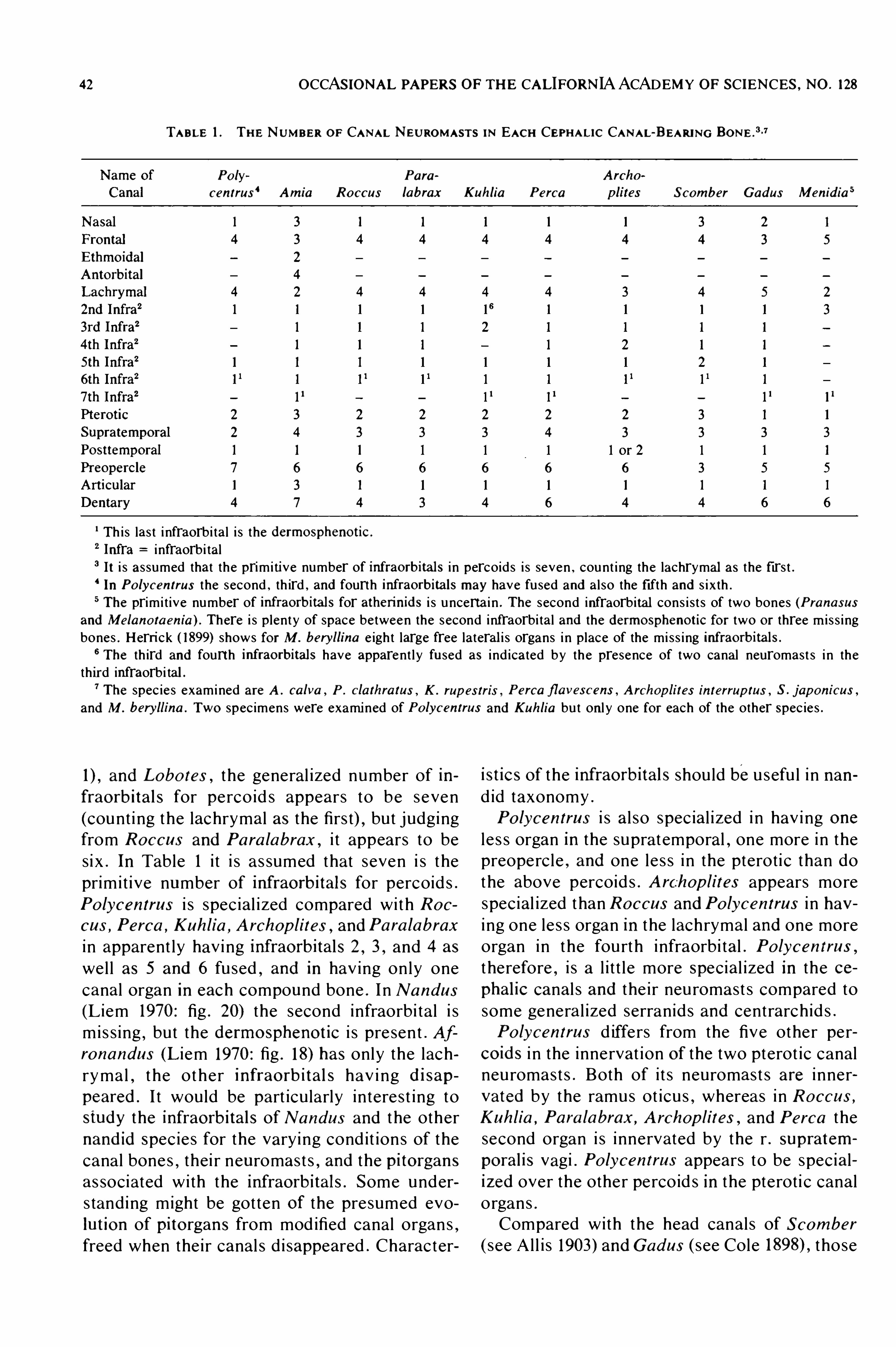
42 OCCASIONAL PAPERS OF THE CALIFORNIA ACADEMY OF SCIENCES , NO . 128
TABLE 1 . THE NUMBER OF CANAL NEUROMASTS IN EACH CEPHALIC CANAL-BEARING BONE .
#-7
Po ly Para
centras‘ Amia Ro ccus labrax Kuhlia Perca Scomber Gadus Menidia s
Th is las t i nfraorbi tal i s the dermospheno tic .
2 Infra i nfraorbi tal3 It i s assumed that the prim i tive number of infraorbitals i n percoids i s seven , count i ng the lachrymal as the firs t .In Po lycentrus the second , th ird , and fourth infraorbita ls may have fused and al so the fifth and s i x th .
5 The prim i t ive number of infraorbitals for atherinids i s u ncerta i n . The second infraorbital cons i s t s of two bones (Pranasusand Melano taenia ) . There i s plenty of space between the second infraorbi tal and the dermospheno tic for two or three m i s s i ngbones . Herri ck ( 1899) shows for M. beryllina e igh t large free lateral i s organs i n place of the mi ss i ng infraorbitals .
6 The th ird and fourth infraorbital s have apparent l y fused as i nd icated by the presence of two canal neuromast s i n thethird infraorbi tal .
7 The spec ies examined are A . calva , P . clathratus , K . rup es tris , Perca fl avescens , Archop lites interrup tus , S . j aponicus ,
and M. beryllina . Two spec imens were examined of Po lycentrus and Kuhlia bu t onl y one for each of the other Spec ies .
and Lobo tes , the general i zed number of in
fraorbitals for perco id s appears to be seven
(count i ng the lachrymal as the firs t) , but j udgingfrom Ro ccus and Para labrax , i t appears to be
s i x . In Table 1 i t i s as sumed that seven i s t he
prim i t i ve number of infraorbitals for percoid s .
Po lycentrus i s spec ial i zed compared wi th Ro ccus , Perca , Kuhlia , Archop lites ,
andPara labrax
i n apparent ly hav i ng infraorbitals 2 , 3 ,and 4 as
wel l as 5 and 6 fu sed , and in hav ing on ly one
canal organ in each compound bone . I n Nandus
(L i em 1970 : fig . 20) the second infraorbi tal i s
m i s s ing , but the dermo spheno tic i s pre sen t . Afro nandus (L i em 1970 : fig . 18) has only the lach
ryma l , t he ot her infraorbitals hav i ng disappeared . I t would be part icu larly in teres t ing to
study the infraorbitals of Nandus and t he other
nandid Spec ie s for the vary i ng cond it ion s of the
cana l bone s , thei r neuromas t s , and the pitorgans
as sociated wi th the infraorbitals . Some under
s tand ing might be gotten Of the presumed evo
lution of pito rgans from mod ified canal organ s ,freed when the i r canal s d i sappeared . Character
istics of the infraorbitals shou ld beu sefu l i n nan
d id taxonomy .
Po lycentrus i s al so spec ial i zed i n hav i ng one
l e s s organ in the suprat emporal , one more i n the
preoperc le, and one le s s i n the pteroti c than do
the above percoid s . Archop lites appears more
spec ial ized than Ro ccus andPo lycentrus i n hav
i ng one le s s organ in the lachrymal and one more
organ in the fourth i nfraorbi tal . Po lycen trus ,
t herefore,i s a l i t t l e more Special i zed in the ce
phalic canal s and the i r neu romast s compared to
some genera l i zed serranid s and centrarch id s .
Po lycentrus d iffers from the five other per
co ids i n the innervat ion Of the two pterot ic canal
neu romas t s . Both of i t s neuromas t s are i nner
vated by the ramus ot icu s , whereas i n Ro ccus ,
Kuhlia , Para labrax , A rchop lites , and Perca the
second organ i s i nnervated by the r . supratem
poralis vagi . Po lycentrus appears to be spec ial
ized over the other perco id s in the pteroti c canal
o rgans .
Compared wi th the head canal s of Scomber
( see Al l i s 1903) andGadus ( see Cole those
44 OCCASIONAL PAPERS OF THE CALIFORN IA ACADEMY OF SCIENCES , NO . 128
imens and i n all publ i shed account s , i s s t ri k i ngl y
d ifferent and spec ial i zed . The characte r con s i s t s
of two (or one) secondary canal neuromas t s in
add i t ion to the s i ngle origi nal (primary ) neuro
mas t . The secondary neuromas t s l i e very cl ose
to the anterior edge of the floor of the nasal ca
nal , one organ near each lateral corner of the
canal , and are i nnervated from the i nfraorbi tal
t runk . The origi nal nasal canal neuromas t l i e s
near the midlength Of the canal and i s i nnervated
from the supraorbi tal t runk .
The beryco ids all have#
th i s characte r (at l eas t
one spec ie s Of each fami ly examined) , but the
stephanoberyco ids , i nc lud ing melamphae ids and
g ibberichthyids , do not . Po lymixiids have what
appears to be an earl i er development of the ber
yco id cond i t ion . I n Po lymixia there are three
free lateral i s organs i n a membranous canal t hat
pas se s acros s but jus t out s ide of the ent rance to
the nasal canal . Thi s membranous canal i s con
nected acros s the t ip of the snout wi th a s im i lar
canal on the other S ide of the head . Another
lateral i s organ l ie s at the midpo in t of the cros s
i ng canal . The stephanoberyco ids have what appears to be a s t i l l more general ized cond i t ion i n
that there i s a row of free lateral i s organ s i n the
Skin acros s the t ip of the snout i n p lace of the
membranous canal OfPo lym ixia and a somewhat
en larged free lateral i s organ in the sk i n ju st out
s ide each lateral corner of the Open ing i n the
nasal canal i n p lace Of the canal organ i n s ide the
nasal canal near each corner i n beryco ids .
The mode of format ion of the secondary nasal
canal organ s of beryco ids i s what make s th i s
nasal cana l s pec ial i zat i on espec ial l y d ifferent
from that i n other fi she s . In beryco ids free lat
eralis organs c lose to the nasal canal open i ng
have apparent l y been s imply engu lfed by the
bony nasal canal wi thou t firs t be i ng formed in
a prenasal o s s i cl e . In carangids , rachycentrids ,
echeneids , coryphaenids , and other groups , the
prenasal o s s i c le or os s ic le s do not fu se onto the
nasal bone . In Scomber a prenasal o s s i c l e un
doubted l y has fused onto the nasal bone . In ber
yco ids there are no i nd icat ion s of such fu s ion ;the one or two added canal neuromas t s l ie very
clo se to the anterior edge of the nasal bone ,there i s no pore mark i ng a fus ion , and t here i s
no bend or i rregu lari ty i nd icat i ng a junct ion of
two bones .
I t appears t hat i n the beryco ids we have an
example , sure ly very rare , of two free t ran sverse
organs (reduced to one organ in some beryco ids)
being ,in effect , captured (engu lfed ) by a na
sal canal bone#The berycoid nasal canal bone
spec ial i zat ion wou ld seemingl y remove the ber
yco ids from be i ng con s ide red t he ance s t ral
source of percoid s . The same stat ement mayhold for the stephanoberyco ids which have what
appears to be an earl ier stage of the pol ymix i i d
cond i t i on . Thi s character s tate in beryciformswi l l be presented in a s eparate paper .
I t may be added that beryciforms have other
spec ial i zat ion s of the cephal ic lateral i s s y stem .
I n stephanoberyco ids the supratemporal canal
i s actual l y i n commun icat ion wi th the nasal ca
nal and the frontal canal Via a large lateral i s
chamber l y i ng in t he i nterorbi tal area over the
frontal s . In melamphaeids t he lachrymal canal
i s in commun icat ion wi th the nasal canal . These
development s are rare or un ique in te l eos t s .
In summary , two general i zat ion s m igh t be
made about the i nnervat ion Of the snout region
for te leos t s from cond i t ion s Observed in Po lycentrus and S i hl er nerve preparat ion s Of representatives Of 85 fami l i e s . The lateral i s i nnerva
t i on of the Supraorb i tal t runk ends wi th the nasal
canal bone . Any pitorgan development on t he
snout wi l l be i nnervated from the i nfraorbi ta l
t runk . Any new canal and canal neuromas t s be
yond the anterior end Of the nasal canal wi l l al so
be i nnervated from the i nfraorbi tal t runk . A Sim
ilar general i zat ion appl ie s to the general cu taneous i nnervat ion of the supraorbi tal t ru nk . In
most of the fi shes Observed , i t ended abou t at
the anterior end of the nasal bone or a l i t t l e be
yond i t . On ly i n the atherinomo rphs and holo
centrids d id i t extend s ign ificant l y fu rther , going
to the upper jaw as a large nerve .
Innervat ion of the Upper Jaw
In Po lycentrus the upper jaw i s suppl i ed wi th
general cu taneous i nnervat ion by the i nferior
ramulu s of the ramus max i l lari s tri geminu s (10 3 ;Fig . 1) and wi th gu s tatory i nnervat ion by the
anterior ramus Of the palat i ne nerve (Fig . NO
other nerves go to the upper jaw ,no t even from
the supraorbi tal t runk , which i t se lf i s someth i ng'
of a surpri se if the upper jaw i s cons idered a part
Of the snou t . Po lycen trus i s representat i ve Of
most tel eos t s in i nnervat ion of the upper jaw .
One except ion i s Men idia (Herrick 1899 ; fig . 3 ,
the yel low branch to the upper jaw) . In Menidia
the supraorbi tal t runk i s conti nued out to the t ip
of the upper jaw ,supply ing i t rather heav i l y wi th
genera l cu taneou s i nnervat ion . The nerve i s
FREIHOFER : CRANIAL NERVES OF POLYCENTRUS SCHOMBURGKII
large , as seen on S i h l er nerve preparat ion s of
various atherinomo rphs . Of 85 fami l ie s , on l y t he
seven atherinomorph fami l i e s examined and the
Holocent ridae had the premax i l lary exten s ion of
the supraorbi tal t runk . In the Holocentridae the
funct ional component i s , presumably , al so gen
eral cu taneous .
Cheek Mu scl e Innervat ion
The patt ern of i nnervat ion of the cheek mu s
cl e i n Po lycentrus (Fig . 6) reflect s the part ial
s ubd iv i s i on of t he adductor mand ibu lae i n to
th ree part s ca l l ed A A 2 , and A3 (Fig .
branches IO2c and 10 2d i nnervat i ng A ] , branch
l0 2e i nnervat i ng A 2 , and t he hat ched part s of
10 2 i nnervat i ng A 3 . The pat tern in Po lycentrus
i s general i zed for percoid s . In fact , most of t he
other perco id s examined had more d i s t i nc t i ve
patte rn s of i nnervat i on Of the cheek mu sc l e than
does Po lycentrus , each being a l i t t l e d ifferent
from the others . The sc iaen id s were e spec ial l y
d i s t i nct,the A 113 muscle i n some spec ie s be i ng
developed as fu l l y as the A B in perCOpsiforms ,i nc lud ing i t s pat tern of i nnervat ion . Atherino
morphs (7 fami l i e s examined) have an i nnerva
t ion pat t ern d i s t i nc t from that of all other fami
l i e s stud ied except gastero steids in whi ch i t i s
t he same . Much can be learned of sy s temat i c
i nterest from the cheek muscle s by study of the
pattern of i nnervat ion wi th i n the mu scle . Resu l t s
of a s tudy of the A 13 musc le are bei ng prepared
for a separate paper .
Correc t Name for
Ramus Late ral i s Acces soriu s
Hem' i ck ( 1899 : fig . 3) desc ri bes three ramu l i
from the r . lateral i s vagi which joi n the r . recu r
rens fac ial i s to form the r . late ral i s acces soriu s
facial i s (RLA) . These ramul i jo i n RLA on t he
pos terior part of the head above the pharyngeal
area and on the anteri or part of the trunk . Her
rick i nd irect l y deri ves the se ramul i from a smal l
bund le of fibers (abou t 20) wh ich detach from
the root of n . glo s sopharyngeu s , th i s smal l bun
d l e jo i n ing i ntracran ial l y the root of the ramus
lat eral i s vagi ( r . lat . vagi) . The fibers are of sma l l
to med ium d iameter , con s iderably smal l er t han
lateral i s fibers to canal neuromas t s , accord i ng to
Herri ck , but larger than average commun i s fi
bers . Herri ck cou ld not t race these med ium
smal l fibers from the glo s sopharyngeu s th rough
the gangl ion of the r . lat . vagi . He as sumed that
fibers of the same S i ze observed leav ing the oth
45
er s ide of the gangl ion were the same as t he one s
observed entering i t,t hat i s , that they were from
the 20 fibers from the glos sopharyngeu s . He
rat her confident l y i d en t ifie s the three ramu l i
from the r . lat . vagi wh ich appear to jo i n RLAas being deri ved from the smal l bund le of fibers
from the root of the glos sopharyngeu s . He fu r
thermo re as s ign s them to the communi s sy stem .
Herrick’
s reason s , others in add i t ion to tho se
ju s t gi ven , are not very conv inc i ng . The use of
the name ramus late ral i s acces soriu s , therefore ,real l y re s t s upon the fact that th i s nerve i s made
up Of gu statory fibers from both the fac ial i s andvagu s (or glOSSOpharyngeus) nerves .
In Po lycen trus no such ramul i from the r . lat .
vagi were Observed to jo i n the recurrent fac ial
ramus . In Po lycentrus there i s an i n t racran ial
vagal ramus that pas se s up to the parietal bone ,
and on some specimens i t pas se s ou t of the cra
n ium through the same foramen as does RLA ,
but on other spec imen s i t pas se s through i t s own
foramen c lose to that for RLA . I n the lat ter
spec imen s i t was observed to pas s to overl y i ng
sk i n and to ski n ly i ng anterior to i t s cran ial ex i t .
As far as coul d be determined from se rial sec
t i on s OfPo lycen trus , i t was seen to be an i n t ra
cran ial part of the rami cu tane i dorsal e s vagi . I t
wou ld be carry i ng general cu taneou s i nnervat i on
to the top of the head . Menidia lacks thi s i ntra
cran ial vaga] ramus . Herri ck equat es t he three
extracran ial ramul i from the r . lat . vagi ofMen
idia with th i s vagal ramus that i s present in many
other fi she s ( see Freihofer 1963 for examples) .
I t may be equ i val ent as Herr ick thought , but
what seems qu i te doubtfu l from Herrick’s d i s
cussion of these three ramul i i s that they are Of
t he commun i s component and that t hey jo i n
RLA and presumably course some di s tance wi th
i t . On a S i h l er nerve preparat ion Of the atherinid
A therinop s affinis ,no ramul i from the r . lat . vagi
were Observed to anas tomose wi th RLA . A th
erinop s al so lacks an i nt racranial vagal ramus to
the pari etal . These three ramul i ofMen idia and
the i nt racranial vagal ramus Of Po lycentrus and
other fi she s need more study . If t hey prove no t
to be commun i s,that i s , if there i s no vagal com
mun i s cont ribut ion to the format ion of RLA ,
there would be reason for changi ng the name of
RLA to s imply that of the ramus recurren s fa
cialis ,i n recogni t ion of i t s pu re l y fac ial com
mun i s compos i t ion . The fac ial part of RLA i s
much larger than i s the vagal in apparent l y all
t e leo s t s havi ng RLA . One d i sadvantage Of u s i ng
46 OCCASIONAL PAPERS OF THE CALIFORNIA ACADEMY OF SCIENCES , NO . 128
the name RLA i s that i t impl ies that the nerve
i s an acces sory lateral l i ne nerve of the acous
t ico - lat eral i s sy stem , which i t i s not . The term
RLA shou ld s tand unt i l the quest ion of the con
tribution of the vagal communi s i s re sol ved .
Trunk Lateral L i ne Nerves
The pat tern of the t runk lateral l i ne nerve s of
Po lycentrus (Fig . 2 1) i s espec ial l y in teres t i ng
becau se i t shows some vent ral and dorsal seg
mental lateral l i ne branches whi ch previou s l y
have been known on ly in variou s lower , non
acanthopterygian fi shes and i n a l e s s - deve loped
s tate in zoarc ids , gobi id s , and mugilids (Fre i
hofer 1970 ,There i s one bas ic d ifference
between the Po lycen trus pattern and that of oth
er fi shes having ventral segmental lateral l i ne
nerves . In Po lycentrus as wel l as in t he othe r 20
or so percoid fami l i e s that have been examined ,
there i s a dorsal longi tud inal col lector late ra l l i ne
nerve . Other perc iformes examined have th i s
col lector a l so , but the zoarcids , gobi id s , and
mugilids do not have i t . The atherinomorphs all
lack i t , as do other groups‘
examined that are
c las s ified lower than the acanthopterygian s and
paracanthopterygians . Some of the paracan
thopterygian fi shes deve lop a dorsal longi tud i nal
col lector lat eral l i ne nerve at l east toward s the
d i stal end of the dorsal longi tud inal ramus . The
ventral segmental lateral l ine branches in Po lycentras , Of which there are five , are a secondary
deve lopment on a bas i c percoid pat tern and i s
appare n t l y a s s o c iat ed w i t h t h e Spe c i a l i ze d
swimming habi t s ofPo lycentrus . I t would be in
teresting to see the lateral l i ne nerve s of nandids
more general i zed than Po lycentrus , espec ial l y
Nandus . I t would take very carefu l l y pre served
specimens to provide the i nformat ion , but t he
d i s t ribut ion Of free lateral i s organs on the head
and t runk Of all nandid genera shou ld be deter
mined . The resu l t s shou ld have sy s temat i c s ig
nificance for nandid c las s ificat i on .
Po lycentrus i s a secret i ve fi sh wh ich s tal k s i t s
prey by an extremely s low forward movement
made by the beat i ng of i t s transparent soft dorsal
and anal fins , i t s mode Of forward progre s s , i t s
colorat i on , and i t s body shape re semb l i ng a
drift i ng dead leaf. When start l ed or afte r at tack
ing a prey fi sh ,Po lycentrus can move qu i te rap
id l y backward s from i t s pos i t ion near the su rface
to i t s ret reat near the bottom in a hol e or pot .
The head and t runk lateral l ine sy stem may re
flect spec ial i zat ion s for such swimming behav
ior . The lateral i s organ s of the t runk are all free
or naked organ s borne on scal e s , a seri es of
about e ight organ s on each such scal e , the lat
eralis- beari ng scal e s be i ng arranged in rows fol
lowing the ma i n contours of the t runk (Fig .
The lateral l i ne on the head i s al so spec ial i zed
i n that there are large neuromas t s in the head
lateral i s canal s as wel l as free organ s arranged
aS'
shown in Fig . 2 1 .
The d i s t ribut ion of free lateral i s organ s on the
t runk of Po lycen trus i l lustrate s to what degree
lateral i s organ s can migrat e and the i r nerves fol
low them . The quest ion ari se s for the zoarc ids ,
gob io id s , and mug ilids as t o whe t he r t he s e
groups have los t all t races of the bas ic percoid
pat tern or whether they ever had them .
The large number (20 to 25) of free lateral i s
organ s on the in s ide of the ch in between the
anterior end s Of t he dentari es i s i nteres t i ng i n
that a s imi lar aggregat ion was Observed on var
i ou s other percoid s . I t i s apparent l y an impor
tan t l ocat i o n for rece i v i ng lat e ra l i s s t i mu l i ,wh ich in Po lycentrus may serve a prey - l ocat i ng
funct ion .
An External - In ternal Lateral L i ne Canal
Nerve Re lat ionsh ip
Worthy of spec ial note i s a branch ing re la
tionship observed on a number of ramul i goi ng
to i nd iv idua l neuromast s of the 'head canal s ( see
branches to second and th i rd pores of lachrymal
canal , Fig . 1 , and SORB 4 , SORB 1 1 , and SORB
1 1a ,Fig . A simi lar relat ionsh ip was al so ob
served for lateral l i ne scale s on the trunk Of vari
ou s fi shes . In th i s re lat i onshi p a neuromas t ramu
l u s detache s from a ma i n nerve ramus and
cours e s al one towards i t s canal neuromas t , but
before reach ing i t s canal neuromas t , the ramulus
i t se lf detaches a branch wh ich curves away and
pas s e s some d i s tance to sk i n o r membran e
around an adjacent canal po re . In some i hs tan ce s , o n S i h l e r n e rv e pre parat i o n s t h i s
branch to the membrane around the pore ended
in branchlet s that re sembled in appearance those
that e l sewhere were known to be i nnervat i ng
free lateral i s organs . Such organs occur i n the
ski n along the supraorb ital and i nfraorb i tal ca
nals (Fig . 17) and el sewhere . If the ramulu s were
carry ing onl y general cu taneous i nnervat ion , i t
would seem as pract ical , a d i st ribut ion to have
such fibers course i n other rami that carry the
mai n cutaneous i nnervat ion .
The arrangement of a canal neuromast ram

FREIHOFER : CRANIAL NERVES OF POLYCENTRUS SCHOMBURGKII
u lu s c lose ly as soc iated wi th a ramu l u s innervat
i ng free lateral i s organ s i n the ski n wou ld al low
for the recept ion of d ifferences between lat eral i s
s t imul i i n s ide and out s ide the canal s for each
such locat ion . I t s deve lopment i n Po lycen trus i s
no doubt as sociated wi th thi s fi sh’s very s low
swimming habi t s , but i t was observed al so in
ot he r fi s he s , even for t he t ru nk lat e ra l l i n e
scal e s . Herrick ( 1899) b ri efly de scribed a few of
the se branches on the head ofMenidia but d id
not comment on them .
Rad ix Profundu s
The complex relat ion ship s of the rad ix pro
fundu s wi th the ocu lomotor nerve and t ri gemi nal
s ympathe t i c gangl i on in Po lycentrus (Fig . 14)
are qu i te S im i lar to those of the perc iform Ura
no scopus (Y oung 193 1 : text - fig . There i s a
d ifference i n Menidia that may be s ign ificant
sy s temat i cal l y . Herri ck ( 1899) found that a rec
ognizable i n tracran ial profundu s root and gan
gl i on are mi s s i ng i n th i s atherinid . S i nce the c i l
iary nerves are present inMenidia ,the root and
gangl ion Of the profundu s mu st be pre sen t al so ,
but apparent ly they are fu sed wi th the t rigemi na l
root and gangl ion , a cond i t ion wh ich may be
representat i ve for atherinomorphs .
A St retch -Receptor Nerve to Bas e Of
Maxi l lary Tendon
E spec ial l y i n terest i ng i s a branch ( l0 2f1 ; Figs .
6 and 10) Of t he r . mand ibu lari s tr igeminu s whi ch
ends i n a den se ramificat ion of nerve fibers in
t h e t end i nou s muco sa on t h e i n s id e Of t h e
mouth , t he i nnervat i on being d irect l y i n front of
the anteri or edge of the quadrat e near t he art i c
ulation head of th i s bone . Thi s tend inou s mem
brane fans out toward s the ventral end Of the
max i l lary tendon . Branch 10 2f] may i nnervat e
s t re tch - receptor organ s in th i s membrane .
#
A
simi lar branch was ob served on a number of Oth
er ki nd s of fi shes . I t was a large branch i n a
goatfish , Parup eneus p orphyreus . A S i m i lar
branch occurs i n Menidia ,but Herri ck ( 1899)
d id not suggest any st retch - receptor i nnervat ion
for i t . I t shou ld be Of i n teres t to funct ional anat
omists s tudy i ng jaw mechani sms .
S impl ify i ng C ran ial Nerve Stud ie s
The fifth , seventh , n i nth , and tenth cranial
nerves are the most complex i n the i r branch ing
and number of nerve component s, and for the se
reasons they hold the most i ntere st for sy s tem
47
atists . Of these four cranial nerves , the fifth and
seventh rank above the n inth and. ten th . B roader
taxonomi c coverage can be had by concentrati ng on ly o n the fifth and seventh nerves
,or by
res tri c t i ng the s tudy even further to a certa i n
t runk , ramus , or even to on ly a Si ngle larger
branch of a ramus . The nerve chosen shou ld be
an i n tegral u n i t , complete i n i t se lf, and not part s
Of two nerve s or character complexe s .
I t i s al so best to chose on ly one nerve com
ponent in a nerve t runk or ramus rather than
i nc l ude all component s wi th all the i r branche s .
For example , one of the character complexe s
se lected for compari son wi th Po lycentrus was
t h e la t e ra l i s componen t i n t h e s u praorb i ta l
t runk . Only the nasal and frontal bone s were
i nc luded . These seemed to form a natu ral mor
pho logical uni t for th i s component i n th i s nerve
trunk .
For pract i cal purpose s comparat i ve s tud ie s
can be made d irect l y from S i hl er nerve prepa
rat ion s wi thout recou rse to serial - sect ion t ech
n ique s and i nvolved neurological re search . Prior
ground ing in the important nerve reference s em
ploy i ng func t ional component anal y s i s i s , of
course , es sent ial . A bri ef l i s t i ng of the mos t im
portant of th e longer s tud ie s i s gi ven in the #
In
troduction . There are huge gaps i n the taxo
nomic coverage in the nerve l i teratu re . The gaps
can be fi l l ed effect i ve l y by a reference col lec t ion
of S i h le r nerve preparat ion s .
The nerve component s of the vari ous t ru nks ,rami , and smal l e r branches can be worked out
wi th re lat i ve as surance in most i n stance s by
s tudy of S i h l er nerve preparat ion s and dissec
t ion s of pre served spec imens . The d i s sect ion s
shou ld expose the root s and gangl ia of the t ri
gem i nu s,fac ial i s , glos sopharyngeu s , and vagu s
nerves . The motor and lateral i s component s are
not d ifficu l t to determine becau se the i r end o r
gan s are re lat i vel y large . Si nce free lateral i s o r
gan s may be lo st i n the hand l i ng and preparat ion
of spec imens,the d i s t ribut ion of these organ s
shou ld be plot ted from fresh ly and carefu l ly pre
served Specimen s (Fig . Such a d rawing i s
usefu l i n help i ng as s ign t ermi nal branche s in a
part i cu lar area of a S i h ler preparat ion to the lat
eralis component . The gus tatory component i s
d ifficu l t to determ ine unles s the fibers course
more or le s s as i ndependent nerves for most of
the i r length . Taste ( term inal) buds located on the
externa l body surface are too smal l to be seen
except m ic roscopical l y . Most of the gustatory

48 OCCASIONAL PAPERS OF THE CALIFORN IA ACADEMY OF SCIENCES , NO . 128
sy stem wi l l be represented by the d i st i nct i ve
branches of the recurrent fac ial and palat i ne
nerves . D i s sect ion of the gen icu late gangl ion
should d i sclose the pre sence of larger bund le s
of gu statory fibers leav ing th i s gangl ion and em
teri ng the Supra and i nfraorbi tal t runks , but
S i nce these fibers d i sappear into the branche s Of
these trunks , appropriate microtechn ique meth
od s are neces sary for determin ing the i r distri
bution . For most sy s temat i c purpose s gus tatory
fibers enteri ng these two trunks can be ignored .
For pract i cal purpose s the last component , the
general cu taneou s , i s what remai n s after the oth
er three component s have been determ i ned . In
fi shes , genera] cu taneous fibers terminate i n freenerve end ings i n the ski n or in Special tact i le
e laborat ion s of Sk in , such as c i rri and other sk i n
flaps , or in barbel s or barbel - l i ke s tructu res .
After determinat ion of the funct ional compo
nents at the periphery of the nerves , the pre s
ence of a component can be Shown d iagram
matically i n drawings by a d i s t i nct i ve symbol ,the fiber bund le s of each branch bei ng brought
together as they course central l y toward s the
cranial root s and gangl ia and to the brai n . An
example in which the se S impl ified approaches
were taken i n a sy stemat i c study i s that by
Spri nger and FreihoferShort cut s such as t hose suggested above are
necessary if nerve s are to be used in the c las
sification of fi shes . Bas ic , descri pt i ve neuro logi cal s tud ie s are s t i l l needed on numerou s groups .
I t may fal l to t he lot of some sy stemat i s t s to do
them . Neurologi s t s have seemingly turned to
other tasks .
SUMMARY
1 . The courses of the t rigemi nu s,fac ia l i s ,
glos sopharyngeu s , and vagus cran ial nerve s of
a percoid fi sh , Po lycen trus schomburgkii , are
desc ribed in detai l , and the funct ional compo
hent s of the nerves determined as far as pos s ib l ew it h the method s used . The c loses t groups wi th
which Po lycentrus cou ld be compared i n pub
lished account s were Scamber (Scombridae) and
Menidia (Atheri n idae) . Compari sons were al so
made from S i h l e r p reparat i on s of numerous
groups ( representat i ves of 85 fami l i e s ava i lab le)for se lected nerves .
2 . Noth ing unu sual was noted i n the olfactory
( I) , Opt i c ( I I) , ocu lomotor ( I I I) , t rochlear ( IV) ,or abducens (VI) , nerve s .
3 . The rad ix profundus in Po lycentrus has the
same re lat ionshi ps to the Gas serian gangl ion ,
t ri gemi nal s ympat he t i c gangl ion , c i l iary gan
gl ion , and ocu lomotor nerves as i t has in Urano scopus , as far as could be determined .
4 . Po lycen trus was compared wi th five per
co ids (Ro ccus , Kuhlia , Pe rca ,Para labrax , and
Archop lites ) and several other groups of fi she sfor cephal ic canal neuromas t s and wi th add i
tional groups for cephal i c free lateral i s organs
(pitorgans) .
5 . Po lycentrus i s more special i zed than the
above perco id s , both i n number of pitline s and
i n the i r S i ze .
6 . A pitline correspond ing to the vert i cal p i t
l i ne OfAmia i s present and i nnervated from the
truncu s hyomandibularis by the ramus buccal i s
acces soriu s . The poste rior end of the infraorb i tal
pitline i s al so i nnervated by a branch of the r .
buccal i s acce s soriu s , which represent s a spe
cialization s ince the infraorbi tal pitline i s u sual l y
i nnervated from the i nfraorbi tal t runk by the r .
buccal i s . Thi s po steri or part of the i nfraorb ital
pitline may represent the horizontal pitline of
Am ia that has moved up to the edge of the ihfraorbital .
7 . Some pitline s Of some fi shes may not be
homologous wi th correspond ing pitlines of other
fi she s . Esoco ids are an example . At leas t three
pitlines in Po lycentrus ( the i nfraorb i tal , subhasal , and Supraorbi tal) are not homologous wi th
pitlines in the same locat ion i n , esoco ids . These
three pitlines i n eso co ids are all i nnervated by
the r . buccal i s acces sorius from the t r . hyoman
dibularis , whereas in Po lycentrus the fi rs t two
are i nnervat ed from the i nfraorbi tal t runk by the
r . buccal i s and the las t pitline ( the Supraorb i ta l)i s i nnervated from the Supraorb i tal t runk .
8 . Reflect i ng perhaps the greate r d eve lop
men t of the i nfraorb i tal pitline i s a spec ial i zat ion
of the i nfraorb i tal canal—beari ng bones , a firsts tep toward s degenerat ion of the infraorbi tal ca
hal s ; the second , t h i rd ,and fourth infrao rbitals
are fused and there i s on ly one cana l neuromas t
i n the compound bone . In other respect s the ce
phalic lateral i s canal - bearing bones are a l i t t l e
more spec ial i zed (one les s neuromas t i n the su
pratemporal and one more in the preopercular
canal s ) t han in Ro ccus , Para labrax , A rcho
p lites , and Kuhlia .
9 . Various c lus ters and l ines of cephal i c p i t
organs Should be u seful in nandid taxonomy .
10 . The i nnervat ion of the snout in fi shes i s animportant area for compari son in sy s temat i c
stud ie s . The snout in te leost s i s i nnervated from
FREIHOFER : CRANIAL NERVES OF POLYCENTRUS SCHOMBURGKII
two source s : t he supraorbital and i nfraorbi tal
t runks . In Po lycentrus the supraorb i tal t runk
suppl i e s one canal lat eral i s organ Of the nasa l
canal , and the i nfraorb i tal t runk innervat e s ap
parently all of the free lat eral i s organ s o n t he
snout dorsomed ial and ventral to the nasal bone .
In some fi she s there are important d ifference s
i n th i s general i zed cond i t ion seen in Po lycen
trus . If there are further development s of the
lateral i s sy s tem on the snou t , such as‘
an exten
s ion anteriorl y of the nasal canal , they are in
nervated by the i nfraorbi tal t runk . Innervat ion
of the nasal canal organ or organ s shou ld be de
termined in sy stemat i c s tud ie s becau se two (or
three) lateral i s organ s in the nasal cana l may i hdicate a fus ion of the nasal canal bone (wi th one
or two canal o rgan s) and a prenasal canal (wi th
one canal o rgan) , the lat ter be ing i nnervated by
the i nfraorbi tal t runk , or i t may have other s ig
nificance . An example Of a group hav i ng a nasal
and two prenasal s ( the anterior one membranou s
and the posterior one bony) i s the Carangidae .
Other percoid s hav ing two prenasal s wi th fea
tu re s ind icat i ng a Shared , derived spec ial i zat ion
wi th the Carangidae are t he Coryphaen idae ( two
bony prenasal s , each separate and free from the
nasal ) , Rachycen t r i dae ( two bony pre nasal s
fused bu t free from nasa l) , and Echeneidae ( same
as Rachycent ri dae) . Some other percoid s hav ing
other Spec ial i zat i on s Of an anterior exten s ion of
the nasal canal are t he Sc iaen idae and Po lynem
i dae (both w i th deep , compl icated membranou s
ext en s ion s) ; Toxot idae (a broad , bony prenasal) ;and Lutjanidae (one membranou s prenasal) . The
Scombri dae i s so far unique i n hav ing a bony
prenasal fused to the nasal .
1 1 . Compared wi th Po lycentrus , other per
co ids , and all te leo s t s examined , the beryco ids
have a qu i te d ifferent spec ia l i zat ion of the nasa l
canal . I t con s i s t s Of one or two nasal canal sec
ondary neuromas t s located clo se to the anteri or
edge of the floor of the canal and i nnervated
from - the i nfraorb i tal t runk . Po lym ixia exh ib i t s
a stage that cou ld be antecedent to that Of ber
yco ids . Stephanoberyco ids have what may be the
most general i zed cond i t ion for beryciforms . I t
appears that i n beryco ids free lateral i s organ s
l y i ng close in front of the open ing of the nasal
canal were d i rect l y i ncorporat ed i nto the ante
ri or end of the nasa l canal wi thou t the prior for
mat ion Of a prenasal o s s i c le that subsequent ly
fu sed onto the nasal canal bone . The#
captu re#
or engu lfing mode of format ion wou ld be rare ,
49
i f not un ique . In any event , the nasal canal s pe
c ialization appears bas ic i n beryco ids . I t i nd i
cate s that ne i ther stephanoberyco ids , po lymix
io ids , nor beryco ids are the ancest ral sou rce Of
perc ifo rms . There are other s t ri k i ng Spec ializa
t ion s of the cephal ic lat eral i s canal s y s t em of
beryc iforms .
12 . The gadoid s and o phidio ids have a shared
spec ial i zat ion of the nasa l canal in which the
anteriormo st frontal canal neuromas t has mi
grat ed i nto the nasal canal , giv ing that canal two
neuromas t s and the frontal canal on ly three , t he
usual number bei ng four for almost all acan
thopterygians examined . S tephanoberyx a l so
has t h i s spec ial i zat ion but not Gibberichthys .
The zoarcids have on l y three frontal canal neu
romasts , but onl y one nasal canal neu romas t .
13 . The general cu taneous component of the
supraorb i tal t runk i n Po lycentrus end s on the
snou t posterior to the upperjaw , which i s where
i t end s i n most te leos t s . In all atherinomo rph
fi she s examined , i t cont i nues anteriorl y as a
large nerve onto the upper jaw . The on ly other
group al so found hav ing a s im i lar large exten s ion
onto the snou t i s the Holocentridae . The pre
maxi l lary exten s ion Of the Supraorb i tal t runk
carri e s genera] cu taneou s fibers i n atherino
morph s . The funct ional component i s not yet
known for ho locentrids ,but i t probabl y i s gen
era l cutaneou s .
14 . The pat te rn of branch ing of the ramul i
from the ramus mand ibu lari s t rigeminus i n to the
adductor mand ibu lae muscle s of the cheek i n
Po lycentrus con s i s t s of two ramul i leav ing the
ramus mand ibulari s t rigem inus c lose together .
The i r ramificat ion s in the subd iv i s ion s of the
cheek musc le mas s reflect the s truc ture Of the
mu sc le . The pat te rn of nerve branch ing in the
adductor mand ibulae musc le can be important
i n unders tand ing the subd iv i s ion s Of th i s mu sc le ,such as the origi n of the A IBmuscle . I t al so mayhave sy s temat i c s ign ificance . An example was
d i scovered i n compari ng the pat tern ofPo lycen
trus wi th that of the atherinidMenidia . The ath
erinomorphs as a group have a nerve pat tern to
the cheek musc le d ifferent from that of all other
fi shes examined except for the gastero ste ids .
15 . The recu rrent fac ial ramus (RLA) i s pres
ent i n one of the bas ic percoid pat tern s ( referred
to as the Serranus pat tern) , characteri zed by an
orbi to - pectora l and a pari e to - dorsal branch .
16 . There are few or. no lateral i s fibers in t he
ramus hyoideu s .
50 OCCASIONAL PAPERS OF THE CALIFORNIA ACADEMY OF SCIENCES , NO . 128
17 . There are apparent ly few or no communi s
fibers in t he ramus mand ibulari s t rigemi nu s and
r . max i l lari s t rigeminus .
18 . The ante rior ramus palat i nu s joi n s the r .
max i l lari s t rigem inus on the upper jaw . Some
groups of fi shes lack th i s anastomos i s (atherino
morphs be i ng one) .
19. There i s a Jacobson’
s anastomos i s .
20 . The ramus mand ibulari s i nternu s fac ial i s
i s present .
2 1 . A nerve pos s ib ly function i ng mai n l y as an
i nnervat ion for s t retch receptors in the mem
brane and tendon s as soc iated wi th the bas e of
the max i l lary tendon was Observed in Po lycen
trus and some other perco id s .
22 . A double type of cephal i c cana l lat eral i s
i nnervat ion was observed on Po lycentrus and
some other acanthopterygians . I t con s i s t s of two
branches detach i ng from a s ingle lateral i s branch
that i nnervat es a canal neu romas t : one of the
two canal branches i nnervat e s the canal neuro
mas t , and the res t of the canal branch pas se s to
the membrane around the adjacent canal pore ,
the membrane at l eas t somet imes observed to
bear free lateral i s organ s .
23 . There i s an i n tracran ial vagal ramus which
in Po lycentrus i s a branch of the ramus cutane i
dorsal e s vagi . Thi s i n tracranial vagal ramus does
no t course wi th the recurrent fac ial ramus . I t
doe s not cont ribute to the lat ter ramus . NO vagal
branch was seen to cont ribute to the recurrent
facial nerve on the nape . The ques t ion of the
i nt racran ia l ramus be ing of general cu taneous or
commun i s component shou ld be s tud ied in other
fi shes . If the i ntracran ial vagal ramus doe s not
cont ribute commun i s fibers to the recu rrent fa
c ial nerve , then the name of ramus lat eral i s ac
cessorius shou ld no t be u sed and the name of
recurrent facial nerve be u sed in i t s s tead for the
ent i re course Of the nerve .
24 . The trunk lateral l i ne nerves Show a bas i c
acanthopterygian pattern of a so - cal l ed dorsal ,longi tud inal col l ector lateral l i ne nerve . In ad
d i t ion there are a number of dorsal and vent ral
segmental branches whi ch i nnervate three rows
of scal e s bearing free lateral i s organ s (pitor
gans) : a row along the base of the dorsal fin ; arow halfway between the dorsal fin and the hor
izontal septum ; and a row al ong the base of the
anal fin . There are two rows of free organ s on
the caudal fin : one on i t s upper lobe and one on
i t s lower lobe . There i s on l y one regu lar tubed
lateral l i ne scal e , the fi rs t o ne . Each Of t hese
separat e rows of free organ s on a scal e may rep
resent a canal neuromas t that has migrated to
ward s the base Of a fin and subd iv ided into about
eight smal l e r free organs ; or each row of free
organs on a s cal e may represent mu l ti pl i cat ion
of a s ingle pitorgan ori gi nal l y as soc iated wi th
each tubed lat era l l i ne scal e . Such pito rgans
were Observed , one per lateral l i ne scal e , on
some lateral l i ne scal e s Of other fishes . I t was
not observed on the s ingle tubed scal e of Po lycentrus . I t i s not known which of the pos s ib l e
ori gi ns i s correct for the development of such
free lat eral i s organ s , e i ther on the head or on
the trunk Of the body .
25 . Some interes t ing sy stemat ic problems on
th e h igher cat ego ry c las s ificat i on of ce rta i n
groups of fi shes were d i sc losed i n the compar
at i ve s tud i es made between Po lycentrus and
groups represented i n the nerve l i teratu re and
from S i h l er nerve preparat ion s .
26 . For sy stemat i c purposes the most u sefu l
cranial nerve s to s tudy are the fifth , seventh ,n inth , and tenth , with the fifth and seventh be i ng
the most u seful , if a choice has to be made .
These cran ial nerves are the most complex and
offer the most characters .
27 . From a con s iderat ion Of the u sefu l nes s
t hat ne rves apparent ly have for stud i e s on the
clas s ificat ion of fi she s and the great gaps there
are in the taxonomic coverage of the nerve l i t
erature for fi shes , i t i s apparent that at l eas t one
bas i c desc ri pt ive s tudy of the cran ial nerve s of
a representat i ve of each order i s defini te ly need
ed . Thi s re search shou ld be carr ied ou t by sy s
tematists . Morphologi st s are no longer i n teres t
ed in descri pt ive nerve s tud ies .
28 . Maki ng the preced ing recommendat ions
much more feas ib l e i s the S i hl er techn ique for
s tai n i ng nerves in a cleared , i ntact spec imen .
ACKNOW LEDGMENTS
My thanks go out to many ind i v idual s who
helped make thi s s tudy poss ibl e . Dr . George S .
Myers gave the ori gi nal breed i ng stock of Po lycentrus and encouraged cont i nuance of the projec t . Dr . Stan ley H . Wei tzman helped locate# popu lat ion s of Po lycen trus i n San Franc i sco
aquarium stores , gave adv ice on techniques and
on other mat ters . D r . R i chard Wi nterbottom
kind ly adv i sed on the mu scle terminology . D rs .
Stanley H . Wei tzman , Wi l l iam N . Eschmeyer,
and Mr . Leonard J . V . Compagno cri t i ca l l y read
the manuscri pt . I as sume ful l respons ibi l i t y for
52 OCCASIONAL PAPERS OF THE CALIFORN IAACADEMY OF SCIENCES , NO . 128
L ITERATURE C ITED
ALLIS , EDW ARD PHEL PS , JR . 1889 . The anatomy and deve lopment of the lateral l ine sy stem in Am ia ca lva . J . Morphol .
463—568 . 10 tex t - figs . pls . 30—4 2 (49
1897 . The cranial mu sc les and cranial and first spi nalnerves in Amia ca lva . J . Morphol . 487—808 , pls . 20
38 (64 figs ) .
1903 . The Sku l l , and the cran ial and first sp inal musc les and nerves in Scomber scomber . J . Morphol . 18 : 45329 , l text - fig , pls . 3—12 .
1910 . The cranial anatomy of the mai l - checked fish
es . Zoologica (Stu ttgart) , 57 : i i i 2 19 , 8 pls .
ATODA , K . 1936 . The innervat ion of the cranial nerves of thecatfish : Paras ilurus aso tus L . Sci . Rep . Tohoku Univ . , Sen
dai 91- 113 .
BAUDELOT , EMILE . 1883 . Recherches sur le sy steme ner
veux des poi ssons . Pari s . 178 pp . , 10 pls .
COLE , FRANK J . 1898 . Observat ions on the structure and
morphology of the cranial nerves and lateral sense organsOf fishes ; with spec ial reference to the genus Gadus . Trans .
L i nn . Soc . Lond . , ser. 2 , Zoo] . 17—22 1 , pls . 2 1—23 .
CUVIER, GEORGES L . C . F . D . , AND ACH ILLE VALENCIENNES . 1828 . His toire naturale des poi ssons , 1 . Pari s .
xv i 573 pp .
D ISLER, N . N . 197 1 Lateral l i ne sense organs andtheir importance in fi sh behavior. Academy of Sciences ofthe Severtsov Inst i tu te of Animal Morphology .
(Trans l . from Russ ian by H . Mi l l s and M. Y an'
v . Israe l Program for Sc ient ific Trans lat ions , Ltd . 328 pp . , 57 figs . )
FRASER, THOMAS H . , AND W . C . FRE IHOFER . 1971 . Tryps inmod ificat ion for Sih ler techn ique of stai n ing nerves for sy stematic stud ies of fi shes . Copeia 574—576 , 1 fig .
FRE IHOFER, WARREN C . 1963 . Pat terns of the ramus lateral i saccessoriu s and the ir sy stemat i c s ignificance in te leosteanfi shes . S tanford Ichthyol . Bu ll . 79—189 , 29 figs .
1966 . The Sih ler techn ique of stai n ing nerves for sy stematic study espec ial l y of fishes . Copeia 470—475 ,
2 figs .
1970 . Some nerve pat terns and their sy s temat ic s ignificance i n paracanthopterygian , sa lmoniform ,
gobioid,
and apogon id fi shes . Proc . Cal if. Acad . Sci . , ser . 4 ,
2 15—264 , 20 figs .
1972 . Trunk lateral l i ne nerves , hyoid arch gi l l rakers ,and olfactory bulb locat ion in atheriniform , mugilid , and
percoid fi shes . Occas . Pap . Cal if . Acad . Sci . 95 : 3 1 pp . , 12
figs .
LEONARD J . V . COMPAGNO , AND WILLIAM ROGERS .
1977 . Add i t ional notes on the use of the S ih ler techn iqueof stai n i ng nerves of smal l , whole spec imens of fishes andother vertebrate s . Cope ia 587—588 .
GILMORE , R . G . , JR . 1972 . The cephal i c nerve pat tern and
lateral l i ne s y s t em of Hypo rhamphus umfas c ia tus and
Strongylura marina (Pi sces : Exocoet idae , Be lon idae) . Mas
ter’s Thes i s . Un ivers i ty of West Flori da . 35 pp . , 7 tex t- figs .
GREENWOOD , P. HUM PHRY , DONN E . ROSEN , STANLE Y H .
WE ITZ MAN , AND GEORGE S . MYERS . 1966 . Phy l et i c studies of teleostean fishes , with a prov is ional c lass ificat ion ofl iv ing forms . Bu l l . Am . Mus . Nat . Hist . 339—456 ,
text- figs . 1- 9, pls . 2 1—23 , chart s 1—32 .
GUPTA , O . P. 1972 . On the ori gin and i nnervat ion of thecran ial and Spinal nerves al ong wi th the autonomic nervou ssy stem of the Ind ian Gar- fish , Xenentodo n cancila
Anat . Anz . 131: 39—50 ,4 figs .
HENN IG , W ILLI . 1966 . Phy logenet ic sys temat ic s . (Trans l . by
D . Dwigh t Dav i s and Ra i ner Z angerl . ) Univers ity of Il l i noi sPres s . 263 pp . , 69 figs .
‘
H ERRICK , C . JUDSON . 1897 . The cranial nerve component sof te leost s . Anat . Anz . l 3( l6) : 425—431 .
1898 . Experimen ts wi th the Weigert method . J .
Comp . Neurol . 8 : XXVI] to XLIV .
1899 . The cranial and first spinal nerves ofMenidia ;
a contribut ion upon the nerve components of the bony fish
es . J . Comp . Neurol . 9: 153—455 , pls . 14—20 .
1900 . A contribut ion upon the cranial nerves of thecodfish . J . Comp . Neurol . 10 : 265—316 , pls . 2 1—22 .
190 1 . The cranial nerves and cutaneous sense organsof the North American si luroid fi shes . J . Comp . Neurol . 1 1 :177- 249, pls . l4—l 7 .
1903 . The doc trine of nerve component s and someof i t s appl icat ions . J . Comp . Neurol . 13 : 30 1—312 .
HOLLISTER, GLORIA . 1934 . C leari ng and dyeing fi sh for bones tudy . Zoologica 89
LAURENT , P. , AND S . DUNEL . 1966 . Recherches sur
l’innervation de la pseudobranchie des t#l#os t#ens #i nFrench , Engl i sh summary] . Arch . Anat . Micro sc . Morphol .Exp . 55 : 633—656 , 9 figs .
LEACH ,E . H . 1946 . Curt i s’ subst i tu te for van Gie son’s stai n .
Stai n Technol . 21 : 107- 1 10 .
LIEM , KARE L . 1970 . Comparat ive funct ional anatomy of theNandidae (Pi sces : Teleoste i) . Fieldiana : Z OO] . 56 : 1- 166 , 66
figs .
MAHESHW ARI , S . C . 1965 . The cranial nerves ofMas tacem
belus arma tus (Lacepede) . Jpn . J . Ichthyol . 89—98 ,
4 figs .
MAN IOK , WOLFGANG . 1934 . Der Trigemino- Facialiskomplex
und d ie Innervat ion der Kopfse iteno rgane der E lri t ze(Phoxinus laevis ) . Z . Morphol . Oeko l . T iere 28: 64—106 , 16
figs .
MITHEL , MANORAMA . 1964a . The cranial nerves of the s isorid catfi sh Bagarias bagarias . Copeia 673—678,
2 figs1964b . The cran ia l nerves of Mystus seengha la
(Sykes) . Agra Univ . J . Res . 67—78 , 7 figs .
NARAW ANE ,D . D . 1965 . Stud ies on the ske leton , muscula
ture and d is tribut ion of nerves in the caudal pedunc le andfins of He teropneus tes fossilis B loch . (Physostomi—S i luridae) . J . Univ . Bombay 64—75 , 7 figs .
NELSON , GARETH J . 1972 . Cephal ic sensory canal s , pitlines ,and the c lass ificat ion of esoco id fishes , with notes on galaxiids and other te leos t s . Am . Mus . Nov it . 2492 : 49 pp . , 23
figs .
NORRI S , HENRY WALDO . 1925 . Observat ions upon the peripheral d i stribut ion of the cranial nerves of cert ai n fishes
(Amia , Lep idos teus , Po lyodon , Scaphirhynchus , and Aci
penser) . J . Comp . Neurol . 39: 345—432 , 12 text - figs , 7 pls .
(figs . 13AND SALL Y P. HUGHES . 1920 . The cranial , occ ipi tal ,
and anteri or spinal nerves of the dogfish , Squalus acan
thias . J . Comp . Neurol . 293-402 , 53 figs .PANCRATz ,
D . S . 1930 . The cran ial nerve component s i n thetoadfish (Opsanus tau) . J . Comp . Neurol . 247—286 ,
2 pls .
PINKUS , FELIX . 1894 . D i e Him nerven des Pro top terus ah
nec tens . Morphol . Arbei t . (Jena) 4 : 275—346 , 7 pls .
RAY , Dre LEE . 1950 . The peri pheral nervous system ofLampanyctus leucopsarus . J . Morphol . 87( l ) : 6 1—178 , pls .
l—17 .
ROSEN ,DONN E . 1964 . The re lationsh ips and taxonomic po
53FREIHOFER: CRANIAL NERVES OF POLYCENTRUS SCHOMBURGKII
sition of the halfb eaks , killifishes , s i lvers ides , and the ir re]at ives . Bu ll . Am . Mus . Nat . H ist . 2 17—268 , figs . 1
23 , pls . 14—15 .
1973 . Interre lat ionsh ips of h igher euteleo stean fi she s ,pp . 397—5 13 , 129 figs . ,
4 tables . In , Greenwood , P. Hum
phry , R . S . Mi l es , and Col i n Pat terson . In terre lat ionsh ipsof fi shes
, Supplement no . 1 to Z OO] . J . L i nn . Soc . 53: 536
PR
AND COLIN PATTERSON . 1969. The structure and re
lationships of the paracanthopterygian fishe s . Bu ll . Am .
Mus . Nat . His t . 14l (3)z 357—474 , figs . 1—74 , pls . 52—78 , ta
bles 1—8 .
SAXENA , P. K . 1966 . Cranial nerves of the common catfi sh ,
Clarias batrachus (L i nn ) . Jpn . J . Ich thyol . 91—98 ,
pls . 8—12 .
1967 . The cran ia l nerves Of the mud eel ,Amphibnouscuchia ActaAnat . 68 : 306—320 , 3 figs . #Summari e sin French and German]
1969a . The cranial nerves ofNandus nandus (Ham )
and Xenentodo n canc ila (Ham ) . Anat . Anz . 142
155 , 3 figs .
1969h . The cran ia l nerves ofNo top terus no top terus
(Pal las) . Anat . Anz . 198—209 , 3 figs .
AND RAKESH K . RASTOGI . 1968 . The cran ial nervesof the flat- fish , Cynog lo ssus bilinea tus Annot . Zoo] .Jpn . 70—76 ,
4 figs .
SHELDON , R . E . 1914 . Paraffine -We igert methods for thes tai n ing of nervous t i s sue , with some new mod ificat ions .
Fol ia Neurobio logica 1—28 .
S PRINGER , V ICTOR G . , AND WARREN C . FRE IHOFER . 1976 .
Study of the monotypic fi sh fami ly Pho lidichthyidae (Perciforrnes) . Smith son . Contrib . 2 16 : iv 43 pp .
, 23
tex t—figs .
STANNIUS , HERMANN . 1849 . Das peripheri sche Nervensys
tem d ie Fi sche , anatom i sch und physiologi sch Untersuch t .Rostock . 156 pp . , 5 pls .
STENSIG, E . A . 1947 . The sensory l i nes and dermal bone s ofthe cheek in fi shes and amph ib ians . K . Sven . Vetenskap
sakad . Hand l . , ser. 3 , 195 pp . , 38 figs .
VASISHT , H . S . , AND I . B . S . UBERO I . 1965 . Anatomy ofMys tus seengha la (Sykes) . IV. Nervous sy stem . Res . Bu l l .Panjab Univ . (new ser. ) 16( I 6 1—85 .
W ILLIAMS , T . W . 1943 . A techn ique for the gross d ifferen t ials tai n ing of peripheral nerves in c leared vertebrate t i s sue .
Anat . Rec . 86 : 189—194 , 1 pl .W INTERBOTTOM , RICHARD . 1974a . A descri ptive synonymyof the s triated musc le s of the Te leoste i . Proc . Acad . Nat .
Sci . Ph i la . 225—3 17 , 56 figs .
1974b . The fami l ial phy logeny of the tetraodont iformes (Acanthopterygi i : Pisces) as ev idenced by the ir comparative myology . Smith son . Contrib . Zoo] . 155 : iv 20 1 ,
185 figs .
YOUNG , J . Z . 193 1 . On the autonom ic nervous sys tem of theteleostean fi sh Urano scopus scaber . Q . J . Micro sc . Sci .
(new ser. ) 74 : 491—535 , 12 text - figs .
54 OCCASIONAL PAPERS OF THE CALIFORNIA ACADEMY OF SCIENCES , NO . 128
ABB REV IAT IONS FOR F IGURES
A A2 , A3— d i v i s ion s of the adductor mand ib
u lae muscle .
AA4 , AAS— m . adducto res
ADD OP— m . adductor operculi .
AAPZ— m . adductor arcu s palat i n i anteri or por
t ion .
AD HYO—m . adductor hyomandibularis .
ADD OP— m . adductor opercularis .
AN— anterior narial opening .
Anasto W— anas tomoses wi th .
AWB 1 ,2 ,3—Oiv isions of the mental i s mu sc l e .
BB ] , BEZ , BB3— basibranchials 1 , 2 , 3 .
br— branch .
br vi s t r of vag— branch ial and v i sceral t ru nks
of nervus vagu s .
CB ] to CBS— ceratobranchials 1 , 2 , 3 ,4
, 5 .
CIR —a c i rcu lar , round i sh bund le of mu sc l e on
top of m . t ran sversu s dorsal i s .
Cor 1 to Cor 6— branches of ramus ophtha lm icu s
t ri geminus to cornea .
EM— longi tud inal o ri entat ion of med ial fibers of
the Sphincter oe sophagi .
EF ] to EP4 — epibranch ial bones 1 to 4 .
fac— fac ial i s .
FR COM— frontal commi s sure of supraorbi tal
canaL
G— nervu s glos sopharyngeu s (n . IX) .
G l— branch of m . glOSSOpharyngeus to m leva
tor i nternu s 2 .
G2— ramus pretrematicus IX .
G3— branch of IXth to m levator externus 1 .
G5— branch of IXth to musc le s of gi l l fi lament s .
G6— branch of IXth to gi l l rakers .
G7— branch of IXth to m . Obl iquus vent ral i s l .
G8— branch of IXth to m . rectus ventral i s 1 .
GAM— area Of at tachment of some gi l l arch
mu scle s .
GANG IX— petrosa] gangl ion of IXth crania l
nerve .
GG4 —gangl ion of 4th vagal branchial ramus .
GLR— ramus po sttrematicus of IX .
H AB— m . hyoide i abductores .
H AD—m . hyoide i adductores .
HB ] to HB3— hypobranch ia l bones 1 to 3 .
INFO ] , 4— canal neuromas t s 1 and 4 Of infraor
bnal canaL
10 1 to IO lflb .
— branches Of ramus buccal i s fa
cialis to lateral i s organs (or to Ski n of cheek ;these branches actual l y belong to the r .
max i l lari s i nferi ori s trigemi nus) .
10 2 to 10 2f— branches of ramus mand ibulari s
trigeminus .
IOZa— ramus opercularis trigem inu s .
10 2C , 10 2d , 10 2e— to adduc to r mand i bu lae
muscle of cheek .
10 2f, 10 2f ] , 2 , 3 —to tend inou s mucosa near
ventral end of max i l lary tendon and adja
cent sk i n .
10 2f4 , a , b— to sk i n over art icu lar and l i p padat posterior end of lower jaw .
IO2f5 thru IO2f5e— to Sk in over art i cu lar , i n ter
opercle , angular , anterior end of preoperc le
and base s Of anteri or branch io stega l ray s .
10 2f6 th ru IO2f9—to sk i n over vent ral su rfacesof art icu lar and dentary bones .
l0 2g ,10 2g2 ,
10 2g2a— ramulu s mand ibu lari s in
ternus trigemi nu s (10 2g) and branche s to m .
protractor hyoid ei and adjacent sk i n and
mucosa (10 2g2) and m . intermandibularis
(10 2g2a) .
l0 2h thru 10 2j— branches of ramulu s mandibu
lari s t ri geminu s externus (10 2h) to lower
l ip s , labia] cart i lage teeth , and sk i n near
symphys i s of lower jaw .
10 3 and IO3a to IO3d— ramus maxi l lar i s i nfer
io ris t ri geminus and i t s branches .
IX— nervus glos sopharyngeu s .
LL pore 1 2 . 3 4— lateral l i ne canal pores 1 to
4 of dentary .
LE 1 , LEZ , LE4— m . l evator externus 1 , 2 ,4 .
LEP— m . l evator poste rior .
LGR— latera l row of gi l l rakers ,L1 ] to LI3 —m . l evator i nternu s 1 , 2 , 3 .
Lig l— palatomaxi l lary l igament .
Lig 4—palatopalatine l igament connect i ng pal
at i ne of each S ide acros s ascend ing proces s
of premax i l lary .
m— muscle .
MGR— med ia] row of gi l l rakers .
MP1 to MPIO— lateral i s organs 1 th rough 10 of
mand ibu lo - preopercular canal .
MTG— patch of free lateral i s organs between
anterior end s of dentari e s .
NLL— nervus l i nae lateral i s vagi and some of i t s
branches .
NLL l— horizontal septum lateral l i ne nerve Of
nervu s l i nae lat eral i s .
NLL ] V l— l s t ventral branch of NLL ] .
NLL2 to NLL2b— the longi tud i nal co l l ec tor lat
eral l i ne nerve (NLL2) and some of i t s
branche s .
OBD3— m . Obl iquu s dorsal i s 3 .
OP— m . Obl iquus posterior .
OV l to OV3— m . Obl iquus ventral i s 1 to 3 .
P2— branch of r . palat i nu s .

FREIHOFER: CRANIAL NERVES OF POLYCENTRUS SCHOMBURGKII 55
PCI— m . pharyngo clav icularis i n ternu s .
PCE— m . pharyngoclav icularis ext ernus .
PCL— Va t ica l row of free lateral i s organ s i n
front of preoperc l e .
PH I to PH3— infrapharyngo- branchials 1 to 3 .
PIV— pharyngo - i nte st i nal t ru nk of nervu s va
gu s .
PN— poste ri or narial open i ng .
PO I— l s t lateral i s organ Of postorb i tal s ect ion
of cephal i c lat eral i s canal sy s tem .
PPR— posterior pa lat i ne ramus .
PR HY— m . protractor hyoide i .
PRP— m . pro tractalis pectoral i s .
PRPB— branch of ramus opercularis vag i to m .
prot ractor pectoral i s .
PT l— lat eral i s organ of posttempora l cana l .
OTAW— quadrate tendon of mental i s mu sc l e .
r— ramus .
RCOM— m . rectu s communi s .
r com V— ramus commun ican s from Gas se rian
gangl ion to t runcu s hyomandibularis .
RETD— m.retractor dorsal i s .
r hyo — ramus hyo ideu s
RIN— r. i n tes t i nal i s and r . card iacu s vagi .
RLA -OP— orbi to - pectoral branch Of ramus lat
eralis acces sori u s ( recu rrent fac ial ne rve) .
RLA—PD—parieto - dorsa] branch of ramus lat
eralis acces so riu s .
r mand fac— r. mand ibu lari s fac ial i s .
r mand ext fac— ramus mand ibu lari s ex t ernu s
fac ial i s .
r mand i nt fac— ramus mand ibu lari s i n ternu s fa
cialis .
r mand trig (10 2)— ramu s mand ibu lari s trigemi
nus .
r Op 1 thru r op 3b— branche s of ramus oper
cularis vagi to Ski n and mucosa of Operc le
and part of suboperc le .
r Oper fac— ramus opercularis fac ial i s .
r Op sup fac— ramus opercularis superficialis fa
cialis .
r oph sup fac— ramus Ophthalm icu s superficialis
fac ial i s .
r ophth sup tri— ramus ophthalm icu s superfici
al i s t ri gemi nu s .
r Oper vagi— ramus opercularis vagi .
R SUPR 1 to R SUPR Id— branches of late ral i s
port ion of ramus SUpratempo ralis vagi wi th
some general cutaneous fibers .
R SUPR 2 and R SUPR 2a— port ion of ramus
supratemporalis vagi conta i n i ng i n part the
rami cu tanei dorsal i s vagi .
RSV— ramus supratemporalis vagi .
RV ] RV4 , RVS— m . rectu s ventral i s 1 , 4 , and9
5 .
SIR- e sophageal ramus of 4th vagal b ranch ialramus .
S0 ] to SOS—c ana l neuromas t s 1 t hrough 5 of
supraorb i tal canal .
SORB 2 to SORB2b— branches of t runcu s su
prao rbitalis to men inges , sk i n , and free lat
eralis organ s above orbi t .
SORB3— jo in s RLA -PD i nt racran ial l y .
SORB4— branch of ramus ophthalm i cu s super
ficialis fac ial i s .
SORB5— branch of r . ophth . sup . trigem inu s to
ski n dorsal to orb i t .
SORB6 to SORB7A— branches of r . ophth . SUp .
t ri geminu s to sk i n of snout .
SORB8 and SORB9— branches of r . oph . sup .
fac ial i s to l s t and 2nd supraorb i tal canal
organ s Of frontal bone .
SORB IO, SORB ] I— I Ia— branche s Of r . Oph .
sup . tri gemi nu s to ski n and free lateral i s
organ s near nasal canal and to lateral i s or
gan Of nasal canal .
ST l— dorsalmost lat eral i s organ Of supratem
poral canal .
STL— Va t i cal l i ne of free lateral i s organ s in
front of suprat emporal canal .
SYM TR— sympathet i c t runk .
T— t endon .
TA ] , TA3— t endons of A , and A3 d iv i s ion s of
m . adductor mand ibu lae .
TAW,83 TA2— tendon Of A2 and Of AWB
3 d i
v i s ion s of mental i s mu scl e .
TD ] , TD2— m . t ransversus dorsal i s 1 and 2 .
TH— t runcu s hyomandibularis .
TH I— posterior palat i ne ramus .
TH2 , TH3— anterior and posterior part s of ra
mus opercularis profundu s fac ial i s .
THAI— ramus opercularis superfic ialis fac ial i s .
TH4AA, TH4BB ,
TH4EE ] , TH4BB2— branch
es of r . oper . sup . fac . i nnervat i ng lateral i s
organ s on operc le and suboperc le .
THS— to canal o rgan # 1 1 of mand .
- preope rcu lar
canal
TH6— to Sk i n between Opercle and i nte roperc le .
TH7— anas tomose s w i th cu taneous branch from
IO l c .
TH8— to canal organ # 10 Ofmand .
- preopercu lar
canaL
TH9 .TH9A ,
TH9B , TH9B 1a— 1e . TH9B0 .
TH9B02—05 . TH9C . TH9C2 . TH9D—TH9E .
TH 10 ,TH IOA—TH IOB4 ,
TH I I— ramus hyo ideus and i t s branches .

56 OCCASIONAL PAPERS OF THE CALIFORNIA ACADEMY OF SCIENCES , NO . 128
TH 12— ramus mand ibu lari s i nternu s .
TH 12A—TH 12C— to mucosa of anterior part ofpalate and to ectopterygo id teeth .
TH 13 , TH 13A—B , TH I4— 15— branches of ramus
hyoideu s .
TH I6— branch of ramus hyoideu s anastomos i ng
with 10 2f.
TH I7—TH l7a,TH I8— lateral i s branche s of r
mand . fac . to mand . preopercu lar canal .
TH 18a— branch of combined r . mand . t ri . r
mand . fac . to mucosa on med ia] s ide of den
tary .
TH I9 , TH 19a—TH 19a4 ,TH 19b- TH 19b4 , TH 19b
4a— branches of combined r . mand .
tri . and r . mand . fac . to skin,taste bud s ,
gums of teeth , and mucosa of anterior part
of lower jaw .
TH2O— lateral i s branch of r . mand . facial i s to
mand ibu lo - preopercu lar canal .TH20a—TH20c— to sk i n on ventral surface of
dentary .
TH2 1 , TH2 1a , TH22— lateral i s and cu taneous
branches of combined r . mand . t ri . r .
mand . fac . to mand ibu lo - preopercu lar canal
and to ski n .
TH23 , TH23a—b , TH24 , TH24a ,TH24b, TH25
lateral i s and cutaneou s branche s Of com
bined r . mand . tri r . mand . fac .
tr— t runcu s .
TS I— c ana] neuromas t of firs t (and on ly) t runcu s
lateral l i ne scal e .
TV4 , TVS— m . t ran s ve r su s ve nt ra l i s of t he
fou rth and fifth branchial arche s , respec
tively .
Vl— Ist vagal branch ial ramus . V 1 i s the prefix
to all branches of th i s firs t ramus .
V IA— branch of 1st branch ial ramus to levator
externu s 2 .
V lB— branch of 1st vagal po sttrematic branchial
ramus to gi l l rakers .
VIC— to m . Obl iquu s ventral i s 2 .
VIE— to l s t epibranch ial gi l l rakers .
VIP— pharyngeal ramus of l s t vagal pretremat i c
branchia] ramus .
V IPO— Ist vagal po sttrematic branch ial ramus .
VIPR— l st vagal pretremat i c branch ial ramus .
V2A— branch Of 2nd vagal po sttrematic bran
ch ial ramus to m . levator poste rior .
V2B— branch of 2nd vagal po sttrematic bran
chial ramus to m . l evator externu s 4 .
V2C— branch Of 2md vagal pretremat i c branchial
ramus to m . Ob l iquu s dorsal i s I I I and m .
t ransversu s dorsa l i s 2 .
V2D— branch of 2nd vagal pretremat ic branch ial
ramus to m . transversus dorsal i s 2 .
V2E— branch ofV2P to ceratobranch ial gi l l rak
ers .
V2F— branch of 2nd po sttrematic vagal b ran
ch ial ramus to gi l l rakers .
V2G— branch of 2nd po sttrematic vagal b ran
ch ia] ramus to m . rec tu s communi s .
V2H— branch Of 2nd po sttrematic vagal b ran
ch ial ramus to m . rectu s ventral i s 4 .
V2J— branch of 2nd vagal branch ia] po sttrema
t i c ramus to m . Obl iquu s vent ral i s 3 .
V2F— pharyngeal ramus of 2nd vagal pretrem
at i c ramus .
V2PO— second vaga l po sttrematic branch ia] ra
mus .
V2PR— 2nd vagal pretremat i c branch ial ramus .
V3A— branch of 3rd vagal branch ia] ramus to m .
retractor dorsal i s .
V3B— branch of 3rd vagal branch ial po sttrema
t ic dorsa l i s ramus to gi l l rakers .
V3E— gi l l raker branch of pharyngeal ramus of
3rd vagal branch ia] ramus .
V3P— pharyngeal ramus of 3rd vagal b ranch ia]
ramus .
V3PO— 3rd vagal po sttrematic branch ia] ramus .
V3PR— 3rd vagal pretremat i c branch ial ramus .
V4A— to mucosa of roof of poster ior end of
pharynx .
V4B— branch of 4th vagal po sttrematic branch i
a] ramus to m . Obl iquu s posterior and ad
ductor 5 .
V4C— branch Of V4PO to teeth and muco sa of
5 th ceratobranchial .V4D—branch of V4PO to m . transversu s ven
tralis IV .
V4E— branch of V4PO to pharyngoclav icularis
externu s and i n ternu s muscle s .
V4PO— 4th vagal po sttrematic branchial ramus .
V4PR— 4 th vagal pretremat i c branchia l ramus .
X— nervus vagu s .
1a to 1h— branches of ramus max i l lari s trigem
i nu s (cours i ng in ramus buccal i s fac ial i s) to
sk i n over cheek and part Of preoperc le .
3b to 3g— branches of ramus buccal i s innervat
i ng free lat eral i s organs on orbi tal s and lach
rymal .
3 BC— branch of 3rd vagal po sttrematic bran
chial ramus to m . adductores 4 .
3 BD— branch of 3rd vagal po sttrematic bran
ch ia] ramus to m . transversu s ventral i s 4 .


OCCASIONAL PAPERS OF THE CALIFORNIA ACADEMY OF SCIENCES , NO .

OCCASIONAL PAPERS OF THE CALIFORNIA ACADEMY OF SCIENCES , NO .

FREIHOFER: CRANIAL NERVES OF POLYCENTRUS SCHOMBURGKII 6 1
SORB 68
SORB 11A
SORB
Tr supraorbita
R OPH SUP FAC R OPH SUP TRI
F IGURE 3 . Po lycentrus schomburgkii . Obl ique V iew of supraorbi tal trunk look ing towards roof of orbital cavi t y .
PREVOMERASPHENOID
PALATINE
PALATINUS RANCH
F IGURE 4 . Po lycentrus schomburgkii . View looking dorsa l ly showing branches of truncus hyomandibularis and glos sopharyngea] nerves to palate and posterior floor of cran ia l area .
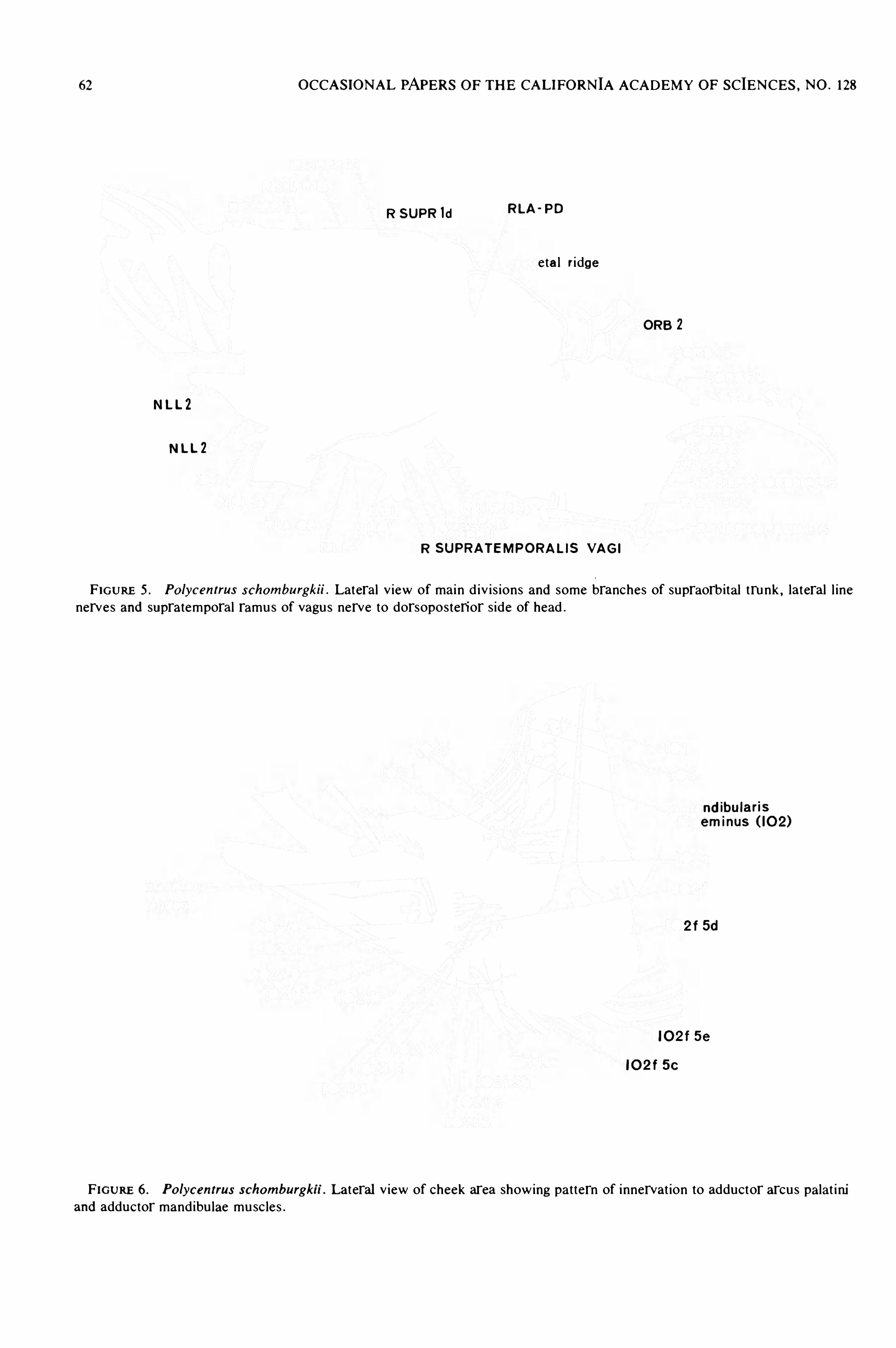
62 OCCASIONAL PAPERS OF THE CALIFORNIA ACADEMY OF SCIENCES , NO . 128
R SUPR 1d RLA ' PD
eta l ri dge
ORB 2
NLL2
NLL2
R SUPRATEMPORALIS VAGI
FIGURE 5 . Po lycentrus schomburgkii . Lateral v iew of mai n d iv i s ions and some branches of supraorbital trunk , lateral l inenerves and supratemporal ramus of vagus nerve to dorsoposterior s ide of head .
nd ibularis
eminus ( 10 2)
2 f 5d
10 2 f 5 e
10 2 f 5 e
FIGURE 6 . Po lycentrus schomburgku . Lateral v iew of cheek area showing pat tern of innervat ion to adductor arcus palat i niand adductor mandibu lae musc les .
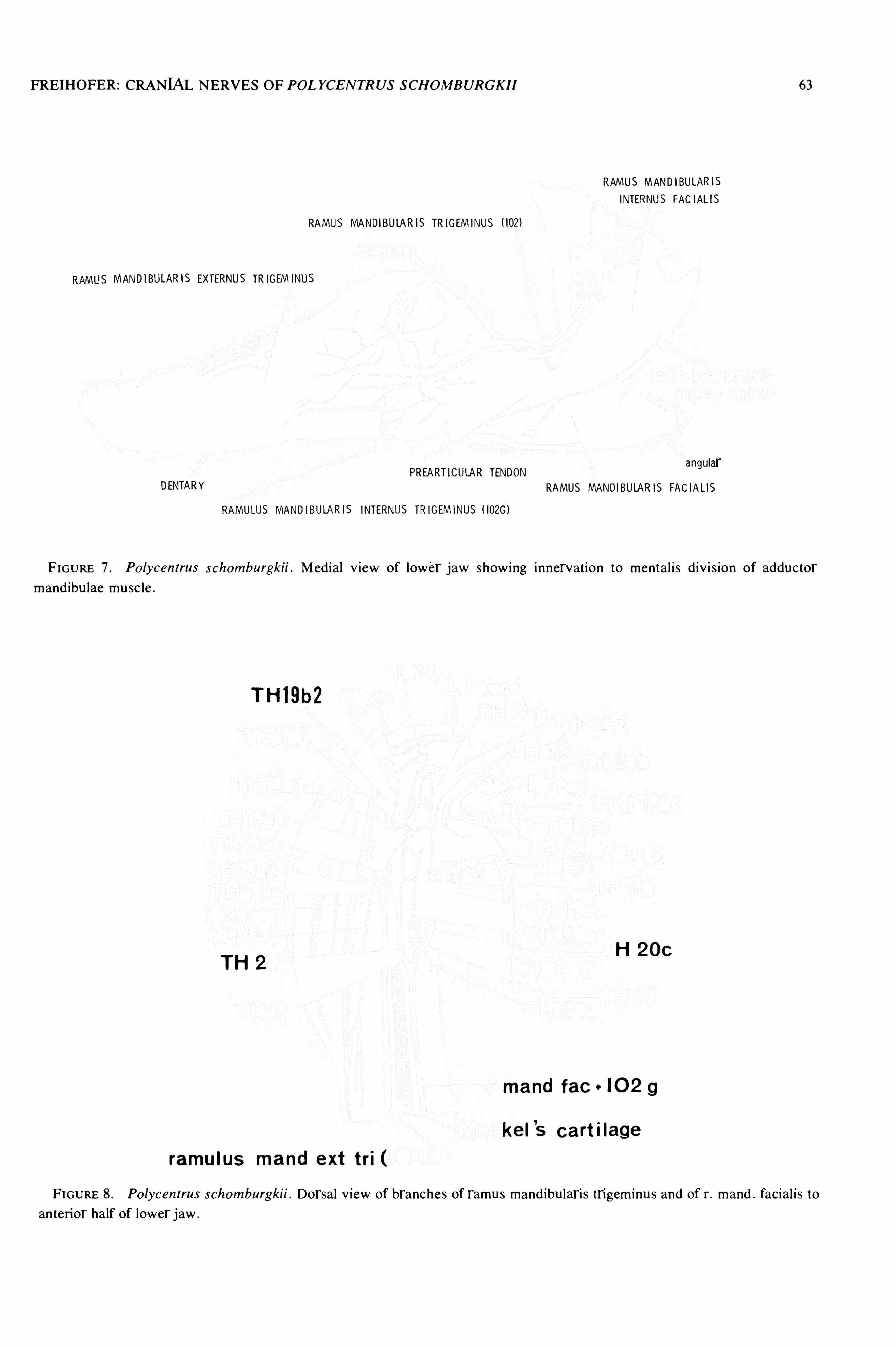
FREIHOFER : CRANIAL NERVES OF POLYCENTRUS SCHOMBURGKII 63
RAMUS MANDIBULARIS
INTERNUS FAC IALIS
RAMUS MANDIBULARIS TRIGEMINUS ( IOZl
MANDIBULARIS EXTERNUS TRIGEMINUS
angularPREARTICULAR TENDON
DENTARY RAMUS MANDlBULARIS FAC IALIS
RAMULUS MANDIBULAR| S INTERNUS TRIGEMINUS ( 1020 )
FIGURE 7 . Po lycentrus schomburgkii . Med ia] V i ew of lower jaw showing innervat ion to mental i s d iv i s ion of adductormand ibulae mu sc le .
TH19b2
TH 2H 2 0 0
ma nd fac 0 10 2 g
ke l’
s cart i lage
ramu l us ma nd ext tri (
FIGURE 8 . Po lycentrus schomburgkii .Dorsal V iew of branches of ramus mand ibulari s trigem i nus and of r. mand . facial i s to
anterior half of lower jaw .
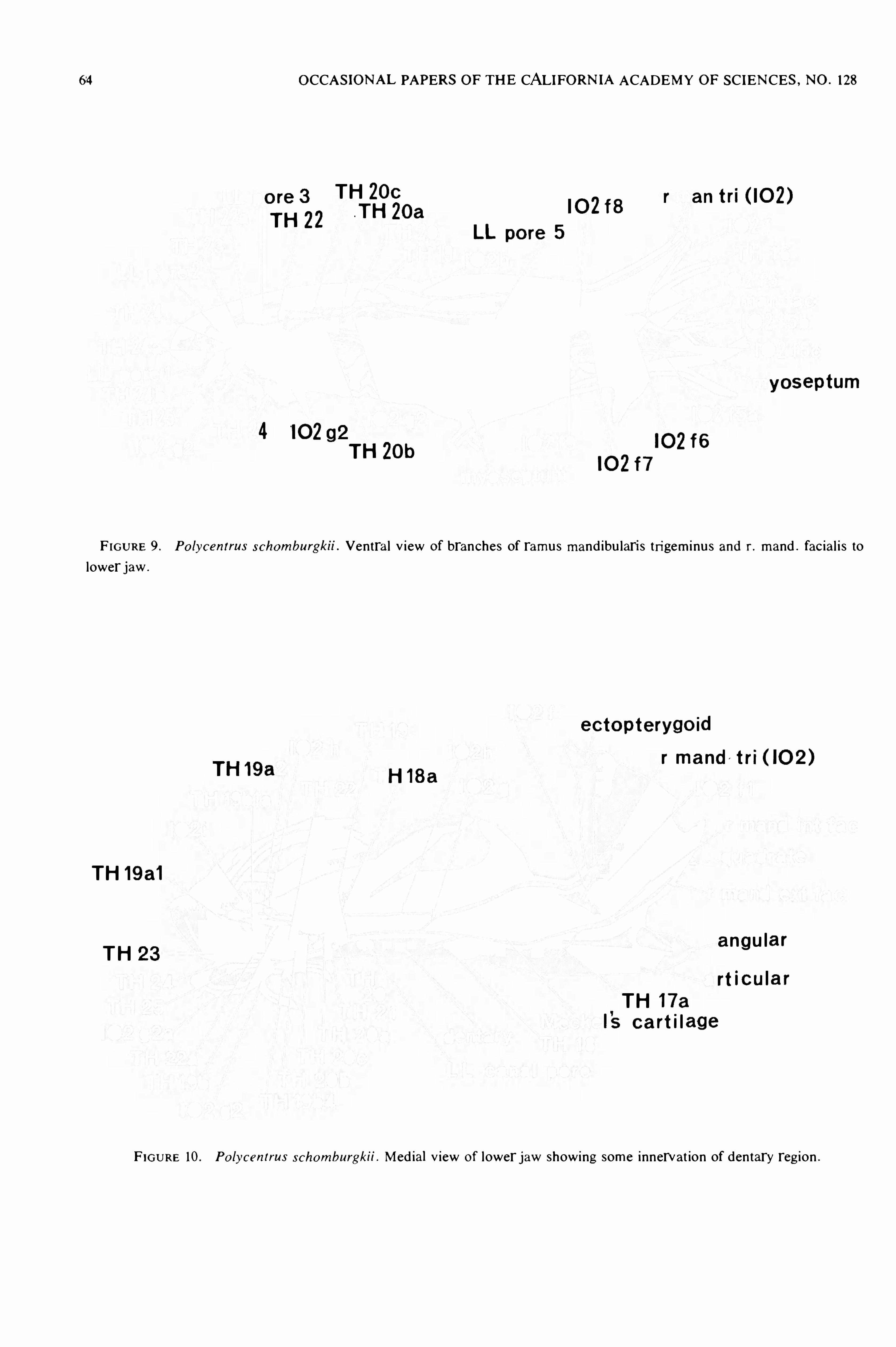
64 OCCASIONAL PAPERS OF THE CALIFORNIA ACADEMY OF SCIENCES , NO . 128
TH ZOco
1
r
§3
22.TH 2oa 10 2f8
r an trr( 10 2)
LL po re 5
yos eptum
4 '0 292TH
10 2f6
10 2f7
F IGURE 9 . Po lycentrus schomburgkii .Ventra l v iew Of branches of ramus mand ibu laris tri gem i nu s and r. mand . fac ial i s to
lower jaw .
ec to pterygo id
r ma nd / t'
ITH 19a H 18a
TI ( 0 2 )
TH 19a1
TH 23angu lar
rt icula r
TH 17a
1'
s cart i l ag e
FIGURE 10 . Po lycentrus schomburgkii . Med ia] V iew Of lower jaw showing some innervat ion of dentary reg ion .

FREIHOFER: CRANIAL NERVES OF POLYCENTRUS SCHOMBURGKII 65

66 OCCASIONAL PAPERS OF THE CALIFORNIA ACADEMY OF SCIENCES ,NO . 128
r vag i
trunc us hyomandibulari
rmandibu laris fac ia l ihyo ide u s (TH 9)
r ma nd i n t facH QB la
H 98 0 3
r mand ext fac H 98 0 2
THH 9D
TH 98 0
10 292
FIGURE 13 . Po lycen trus schomburgkii . Med ia] V iew of hyoid arch and Opercu lar bones showing di stribut ion of ramushyoideus .
NERVUS OPTICUS ( N l l )SUPERIOR RECTUS
C ILIARIS BREV IS
SUPERIOR OBLIQU NERVUS TROCHEARUS ( N. W )
ClLlARlS LONGUS
ClLIARY GANGLION
GASSERIAN GANGLION
PROFUNDUS GANGLION
RADIX PROFUNDUS
INFERIOR OBLlQUENTERNAL RECTUS
RADIX LONGUS
EXTERNAL RECTUSINTERNAL RECIUS
INFERIOR RECIUS
FIGURE 14 . Po lycentrus schomburgkii . Med ia] V i ew of eyebal l and eye musc les showing di stri but ion of cranial nerves III ,IV, and VI , and ramus profundus .
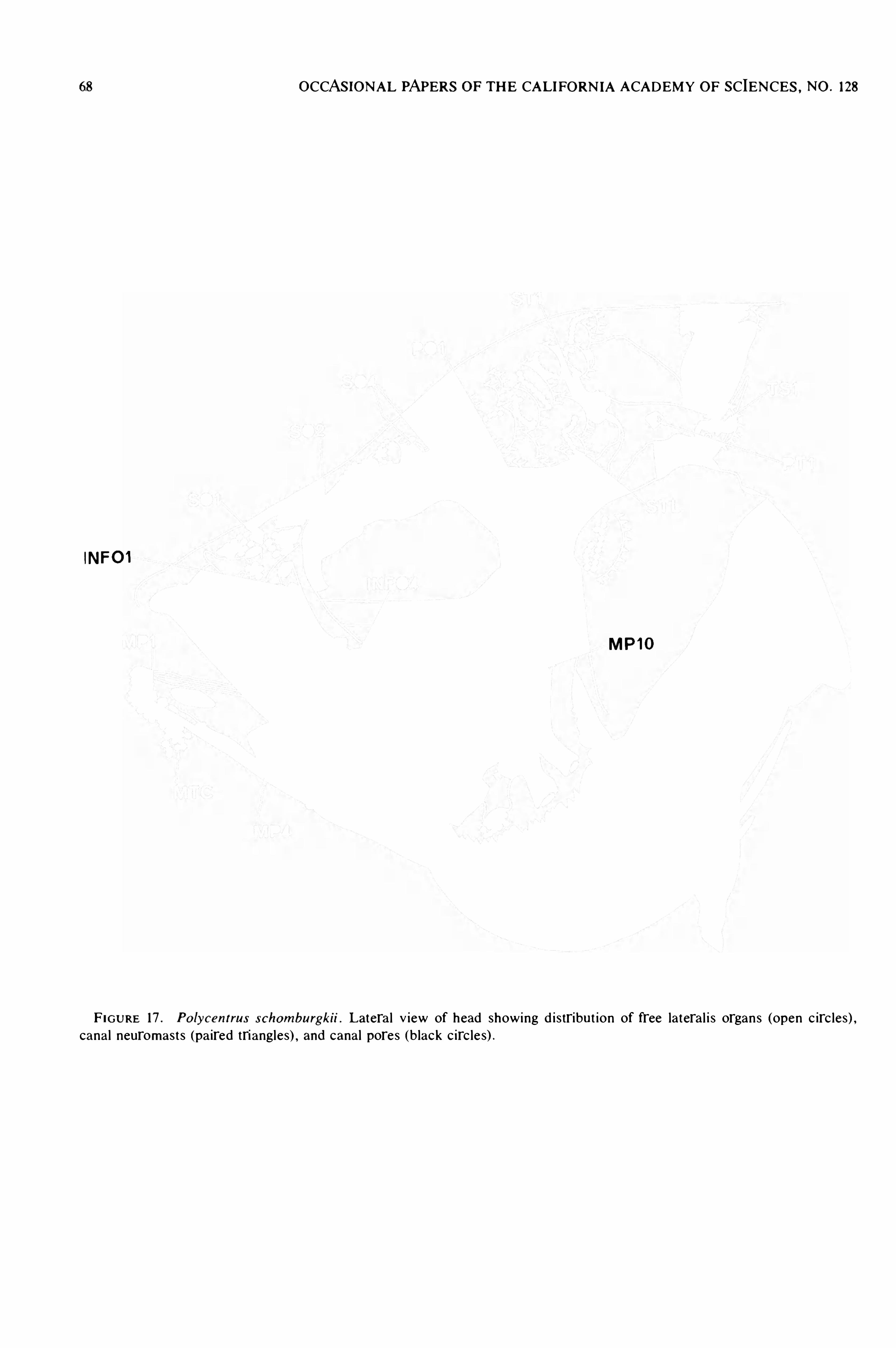
68 OCCASIONAL PAPERS OF THE CALIFORN IA ACADEMY OF SCIENCES , NO . 128
lNFOl
MF 10
F IGURE 17 . Po lycentrus schomburgkii . Latera l V iew of head showing d istribut ion of free lateral i s organs (open c irc les) ,canal neuromast s (pa ired triangles) , and canal pores (black c irc le s) .

FREIHOFER : CRANIAL NERVES OF POLYCENTRUS SCHOMBURGKII 69
SUPRACLEITHRUM
C LEITHRUM
PSEUDOBRANCH
GLOSSOHYAL
HYPOHYA I.
ENTRAL HY POHYAL
UROHYAL
FIGURE 18 . Po lycentrus schomburgkii . Lateral v i ew of gi l l arches and pectoral g irdle showing assoc iated gi l l arch musc le s .
Anterior to righ t .
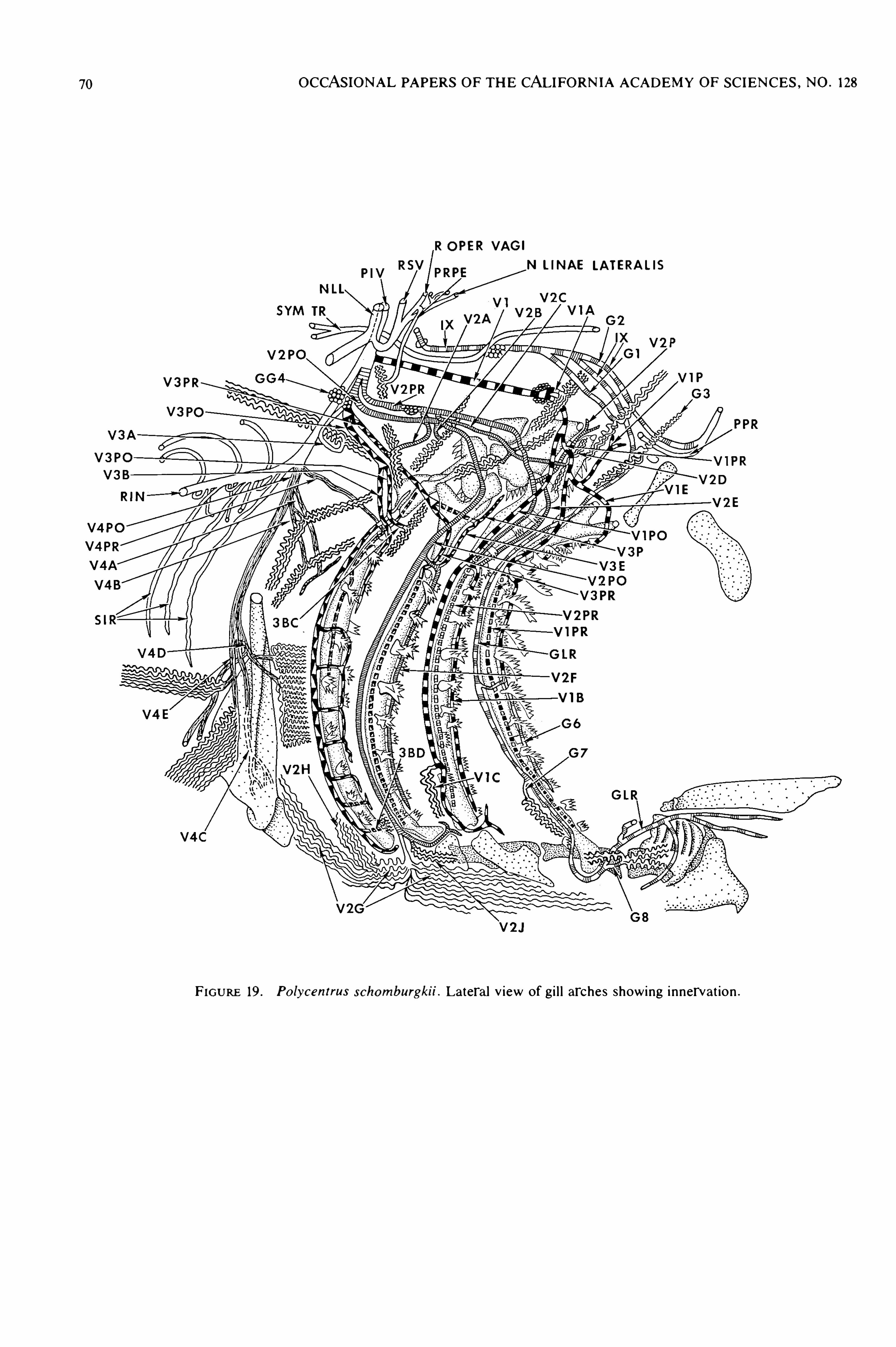
70 OCCASIONAL PAPERS OF THE CALIFORNIA ACADEMY OF SCIENCES , NO . 128
R OPER VAGI
N LINAE LATERALIS
V3PR
FIGURE 19 . Po lycentrus schomburgkii . Lateral v iew of gi l l arches showing innervat ion .
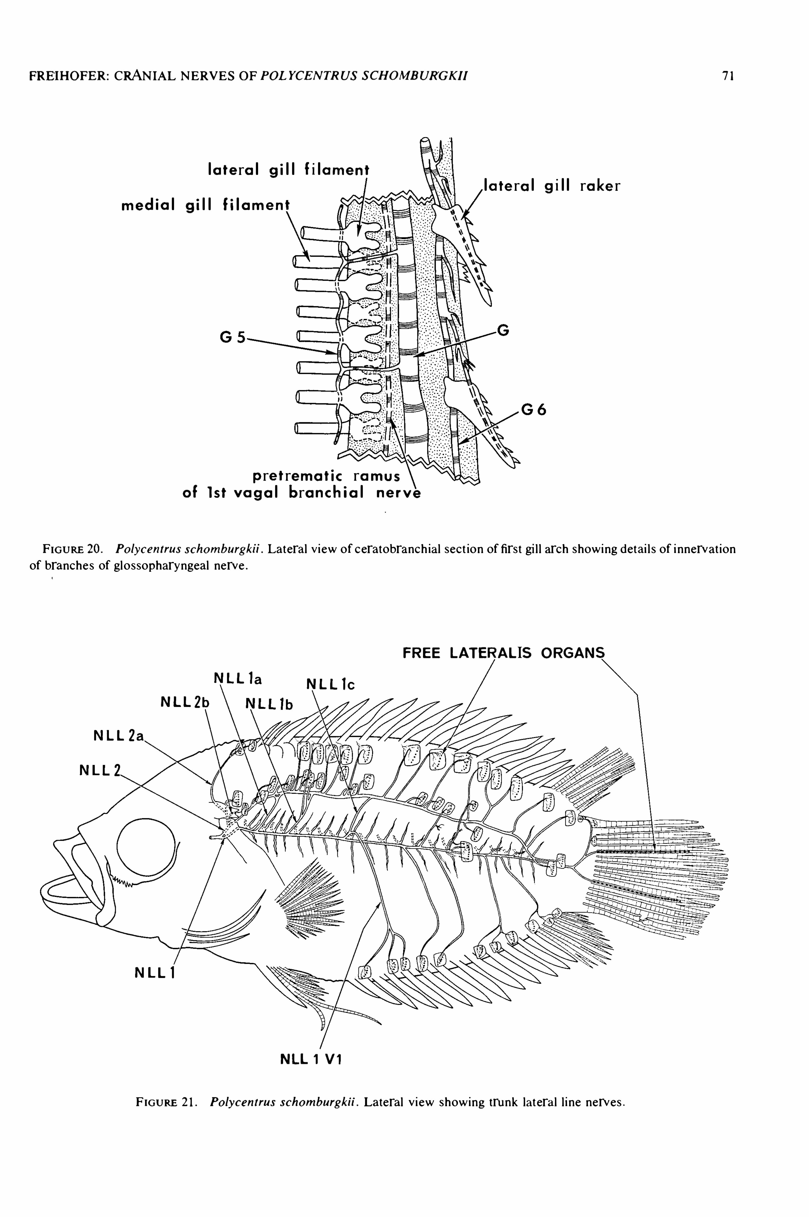
FREIHOFER : CRANIAL NERVES OF POLYCENTRUS SCHOMBUR'
GKII 7 ]
l a te ra l g i l l fi lame ntla te ra l g i l l ra ke r
med ia l g i l l Ii lam e n
p re t rema t ic ra mus
o i l s t v a g a l bro nch ia l ne rv e
FIGURE 20 . Po lycentrus schomburgku . Lateral V i ew of ceratobranch ial sect ion of first gi l l arch showing detai l s of i nnervat ionof branches of glossopharyngeal nerve .
FREE LATERALIS ORGAN
NLL la NLL lC
NLLZb
NLL2a
NLL ]
NLL 1 VI
F IGURE 21 . Po lycentrus schomburgku . Lateral v i ew showing trunk latera l l i ne nerves .
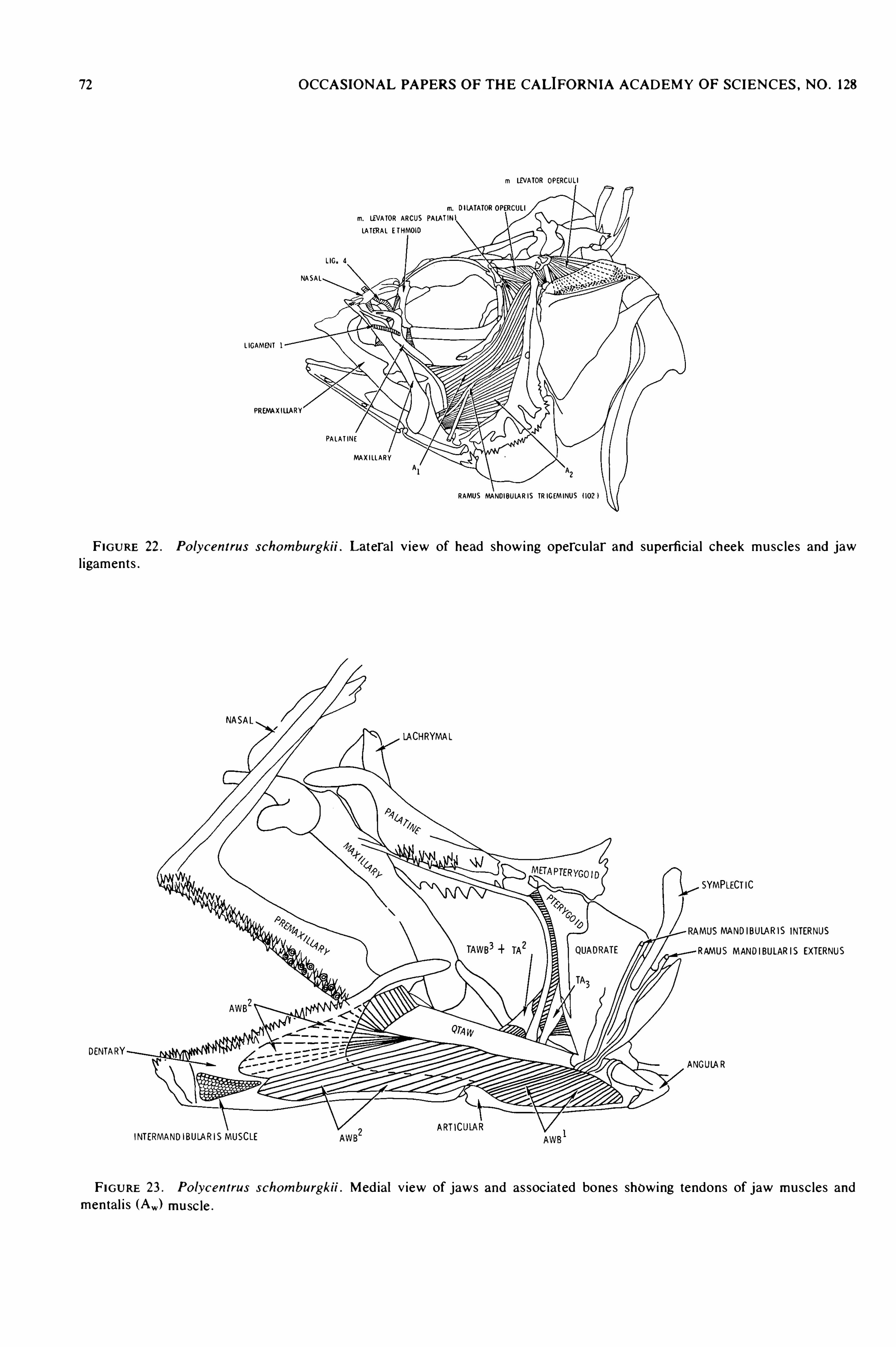
72 OCCASIONAL PAPERS OF THE CALIFORN IA ACADEMY OF SCIENCES , NO . 128
m LB/ATOR OPERCULI
m. Di lATATOROP
m. LEVA TOR ARCUS PAIA TlNLA TERA L ETHMOlD
LIG. 4
NASA L
L IGAMENT l
PREMAXILIARY
PA LA T INEMAXI LL A RY
RAMUS MANDIBULA R IS TR IGEMINUS ( 102 1
FIGURE 22 . Po lycentrus schomburgkii . Lateral v iew of head showing opercu lar and superficia l cheek musc les and jawl igaments .
NASAL
LACHRYMAL
SYMPLECTIC
MUSMANDIBUIA RIS INTERNUS
RAMUS MANDIBULARIS EXTERNUS
DENTARYANGUIA R
ARTICINTERMANDIBULARISMUSCLE
FIGURE 23 . Po lycentrus schomburgkn . Med ia] V iew of jaws and as soc iated bones showing tendons of jaw musc les andmental i s (Aw ) musc le .

I'
HOFER: CRANIAL NERVES OF POLYCENTRUS SCHOMBURGKII 73
lNTERMANDIPRMRTICULAR
MECKEL'S CARTILAG
RAMUS MANDIBULARIS TRIGEMINUS
GURE 24 . Po lycentrus schomburgkn . Med ia] V iew of jaws Showing insert ion of adductor mand ibu lae musc le s and tendons .
INT ERHYA L
HYOIDE I ADDUCTORESEPIHYAL
CERATCHYAL
HYOlDEl ABDUCTOR ES MUSCLE
FIGURE 25 . Po lycentrus schomburgkii . Med ia] V iew of opercle and hyoid arch showing hyohyo ideus musc le .
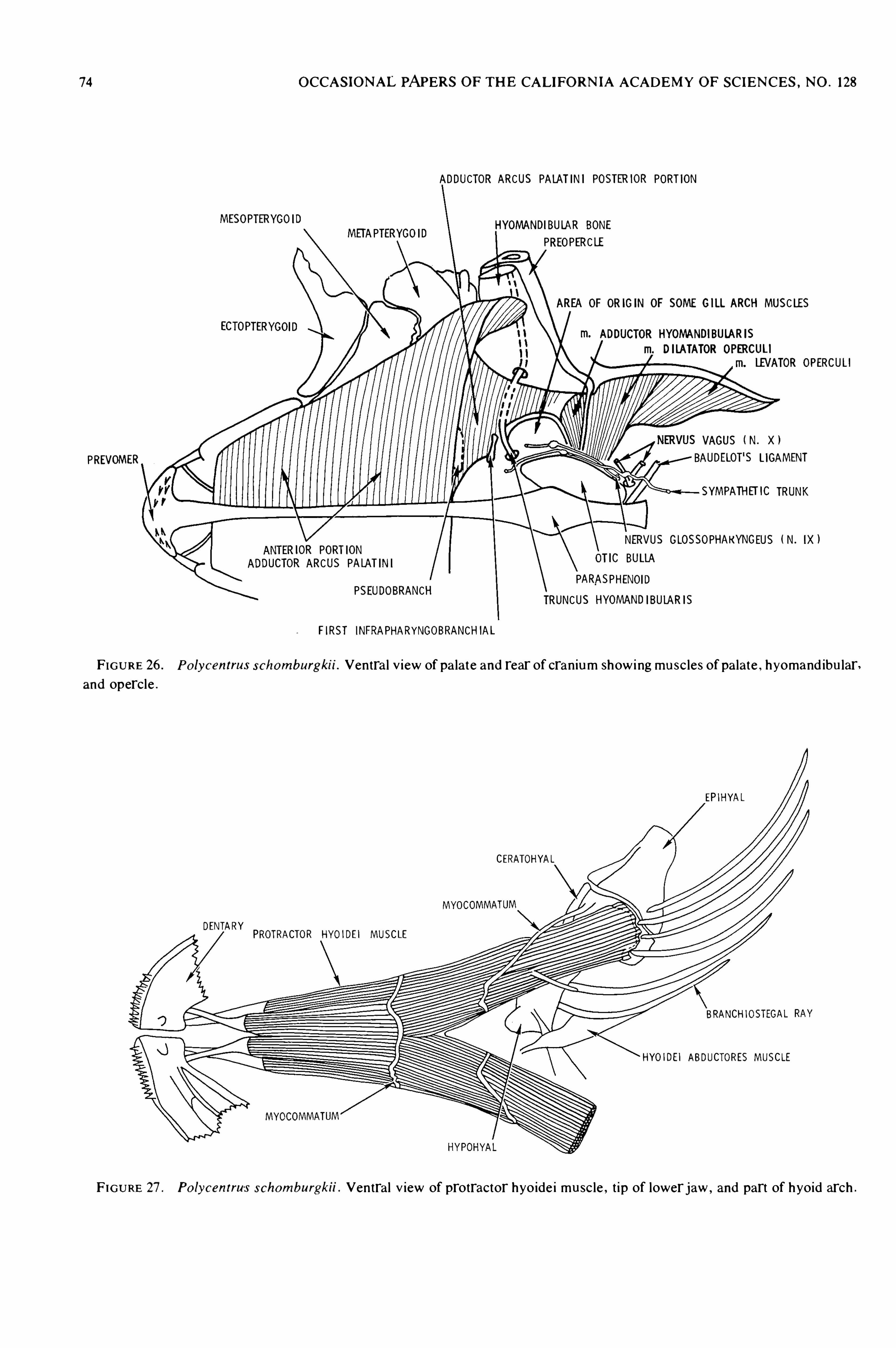
74 OCCASIONAL PAPERS OF THE CALIFORNIA ACADEMY OF SCIENCES , NO . 128
ADDUCTOR ARCUS PALATINI POSTERIOR PORTION
MESOPTERYGOIDHYOMANDIBULAR BONE
METAPTERYGOIDPREOPERCLE
OF ORIGIN OF SOME GILLARCH MUSCLES
ECTOPTERYGOADDUCTOR HYOMANDIBULARIS
DILATATOR OPERCULI
R OPERCULI
VAGUS ( N. X )
PREVOMER AUDELOT'S LIGAMENT
SYMPATHH IC TRUNK
NERVUS GLOSSOP EUS IXlOR PORTION
HAKYNG ( N
UCTOR ARCUS PALATINI OT'C BULLA
PARASPHENOIDPSEUDOBRANCH
TRUNCUS HYOMANDIBULARIS
FIRST INFRAPHARYNGOBRANCHIAL
FIGURE 26 . Po lycentrus schomburgkii . Ventral v iew of pa late and rear of cranium showing musc les of pa late , hyomand ibu larand opercle .
EP IHYAL
CERATOHYAL
MYOCOMMATUM
DENTARY PROTRACTOR HYO1DE] MUSCLE
BRANCHIOSTEGAL
HYOIDEI ABDUCTORES MUSCLE
MYOCOMMATUM
FIGURE 27 . Po lycentrus schomburgkii .Ventral V iew of protractor hyoide i musc le , t ip of lower jaw ,
and part of hyoid arch .

76 OCCASIONAL PAPERS OF THE CALIFORNIA ACADEMY OF SCIENCES , NO . 128
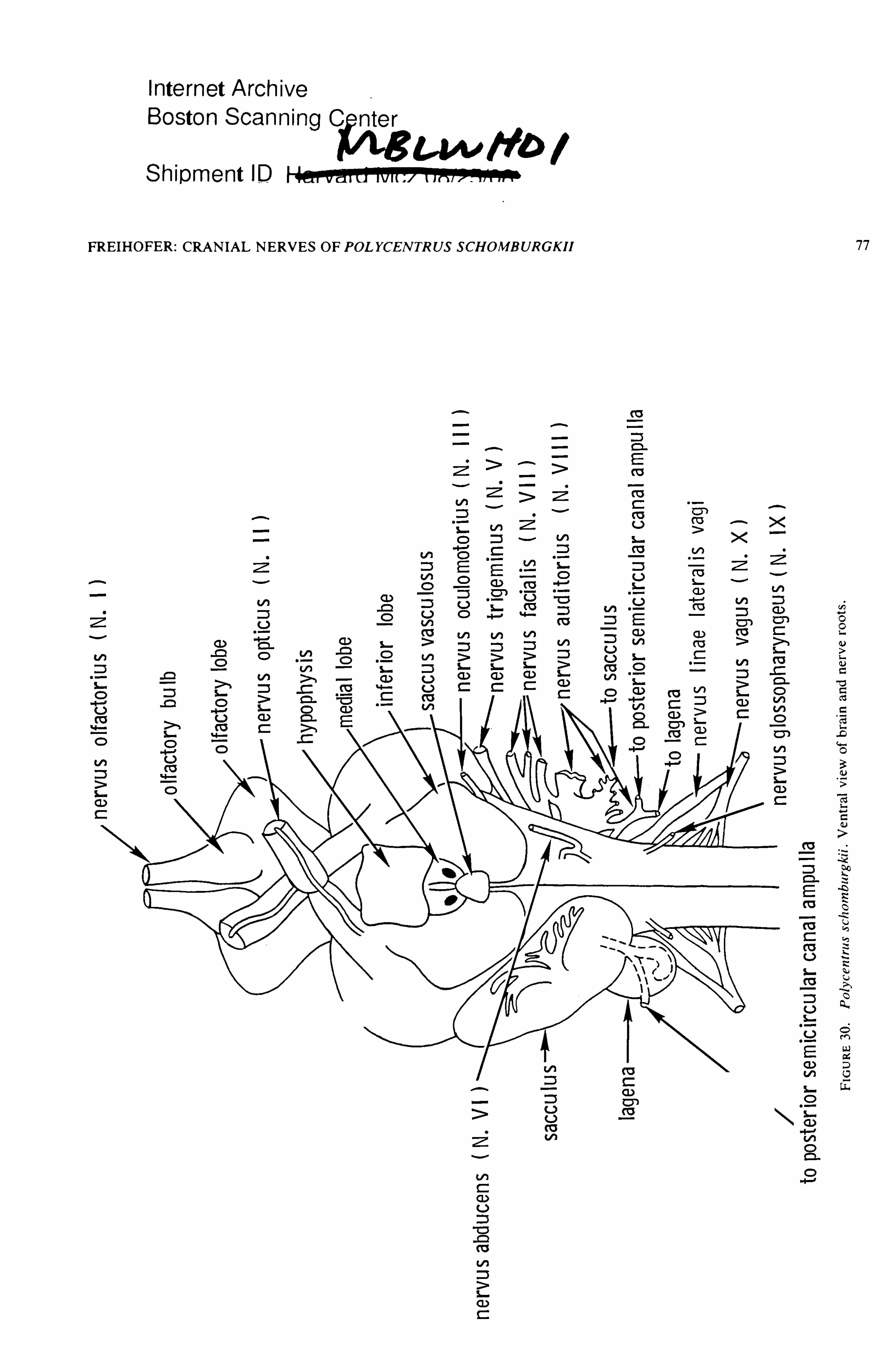
Internet Archive
Boston Scan
Shipment ID Han -3M
FREIHOFER : CRANIAL NERVES OF POLYCENTRUS SCHOMBURGKII

78 OCCASIONAL PAPERS OF THE CALIFORNIAACADEMY OF SCIENCES , NO .
![Complete genome sequence of Archaeoglobus profundus...A. fulgi-dus [1]. However, A. profundus differs from l-A. fu gidus in the composition of complex lipids, con-sisting of two phosphoglycolipids](https://img.pdfslide.us/doc/110x75/60ba70d6e5e2eb554115704d/complete-genome-sequence-of-archaeoglobus-profundus-a-fulgi-dus-1-however.jpg)