Embed Size (px)
DESCRIPTION
Citation preview
ROLE OF HISTONES IN DNA PACKAGING
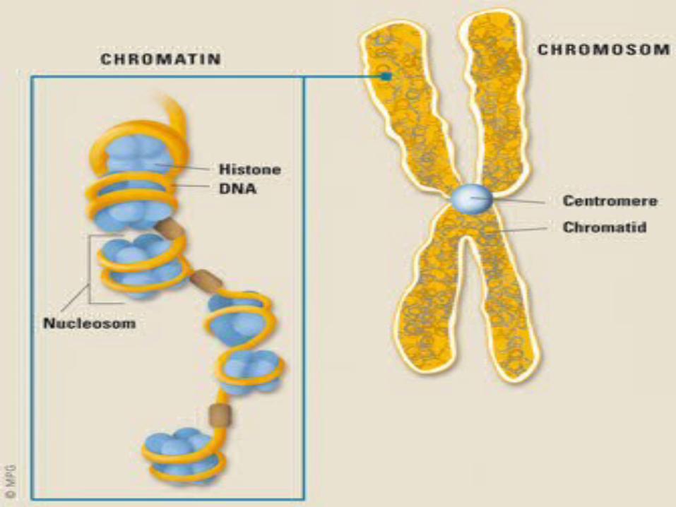
CHROMOSOMES:
• “Chromosomes are thread like structures; appear at the time of cell division, in the nucleus.”
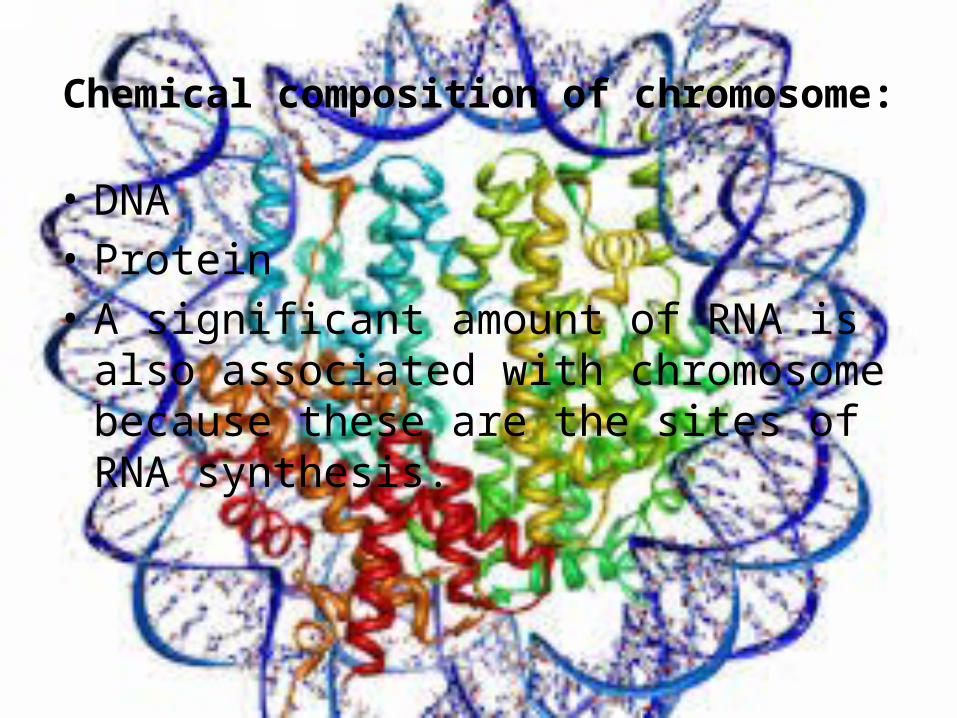
Chemical composition of chromosome:
• DNA• Protein• A significant amount of RNA is also associated
with chromosome because these are the sites of RNA synthesis.
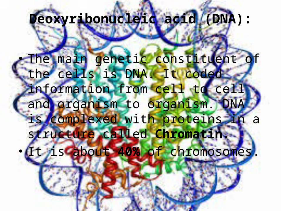
Deoxyribonucleic acid (DNA):
• The main genetic constituent of the cells is DNA. It coded information from cell to cell and organism to organism. DNA is complexed with proteins in a structure called Chromatin.
• It is about 40% of chromosomes.
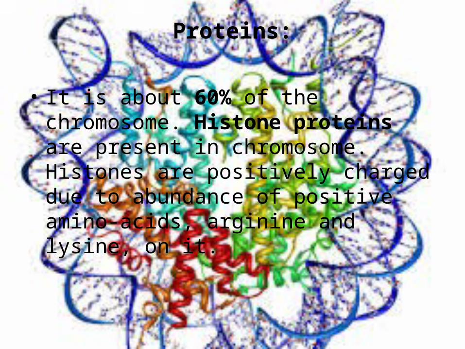
Proteins:
• It is about 60% of the chromosome. Histone proteins are present in chromosome. Histones are positively charged due to abundance of positive amino-acids, arginine and lysine, on it.

Structural features:
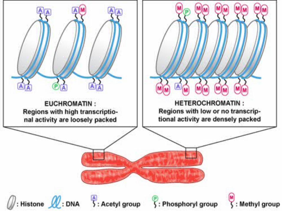
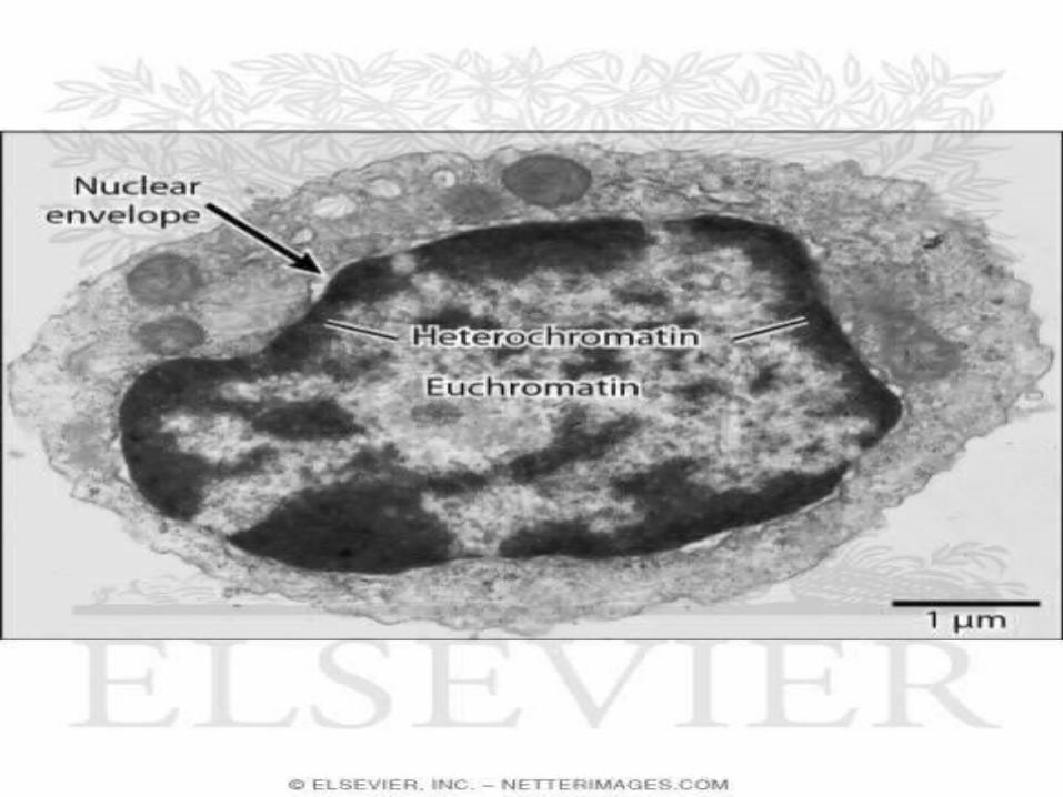
Condensed and non-condensed portions:Heterochromatin:
• These are highly condensed portions of chromosomes.
Euchromatin:• These are portions other than
heterochromatin.lightly stained.
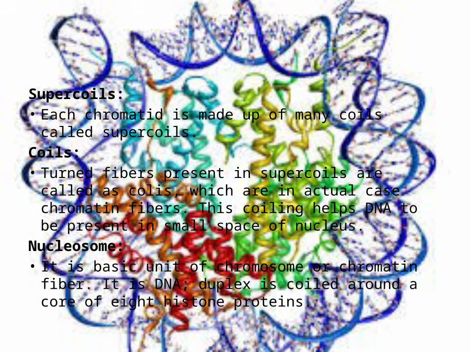
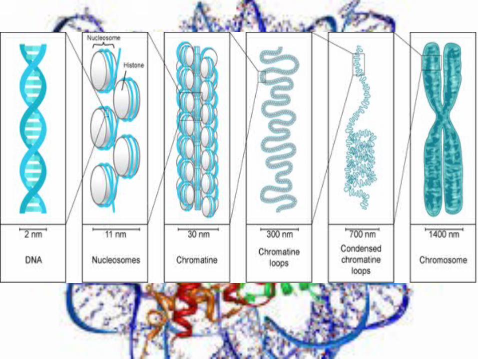
Supercoils:• Each chromatid is made up of many coils called supercoils.Coils:• Turned fibers present in supercoils are called as colis,
which are in actual case chromatin fibers. This coiling helps DNA to be present in small space of nucleus.
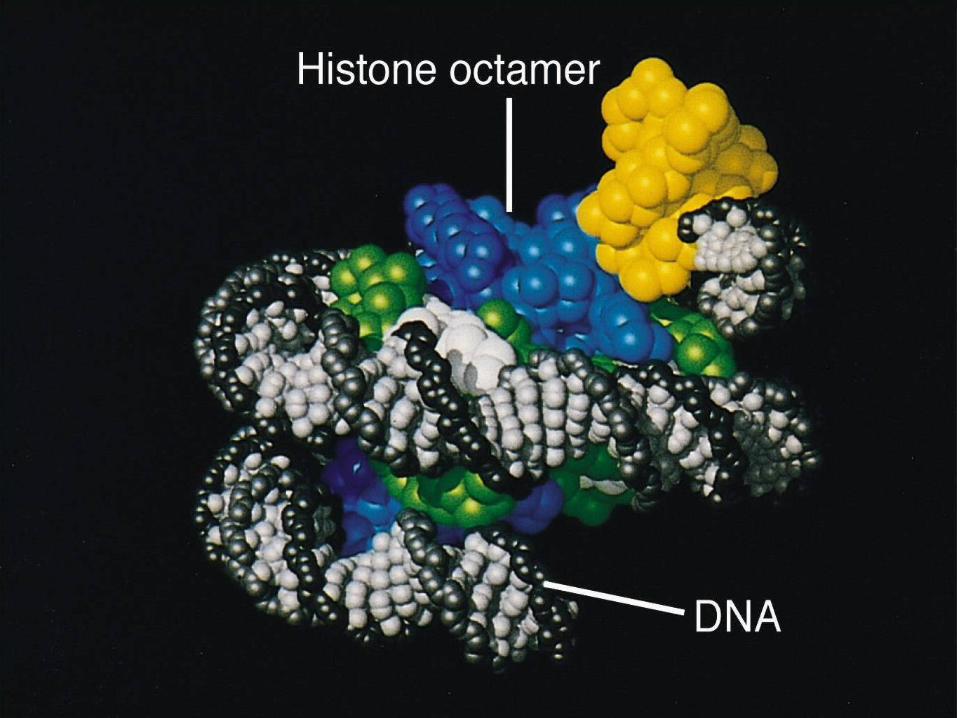
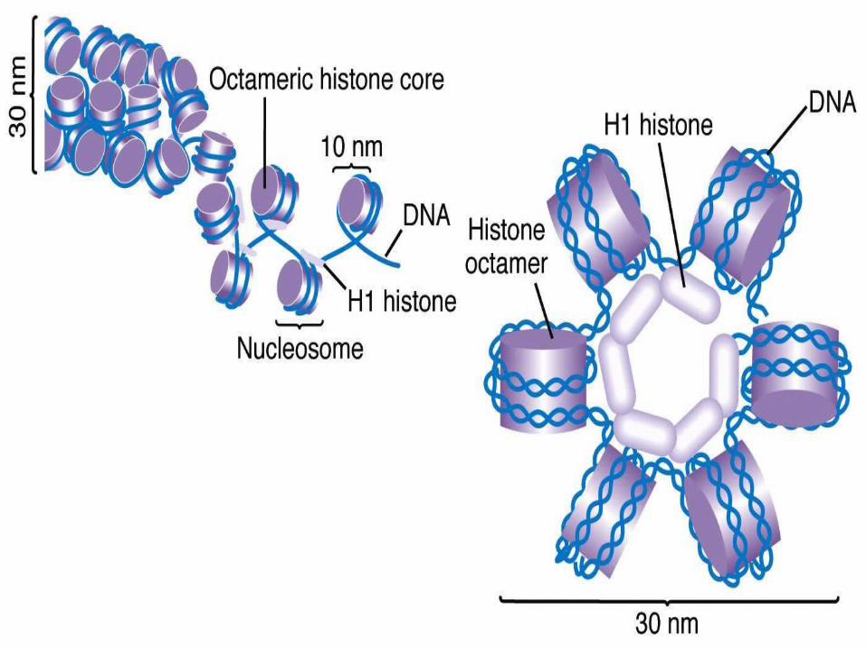
Nucleosome:• It is basic unit of chromosome or chromatin fiber. It is
DNA; duplex is coiled around a core of eight histone proteins.
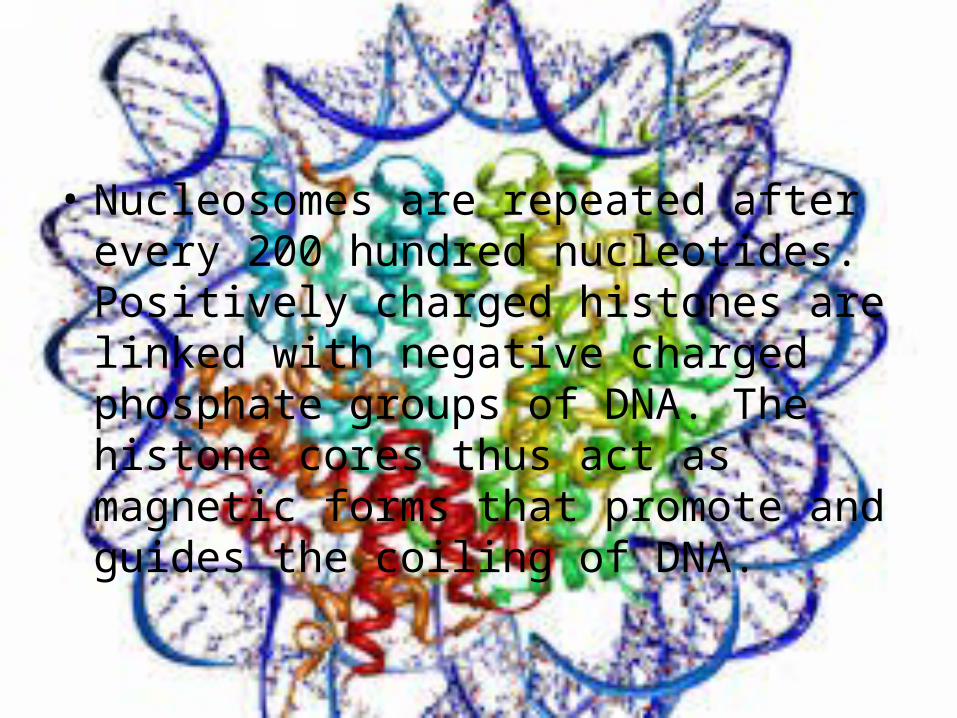
• Nucleosomes are repeated after every 200 hundred nucleotides. Positively charged histones are linked with negative charged phosphate groups of DNA. The histone cores thus act as magnetic forms that promote and guides the coiling of DNA.
Histones or Histone proteins
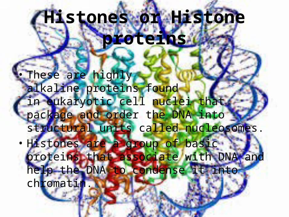
• These are highly alkaline proteins found in eukaryotic cell nuclei that package and order the DNA into structural units called nucleosomes.
• Histones are a group of basic proteins that associate with DNA and help the DNA to condense it into chromatin.
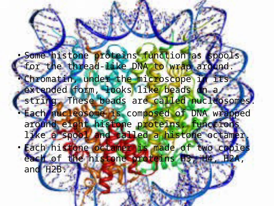
• Some histone proteins function as spools for the thread-like DNA to wrap around.
• Chromatin, under the microscope in its extended form, looks like beads on a string. These beads are called nucleosomes.
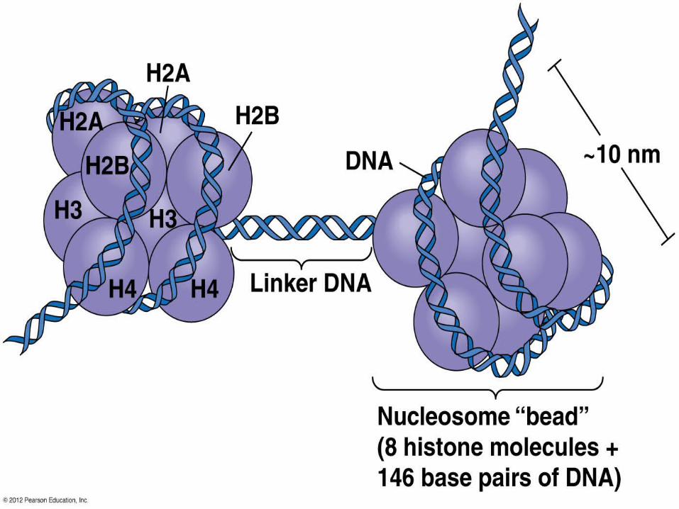
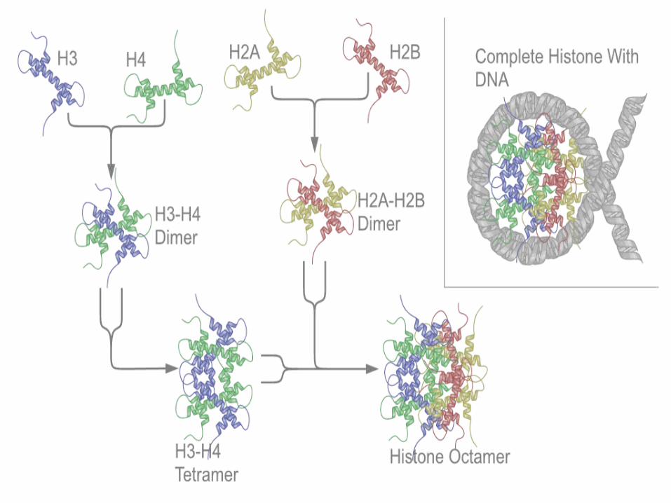
• Each nucleosome is composed of DNA wrapped around eight histone proteins, functions like a spool and called a histone octamer.
• Each histone octamer is made of two copies each of the histone proteins H3, H4, H2A, and H2B.
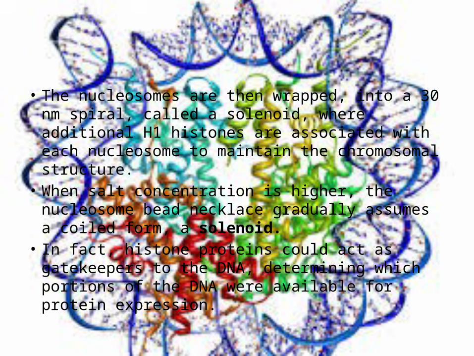
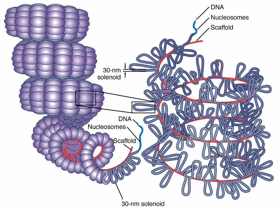
• The nucleosomes are then wrapped, into a 30 nm spiral, called a solenoid, where additional H1 histones are associated with each nucleosome to maintain the chromosomal structure.
• When salt concentration is higher, the nucleosome bead necklace gradually assumes a coiled form, a solenoid.
• In fact, histone proteins could act as gatekeepers to the DNA, determining which portions of the DNA were available for protein expression.
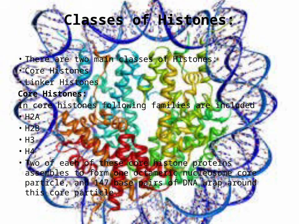
Classes of Histones:
• There are two main classes of Histones:• Core Histones• Linker HistonesCore Histones:In core histones following families are included• H2A• H2B• H3• H4• Two of each of these core histone proteins assembles to form one
octameric nucleosome core particle, and 147 base pairs of DNA wrap around this core particle.
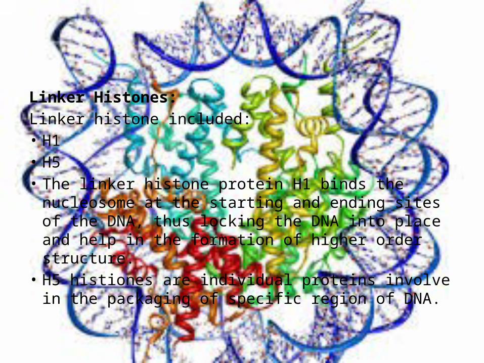
Linker Histones:Linker histone included:• H1• H5• The linker histone protein H1 binds the nucleosome at
the starting and ending sites of the DNA, thus locking the DNA into place and help in the formation of higher order structure.
• H5 histiones are individual proteins involve in the packaging of specific region of DNA.
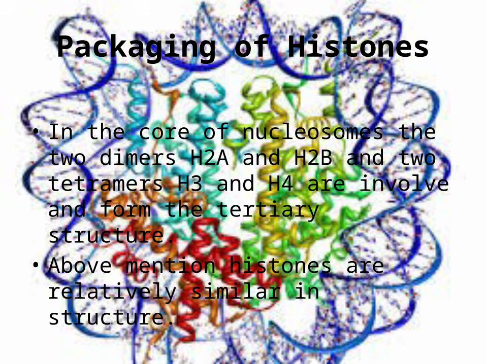
Packaging of Histones
• In the core of nucleosomes the two dimers H2A and H2B and two tetramers H3 and H4 are involve and form the tertiary structure.
• Above mention histones are relatively similar in structure.
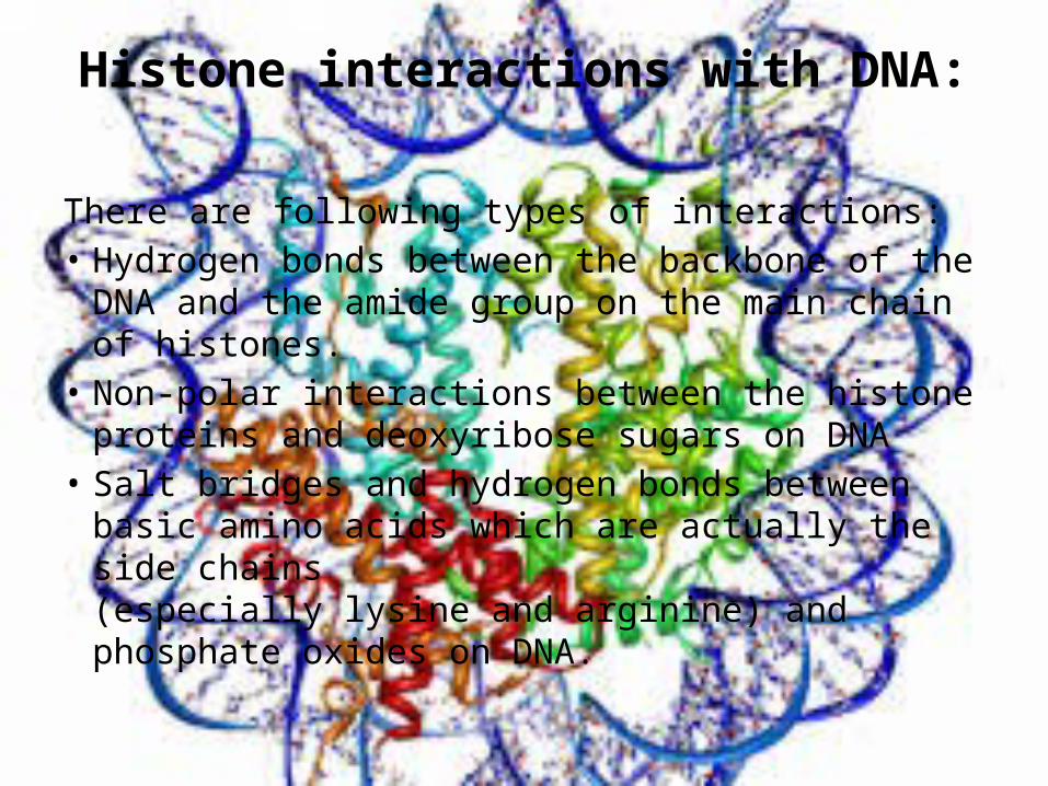
Histone interactions with DNA:
There are following types of interactions:• Hydrogen bonds between the backbone of the DNA
and the amide group on the main chain of histones.• Non-polar interactions between the histone
proteins and deoxyribose sugars on DNA• Salt bridges and hydrogen bonds between basic
amino acids which are actually the side chains (especially lysine and arginine) and phosphate oxides on DNA.
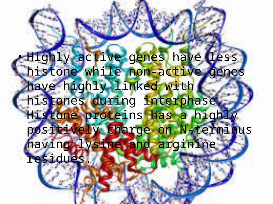
• Highly active genes have less histone while non-active genes have highly linked with histones during interphase. Histone proteins has a highly positively charge on N-terminus having lysine and arginine residues.
Types of modification in Histones:
• Histones can be changed to alter how much packing the DNA is capable of. There are many modifications that affect how well DNA is packaged.
• The three main types of modifications can be seen in the following table:
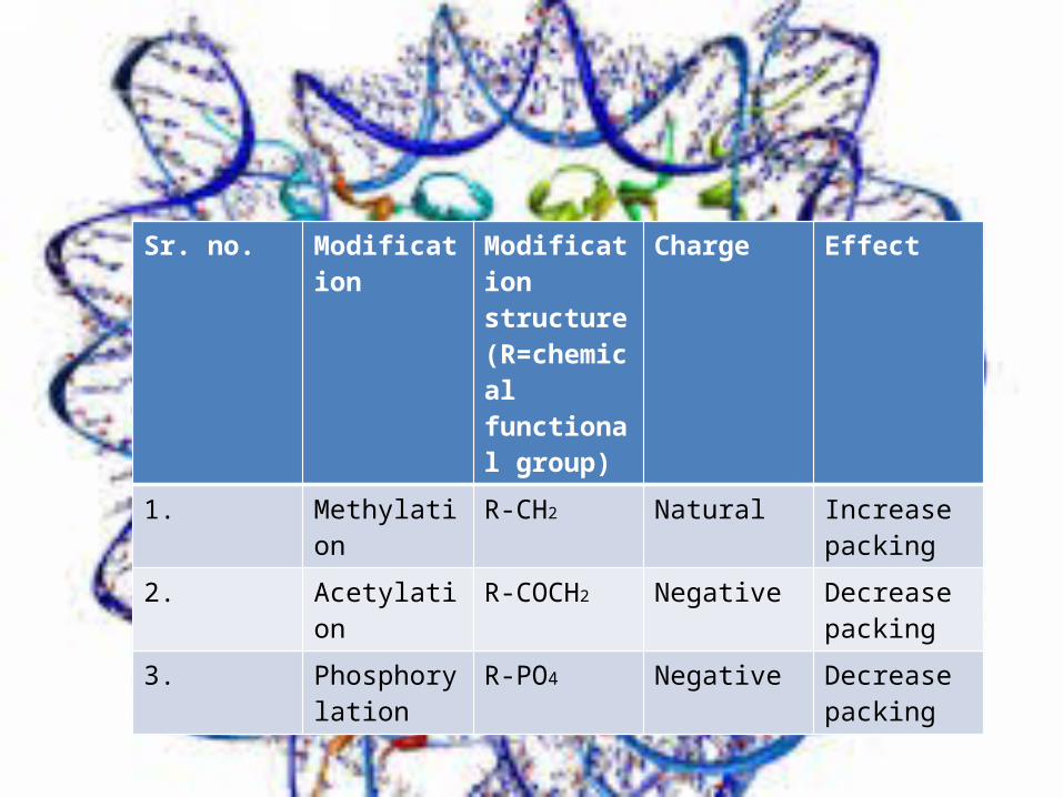
Sr. no. Modification Modification structure (R=chemical functional group)
Charge Effect
1. Methylation R-CH2 Natural Increase packing
2. Acetylation R-COCH2 Negative Decrease packing
3. Phosphorylation
R-PO4 Negative Decrease packing
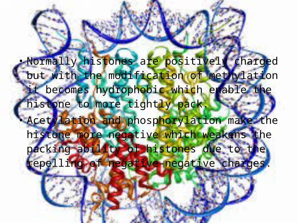
• Normally histones are positively charged but with the modification of methylation it becomes hydrophobic which enable the histone to more tightly pack.
• Acetylation and phosphorylation make the histone more negative which weakens the packing ability of histones due to the repelling of negative-negative charges.
Chromatin
• It is complex nucleic acid and protein which condenses to form chromosome during cell division.
• In eukaryotes it is found within cell nucleus whereas in case of prokaryotes it is present in nucleoid.
• It can easily recognize through staining therefore its name, literally means colored material.
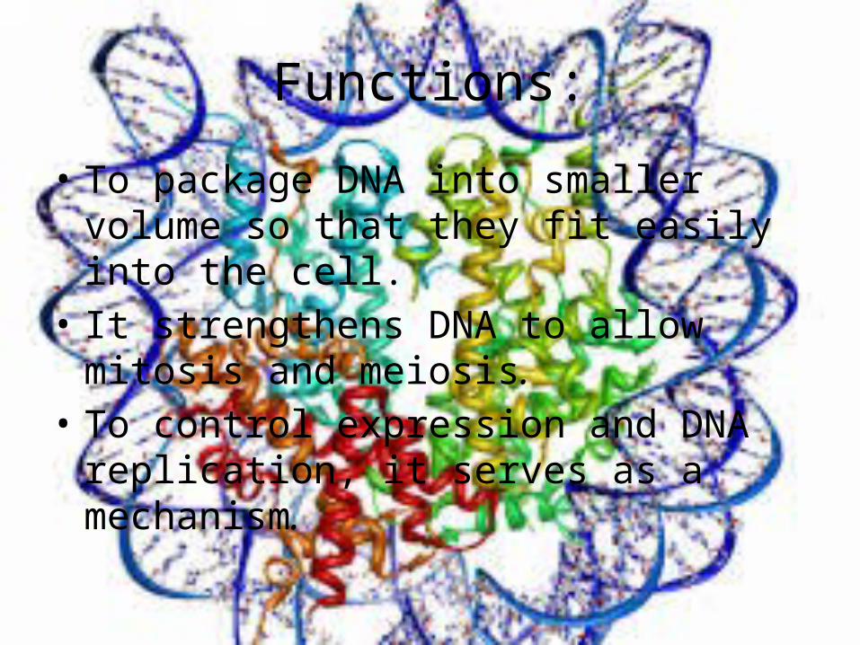
Functions:
• To package DNA into smaller volume so that they fit easily into the cell.
• It strengthens DNA to allow mitosis and meiosis.
• To control expression and DNA replication, it serves as a mechanism.
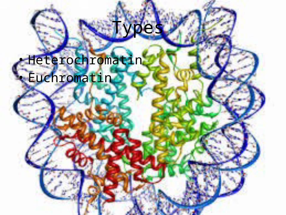
Types
• Heterochromatin • Euchromatin
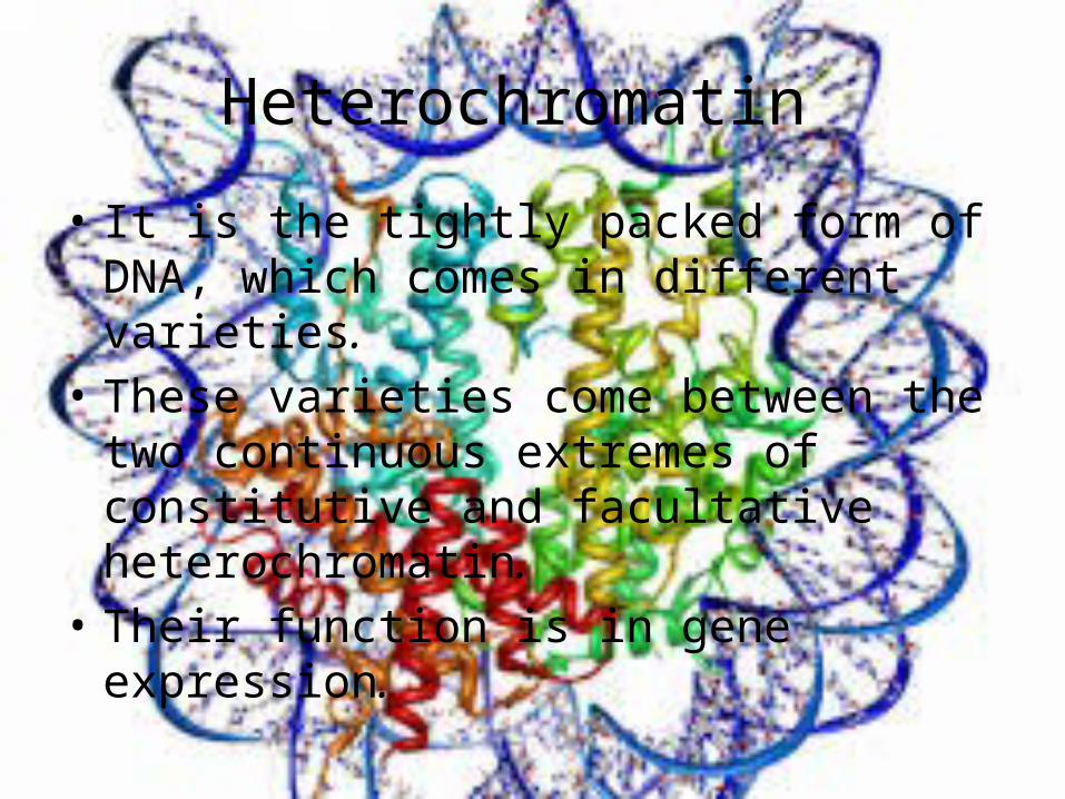
Heterochromatin
• It is the tightly packed form of DNA, which comes in different varieties.
• These varieties come between the two continuous extremes of constitutive and facultative heterochromatin.
• Their function is in gene expression.
• It is not active and under specific environmental and developmental signaling, loses its condensed structure and become active.
• Centromere and telomere both are heterochromatin as in the Barr body off, second inactivated X chromosome in female.
function
• Gene regulation and protection of chromosome integrity
• Dense packing of DNA makes less accessible to protein factors that bind with DNA or its associated sites.
• It results in formation of epigenetic inheritance.
Constitutive Heterochromatin
• All of its cells pack in the same region of DNA so any gene in all cells would be poorly expressed.i.e.1, 9, 16 and Y human chromosome contain large region of constitutive chromatin.
• In most organisms, it is present around chromosome centromere and near telomeres.
• Constitutive chromatin affect the nearer genes and usually repetitive and form centromere or telomeres in addition to acting as an attractor for gene expression and repression signals
Facultative heterochromatin
• Formation of facultative heterochromatin is regulated and associated with morphogenesis or differentiation.i.e. X chromosome inactivation in female mammals.
• facultative chromatin is the result of genes silenced in a mechanism of histone methylation.
• One X chromosome is packs as silences in case of facultative and other cell packed as euchromatin and expressed.
Euchromatin
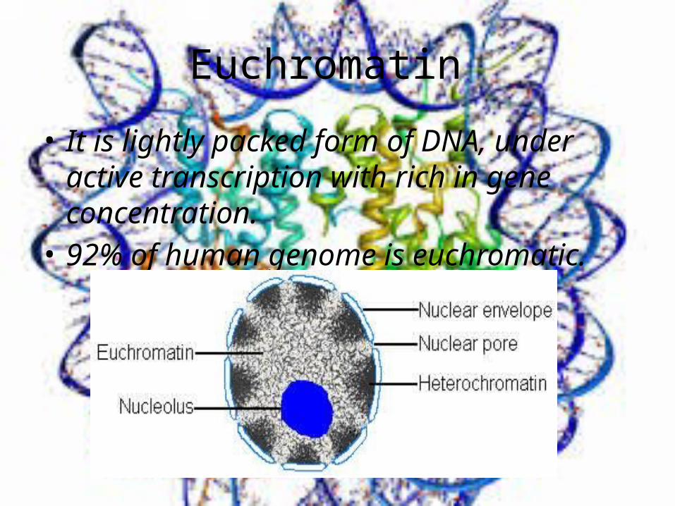
• It is lightly packed form of DNA, under active transcription with rich in gene concentration.
• 92% of human genome is euchromatic.
Functions
• Active transcription of DNA to mRNA products.• Its unfolded structure allows the gene
regulatory proteins and RNA polymerase to bind with DNA sequence so to initiate the transcription process.
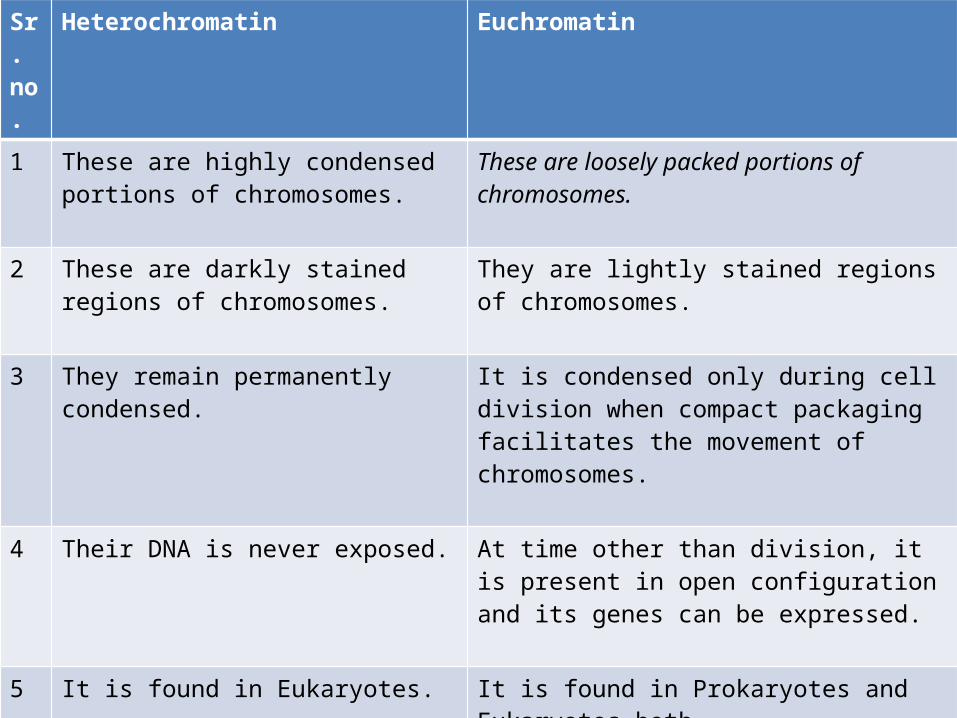
Sr. no.
Heterochromatin Euchromatin
1 These are highly condensed portions of chromosomes.
These are loosely packed portions of chromosomes.
2 These are darkly stained regions of chromosomes.
They are lightly stained regions of chromosomes.
3 They remain permanently condensed. It is condensed only during cell division when compact packaging facilitates the movement of chromosomes.
4 Their DNA is never exposed. At time other than division, it is present in open configuration and its genes can be expressed.
5 It is found in Eukaryotes. It is found in Prokaryotes and Eukaryotes both.
6 It is genetically inactive form of chromatin.
It is genetically active form of chromatin.
7 It replicates late. It is earlier replicative


![Comprehensive analysis of histone post-translational … · 2016. 12. 1. · DNA repair, enhancer licensing, cell differentiation, and regulation of disease [1514]. Specific histone](https://img.pdfslide.us/doc/110x75/60af24203ba57e2b470129c5/comprehensive-analysis-of-histone-post-translational-2016-12-1-dna-repair.jpg)