Embed Size (px)
Citation preview
Antigen and
Antibody Reaction
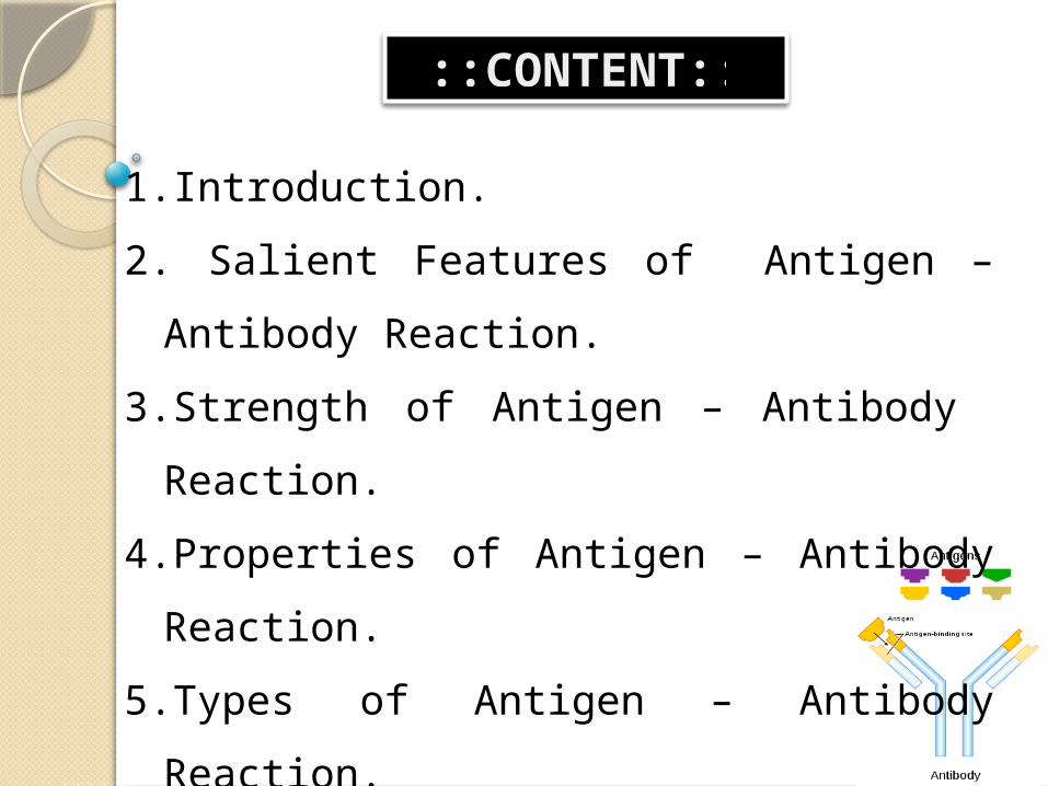
::CONTENT::
1.Introduction.2. Salient Features of Antigen – Antibody
Reaction.3.Strength of Antigen – Antibody
Reaction.4.Properties of Antigen – Antibody
Reaction.5.Types of Antigen – Antibody Reaction.6.Application of Antigen – Antibody
Reaction.7.Conclusion.
INTRODUCTION:
• The antigens and the antibodies combine specifically with each other. This interaction between them is called Antigen-Antibody reaction.
• It may be abbreviated as Ag – Ab reaction.
• These form the basis for humoral immunity or antibody mediated immunity.
• These reactions form the basis for detection of infectious disease causing agents and also some non-specific Ag’s like enzymes.
• When Ag – Ab reactions occur invitro, they are known as serological reactions.
• The reactions between Ag and Ab occur in three stages. In first stage the reaction involves formation of
Ag-Ab complex. The second stage leads to visible events like
precipitation, agglutination etc.The third stage includes destruction of Ag or its
neutralization
Salient Features of Antigen – Antibody Reaction:
• Specificity of Antigen – Antibody Reaction.
• Immune complex.
• Binding Site of Antigen – Antibody Reaction.
• Binding Force of Antigen – Antibody Reaction.
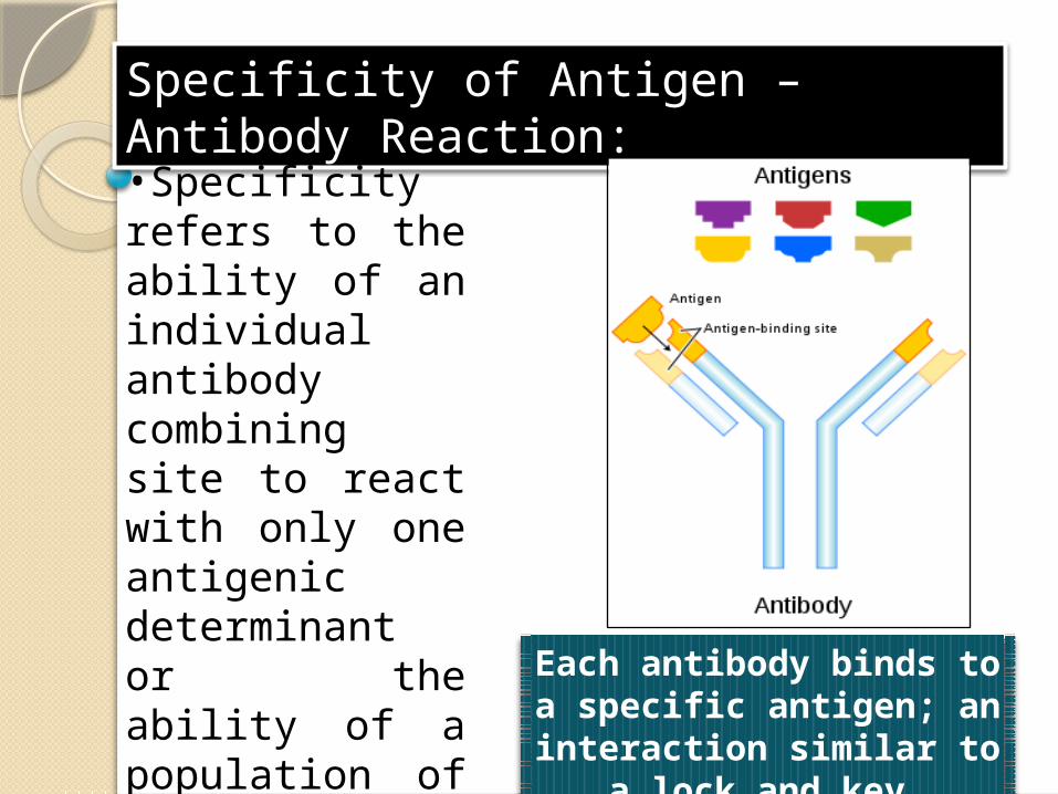
Specificity of Antigen – Antibody Reaction:
•Specificity refers to the ability of an individual antibody combining site to react with only one antigenic determinant or the ability of a population of antibody molecules to react with only one antigen.
Each antibody binds to a specific antigen; an interaction
similar to a lock and key.
•For example, the antibody produced against lens antigen will react only with lens-antigen. Similarly, the antibody produced against kidney antigen will react with only kidney- antigen. A standard lock can be opened by its own key only as one antibody can react with its own antigen.
Immune Complex:
•An immune complex is formed from the integral binding of an antibody to a soluble antigen. •The bound antigen acting as a specific epitope, bound to an antibody is referred to as a singular immune complex.
•Mechanisms of antigen-antibody interaction leading to inflammation. Antigen-antibody immune complex formation results in complement activation, opsonization of target cells, assembly of membrane attack complexes and release of complement activators for chemotaxis. •Fc receptor mediated cell activation triggers cellular responses, such as phagocytosis, antibody-dependent cellular cytotoxicity (ADCC) and release of inflammatory mediators.
Ag + Ab Ag-Ab complexAntigen – Antibody Reaction
Binding Site of Antigen – Antibody Reaction:• In antigen - antibody reaction, the antibody attaches with the antigen.
• The part of antigen which combines with antibody is called Epitope.
• An epitope, also known as antigenic determinant, is the part of an antigen that is recognized by the immune system, specifically by antibodies, B cells, or T cells.
•The part of an antibody that recognizes the epitope is called a paratope.

Antigen and Antibody to Show Epitope And Paratope
Binding Force of Antigen – Antibody Reaction:• The binding between antigen and antibody in ag – ab reaction is due to three factors namely:
Closeness between antigen and antibody. Non – covalent bonds or Intermolecular forces . Affinity of antibody.
Closeness between antigen and antibody: When antigen and antibody are closely fit, the strength of binding is great. When they are apart binding strength low.
Non – Covalent Bonds: The bonds that hold the antigen to the antibody combining site are all non-covalent in nature. These include hydrogen bonds, electrostatic bonds, Van der Waals forces and hydrophobic bonds.
Affinity of antibody: Antibody affinity is the strength of the reaction between a single antigenic determinant and a single combining site on the antibody.
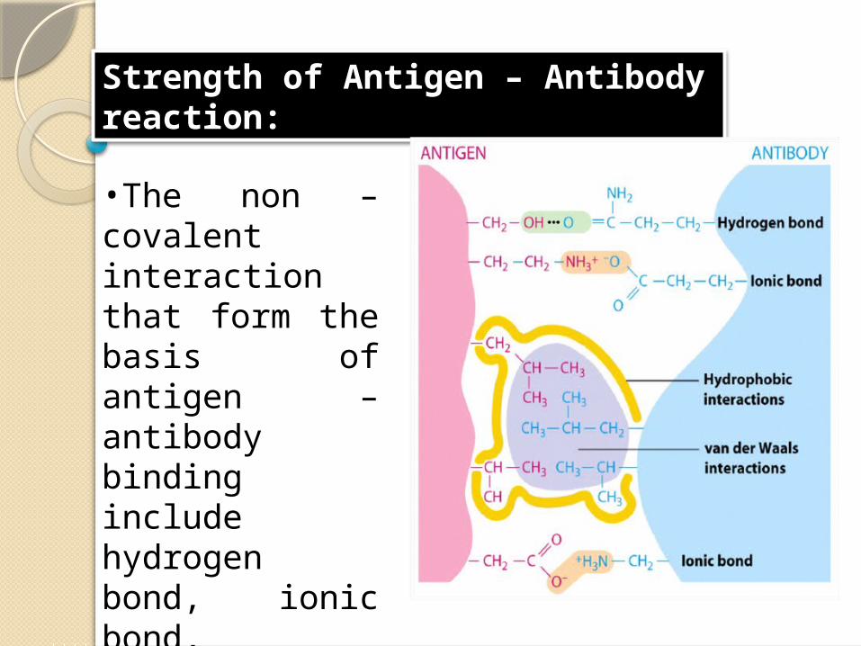
Strength of Antigen – Antibody reaction:
•The non – covalent interaction that form the basis of antigen – antibody binding include hydrogen bond, ionic bond, hydrophobic interaction and Van der Waals interaction.
•A strong antigen – antibody interaction depends on a very close fit between the antigen and antibody which requires high degree of specificity.
Properties of Antigen – Antibody Reaction:
The properties of antigen and antibody can be explained with the help of three points. They are:
• Antibody Affinity.• Antibody Avidity• Cross reaction.
Affinity of Antibody:
Interactions between antigen and antibody involve non-covalent binding of an antigenic determinant (epitope) to the variable region (complementarity determining region, CDR) of both the heavy and light immunoglobulin chains. These interactions are analogous to those observed in enzyme-substrate interactions and they can be defined similarly. To describe the strength of the antigen-antibody interaction, one can define the affinity constant (K) as shown:
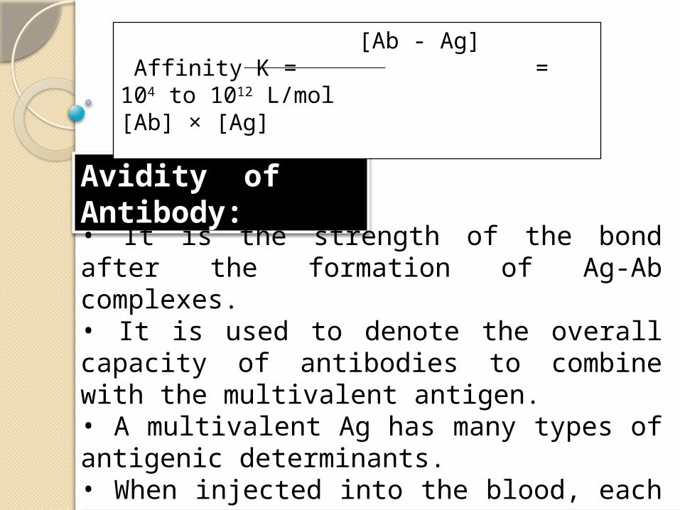
Avidity of Antibody:
• It is the strength of the bond after the formation of Ag-Ab complexes. • It is used to denote the overall capacity of antibodies to combine with the multivalent antigen. • A multivalent Ag has many types of antigenic determinants. • When injected into the blood, each antigenic determinant stimulates the production of a particular antibody.
[Ab - Ag] Affinity K = = 104 to 1012 L/mol
[Ab] × [Ag]
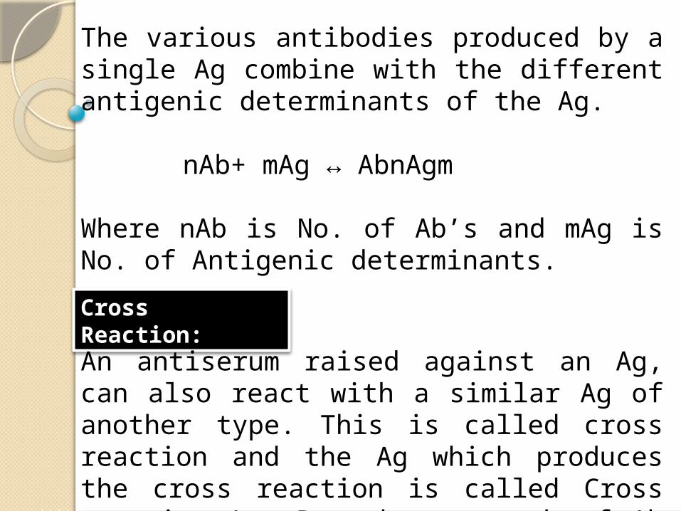
The various antibodies produced by a single Ag combine with the different antigenic determinants of the Ag.
nAb+ mAg ↔ AbnAgm
Where nAb is No. of Ab’s and mAg is No. of Antigenic determinants.
Cross Reaction:
An antiserum raised against an Ag, can also react with a similar Ag of another type. This is called cross reaction and the Ag which produces the cross reaction is called Cross reactive Ag. But the strength of Ab raised against its own Ag is strong.
• The bonds involved in cross reactions are weak.
Example: The serum raised against albumin of hen’s egg can cross react with albumin obtained from duck’s egg.The antiserum raised against human insulin will react with the insulin of Pig, Sheep, whale etc.The antiserum raised against Pneumococcal polysaccharides will react with E.coli, blood group A and collagen Ag’s.
Types of Antigen – Antibody Reaction:
• Precipitation Reaction.
• Agglutination Reaction.
• Complement Fixation.
• ELISA – Enzyme Linked ImmunoSorbent Assay.
• Immunofluorescence.
The types of antigen – antibody reactions are:
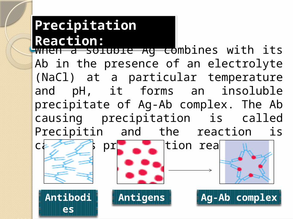
Precipitation Reaction:
When a soluble Ag combines with its Ab in the presence of an electrolyte (NaCl) at a particular temperature and pH, it forms an insoluble precipitate of Ag-Ab complex. The Ab causing precipitation is called Precipitin and the reaction is called as precipitation reaction.
Antibodies Antigens Ag-Ab complex
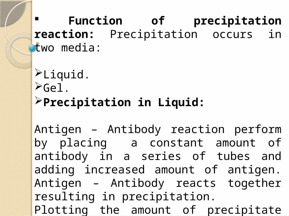
Function of precipitation reaction: Precipitation occurs in two media:
Liquid.Gel.
Precipitation in Liquid:
Antigen – Antibody reaction perform by placing a constant amount of antibody in a series of tubes and adding increased amount of antigen. Antigen – Antibody reacts together resulting in precipitation.Plotting the amount of precipitate against increasing antigen conc. Yeilds a precipitation curve.
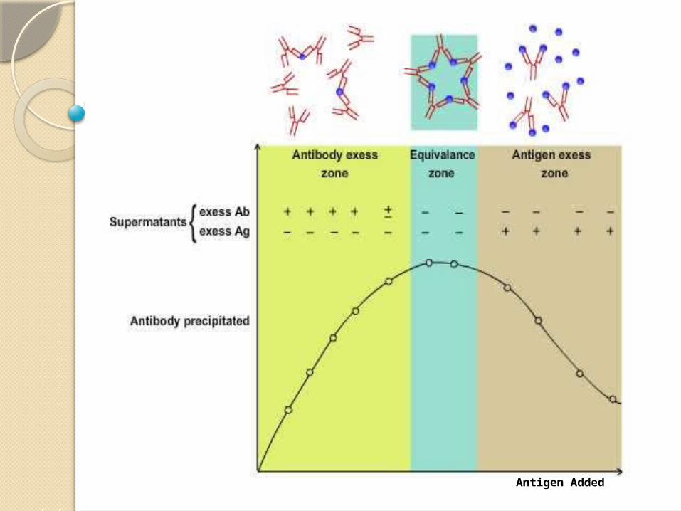
Antigen Added
Precipitation in gel:
Radial Immunodiffusion (Mancini) :In these methods agar gel or similar gels are used on plates or petriplates. Both Ag and Ab diffuse freely in the gel system in all directions. At a certain point depending on the rate of diffusion and concentration of the reactants, a zone of equivalence will be formed, which is seen as a visible precipitation.
Precipitation curve shows three zones:
1. Zone of Ab axis.2. Zone of equivalence.3. Zone of Ag axis.
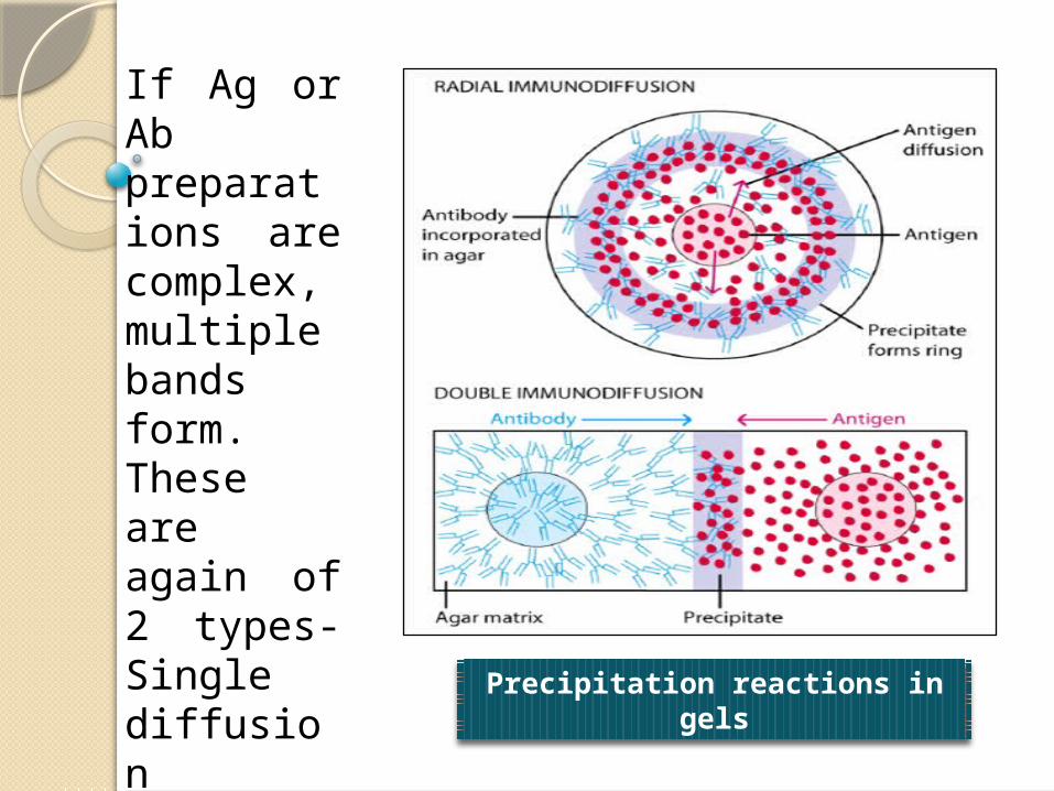
If Ag or Ab preparations are complex, multiple bands form. These are again of 2 types- Single diffusion methods and double diffusion methods. Precipitation reactions in gels
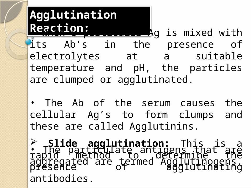
Agglutination Reaction:• When a particular Ag is mixed with its Ab’s in the presence of electrolytes at a suitable temperature and pH, the particles are clumped or agglutinated.
• The Ab of the serum causes the cellular Ag’s to form clumps and these are called Agglutinins.
• The particulate antigens that are aggregated are termed Agglutinogens. Slide agglutination: This is a rapid method to determine the presence of agglutinating antibodies.
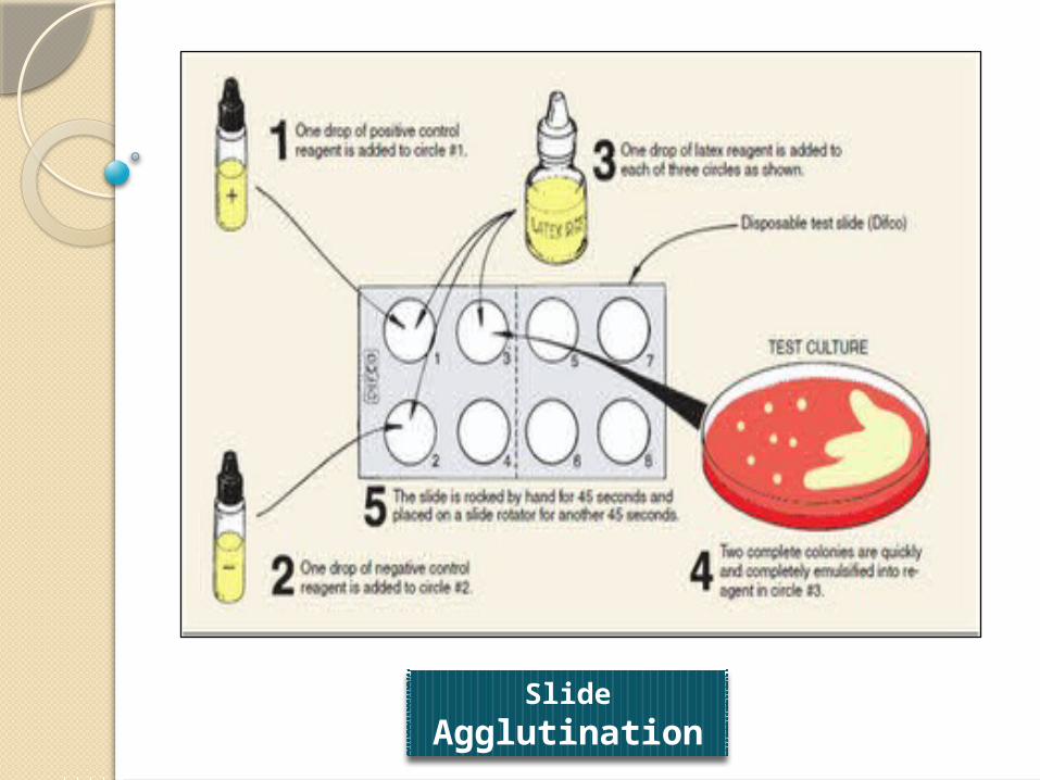
Slide Agglutination
To a uniform suspension of particulate Ag, a drop of saline is added and then a drop of antiserum is added.
The slide is gently rocked or a fine loop is used to mix the contents. If granulation occurs the test is Positive. It takes a minute for the test to complete and is visible to the naked eye. Some times confirmation may be done by observing slide under microscope.
This test is used for blood grouping (Haemagglutination) and cross matching.
Tube agglutination: This is a standard method for quantitative estimation of Ab. The serum containing Ab is diluted serially with saline in several small test tubes, to which a constant volume of Ag suspension is added.
A control tube is kept which has no antiserum. The tubes are incubated until visible agglutination is observed. The tube showing highest agglutination is referred to as the titre.
Tube agglutination is employed for the serological diagnosis of typhoid, brucellosis and typhus fever. Widal test is used for the estimation of typhoid fever.
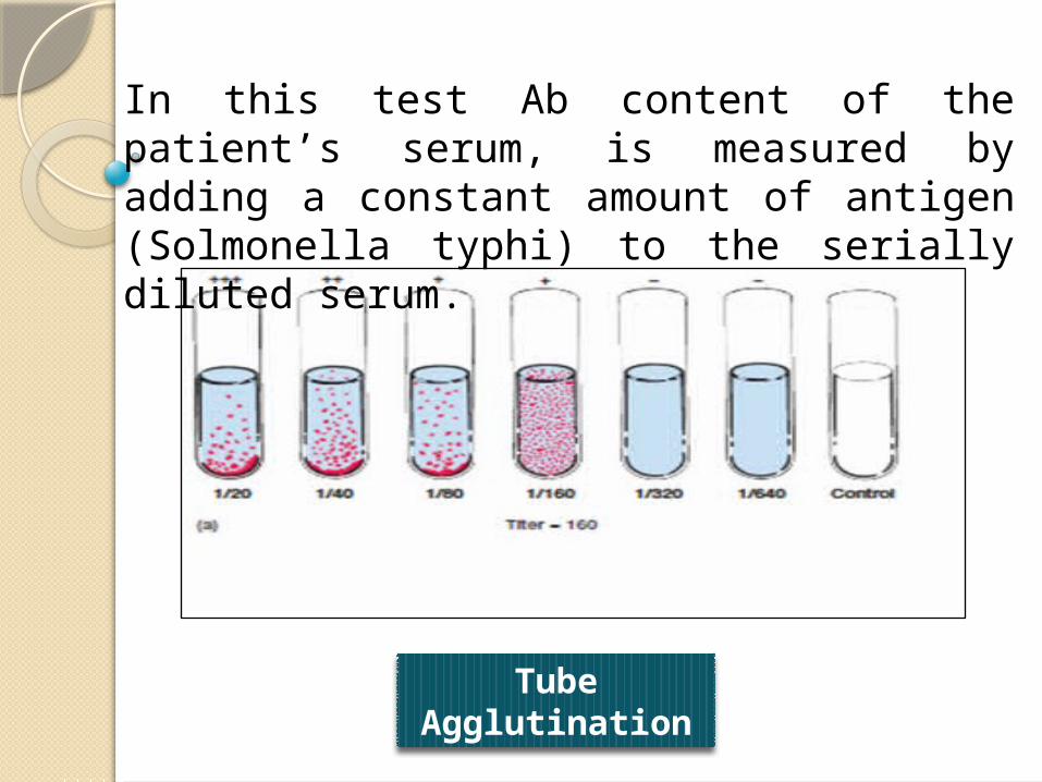
Tube Agglutination
In this test Ab content of the patient’s serum, is measured by adding a constant amount of antigen (Solmonella typhi) to the serially diluted serum.
Passive agglutination test: It is similar to haemagglutination test but the physical nature of the reaction is altered.
The Ag is coated on the surface of a carrier particle and thereby helps to convert a precipitation reaction into an agglutination reaction making the reaction more sensitive. The carrier particles used can be RBC, latex particles or bentonite. Some times RBC coated with polystyrene (tanned RBC) can be used.
When patients serum is mixed with these, it leads to agglutination. This test is used for the diagnosis of Rheumatoid arthritis.

Passive Agglutination
Carrier Particle
Soluble Antigen
Coated Particle
Coated Particle Antibody Agglutination

Provides a highly sensitive assay for small quantities of an Antigen.Example: First home pregnancy test
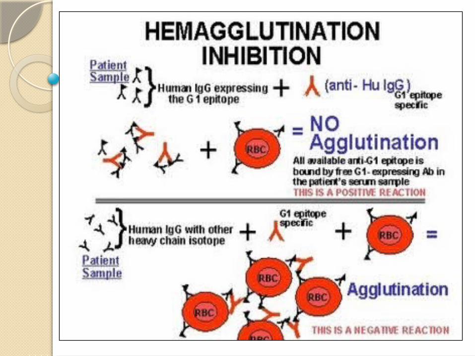
Agglutination Inhibition:

Complement Fixation: • Lysis of RBC or bacteria requires some non-specific unstable components of fresh serum which are called complement.
•This complement system comprises of 11 proteins and are present in ever individual. They bind to Fc component of Ab involved in Ag-Ab complex. This ability of the Ag-Ab complex to fix complement is used in complement Fixation tests.
•In the first stage, the test Ag and the antiserum (heated to 56oC to inactivate complement) are mixed in the presence of known amount of complement. This is incubated at 4oC for 18h.

• If Ab specific for the Ag is present in the serum, Ag-Ab complex will be formed that will fix the complement.

• In 1971, enzyme labeled Ag’s and Ab’s were developed as serological reagents for the assay of Ab’s and Ag’s.
• These are very simple, sensitive, economic and less hazard when compared to RIA.
•The ligand used here is a molecule which can detect the Ab and is covalently coupled to an enzyme such as peroxidase, betagalactosidase, alkaline phosphatase etc.
ELISA – Enzyme Linked ImmunoSorbent Assay:

ELISA is of 3types.
Indirect ELISA: This technique is used for the detection of HIV. The envelop proteins are developed by recombinant technology and coated on the surface of the of microtire plates. Suspects serum is added, and unbound proteins are washed off.
Sandwich ELISA: Used to detect the presence of Ag in a sample. The well is coated with Ab specific to the Ag and then suspect serum is added allowed to react. The wells are washed to remove unbound Ag’s.

Types of ELISA

Competitive ELISA: Another variation for measuring amounts of antigen is competitive ELISA. In this technique, antibody is first incubated in solution with a sample containing antigen.
The antigen-antibody mixture is then added to an antigen coated micro titer well.
Then a labeled Ab against a different epitope of the Ag is added. Unbound Ab’s are removed by washing and this is followed by addition of colored substrate and development of color. The intensity of color is directly proportional to the concentration of the Ag in the serum.

The more antigen present in the sample, the less free antibody will be available to bind to the antigen-coated well. Addition of an enzyme-conjugated secondary antibody (Ab2) specific for the isotype of the primary antibody can be used to determine the amount of primary antibody bound to the well as in an indirect ELISA.
Immunofluorescence:
•Fluorescence is the property of absorbing light rays of one particular wavelength and emitting rays with a different wave length.
• Fluorescent dyes show up brightly under UV light as they convert into visible light.

Coons et al (1942) showed that labeled dyes can be conjugated to Ab’s and these labeled antibodies can be used to detect Ag’s.•Dyes that are commonly used include:
Fluorescein, an organic dye that is the most widely used label for immunofluorescence procedures, absorbs blue light (490 nm) and emits an intense yellow-green fluorescence (517 nm).
Phycoerythrin is an efficient absorber of light (~30-fold greater than fluorescein) and a brilliant emitter of red fluorescence, stimulating its wide use as a label for immunofluorescence.

Direct and indirect Immunofluorescence

The chief use of antigen-antibody reactions are:
• Determination of blood groups for transfusion.• Serological ascertainment of exposure to infectious agents.• Development of immunoassays for the quantification of various substances.• To detect the presence or absence of protein in serum.• Determining the characteristics of certain immuno-deficiency disease.
Application of Antigen – Antibody Reaction:

Conclusion:
Thereby we conclude up this topic with a quick review about Antigen – Antibody Reaction. The antigens and the antibodies combine specifically with each other. This interaction between them is called Antigen-Antibody reaction. As per the current requirement of the syllabus, the topic has been included, right from its features till the types of reaction . We show our gratitude towards all of you . Hope this project satisfy the required information by you and any resemblance of mistakes are duly apologized.

Thank You