Embed Size (px)
Citation preview


Antigen-Antibody Properties
• You must remember Antibody affinity(single) VS avidity (multiple)
• Cross-reactivity: occurs when two different antigens share an identical or very similar epitope. The antibody’s affinity for the cross-reacting epitope will be _____ than for the original epitope.
• ABO blood groups and infectious diseases (Jenner?)

Precipitin reactions
The interaction of antibody with antigen in solution may cause formation of an insoluble lattice that will precipitate out of solution.
This precipitate will only form if:
- the antibody is bivalent or polyvalent- the antibody or antibody mixture can bind to at least two different sites on the antigen (either two different epitopes or two identical epitopes)
Monoclonal antibodies are likely to be less efficient at immunoprecipitation than polyclonal antibodies.
Kuby Figure 6-4a

Antibody excess -
too much antibody
prevents efficient cross-
linking
Antigen excess -
too much antigen
prevents efficient cross-
linking
Equivalence - ratio
of antibody to antigen is
optimal and a maximum
amount of precipitate is
formed.
Kuby Figure 6-4b
Formation of the precipitate also requires that the antigen and antibody be present at
appropriate concentrations relative to each other.

Surface Plasmon Resonance (SPR)


Kuby Figure 6-5
Double Immunodiffusion
Diffusion of antibody and antigen towards each other in an agarose gel.
A line of precipitate will form if the antibody binds to antigen.
Used to determine if an antigen or antibody is present.

Volume 271, Number 30, Issue of July 26, 1996 pp. 18054-18060JBC
http://www.fbr.org/swksweb/immunolist.html
Ag1
Ag2
Ag3
Ag4
Ag5
Ag6
Ab
Kuby Figure 6-5
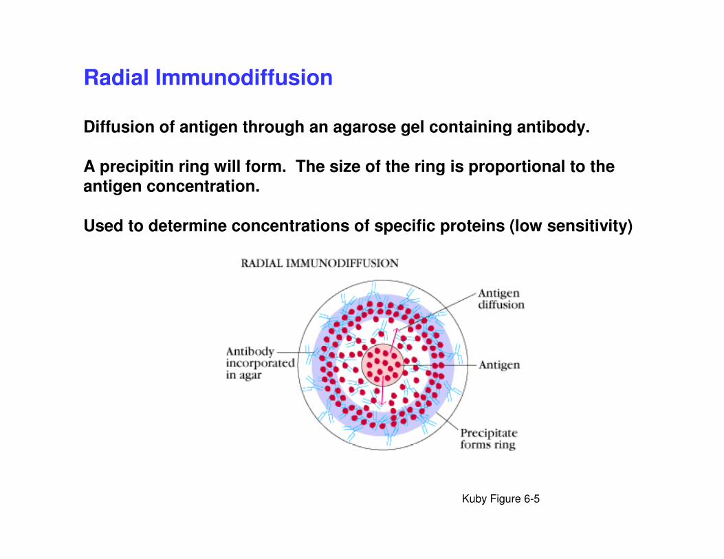
Radial Immunodiffusion
Diffusion of antigen through an agarose gel containing antibody.
A precipitin ring will form. The size of the ring is proportional to the antigen concentration.
Used to determine concentrations of specific proteins (low sensitivity)

http://www.fbr.org/swksweb/immunolist.html
http://leahi.kcc.hawaii.edu/~johnb/micro/medmicro/medmicro.11.html

Hemagglutination
Antibody can also cross-link cells or beads.
Cross-linking of red cells is called hemagglutination.
Non-cross-linked cells settle in a bead to the bottom of the well.
Cross-linked cells settle in a diffuse pattern.
Used to measure antibody presence and level (titre).
Used to measure antibodies to red cell antigens or to other antigens bound to
the surface of red cells.
Kuby Figure 6-8

Figure 6.7

Immunoelectrophoresis

Enzyme-linked immunosorbent assay (ELISA)
Used to measure antigen or antibody presence and concentration.
Far more sensitive than precipitin or agglutination techniques.
Relies on the ability to covalently conjugate chemicals to the Fc region of Ig
without interfering with antigen binding (“enzyme-linked”) and the ability of
plastic to nonspecifically bind proteins (immunosorbent).
In ELISA, an enzyme is bound to the Fc region - usually horseradish
peroxidase or alkaline phosphatase. Enzyme presence can be determined by
use of colorimetric substrates.
Final measurement is an absorbance.
Comparison with standard curves indicates concentration of antigen or
antibody.
Various assay formats are possible.


http://www.supercolostrum.com/colostrum/Information/information9.htm

Sample Absorbance
Positive control 1.689
Negative control 0.153
Assay control 0.123
Patient A 0.055
Patient B 0.412
Patient C 1.999
http://www.supercolostrum.com/colostrum/Information/information9.htm


Kuby Figure 6-11

Relative sensitivities:
Precipitin reactions < Agglutination reactions
< ELISA
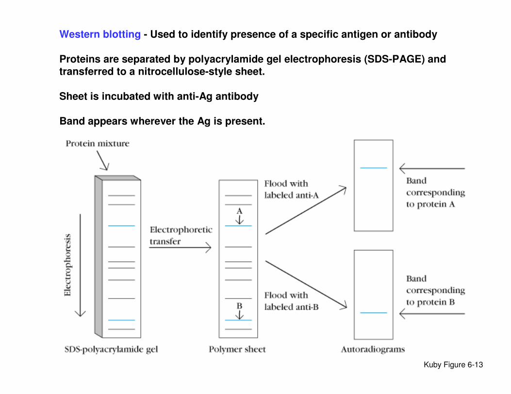
Western blotting - Used to identify presence of a specific antigen or antibody
Proteins are separated by polyacrylamide gel electrophoresis (SDS-PAGE) and
transferred to a nitrocellulose-style sheet.
Sheet is incubated with anti-Ag antibody
Band appears wherever the Ag is present.
Kuby Figure 6-13

http://www.biology.arizona.edu/immunology/activities/western_blot/west2.html
HIV Western Blot
1.Lane 1, HIV+ serum (positive control)
2.Lane 2, HIV- serum (negative control)
3.Lane A, Patient A
4.Lane B, Patient B
5.Lane C, Patient C
No bands present………………………………...Negative
Bands at either p31 OR p24 AND bands
present at either gp160 OR gp120……………..Positive
Bands present, but pattern does not
Meet criteria for positivity………………………...Indeterminate


ELISPOT
1) Detects cell actively secreting cytokines
2) Capture antibody
3) Activated cells

4) Anti-cytokine antibody linked to reporter enzyme
5) Substrate
6) Quantify spots. Each spots represent the an actively secreting cell
ELISPOT

Immuno-magnetic separation
(IMAS)
• Technique used to separate cells, proteins,
nucleic acids using antibodies or ligands-
bound to magnetic beads.
• Removed out from mix suspension using a
magnet.

Rabbit
anti-CD4
Goat anti-
rabbit Fc
specific
Remove:
positive or
negative selection


Immunofluorescence
1 2 3


Fluorescence Acitvated Cell Sorter
(FACS)
• Mixed cell population
• Requires two different fluorochromes
• Commonly used: FITC, PE, Texas Red, etc


FACS Analysis
Reagents:
CD4-PE Phycoerythrin (PE)
VS
CD3 Fuorecein (FITC)


695490,675TruRed
625615589Texas Red
548576570Rhodamine
444572547TRITC
512503BODIPY-FL
389519495Fluorescein (FITC)
240 k578480;565Phycoerythrin (PE)
528425Lucifer yellow
596423375;400Cascade Blue
Reactive and conjugated probes
MWEm (nm)Ex (nm)Probe
Commonly Used Fluorochromes