Embed Size (px)
Citation preview
Motor unit
SR2002
Dr. Arimantas Lionikas
October 28, 2013
Motor Unit (MU)Plan
• Structural organization• Electromyography (EMG)• Recruitment threshold of MUs• MU types• MU recruitment during exercise• Reading list:
1. Enoka R. Neuromechanics of human movement. 2002. Publishers: Human Kinetics, p. 278-2972. MacIntosh, B.R., Gardiner, P.F. McComas, A.J. Skeletal muscle, 2nd edition. 2006. Publishers: Human Kinetics, p. 32-39, 126-150.3. McArdle W.D. et al. Exercise Physiology: energy, nutrition, human performance. 2001. Publisher: Lippincott Williams & Wilkins, p. p. 374-382, 394-400.
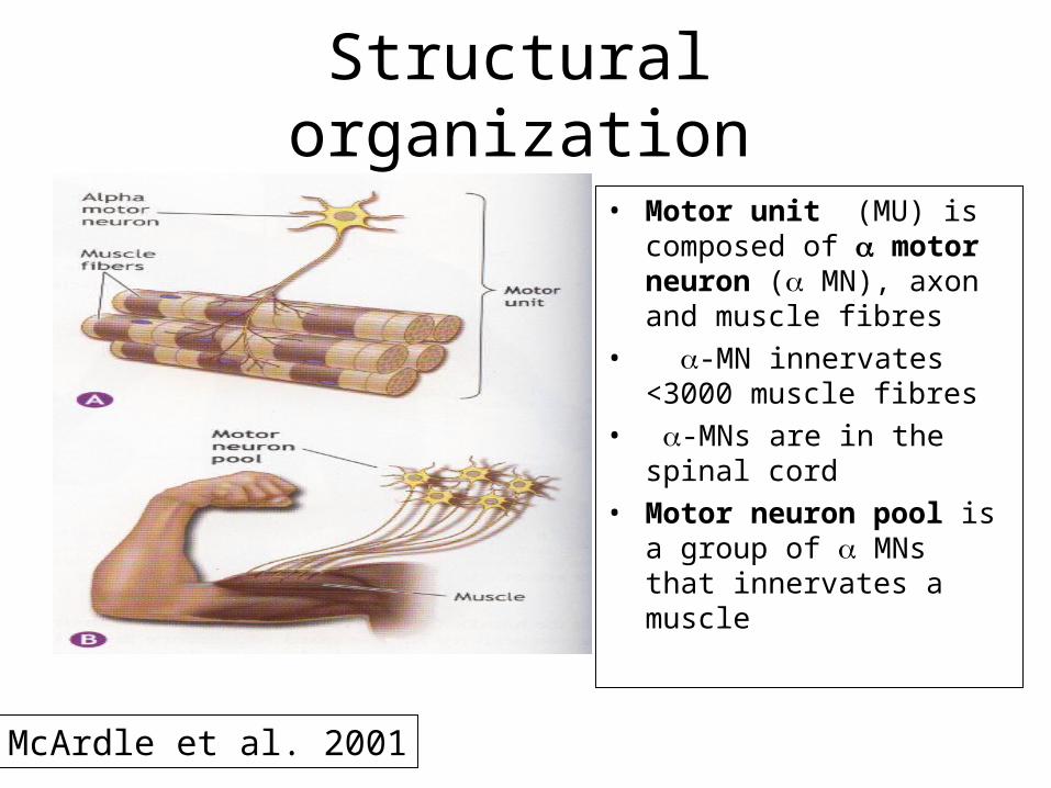
• Motor unit (MU) is composed of motor neuron ( MN), axon and muscle fibres
• -MN innervates <3000 muscle fibres
• -MNs are in the spinal cord
• Motor neuron pool is a group of MNs that innervates a muscle
Structural organization
McArdle et al. 2001
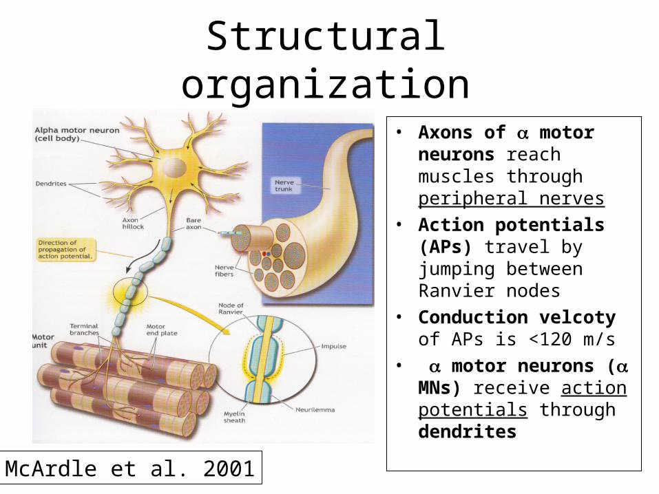
• Axons of motor neurons reach muscles through peripheral nerves
• Action potentials (APs) travel by jumping between Ranvier nodes
• Conduction velcoty of APs is <120 m/s
• motor neurons ( MNs) receive action potentials through dendrites
Structural organization
McArdle et al. 2001
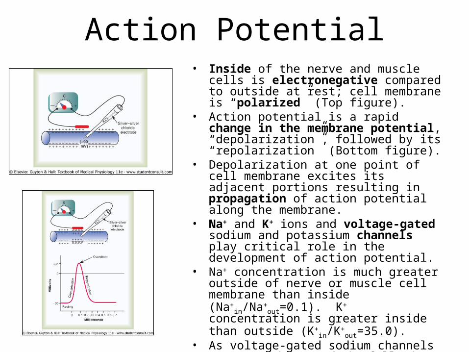
• Inside of the nerve and muscle cells is electronegative compared to outside at rest; cell membrane is “polarized” (Top figure).
• Action potential is a rapid change in the membrane potential, “depolarization”, followed by its “repolarization” (Bottom figure).
• Depolarization at one point of cell membrane excites its adjacent portions resulting in propagation of action potential along the membrane.
• Na+ and K+ ions and voltage-gated sodium and potassium channels play critical role in the development of action potential.
• Na+ concentration is much greater outside of nerve or muscle cell membrane than inside (Na+
in/Na+out=0.1).
K+ concentration is greater inside than outside (K+
in/K+out=35.0).
• As voltage-gated sodium channels open, Na+ ions rush in following the concentration gradient causing “depolarization”. Infulx of Na+ ceases with inactivation of the channels.
• “Depolarization” triggers opening of potassium channels leading to outward diffusion of K+ ions and “Repolarization” of the membrane.
Action Potential
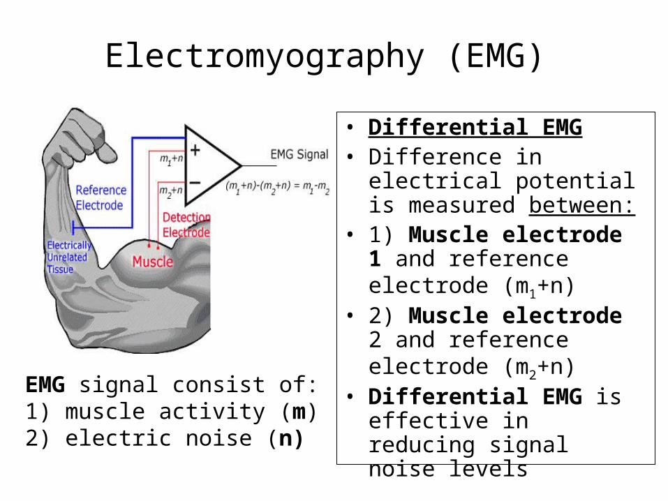
Electromyography (EMG) • Differential EMG• Difference in electrical
potential is measured between:
• 1) Muscle electrode 1 and reference electrode (m1+n)
• 2) Muscle electrode 2 and reference electrode (m2+n)
• Differential EMG is effective in reducing signal noise levels
EMG signal consist of:1) muscle activity (m)2) electric noise (n)
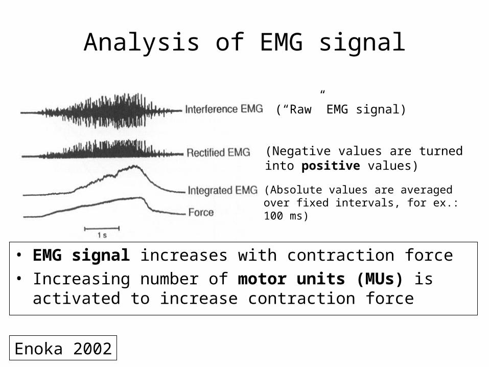
Analysis of EMG signal
• EMG signal increases with contraction force• Increasing number of motor units (MUs) is
activated to increase contraction force
(“Raw” EMG signal)
(Negative values are turned into positive values)(Absolute values are averaged over fixed intervals, for ex.: 100 ms)
Enoka 2002
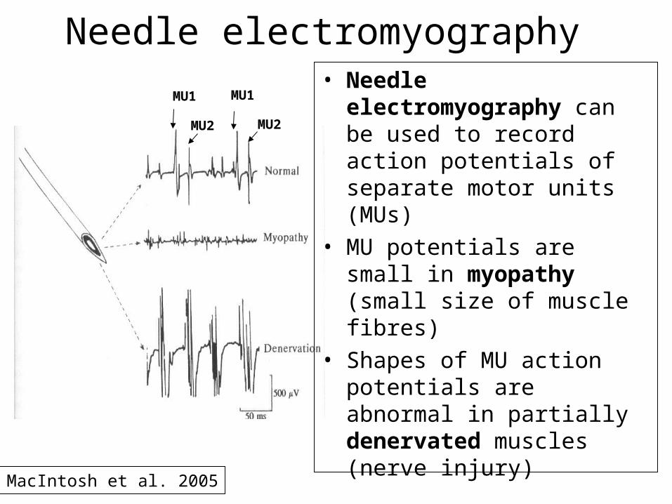
Needle electromyography• Needle
electromyography can be used to record action potentials of separate motor units (MUs)
• MU potentials are small in myopathy (small size of muscle fibres)
• Shapes of MU action potentials are abnormal in partially denervated muscles (nerve injury)
MU1 MU1
MU2 MU2
MacIntosh et al. 2005
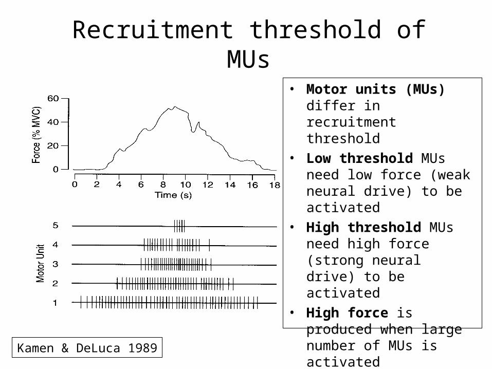
Recruitment threshold of MUs• Motor units (MUs)
differ in recruitment threshold
• Low threshold MUs need low force (weak neural drive) to be activated
• High threshold MUs need high force (strong neural drive) to be activated
• High force is produced when large number of MUs is activated
Kamen & DeLuca 1989
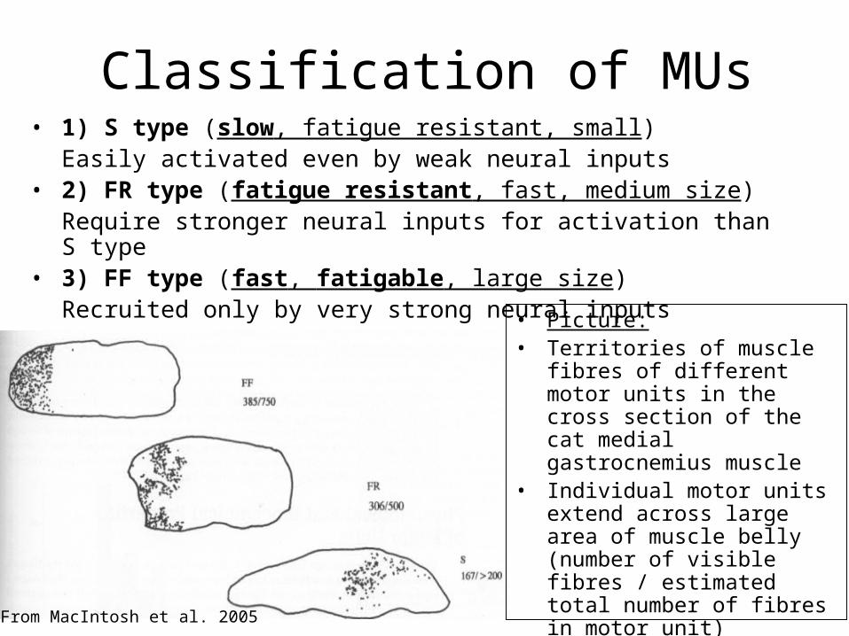
Classification of MUs• 1) S type (slow, fatigue resistant, small)
Easily activated even by weak neural inputs• 2) FR type (fatigue resistant, fast, medium size)
Require stronger neural inputs for activation than S type • 3) FF type (fast, fatigable, large size)
Recruited only by very strong neural inputs• Picture:• Territories of muscle fibres
of different motor units in the cross section of the cat medial gastrocnemius muscle
• Individual motor units extend across large area of muscle belly (number of visible fibres / estimated total number of fibres in motor unit)
From MacIntosh et al. 2005
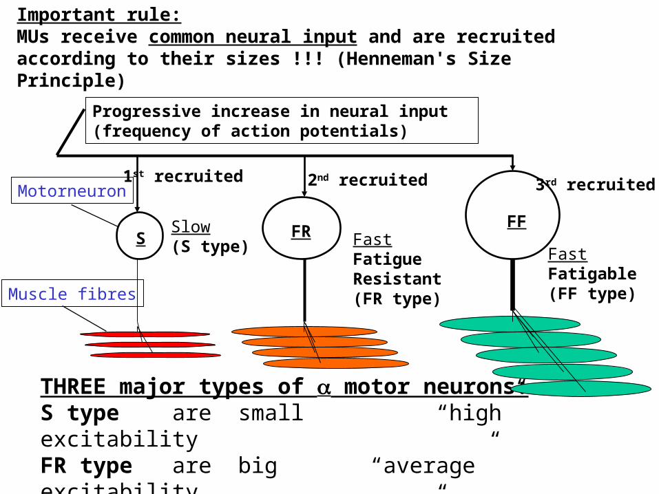
Progressive increase in neural input (frequency of action potentials)
S FR FFSlow(S type) Fast
FatigueResistant (FR type)
FastFatigable (FF type)
1st recruited 2nd recruited 3rd recruited
Important rule:MUs receive common neural input and are recruited according to their sizes !!! (Henneman's Size Principle)
THREE major types of motor neurons: S type are small “high” excitability FR type are big “average” excitabilityFF type are very big “low” excitability
Motorneuron
Muscle fibres
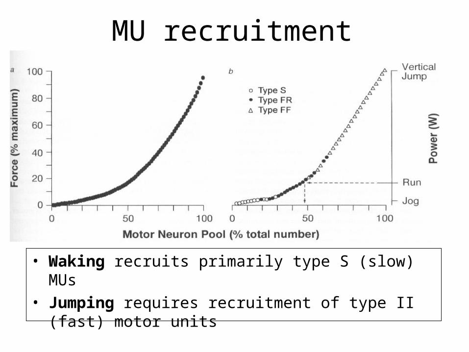
MU recruitment
• Waking recruits primarily type S (slow) MUs• Jumping requires recruitment of type II (fast)
motor units
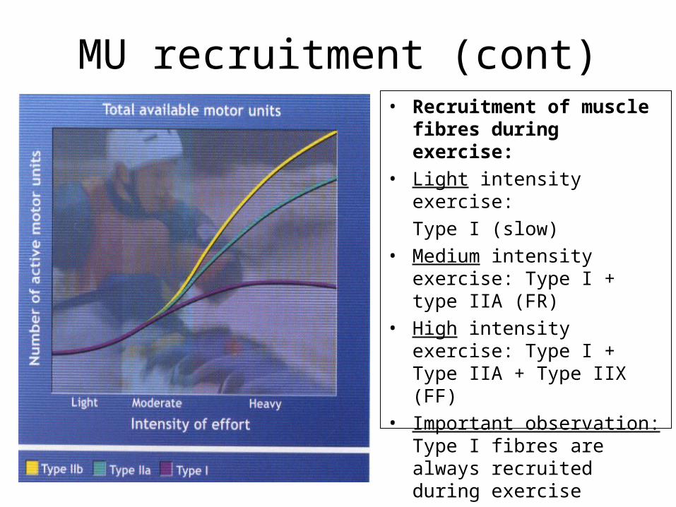
MU recruitment (cont)• Recruitment of muscle
fibres during exercise:• Light intensity exercise:
Type I (slow)• Medium intensity
exercise: Type I + type IIA (FR)
• High intensity exercise: Type I + Type IIA + Type IIX (FF)
• Important observation: Type I fibres are always recruited during exercise
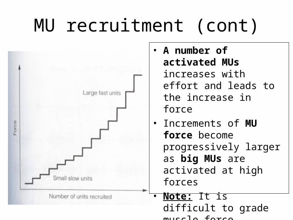
MU recruitment (cont)• A number of
activated MUs increases with effort and leads to the increase in force
• Increments of MU force become progressively larger as big MUs are activated at high forces
• Note: It is difficult to grade muscle force precisely when high force is produced
Motor unitSummary
• A Motor unit (MU) is composed of motor neuron, axon and muscle fibres
• Electromyography can be used to study motor units (MUs)
• There are three main types of MUs (S, FR and FF)
• MUs are recruited according to their sizes in the following order: S => FR => FF

![UGA Physics and Astronomy • Physics and Astronomy ......1998], or between primary motor cortex and supple-mentary motor area [Biswal et al., 1995; Xiong et al., 1999]. Even if the](https://img.pdfslide.us/doc/110x75/603bdb6f700ffd46e847f62b/uga-physics-and-astronomy-a-physics-and-astronomy-1998-or-between-primary.jpg)