1
Pathological role of amyloid peptide in neuromuscular disease, glutamate and glucose involvement. Maud Combes #1 , Philippe Poindron # , Jean Mariani 1 and Noelle Callizot #2 # Neuro-Sys SAS, 410 Chemin Départemental 60, 13120 Gardanne, France 1 UPMC, UFR 927, 4 place Jussieu, 75252 Paris, France 2 Corresponding author : [email protected] In amyotrophic lateral sclerosis (ALS), the progressive loss of motor neurons is accompanied by extensive muscle denervation, resulting in paralysis and ultimately death. Disturbances in glutamate homeostasis, which lead to toxic accumulation of this excitatory neurotransmitter in the synaptic cleft, are observed in several neuropathologies notably in ALS. It has been recently shown that neuromuscular junction (NMJ) loss and motor neuron degeneration are substantially reduced in SOD1G93A mice when APP is genetically ablated. Thus, endogenous APP and Aβ may contribute to ALS pathology in humans (Bryson et al., 2012). Additionally, Aβ1–42 accumulations were observed in motor neurons and spinal cords of sporadic and familial cases of amyotrophic lateral sclerosis (ALS). In previous results, using the nerve/muscle coculture, we showed that glutamate and Aβ act in an interlinked manner, mutually influencing their release (Combes et al., 2015). We showed that NMJs are highly sensitive to Aβ peptide, that the toxic pathway involves glutamate and NMDAR receptors (see Poster Combes et al., JSFM, Lyon, Nov. 2015). In light of these results, we wanted to better understand how the toxicity occurred on each cellular protagonist. Using a primary motor neurons culture and myotubes culture, we studied separately the toxicity of Aβ and the mechanisms involved in the process of cell death. Introduction Culture of motor neurons: Rat SC motor neurons (E14) were cultured as described by Martinou et al., 1992 and Wang et al., 2013. The cells were seeded at a density of 20,000 per well in 96-well plates (for immuno-staining) and of 110,000 per well in 24-wells plates (for Western-blot (WB)) precoated with poly-L-lysine and cultured with medium consisting of Neurobasal medium with a 2% solution of B27 supplement, 2 mmol/liter of L-glutamine, 2 % of penicillin and streptomycin solution, and 10 ng/ml of brain-derived neurotrophic factor (BDNF). Cells were cultured at 37°C in an air (95 %) – CO2 (5 %) incubator. Culture of muscle cells: Human muscle cells (SkMC) were seeded at a density of 12,000 per well in 96-wells plates (for immuno-staining) and of 67,000 per well in 24-wells plates (for Western-blot (WB)) pre-coated with gelatin and cultured for 5 days with proliferation medium consisting of mix of 62 % of MEM medium and 25 % of M199 medium supplemented with glutamine 2 mM, human insulin 10 µg/ml, Human recombinant Epidermal growth factor 10 ng/ml (EGF), human recombinant Fibroblast growth factor basic 2 ng/ml (bFGF), foetal calf serum 10 % and 2 % of PS. After that the medium was replaced by a medium consisting of mix (67 %/25 %) medium composed of MEM and medium 199, supplemented with 5 % FCS, insulin 5 µg/ml, glutamine 2 mM and 2 % PS. Cells were cultured at 37°C in an air (95 %) – CO2 (5 %) incubator. Pharmacological treatments: On day 13 (motor neurons) or day 12 (muscular cells), the cultures were injured with Aβ solution (Callizot et al., 2013) at 2.5, 5, 10 and 20 µmol/L for 4, 8, 16 and 24 h or glutamate at 0.5, 1, 5 and 10 µmol/L for 20 min. After 20 min, glutamate was washed and fresh culture medium was added for additional 4, 8, 16 and 24 h. Untreated cultures served as controls. MK801 at 20 µmol/L, Ifenprodil at 1 µmol/L, and Memantine at 5 µmol/L, were diluted in culture medium and pre-incubated 1 h before Aβ intoxication. Immunostaining: After 4, 8, 16 or 24 h, cells were fixed by a cold solution of ethanol (95 %) and acetic acid (5 %). Motor neurons were incubated with monoclonal anti-microtubule-associated-protein 2 (MAP-2). The muscle cells were incubated with monoclonal anti-myosin. These antibodies were revealed with Alexa Fluor 488 Goat anti-mouse IgG. The immune-labelled cultures were examined with MetaXpress (Molecular Devices, USA) at X 20 magnification. For each condition, 30 fields per well were observed (representing the total surface of the well), and six wells per conditions were analyzed. Quantification of glutamate in supernatants: After 4, 8, 16 and 24 h, glutamate was dosed in supernatant using Amplex red Glutamic acid assay kit (Molecular probes) and following manufacturer’s recommendations. Quantification of glucose uptake: After 24 h of Aβ intoxication, glucose uptake was measured with glucose uptake-Glo assay kit (Promega) and following manufacturer’s recommendations. GluT1 evaluation (WB): After 24 h of Aβ treatment, cells were lysed with Cellytic and immediately frozen at -80°C. All reagents were prepared and used according to manufacturer’s recommendations (Automatic western Blot, Simon™ - ProteinSimple - www.proteinsimple.com). Anti-GluT1, primary antibody was used for WB analysis. Methods Results For the first time we show in this study that: Aβ induced motor neuron injuries (dose and time dependent) these injuries involved a large Glutamate production, and NMDAR involvement. Aβ induced motor neurons injuries following similar mechanisms of toxicity that those observed on CNS neurons (for review Callizot et al., 2013 and Combes et al., JSFM, Lyon, Nov. 2015). Aβ was also toxic for muscle cells, this toxicity did not involve any glutamate. A decrease of glucose uptake was observed associated with increase of GluT-1 levels. Conclusions . . . . . . . . . . . . . . . . . . . . . . . . © Neuro-Sys SAS - Myology 2016 - March 2016 INNOVATIVE RESEARCH www.neuro-sys.fr Effect of Aβ on motor neurons: Aβ induced a significant motor neuron death that depends of the dose and time of exposure. Aβ applied for 8 h did not induced any cell death but a slight decrease of neurite network was observed at the highest concentration (20 µmol/L). By contrast, a large and significant toxicity (neurons and neurites) was observed after 16 h and 24 h Aβ application. References: Combes et al., (2015) J Neurosci Res. 2015 Apr;93(4):633-43 Callizot et al., (2013) J Neurosci Res. 2013;91(5):706-16 Bryson et al., (2012) Hum Mol Genet. 1;21(17):3871-82 Martinou et al., (1992) Neuron 1992 Apr:8(4) 737-44 Wang et al., Mol Genet 2013 Dec 1:22(23):4706-19 Effect of Glutamate on motor neurons: A large and significant cell death was observed after glutamate application. These effects were depending of the time of exposure and the dose applied. Interestingly, the damages were observed shortly after glutamate application (from 4 h). Release of glutamate after Aβ application on motor neurons: After 8 h of Aβ application (20 µmol/L) a large production of glutamate was observed in the culture supernatant. This release occurred shortly after peptide application. Protective effect of MK801, Ifenprodil and Memantine on motor neurons injured with Aβ: MK801 (20 µmol/L, a non competitive N-methyl- D-aspartate receptor [NMDAR]), Ifenprodil (1 µmol/L, Non-competitive NMDAR antagonist displays GluN2B [formerly NR2B] subunit selectivity), and Memantine (5 µmol/L, NMDAR antagonist with extrasynaptic localization selectivity) partially protected motor neurons against Aβ injury (20 µmol/L). These results were in favor of glutamate involvement in the death of motor neurons induced by Aβ. Aβ (20 µM, 24 h) Control Glutamate (10 µM, 20 min) Effect of Aβ on muscle cells: A significant and large cell death was observed on muscular cells after application of Aβ at the highest concentrations (20 µmol/L) in the first 8 h. This effect was dependent of the dose of peptide. At the lowest doses, Aβ did not induced any cell death. Glucose uptake after Aβ application on muscle cells: A clear and significant decrease of glucose intake was observed in presence of Aβ (24 h after application). Interestingly the disturbances of intake were observed before any cell death. Effect of Glutamate on muscle cells: Glutamate did not show any effect on muscle cells at the dose and time of application for which motor neurons injuries were observed. GluT-1 evaluation: Increase of Glucose Transporter 1 level was observed after 24 h of Aβ (20 µmol/L) application on muscle cells.

and glucose involvement. - Neuro-Sys · Introduction Culture of motor neurons: Rat SC motor neurons (E14) were cultured as described by Martinou et al., 1992 and Wang et al., 2013

  • Upload
    others

  • View
    3

  • Download
    0

Embed Size (px)

Citation preview

Pathological role of amyloid peptide in neuromuscular disease, glutamate and glucose involvement.Maud Combes#1, Philippe Poindron#, Jean Mariani1 and Noelle Callizot#2

#Neuro-Sys SAS, 410 Chemin Départemental 60, 13120 Gardanne, France ● 1 UPMC, UFR 927, 4 place Jussieu, 75252 Paris, France2 Corresponding author : [email protected]

In amyotrophic lateral sclerosis (ALS), the progressive loss of motor neurons is accompanied by extensive muscle denervation, resulting in paralysis and ultimately death. Disturbances in glutamate homeostasis, which lead to toxic accumulation of this excitatory neurotransmitter in the synaptic cleft, are observed in several neuropathologies notably in ALS. It has been recently shown that neuromuscular junction (NMJ) loss and motor neuron degeneration are substantially reduced in SOD1G93A mice when APP is genetically ablated. Thus, endogenous APP and Aβ may contribute to ALS pathology in humans (Bryson et al., 2012). Additionally, Aβ1–42 accumulations were observed in motor neurons and spinal cords of sporadic and familial cases of amyotrophic lateral sclerosis (ALS). In previous results, using the nerve/muscle coculture, we showed that glutamate and Aβ act in an interlinked manner, mutually influencing their release (Combes et al., 2015). We showed that NMJs are highly sensitive to Aβ peptide, that the toxic pathway involves glutamate and NMDAR receptors (see Poster Combes et al., JSFM, Lyon, Nov. 2015). In light of these results, we wanted to better understand how the toxicity occurred on each cellular protagonist. Using a primary motor neurons culture and myotubes culture, we studied separately the toxicity of Aβ and the mechanisms involved in the process of cell death.

IntroductionCulture of motor neurons: Rat SC motor neurons (E14) were cultured as described by Martinou et al., 1992 and Wang et al., 2013. The cells were seeded at a density of 20,000 per well in 96-well plates (for immuno-staining) and of 110,000 per well in 24-wells plates (for Western-blot (WB)) precoated with poly-L-lysine and cultured with medium consisting of Neurobasal medium with a 2% solution of B27 supplement, 2 mmol/liter of L-glutamine, 2 % of penicillin and streptomycin solution, and 10 ng/ml of brain-derived neurotrophic factor (BDNF). Cells were cultured at 37°C in an air (95 %) – CO2 (5 %) incubator. Culture of muscle cells: Human muscle cells (SkMC) were seeded at a density of 12,000 per well in 96-wells plates (for immuno-staining) and of 67,000 per well in 24-wells plates (for Western-blot (WB)) pre-coated with gelatin and cultured for 5 days with proliferation medium consisting of mix of 62 % of MEM medium and 25 % of M199 medium supplemented with glutamine 2 mM, human insulin 10 µg/ml, Human recombinant Epidermal growth factor 10 ng/ml (EGF), human recombinant Fibroblast growth factor basic 2 ng/ml (bFGF), foetal calf serum 10 % and 2 % of PS. After that the medium was replaced by a medium consisting of mix (67 %/25 %) medium composed of MEM and medium 199, supplemented with 5 % FCS, insulin 5 µg/ml, glutamine 2 mM and 2 % PS. Cells were cultured at 37°C in an air (95 %) – CO2 (5 %) incubator. Pharmacological treatments: On day 13 (motor neurons) or day 12 (muscular cells), the cultures were injured with Aβ solution (Callizot et al., 2013) at 2.5, 5, 10 and 20 µmol/L for 4, 8, 16 and 24 h or glutamate at 0.5, 1, 5 and 10 µmol/L for 20 min. After 20 min, glutamate was washed and fresh culture medium was added for additional 4, 8, 16 and 24 h. Untreated cultures served as controls. MK801 at 20 µmol/L, Ifenprodil at 1 µmol/L, and Memantine at 5 µmol/L, were diluted in culture medium and pre-incubated 1 h before Aβ intoxication. Immunostaining: After 4, 8, 16 or 24 h, cells were fixed by a cold solution of ethanol (95 %) and acetic acid (5 %). Motor neurons were incubated with monoclonal anti-microtubule-associated-protein 2 (MAP-2). The muscle cells were incubated with monoclonal anti-myosin. These antibodies were revealed with Alexa Fluor 488 Goat anti-mouse IgG. The immune-labelled cultures were examined with MetaXpress (Molecular Devices, USA) at X 20 magnification. For each condition, 30 fields per well were observed (representing the total surface of the well), and six wells per conditions were analyzed.Quantification of glutamate in supernatants: After 4, 8, 16 and 24 h, glutamate was dosed in supernatant using Amplex red Glutamic acid assay kit (Molecular probes) and following manufacturer’s recommendations.Quantification of glucose uptake: After 24 h of Aβ intoxication, glucose uptake was measured with glucose uptake-Glo assay kit (Promega) and following manufacturer’s recommendations. GluT1 evaluation (WB): After 24 h of Aβ treatment, cells were lysed with Cellytic and immediately frozen at -80°C. All reagents were prepared and used according to manufacturer’s recommendations (Automatic western Blot, Simon™ - ProteinSimple - www.proteinsimple.com). Anti-GluT1, primary antibody was used for WB analysis.

Methods

Results

For the first time we show in this study that:

• Aβ induced motor neuron injuries (dose and time dependent) these injuries involved a large Glutamate production, and NMDAR involvement.

• Aβ induced motor neurons injuries following similar mechanisms of toxicity that those observed on CNS neurons (for review Callizot et al., 2013 and Combes et al., JSFM, Lyon, Nov. 2015).

• Aβ was also toxic for muscle cells, this toxicity did not involve any glutamate. A decrease of glucose uptake was observed associated with increase of GluT-1 levels.

Conclusions

. . . . . . . . . . . . . . . . . . . . . . . .

© Neuro-Sys SAS - Myology 2016 - March 2016

INNOVATIVE RESEARCHwww.neuro-sys.fr

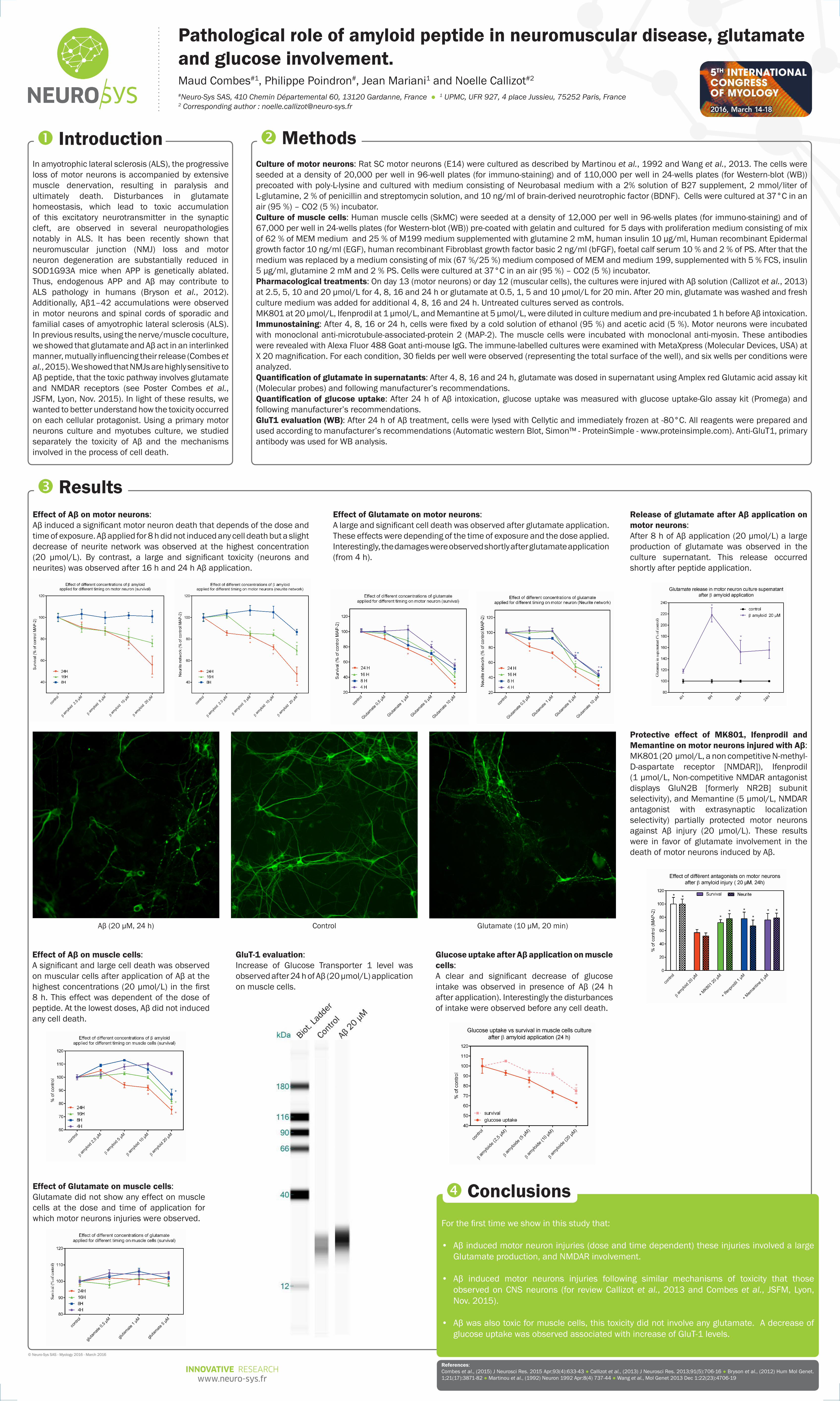
Effect of Aβ on motor neurons:Aβ induced a significant motor neuron death that depends of the dose and time of exposure. Aβ applied for 8 h did not induced any cell death but a slight decrease of neurite network was observed at the highest concentration (20 µmol/L). By contrast, a large and significant toxicity (neurons and neurites) was observed after 16 h and 24 h Aβ application.

References:Combes et al., (2015) J Neurosci Res. 2015 Apr;93(4):633-43 ● Callizot et al., (2013) J Neurosci Res. 2013;91(5):706-16 ● Bryson et al., (2012) Hum Mol Genet. 1;21(17):3871-82 ● Martinou et al., (1992) Neuron 1992 Apr:8(4) 737-44 ● Wang et al., Mol Genet 2013 Dec 1:22(23):4706-19

Effect of Glutamate on motor neurons:A large and significant cell death was observed after glutamate application. These effects were depending of the time of exposure and the dose applied. Interestingly, the damages were observed shortly after glutamate application (from 4 h).

Release of glutamate after Aβ application on motor neurons:After 8 h of Aβ application (20 µmol/L) a large production of glutamate was observed in the culture supernatant. This release occurred shortly after peptide application.

Protective effect of MK801, Ifenprodil and Memantine on motor neurons injured with Aβ:MK801 (20 µmol/L, a non competitive N-methyl-D-aspartate receptor [NMDAR]), Ifenprodil (1 µmol/L, Non-competitive NMDAR antagonist displays GluN2B [formerly NR2B] subunit selectivity), and Memantine (5 µmol/L, NMDAR antagonist with extrasynaptic localization selectivity) partially protected motor neurons against Aβ injury (20 µmol/L). These results were in favor of glutamate involvement in the death of motor neurons induced by Aβ.

Aβ (20 µM, 24 h) Control Glutamate (10 µM, 20 min)

Effect of Aβ on muscle cells:A significant and large cell death was observed on muscular cells after application of Aβ at the highest concentrations (20 µmol/L) in the first 8 h. This effect was dependent of the dose of peptide. At the lowest doses, Aβ did not induced any cell death.

Glucose uptake after Aβ application on muscle cells:A clear and significant decrease of glucose intake was observed in presence of Aβ (24 h after application). Interestingly the disturbances of intake were observed before any cell death.

Effect of Glutamate on muscle cells:Glutamate did not show any effect on muscle cells at the dose and time of application for which motor neurons injuries were observed.

GluT-1 evaluation:Increase of Glucose Transporter 1 level was observed after 24 h of Aβ (20 µmol/L) application on muscle cells.