Embed Size (px)
Citation preview

25-1
Respiratory
System

25-2
Organization and Functions of the Respiratory System Structural classifications:
upper respiratory tract lower respiratory tract.
Functional classifications: Conducting portion: transports air.
Nose nasal cavity Pharynx Larynx Trachea progressively smaller airways, from the primary bronchi
to the bronchioles

25-3
Organization and Functions of the Respiratory System
Respiratory portion: carries out gas exchange. respiratory bronchioles alveolar ducts alveoli
Upper respiratory tract is all conducting Lower respiratory tract has both conducting
and respiratory portions

4

25-5
Respiratory System Functions Breathing (pulmonary ventilation): the
moving in and out of air consists of two cyclic phases:
inhalation, also called inspiration exhalation, also called expiration
Inhalation draws gases into the lungs. Exhalation forces gases out of the lungs.

25-6
Respiratory System Functions Gas exchange: O2 and CO2
External respiration External environment and blood (air sacs)
Internal respiration (cell) Blood and cells
Respiration should not be confused with breathing!
Breathing is the taking in of oxygen by inhaling andthe giving off of carbon dioxide by exhaling

25-7
Respiratory System Functions Gas conditioning:
Warmed Humidified Cleaned of particulates
Sound production: Movement of air over true vocal cords Also involves nose, paranasal sinuses, teeth,
lips and tongue Olfaction:
Olfactory epithelium over superior nasal conchae
Defense: vibrissae, mucus, lymphoid tissue

25-8
Upper Respiratory Tract Composed of
the nose the nasal cavity the paranasal sinuses the pharynx (throat) and associated structures.
All part of the conducting portion of the respiratory system.

9

Nose Nose: external nares nasal cavity
internal nares Nasal septum splits in two Nasal conchae swirl air over mucus
membrane Designed to: Filter, Warm, Humidify Trap dust and infectious agents Detect olfactory stimuli Modify vocal sounds
25-11
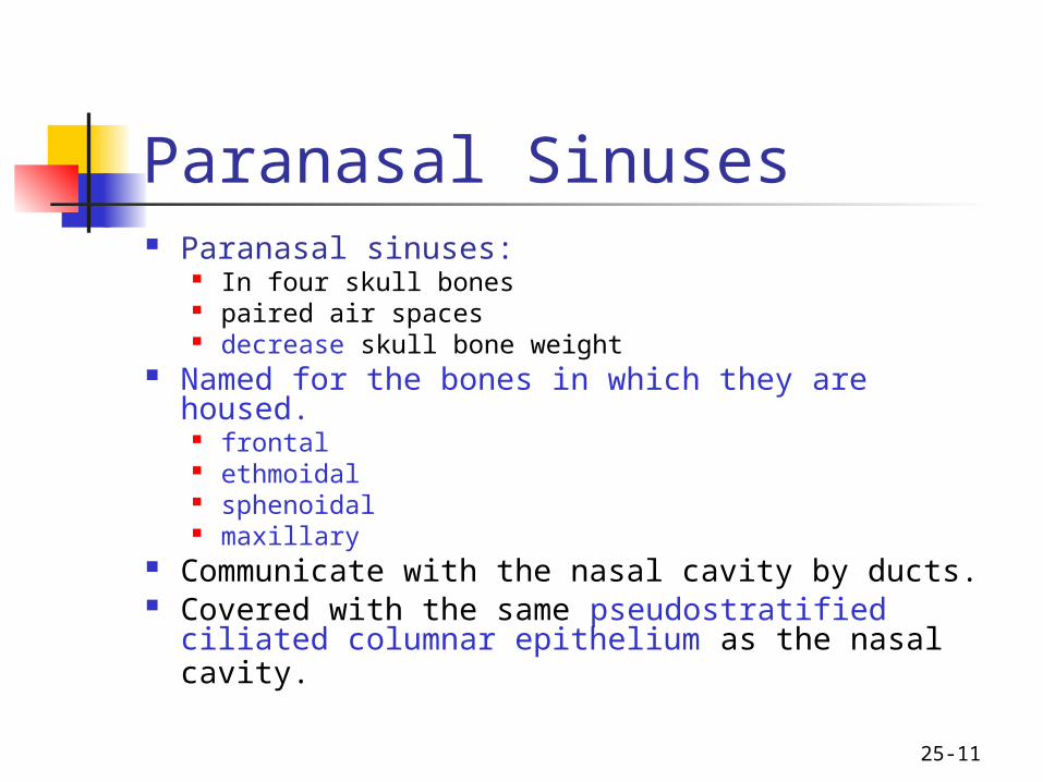
Paranasal Sinuses Paranasal sinuses:
In four skull bones paired air spaces decrease skull bone weight
Named for the bones in which they are housed. frontal ethmoidal sphenoidal maxillary
Communicate with the nasal cavity by ducts. Covered with the same pseudostratified
ciliated columnar epithelium as the nasal cavity.

12

25-13
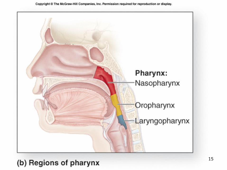
Pharynx Common to both the respiratory and
digestive systems. Commonly called the throat. Funnel-shaped Funnel shaped tube from
internal nares to larynx slightly wider superiorly and narrower
inferiorly.

25-14
Pharynx With flexible, distensible lateral walls:
lined by a mucosa contain skeletal muscles primarily used for
swallowing. to force swallowed food into the esophagus.
Partitioned into three adjoining regions: Nasopharynx – upper part Oropharynx – middle part Laryngopharynx – lower part
15

25-16
Nasopharynx Superiormost region of the pharynx. Location:
posterior to the nasal cavity superior to the soft palate
separates it from the posterior part of the oral cavity. Normally, only air passes through. Soft palate
Blocks material from the oral cavity and oropharynx elevates when we swallow.
Auditory tubes paired In the lateral walls of the nasopharynx connect the nasopharynx to the middle ear.
Pharyngeal tonsil posterior nasopharynx wall single commonly called the adenoids.

25-17
Oropharynx The middle pharyngeal region. Location:
Between the uvula & top of epiglottis Common respiratory and digestive pathway
both air and swallowed food and drink pass through. Lymphatic organs
provide the “first line of defense” against ingested or inhaled foreign materials.
Palatine tonsils on the lateral wall between the arches
Lingual tonsils At the base of the tongue.

25-18
Laryngopharynx Inferior, narrowed region of the pharynx. Location:
Connects with both esophagus & larynx The larynx (voice box) forms the anterior wall Lined with a non-keratinized stratified
squamous epithelium (mucus membrane)
25-19
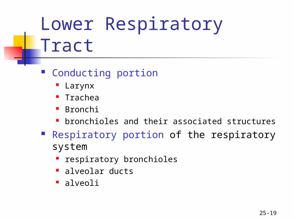
Lower Respiratory Tract Conducting portion
Larynx Trachea Bronchi bronchioles and their associated structures
Respiratory portion of the respiratory system respiratory bronchioles alveolar ducts alveoli

25-20
Larynx Short, somewhat cylindrical airway Location:
bounded posteriorly by the laryngopharynx,
inferiorly by the trachea. Prevents swallowed materials from
entering the lower respiratory tract. Conducts air into the lower
respiratory tract. Produces sounds.

25-21
Larynx Nine pieces of cartilage
three individual pieces Thyroid cartilage - anterior = “Adam's apple” Cricoid cartilage – forms the inferior wall Epiglottis - upper leaf-shaped piece
During swallowing larynx moves up and epiglottis covers opening to trachea
three cartilage pairs Arytenoids: above the cricoid, attach to vocal cords &
pharyngeal muscles Corniculates: attach to arytenoids Cuneiforms: in aryepiglottic fold
held in place by ligaments and muscles. Intrinsic muscles: regulate tension on true vocal
cords Extrinsic muscles: stabilize the larynx

22
25-23
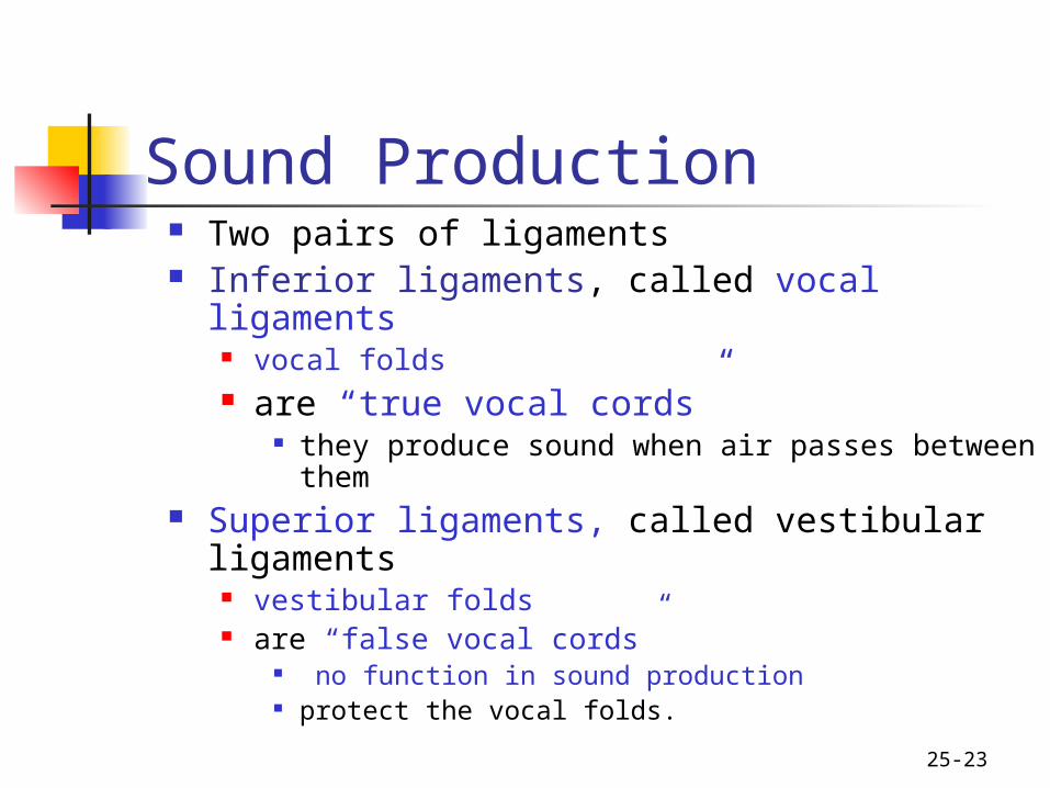
Sound Production Two pairs of ligaments Inferior ligaments, called vocal ligaments
vocal folds are “true vocal cords”
they produce sound when air passes between them
Superior ligaments, called vestibular ligaments vestibular folds are “false vocal cords”
no function in sound production protect the vocal folds.

25-24
Sound Production The tension, length, and position
of the vocal folds determine the quality of the sound. Longer vocal folds produce lower
sounds More taunt, higher pitch Loudness based on force of air
Rima glottidis: opening between the vocal folds

25

26

25-27
Trachea A flexible, slightly rigid tubular organ
often referred to as the “windpipe.” Location:
immediately anterior to the esophagus inferior to the larynx superior to the primary bronchi of the lungs.
Anterior and lateral walls of the trachea are supported by 15 to 20 C-shaped tracheal cartilages.
cartilage rings reinforce and provide some rigidity to the tracheal wall to ensure that the trachea remains open (patent) at all times
cartilage rings are connected by elastic sheets called anular ligaments

28

25-29
Trachea At the level of the sternal angle, the trachea
bifurcates into two smaller tubes, called the right and left primary bronchi.
Each primary bronchus projects laterally toward each lung.
The most inferior tracheal cartilage separates the primary bronchi at their origin and forms an internal ridge called the carina.
Lined with pseudostratified ciliated mucous membrane dust protection – move toward pharynx

25-30
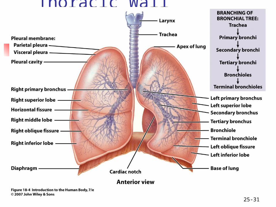
Bronchial Tree A highly branched system
air-conducting passages originate from the left and right primary bronchi.
Progressively branch into narrower tubes as they diverge throughout the lungs before terminating in terminal bronchioles.
Primary bronchi Incomplete rings of hyaline cartilage ensure that
they remain open. Right primary bronchus
shorter, wider, and more vertically oriented than the left primary bronchus.
Foreign particles are more likely to lodge in the right primary bronchus.
25-31
Thoracic Wall Dimensional Changes During Respiration Lateral dimensional changes occur with rib
movements. Elevation of the ribs increases the lateral
dimensions of the thoracic cavity, while depression of the ribs decreases the lateral dimensions of the thoracic cavity.

25-32
Bronchial Tree Primary bronchi
enter the hilum of each lung Also entering hilum:
pulmonary vessels lymphatic vessels Blood vessels nerves.
Secondary bronchi (or lobar bronchi) Branch of primary bronchus one for each lobe of lung left lung:
two lobes two secondary bronchi
right lung three lobes three secondary bronchi.
Tertiary bronchi (or segmental bronchi) Branch of secondary bronchi left lung is supplied by 8 to 10 tertiary bronchi. right lung is supplied by 10 tertiary bronchi supply a part of the lung called a bronchopulmonary segment.

33

Alveoli Cup-shaped out pouch of sac
Lined with thin alveolar cells (simple squamous)
Scattered surfactant secreting cells Lowers surface tension & humidifies
Alveolar macrophages- “cleaners” Gases diffuse across combined
epithelia of alveolus & capillary

35
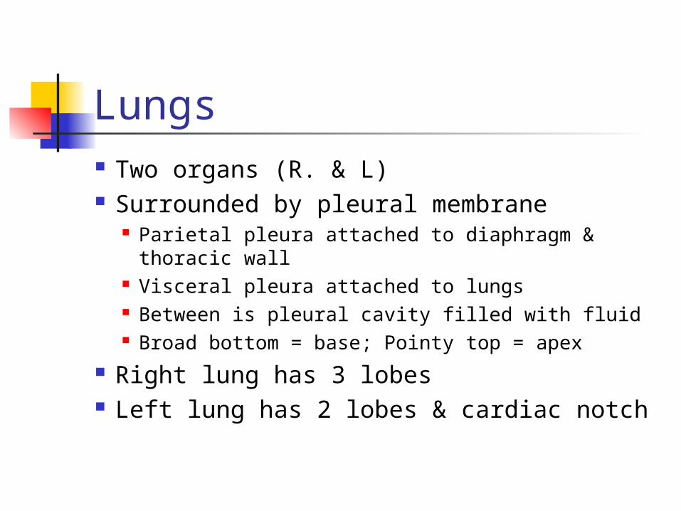
Lungs Two organs (R. & L) Surrounded by pleural membrane
Parietal pleura attached to diaphragm & thoracic wall
Visceral pleura attached to lungs Between is pleural cavity filled with fluid Broad bottom = base; Pointy top = apex
Right lung has 3 lobes Left lung has 2 lobes & cardiac notch

25-37
The lungs are the main organs of respiration in man.
They are never empty.
There is always air inside until after death
The amount of air that goes in and out of the lungs(about 500 cc) is called tidal air
Respiratory movements are controlled in the brain stem particularly in the medulla oblongata.

25-38
Gross Anatomy of the Lungs Each lung has a conical shape. Its wide, concave base rests upon the muscular
diaphragm. Its relatively blunt superior region, called the apex or
(cupola), projects superiorly to a point that is slightly superior and posterior to the clavicle.
Both lungs are bordered by the thoracic wall anteriorly, laterally, and posteriorly, and supported by the rib cage.

39

25-40
Pleura and Pleural Cavities The outer surface of each lung and the
adjacent internal thoracic wall are lined by a serous membrane called pleura, which is formed from simple squamous epithelium.
The outer surface of each lung is tightly covered by the visceral pleura, while the internal thoracic walls, the lateral surfaces of the mediastinum, and the superior surface of the diaphragm are lined by the parietal pleura.

41

25-42
Pleura and Pleural Cavities The outer surface of each lung is tightly covered by the
visceral pleura, while the internal thoracic walls, the lateral surfaces of the mediastinum, and the superior surface of the diaphragm are lined by the parietal pleura.
The potential space between these serous membrane layers is a pleural cavity.
The pleural membranes produce a thin, serous fluid called serum that circulates in the pleural cavity and acts as a lubricant, ensuring minimal friction during breathing.

25-43
Lymphatic Drainage Lymph nodes and vessels are located within
the connective tissue of the lung as well as around the bronchi and pleura.
The lymph nodes collect carbon, dust particles, and pollutants that were not filtered out by the pseudostratified ciliated columnar epithelium.

44
25-45
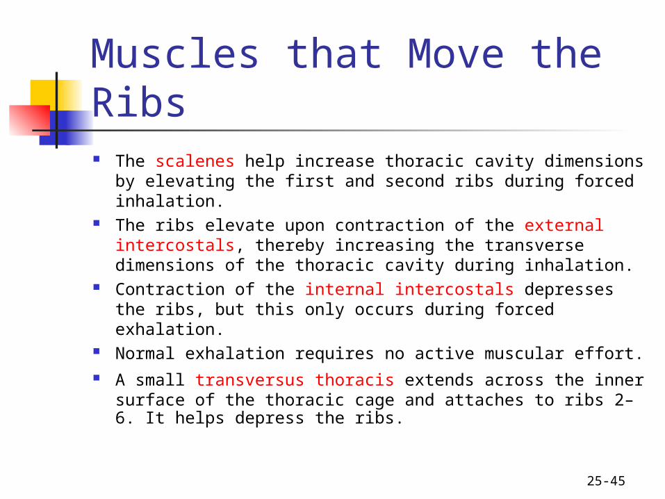
Muscles that Move the Ribs The scalenes help increase thoracic cavity dimensions
by elevating the first and second ribs during forced inhalation.
The ribs elevate upon contraction of the external intercostals, thereby increasing the transverse dimensions of the thoracic cavity during inhalation.
Contraction of the internal intercostals depresses the ribs, but this only occurs during forced exhalation.
Normal exhalation requires no active muscular effort. A small transversus thoracis extends across the inner
surface of the thoracic cage and attaches to ribs 2–6. It helps depress the ribs.

25-46
Muscles that Move the Ribs Two posterior thorax muscles also assist with
respiration. The serratus posterior superior elevates ribs 2–5 during
inhalation, and the serratus posterior inferior depresses ribs 8–12 during exhalation.
In addition, some accessory muscles assist with respiratory activities.
The pectoralis minor, serratus anterior, and sternocleidomastoid help with forced inhalation,
while the abdominal muscles (external and internal obliques, transversus abdominis, and rectus abdominis) assist in active exhalation.

47

25-48
Aging and the Respiratory System Becomes less efficient with age due to several structural
changes. Decrease in elastic connective tissue in the lungs and the
thoracic cavity wall. Loss of elasticity reduces the amount of gas that can be
exchanged with each breath and results in a decrease in the ventilation rate.
Emphysema may cause a loss of alveoli or their functionality
Reduced capacity for gas exchange can cause an older person to become “short of breath” upon exertion.
Carbon, dust, and pollution material gradually accumulate in our lymph nodes and lungs.

Aging Everything becomes less elastic Decrease in Vital capacity Can decrease blood O2 level Decreased exercise capacity Decreased macrophage activity Increased susceptibility to
pulmonary disease


![Respiratory system roadmap.pptx [Repaired] - Loginanatomical-sciences.health.wits.ac.za/roadmaps/Respiratory system... · DIVISION OF THE RESPIRATORY SYSTEM CONDUCTING PORTION Nasal](https://img.pdfslide.us/doc/110x75/5a78c3d87f8b9ae6228c9db0/respiratory-system-repaired-loginanatomical-scienceshealthwitsaczaroadmapsrespiratory.jpg)
![Respiratory System [โหมดความเข้ากันได้] · PATHOLOGY OF RESPIRATORY SYSTEM นพ. อรรณพ นาคะป ท Respiratory system U it](https://img.pdfslide.us/doc/110x75/5fa578efd4e80f055f6b3401/respiratory-system-aaaaaaaaaaaaaaaaaa-pathology.jpg)


![Anatomy and Physiology Respiratory System [Tab 2] Respiratory System](https://img.pdfslide.us/doc/110x75/56649ebd5503460f94bc631f/anatomy-and-physiology-respiratory-system-tab-2-respiratory-system.jpg)