Embed Size (px)
Citation preview

IMPLANTATION
&
PLACENTATIONPranjal Gupta
Bsc(H) Zoology, Ramjas College

OVERVIEW
Human embryo cleavages and blastulation
Human implantation
Human basic gastrulation
Extra-embryonic membranes in birds
Extra-embryonic membranes in mammals
Classification of placenta
Human placental physiology

HUMAN EMBRYO CLEAVAGES
1st cleavage occur in first 30h; Upper oviduct
Next within 40hrs after fertilization
Third about 3days after fertilization, i.e. 8celled
embryo.
By the end of 3rd day, 8-16 celled morula reaches
uterus
Holoblastic cleavage
Enclosed within zona pellucida

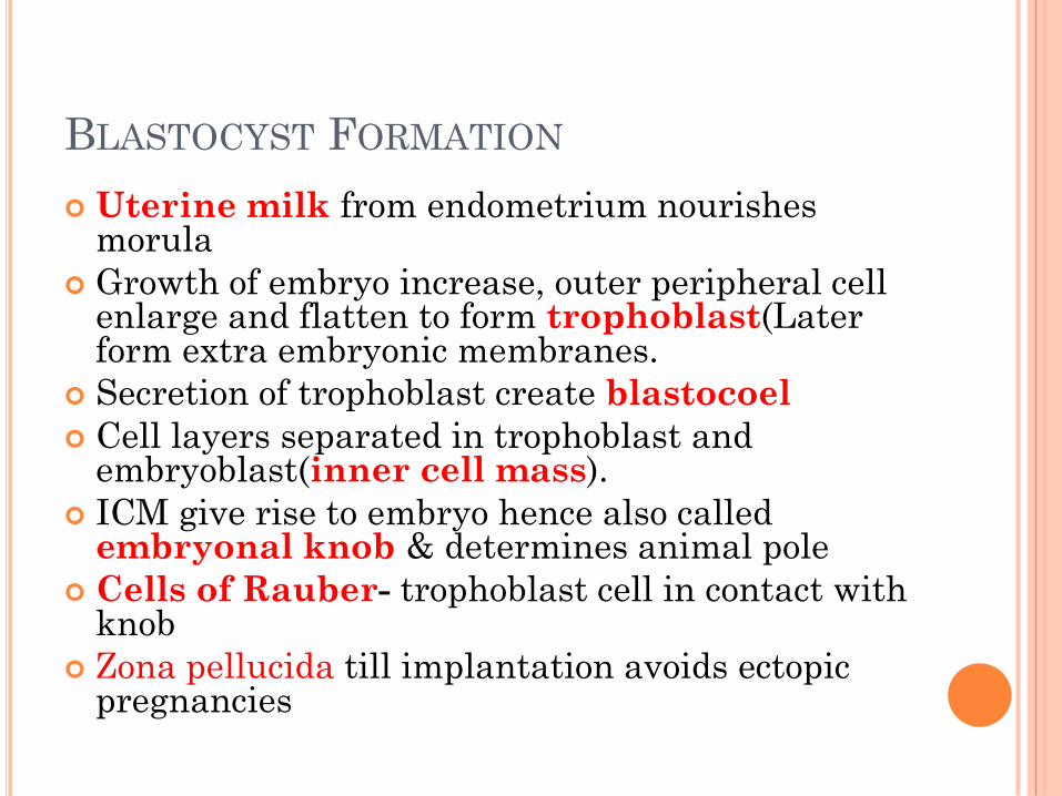
BLASTOCYST FORMATION
Uterine milk from endometrium nourishes morula
Growth of embryo increase, outer peripheral cell enlarge and flatten to form trophoblast(Later form extra embryonic membranes.
Secretion of trophoblast create blastocoel
Cell layers separated in trophoblast and embryoblast(inner cell mass).
ICM give rise to embryo hence also called embryonal knob & determines animal pole
Cells of Rauber- trophoblast cell in contact with knob
Zona pellucida till implantation avoids ectopic pregnancies
IMPLANTATION
Anchoring or embedding of the blastocyst into
endometrium of uterus.
7th day post fertilization, and a 3 day long
process.
Zona hatching
Trophoblast not exposed till proper site reached
Implantation allows the ICM region to be
engraved inside endometrium
Local endometrial lysis by trophoblastic enzymes
Formation of Chorionic villi from trophoblast.


Embedded embryo
inside endometrium
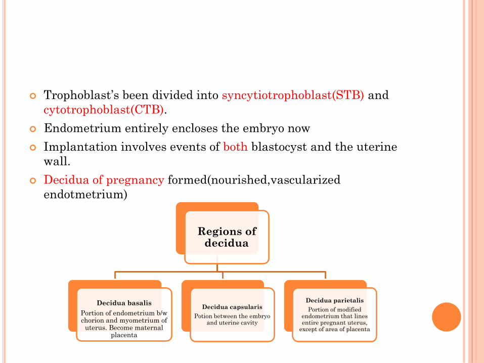
Trophoblast’s been divided into syncytiotrophoblast(STB) and
cytotrophoblast(CTB).
Endometrium entirely encloses the embryo now
Implantation involves events of both blastocyst and the uterine
wall.
Decidua of pregnancy formed(nourished,vascularized
endotmetrium)
Regions of decidua
Decidua basalis
Portion of endometrium b/w chorion and myometrium of
uterus. Become maternal placenta
Decidua capsularis
Potion between the embryo and uterine cavity
Decidua parietalis
Portion of modified endometrium that lines entire pregnant uterus,
except of area of placenta
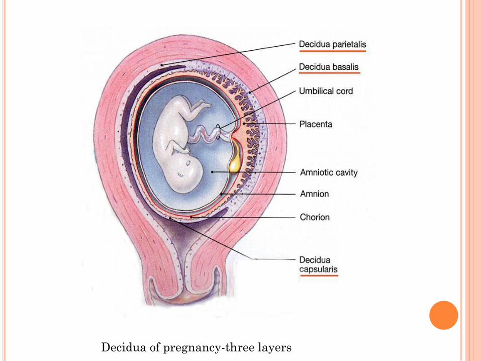
Decidua of pregnancy-three layers

CHANGES IN POSITIONS DURING
PREGNANCY

ECTOPIC PREGNANCIES

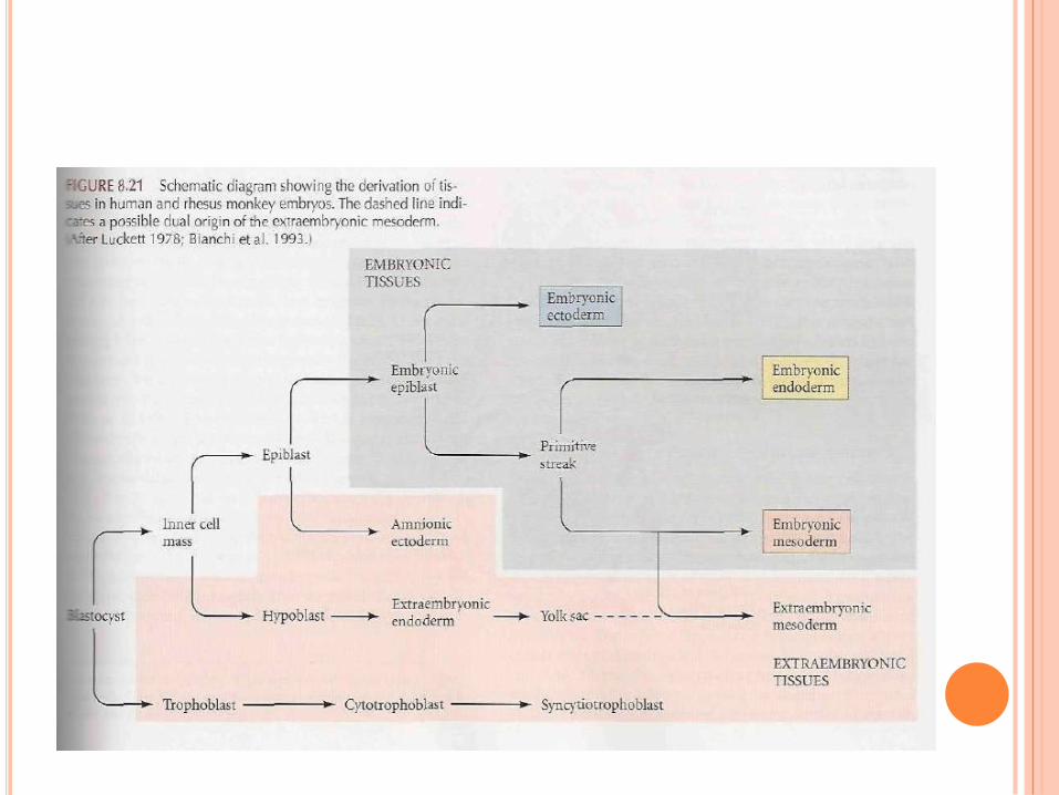
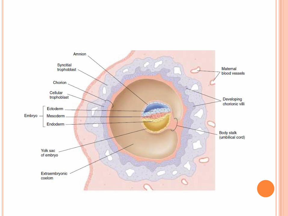
GASTRULATION
By morphogenetic movements three germ layers are separated
Trophoblast divided into two, and CTB directly cover the ICM while STB spreads in endometrium.
CTB give rise to extra-embryonic mesoderm(EEM).
EEM diff. to outer or parietal or Somatopleuric EEMand inner Splanchnopleuric EEM.
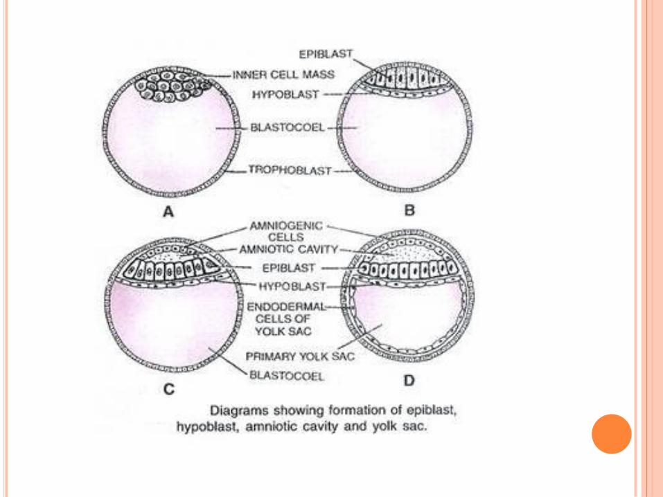
ICM differentiated to epiblast and hypoblast, both form a disk called embryonic disk.
Epiblast divides to amnion and embryonic ectoderm.
Chorion formed by Somatopleure inside and trophoblast outside
Later chorion becomes essential part of placenta.
Hypoblast give rise to EE endodermal cells lining primary yolk sac.
Yolk sac become smaller to form secondary yolk sac
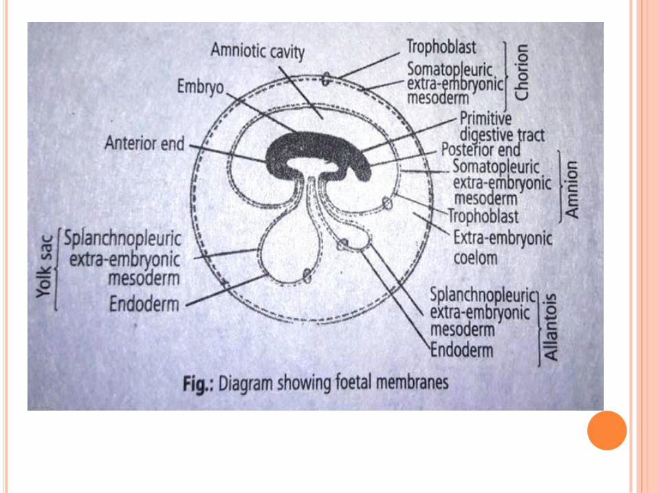
EXTRA EMBRYONIC MEMBRANES
It is an adaptation to terrestrial mode of life.
Situated outside boundaries of embryo hence so called.
4 major membranes- Chorion. Amnion, Allantois and Yolk sac.
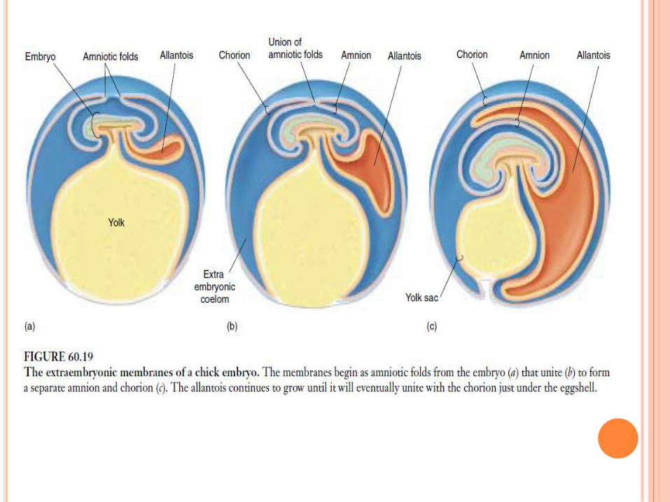
In birds
Amnion and chorion arises from two folds that grow to surround
embryo.
Amnion, inside surround embryo and suspend it in amniotic fluid
Chorion is in contact with the shell and separated from other
membranes through EE coelom
Yolk here is utilized for nourishment unlike mammals
Allantois is derived as out pouch of gut and store uric acid excreted.
Allantois later expands to form a sac that fuses with chorion under
eggshell, a functional unit of embryonic blood vessels inside allantois
is formed.
Allantois now in direct contact with the porous shell act as ‘lung’ for
chick embryo.
In mammals
Embryonic membranes arise from trophoblast.
First prior to implantation trophoblast form chorion
Chorion has great role in placentation. Made of Trophoblast outside and Somatopleure inside
Yolk sac here is not involved in nutrition for long run only initial vitelline circulation. Made of endoderm inside and splanchnopleure outside
Allantois contributes blood vessels to structure that forms the umbilical cord. Endoderm inside and splanchnopleure outside.
Amnion, consist of trophoblast inside and somatopleure outside. It cover the entire embryo. Shock absorber and avoid desiccation.
Umbilical cord is formed of stalk of yolk sac and allantois.
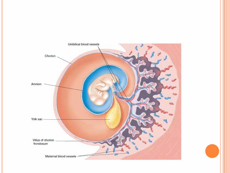
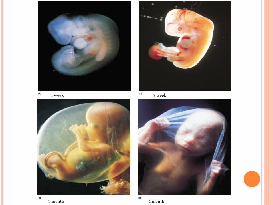
PLACENTATION
3 month
7 week4 week
4 month
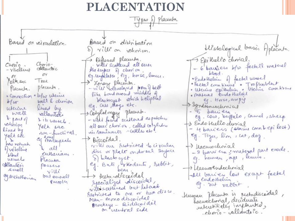
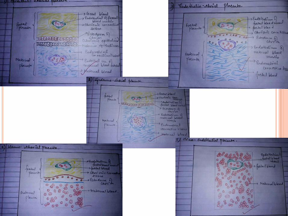
PLACENTATION
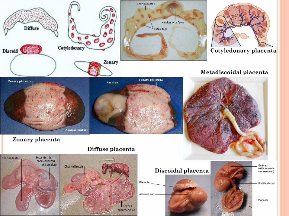
Zonary placenta
Diffuse placenta
Discoidal placenta
Metadiscoidal placenta
Cotyledonary placenta

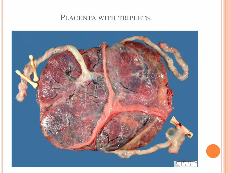
PLACENTA WITH TRIPLETS.
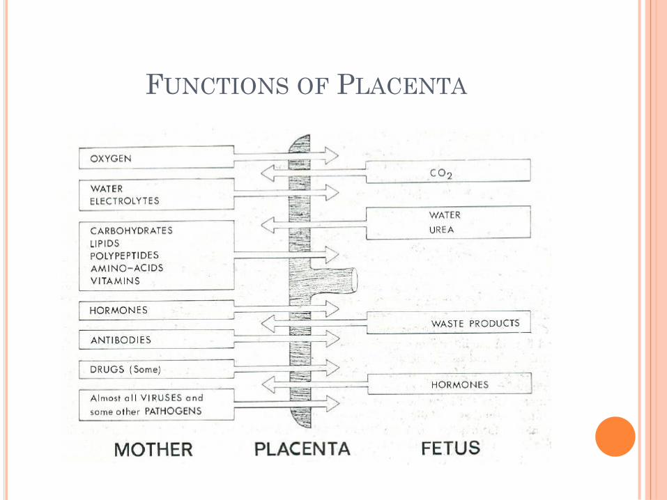
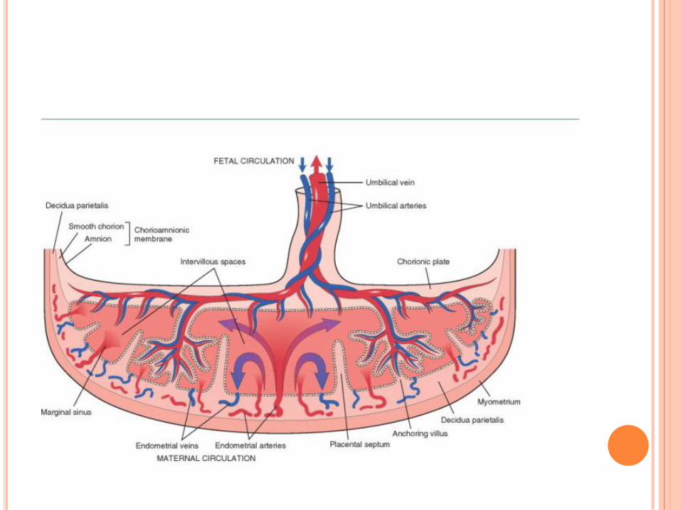
FUNCTIONS OF PLACENTA
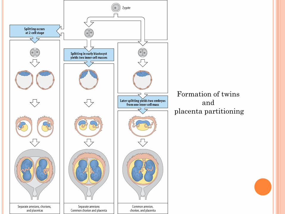
Formation of twins
and
placenta partitioning