Embed Size (px)
Citation preview

8894 Phys. Chem. Chem. Phys., 2011, 13, 8894–8899 This journal is c the Owner Societies 2011
Cite this: Phys. Chem. Chem. Phys., 2011, 13, 8894–8899
Designing an enzyme-based nanobiosensor using molecular
modeling techniques
Eduardo F. Franca,*aFabio L. Leite,
bRichard A. Cunha,
aOsvaldo N. Oliveira Jr.
c
and Luiz C. G. Freitasd
Received 14th February 2011, Accepted 3rd March 2011
DOI: 10.1039/c1cp20393b
Nanobiosensors can be built via functionalization of atomic force microscopy (AFM) tips with
biomolecules capable of interacting with the analyte on a substrate, and the detection being
performed by measuring the force between the immobilized biomolecule and the analyte.
The optimization of such sensors may require multiple experiments to determine suitable
experimental conditions for the immobilization and detection. In this study we employ molecular
modeling techniques to assist in the design of nanobiosensors to detect herbicides. As a proof
of principle, the properties of acetyl co-enzyme A carboxylase (ACC) were obtained with
molecular dynamics simulations, from which the dimeric form in an aqueous solution was
found to be more suitable for immobilization owing to a smaller structural fluctuation than
the monomeric form. Upon solving the nonlinear Poisson–Boltzmann equation using a
finite-difference procedure, we found that the active sites of ACC exhibited a positive surface
potential while the remainder of the ACC surface was negatively charged. Therefore, optimized
biosensors should be prepared with electrostatic adsorption of ACC onto an AFM tip
functionalized with positively charged groups, leaving the active sites exposed to the analyte.
The preferential orientation for the herbicides diclofop and atrazine with the ACC active site
was determined by molecular docking calculations which displayed an inhibition coefficient
of 0.168 mM for diclofop, and 44.11 mM for atrazine. This binding selectivity for the herbicide
family of diclofop was confirmed by semiempirical PM6 quantum chemical calculations which
revealed that ACC interacts more strongly with the herbicide diclofop than with atrazine,
showing binding energies of �119.04 and +8.40 kcal mol�1, respectively. The initial
measurements of the proposed nanobiosensor validated the theoretical calculations and
displayed high selectivity for the family of the diclofop herbicides.
1. Introduction
Many analytical techniques have been used to detect pesticides
and other residues in the environment, but new prospects for
detection have emerged recently with nanobiosensors,1,2 which
basically comprise a biological component (e.g. enzyme, antibody)
immobilized on a nanoscale detection device. Of particular impor-
tance are the nanobiosensors obtained by deposition of a receptor
layer (protein) on microcantilevers with analytes detected at a
concentration as low as 10�18 mol L�1 using an atomic force
microscope (AFM).3 The functionalization of AFM tips has also
been exploited, with whichmolecular interactions can bemeasured
with a resolution of 10�12 N, thus suggesting the possible
measurement of single molecule interactions using the force curve
in the so-called atomic force spectroscopy (AFS).4
The fabrication of optimized nanobiosensors requires prior
knowledge of various features. In addition to an adequate choice
of the biomolecule to be immobilized which would interact
specifically with the analyte of interest, the method of immobili-
zation and the experimental conditions must be determined. For
enzyme-based sensors, the layer-by-layer (LbL)5,6 technique
has been used owing to its simplicity and versatility.7,8 For the
successful deposition of the LbL film on an AFM tip, the
important issues are the charge distribution over the enzyme
surface and the charge of the active sites, as the latter need to be
exposed to the analyte. It is clear therefore that multiple experi-
ments need to be performed for reaching the optimized conditions,
which has been the motivation for the use of molecular modeling
techniques to predict the characteristics of specific systems.
a Instituto de Quımica, Universidade Federal de Uberlandia,38.400-902, Uberlandia, MG, Brazil.E-mail: [email protected]; Fax: +55 34 3239 4208;Tel: +55 34 3239 4143
bUniversidade Federal de Sao Carlos, 18052-780, Sorocaba, SP,Brazil
c Instituto de Fısica de Sao Carlos, USP, 13560-970, Sao Carlos, SP,Brazil
dDepartamento de Quımica, Universidade Federal de Sao Carlos,13565-905, Sao Carlos, SP, Brazil
PCCP Dynamic Article Links
www.rsc.org/pccp PAPER
This journal is c the Owner Societies 2011 Phys. Chem. Chem. Phys., 2011, 13, 8894–8899 8895
In this study we investigate the properties of the enzyme
acetyl co-enzyme A carboxylase (ACC), which is promising for
the detection of herbicides. The family of acetyl-coenzyme A
carboxylases (ACCs) have a crucial role in the metabolism of
fatty acids in most living organisms.9–11 Acetyl-CoA carboxyl-
ase (ACC), in particular, catalyses the first step, namely, the
carboxylation of acetyl-CoA to malonyl-CoA. This enzyme
comprises a biotin carboxyl carrier protein (BCCP), biotin
carboxylase (BC), and carboxyltransferase (CT).12 The CT
domain containsB800 residues (90 kDa), which constitute the
C-terminal, and corresponds to one-third of the eukaryotic
multidomain ACCs.13 This domain (CT) is the active site for
two classes of widely used commercial herbicides,13–16 represented
by haloxyfop and diclofop (FOPs) and sethoxydim (DIMs).
These compounds are potent inhibitors of ACCs of plants that
are killed by ACC-inhibiting herbicides because the biosynthesis
of fatty acids is hampered.
Molecular dynamics simulations were used here to deter-
mine the most suitable conformations of ACC in an aqueous
solution and both the monomeric and dimeric forms of the
enzyme were investigated. In order to predict the type of
functionalization of the AFM tip required for the electrostatic
adsorption of ACC, the surface potential of the enzyme
was calculated by solving the nonlinear Poisson–Boltzmann
equation.18,19 Then, the interaction energies between ACC and
two herbicides, namely diclofop and atrazine, were calculated
using molecular docking and semiempirical quantum chemical
calculations. We shall show that ACC-based nanobiosensors
should be selective for the diclofop family of compounds, in
good agreement with experimental results.13,20,21
2. Methodology
2.1 Molecular systems
The X-ray crystallographic structure of the ACC enzyme, used
for the initial models, was obtained from the CT domain in
the Protein Data Bank, PDB ID: 1UYR. The missing residues
in the a and b subunits of 1UYR were added using the
Swiss-PdbViewer.17 This completed ACC structure was the
initial model for the ACC dimer, whereas only the a subunit
was used for the initial model of the ACC monomer. All water
molecules of crystallization were removed, and hydrogen
atoms were added to create an all-atoms model for the
Molecular Dynamics and Docking calculations. The systems
simulated by Molecular Dynamics are summarized in Table 1.
2.2 Molecular dynamics simulation
The modeled systems (monomer and dimer) were solvated by
filling the appropriated simulation box with SPC (single point
charges) water model molecules.18 Sodium and chloride ions
were used to achieve the ionic strength of 100 mM for each
system, which were energy minimized using 10 000 steps with
the steepest descent method. After minimization the solvent
was equilibrated by performing 100 ps molecular dynamics
simulation at 50, 150 and 298 K, with non-hydrogen atoms
positionally restrained (force constant 1.0� 103 kJ mol�1 nm�2).
Following the solvent equilibration step, for each tempera-
ture a total of 10 ns molecular dynamics simulations were
performed in an isothermal–isobaric (NPT) ensemble using
the leapfrog algorithm19 with a 2 fs time step. The configura-
tions were recorded every 1 ps for analysis. The temperature
was kept at 298 K by coupling the system to a Berendsen
thermostat20 with a relaxation time of 0.1 ps. The pressure was
maintained at 1 bar by coupling to a Berendsen barostat20
via isotropic coordinate scaling with a relaxation time of 10 ps
and a compressibility of 4.5 � 10�6 (kJ mol�1 nm�3)�1. The
stretching and bending motions of the system were constrained
using the LINCS algorithm.21 A 1.4 nm cutoff was used for the
short-range electrostatics and van der Waals interactions.
Long-range electrostatic contributions were treated via the
particle mesh Ewald (PME) method.22 All simulations were
carried out using the OPLS-AA23 force field within the
GROMACS 4.0.4 program.24
2.3 Electrostatic potential and solvation free energy
calculations
The electrostatic potential and the hydration free energy
for ACC were estimated by solving numerically the non-
linear Poisson–Boltzmann equation using a finite-difference
procedure25–28 with the APBS (Adaptive Poisson–Boltzmann
Solver) program29 in conjunction with the OPLS-AA force
field parameters set.30 The structures in the aqueous solution
were obtained using a dielectric constant of 78.54 for the solvent
with a solvent radius of 1.4 nm, surface tension of 0.105 N m�1,
and ionic strength of 100 mM. The internal dielectric constant
of the solute was set to 2, and the apolar contribution to the
solvation free energy was calculated using a surface tension
coefficient of 0.105 kJ mol�1. The three-dimensional potentials
were obtained using 129 grid points in x, y and z directions.
2.4 Molecular docking
The ACC structure and the herbicides atrazine and diclofop
were prepared using the ADT (AutoDockTools) program.31
The partial charges for the ligands (herbicides) were calculated
using the Gasteiger–Huckel method32 implemented in the
ADT program. A 3D grid was created by the AutoGrid
algorithm (a subprogram of AutoDock) to evaluate the binding
energies between ACC and the herbicides. Grid maps con-
taining 56 � 56 � 126 points for the herbicides were used to
constrain them within the active sites of ACC. The Lamarckian
genetic algorithm (LGA) was applied to search the confor-
mational space of the herbicides, while keeping the ACC struc-
ture rigid. For each run a set containing ten LGA docked
structures was obtained, from which two clusters of docked
Table 1 Description of the simulated systems
System Number of solute atoms Number of ions Number of solvent molecules Ionic strength/mol L�1
ACC monomer 11 609 210 55 470 0.100ACC dimer 23 185 254 66 782 0.100
8896 Phys. Chem. Chem. Phys., 2011, 13, 8894–8899 This journal is c the Owner Societies 2011
structures were chosen based on two criteria: (i) lowest
ETotal values; and (ii) clusters within a root mean square
deviation (RMSD) limit of 2.0 A.
2.5 Quantum chemical calculations
In order to evaluate the binding energy differences between
diclofop and atrazine herbicides, single point quantum chemical
calculations were performed for the ACC active site cavity
model, the herbicide and the complex (ACC active site cavity +
herbicides). The calculations were performed using the PM6
semiempirical Hamiltonian implemented in the MOPAC2009
software package.33 In order to build the ACC active site
cavity model, all the residues within 10.0 A of the cavity
were retained and all the others were deleted. The remaining
binding was completed by adding hydrogen atoms. Thus, the
binding energy was calculated by subtracting the heat of
formation of the ACC active site cavity and the herbicide
from the complex (ACC active site cavity + herbicide).
2.6 Atomic force microscopy
A Nanoscope V (VEECO) SPM instrument was used to
characterize the enzymes and herbicides film surfaces. AFM
has the advantage of providing images of polymer and organic
materials coated on insulating substrates, which prompted us
to choose it as a characterization method for studies involving
protein films. The enzyme was immobilized onto a thiol self-
assembled monolayer (SAM) formed on the gold-coated AFM
tip. For quantitative force experiments the geometry of the tip
and spring constant, kc, of the cantilever need to be known.
Several procedures have been applied to characterize the size
and shape of tips and cantilevers.38
3. Results and discussion
The biosensor under design requires the immobilization of
ACC on an AFM tip to detect enzyme-inhibiting herbicides,
such as diclofop. According to the literature,9 ACC may exist
in solution as a monomer and a dimer, both of which have
catalytic activity, suggesting that dimerization is not an
absolute requirement for the catalytic activity. Experimental
studies showed that the dimer form is the wild-type enzyme
and specific mutations on its interface are responsible for
inducing monomeric behavior in solution, whose catalytic
activity was weaker than that observed for the wild-type
enzyme. It is therefore necessary to know the intrinsic charac-
teristics of the monomer and dimer of ACC in solution to
select the suitable enzymatic structure (monomer or dimer) for
interaction with the pesticide. This interaction is strongly
related to the conformational fluctuation of the enzyme in
solution, from which one infers that the structural dynamics
of ACC must be determined for an optimized design of the
nanobiosensor.
The structural dynamics of ACC was evaluated by computing
its root mean square displacement (RMSD) during the molecular
dynamics simulation, in relation to its initial structure. Fig. 1
shows clearly the difference in the structural fluctuation
between the monomer and dimer in an aqueous solution.
The monomer completely loses its initial structure, and struc-
tural equilibrium is not achieved even after 9 ns of molecular
dynamics simulation. The lability of this monomeric structure
suggests that interaction forces between ACC and an herbicide––
at the reactive site––can be weakened by these conformational
changes, resulting in poor force propagation and inaccurate
detection. In contrast, the ACC dimer exhibited only 0.3 nm
of conformational fluctuation, showing a relative structural
rigidity in the aqueous solution with equilibrium being reached
after only 3 ns. One then infers that the interaction force
between the dimeric ACC and a pesticide at the active sites is
more likely to be transmitted to the AFM tip due to the
rigidity of this wild-type enzyme, which avoids absorption of
the greatest part of mechanical perturbation produced by
pesticide–ACC interactions.
The structural fluctuations and the differences between the
monomer and dimer of ACC can be attributed to two main
factors. The first is the electrostatic interaction between
charged amino acids such as arginine (ARG), lysine (LYS),
aspartic acid (ASP) and glutamic acid (GLU). The second is
the hydration of the polar amino acids, increasing the protein
mobility.
To evaluate the electrostatic repulsion between charged
groups with the same signal, the total translation of the amino
acids ARG, LYS, ASP and GLU was calculated from the
entire molecular dynamics trajectory. This calculation revealed
that amino acid groups with like charges tend to stay far from
each other. As a consequence, the variation in distance was
0.56 nm, at least 7% greater than the average mean distance
variation of all the atoms in ACC. However, electrostatic
attraction between oppositely charged amino acids leads to
salt bridges that stabilize the initial structure of both mono-
meric and dimeric enzyme structures. The presence of salt
bridges was assumed only when the distance between ARG
and ASP, ARG and GLU, LYS and ASP, and LYS and GLU
was less than 0.32 nm. Table 2 displays the number of salt
bridges in the beginning and at the end (10 ns) of the molecular
dynamics simulation. One notes that the number of salt
bridges in the dimer is more than double that in the monomer.
Therefore, the electrostatic interaction among oppositely charged
amino acids, e.g. ARG, LYS, ASP and GLU, on the border
Fig. 1 Time evolution of the root mean square deviation (RMSD) of
ACC atoms from the initial structure.

This journal is c the Owner Societies 2011 Phys. Chem. Chem. Phys., 2011, 13, 8894–8899 8897
between the two monomers in the dimer yielded the small
structural fluctuations shown in Fig. 1, with a rigid structure in
the aqueous solution. Nevertheless, the number of salt bridges
decreases during the simulation (see Table 2), which should be
ascribed mainly to the interaction between water molecules
and these charged amino acids. This solvation effect is
best illustrated by analyzing the number of hydrogen bonds
between water molecules and ACC given in Table 2.
It is clear from Table 2 that the number of salt bridges is
inversely related to the number of hydrogen bonds as a result
of water solvation of these hydrophilic groups. The charged
amino acids induce new hydrogen bonds between water
molecules and ACC, thus decreasing the number of salt bridges
and leading to the structural fluctuation in the enzyme struc-
ture. The intrinsic feature of the ACC monomer in having
more accessible area for the solvent per atom than the dimer
implies that a larger number of charged amino acids are
exposed to water molecules under thermal motion. Therefore,
the solvation effect seems to be the main factor for the
fluctuation.
To further describe the solvation effect on the structural
fluctuation and the structure stabilization of ACC by the water
molecules, the electrostatics and apolar contributions to the
free energy of solvation of the ACC monomer and dimer were
calculated via Poisson–Boltzmann electrostatics using the
APBS program.34 The initial structure (crystallographic) and
the final structure, from the last 10 ns of simulation, were
selected and calculated with the same ionic strength, so that
the conformational fluctuation was the only variable. There
are no experimental values in the literature to compare with
the calculated solvation free energy. These calculations can be
potentially used to predict relative solvation stabilization for
the different forms of the ACC enzyme. According to the
results given in Table 3, the solvation free energy is mainly
governed by the electrostatic contribution, which is consistent
with the relatively large number of charged amino acids in the
enzyme structure. The increase (in modulus) in the electro-
static contributions, from 0 to 10 ns, of 1680 kJ mol�1 and
3500 kJ mol�1 for the monomer and dimer, respectively,
explains the increase in the number of water molecules hydrogen
bonded to the charged groups during the molecular dynamics
simulation. On the other hand, the apolar contribution varied
only slightly during the simulation, viz. 20 and 140 kJ mol�1
for the monomeric and the dimeric structures, respectively.
The total free energy of solvation given in Table 3 confirmed
that the relaxed enzymatic structure is energetically stabilized
in an aqueous solution, reinforcing the hypothesis that
solvation effects are important for the structural fluctuations.
This suggests that the exposed charges on the ACC surface are
strongly solvated by water molecules that diffused within the
protein structure, reducing the direct electrostatic repulsion
among like charged groups, and inducing conformational
changes in the protein structure.
The results presented so far confirmed that the dimeric
structure is the most probable for ACC in aqueous solutions,
and to be immobilized on an AFM tip to produce a nano-
biosensor and detect herbicides. The next step is to decide how
the immobilization should take place, i.e. which would be the
best orientation to adsorb on the AFM tip while leaving the
active sites exposed to the solution in contact with the tip.
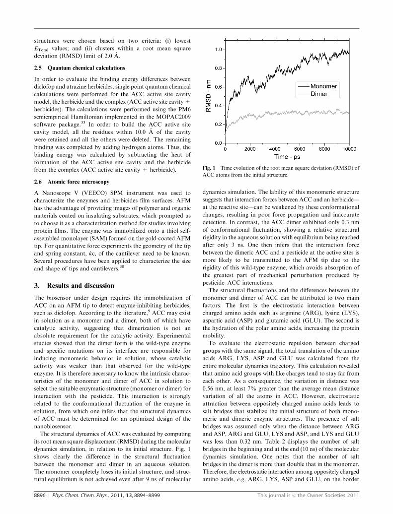
Because adsorption with electrostatic interactions between
oppositely charged molecules is one of the main techniques
used for biosensing,35,36 the electrostatic potential of ACC was
calculated by solving the Poisson–Boltzmann equation using
the APBS program34 (see Methodology). The electrostatic
landscape shown in Fig. 2 indicates that ACC has positively
and negatively charged groups located at distinct, specific
regions on its surface due to the presence of charged amino
acids such as ARG, LYS, ASP and GLU. According to the
crystallographic data for ACC complexed with the herbicide
diclofop13 and the electrostatic potential, the positive charges
are mainly located close to the active sites, which present high
percentage of the positively charged amino acid: LYS. This
suggests that AFM tips functionalized by negatively charged
functional groups (R�COO� or SiO2 tips) are not suitable to
immobilize ACC. On the other hand, the negative groups
(ASP and GLU) far from the active sites shown in Fig. 2
Table 2 Number of salt bridges in ACC and number of water molecules making hydrogen bonds with the enzyme
System Time Number of salt bridgesNumber of hydrogen bonds betweenACC and water molecules
Monomer 0 ns 41 147110 ns 35 1675
Dimer 0 ns 85 279710 ns 78 3134
Table 3 Free energy of solvation (kJ mol�1) for the monomer anddimer of ACC enzyme at the beginning and after 10 ns of moleculardynamics simulation
System DGsolv(electrostatic) DGsolv(apolar) DGsolv(total)
Monomer 0 ns �2.73 � 104 3.94 � 103 �2.34 � 104
10 ns �2.90 � 104 3.96 � 103 �2.50 � 104
Dimer 0 ns �4.63 � 104 6.52 � 103 �3.98 � 104
10 ns �4.98 � 104 6.66 � 103 �4.31 � 104
Fig. 2 Electrostatic potential (�5kBT/e to +5kBT/e) of the ACC
enzyme, which shows the most negative potential as the probable
immobilization area.
8898 Phys. Chem. Chem. Phys., 2011, 13, 8894–8899 This journal is c the Owner Societies 2011
also suggest that AFM tips functionalized with positively charged
groups, such as R–NH3+ and aminopropylethoxysilanes, would
unblock the active sites, leaving them free to interact with the
herbicide.
On the basis of the electrostatic potential for the ACC surface
and assuming an AFM tip functionalized with positively
charged groups, it is possible to suggest the possible orientation
of the enzymes on the tip, as displayed in Fig. 3. In the latter,
many ACC enzymes (each one shown in a different color) are
adsorbed on the functionalized surface of the AFM tip.
The interaction between the ACC dimer and herbicides can
be calculated using molecular docking, whose use is justified
by the small structural fluctuation of the ACC dimer shown in
Fig. 1. The two herbicides used were atrazine and diclofop
shown on the top of Fig. 5, which were made to dock to
the active site of ACC. The docking calculations resulted in
10 possible conformations for each herbicide in the active site.
From the docked herbicides on the ACC active site, the most
favorable clusters were used to calculate the binding energy
using the semiempirical PM6 Hamiltonian implemented in the
MOPAC2009 program. Binding energies of �119.04 and
+8.40 kcal mol�1 for diclofop and atrazine, respectively, were
found. These results are consistent with the experimental
finding that diclofop belongs to a specific class of inhibitors
for ACC, thus exhibiting a more favorable binding energy
than atrazine.13 The positive binding energy for atrazine is
expected, since it is not known as an inhibitor of ACC, but an
inhibitor of phosphodiesterase.37 These theoretical predictions
have been confirmed with preliminary results for the proposed
nanobiosensor, as indicated in Fig. 4. The average interaction
force between the diclofop and ACC enzyme is ca. 3 times the
force between atrazine and protein.
The perfect binding of diclofop to the ACC active sites is
illustrated in Fig. 5, obtained from the conformation of the
Fig. 3 Artwork of the enzymatic nanobiosensor. (A) Schematic representation of the detecting principle for the nanobiosensor. The ACC
molecules were adsorbed on the AFM tip from a solution owing to electrostatic attraction. The functionalized tip was then immersed in a liquid
cell containing a solid support coated with a layer of a herbicide. The interaction between ACC and the herbicide was obtained by measuring the
force curve as the AFM tip approached the herbicide-containing sample and was later withdrawn. (B) Various immobilized ACC molecules
oriented on the surface of a functionalized AFM tip. The change in color was just to facilitate visualization.
Fig. 4 Results for the adhesion forces obtained from the nano-
biosensor suggested by the molecular modeling techniques. These
forces were obtained by taking the force curves in an atomic force
microscope Nanoscope V (Veeco).
Fig. 5 Two diclofop herbicides efficiently docked to the ACC active
sites. The inset shows the structures of the herbicides (A) atrazine and
(B) diclofop.
This journal is c the Owner Societies 2011 Phys. Chem. Chem. Phys., 2011, 13, 8894–8899 8899
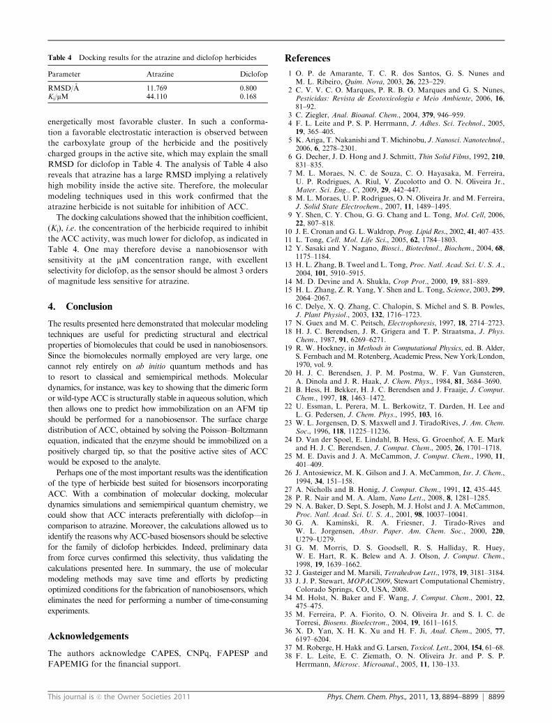
energetically most favorable cluster. In such a conforma-
tion a favorable electrostatic interaction is observed between
the carboxylate group of the herbicide and the positively
charged groups in the active site, which may explain the small
RMSD for diclofop in Table 4. The analysis of Table 4 also
reveals that atrazine has a large RMSD implying a relatively
high mobility inside the active site. Therefore, the molecular
modeling techniques used in this work confirmed that the
atrazine herbicide is not suitable for inhibition of ACC.
The docking calculations showed that the inhibition coefficient,
(Ki), i.e. the concentration of the herbicide required to inhibit
the ACC activity, was much lower for diclofop, as indicated in
Table 4. One may therefore devise a nanobiosensor with
sensitivity at the mM concentration range, with excellent
selectivity for diclofop, as the sensor should be almost 3 orders
of magnitude less sensitive for atrazine.
4. Conclusion
The results presented here demonstrated that molecular modeling
techniques are useful for predicting structural and electrical
properties of biomolecules that could be used in nanobiosensors.
Since the biomolecules normally employed are very large, one
cannot rely entirely on ab initio quantum methods and has
to resort to classical and semiempirical methods. Molecular
dynamics, for instance, was key to showing that the dimeric form
or wild-type ACC is structurally stable in aqueous solution, which
then allows one to predict how immobilization on an AFM tip
should be performed for a nanobiosensor. The surface charge
distribution of ACC, obtained by solving the Poisson–Boltzmann
equation, indicated that the enzyme should be immobilized on a
positively charged tip, so that the positive active sites of ACC
would be exposed to the analyte.
Perhaps one of the most important results was the identification
of the type of herbicide best suited for biosensors incorporating
ACC. With a combination of molecular docking, molecular
dynamics simulations and semiempirical quantum chemistry, we
could show that ACC interacts preferentially with diclofop—in
comparison to atrazine. Moreover, the calculations allowed us to
identify the reasons whyACC-based biosensors should be selective
for the family of diclofop herbicides. Indeed, preliminary data
from force curves confirmed this selectivity, thus validating the
calculations presented here. In summary, the use of molecular
modeling methods may save time and efforts by predicting
optimized conditions for the fabrication of nanobiosensors, which
eliminates the need for performing a number of time-consuming
experiments.
Acknowledgements
The authors acknowledge CAPES, CNPq, FAPESP and
FAPEMIG for the financial support.
References
1 O. P. de Amarante, T. C. R. dos Santos, G. S. Nunes andM. L. Ribeiro, Quim. Nova, 2003, 26, 223–229.
2 C. V. V. C. O. Marques, P. R. B. O. Marques and G. S. Nunes,Pesticidas: Revista de Ecotoxicologia e Meio Ambiente, 2006, 16,81–92.
3 C. Ziegler, Anal. Bioanal. Chem., 2004, 379, 946–959.4 F. L. Leite and P. S. P. Herrmann, J. Adhes. Sci. Technol., 2005,19, 365–405.
5 K. Ariga, T. Nakanishi and T.Michinobu, J. Nanosci. Nanotechnol.,2006, 6, 2278–2301.
6 G. Decher, J. D. Hong and J. Schmitt, Thin Solid Films, 1992, 210,831–835.
7 M. L. Moraes, N. C. de Souza, C. O. Hayasaka, M. Ferreira,U. P. Rodrigues, A. Riul, V. Zucolotto and O. N. Oliveira Jr.,Mater. Sci. Eng., C, 2009, 29, 442–447.
8 M. L. Moraes, U. P. Rodrigues, O. N. Oliveira Jr. andM. Ferreira,J. Solid State Electrochem., 2007, 11, 1489–1495.
9 Y. Shen, C. Y. Chou, G. G. Chang and L. Tong, Mol. Cell, 2006,22, 807–818.
10 J. E. Cronan and G. L. Waldrop, Prog. Lipid Res., 2002, 41, 407–435.11 L. Tong, Cell. Mol. Life Sci., 2005, 62, 1784–1803.12 Y. Sasaki and Y. Nagano, Biosci., Biotechnol., Biochem., 2004, 68,
1175–1184.13 H. L. Zhang, B. Tweel and L. Tong, Proc. Natl. Acad. Sci. U. S. A.,
2004, 101, 5910–5915.14 M. D. Devine and A. Shukla, Crop Prot., 2000, 19, 881–889.15 H. L. Zhang, Z. R. Yang, Y. Shen and L. Tong, Science, 2003, 299,
2064–2067.16 C. Delye, X. Q. Zhang, C. Chalopin, S. Michel and S. B. Powles,
J. Plant Physiol., 2003, 132, 1716–1723.17 N. Guex and M. C. Peitsch, Electrophoresis, 1997, 18, 2714–2723.18 H. J. C. Berendsen, J. R. Grigera and T. P. Straatsma, J. Phys.
Chem., 1987, 91, 6269–6271.19 R. W. Hockney, in Methods in Computational Physics, ed. B. Alder,
S. Fernbach andM. Rotenberg, Academic Press, New York/London,1970, vol. 9.
20 H. J. C. Berendsen, J. P. M. Postma, W. F. Van Gunsteren,A. Dinola and J. R. Haak, J. Chem. Phys., 1984, 81, 3684–3690.
21 B. Hess, H. Bekker, H. J. C. Berendsen and J. Fraaije, J. Comput.Chem., 1997, 18, 1463–1472.
22 U. Essman, L. Perera, M. L. Berkowitz, T. Darden, H. Lee andL. G. Pedersen, J. Chem. Phys., 1995, 103, 16.
23 W. L. Jorgensen, D. S. Maxwell and J. TiradoRives, J. Am. Chem.Soc., 1996, 118, 11225–11236.
24 D. Van der Spoel, E. Lindahl, B. Hess, G. Groenhof, A. E. Markand H. J. C. Berendsen, J. Comput. Chem., 2005, 26, 1701–1718.
25 M. E. Davis and J. A. McCammon, J. Comput. Chem., 1990, 11,401–409.
26 J. Antosiewicz, M. K. Gilson and J. A. McCammon, Isr. J. Chem.,1994, 34, 151–158.
27 A. Nicholls and B. Honig, J. Comput. Chem., 1991, 12, 435–445.28 P. R. Nair and M. A. Alam, Nano Lett., 2008, 8, 1281–1285.29 N. A. Baker, D. Sept, S. Joseph, M. J. Holst and J. A. McCammon,
Proc. Natl. Acad. Sci. U. S. A., 2001, 98, 10037–10041.30 G. A. Kaminski, R. A. Friesner, J. Tirado-Rives and
W. L. Jorgensen, Abstr. Paper. Am. Chem. Soc., 2000, 220,U279–U279.
31 G. M. Morris, D. S. Goodsell, R. S. Halliday, R. Huey,W. E. Hart, R. K. Belew and A. J. Olson, J. Comput. Chem.,1998, 19, 1639–1662.
32 J. Gasteiger and M. Marsili, Tetrahedron Lett., 1978, 19, 3181–3184.33 J. J. P. Stewart, MOPAC2009, Stewart Computational Chemistry,
Colorado Springs, CO, USA, 2008.34 M. Holst, N. Baker and F. Wang, J. Comput. Chem., 2001, 22,
475–475.35 M. Ferreira, P. A. Fiorito, O. N. Oliveira Jr. and S. I. C. de
Torresi, Biosens. Bioelectron., 2004, 19, 1611–1615.36 X. D. Yan, X. H. K. Xu and H. F. Ji, Anal. Chem., 2005, 77,
6197–6204.37 M. Roberge, H. Hakk and G. Larsen,Toxicol. Lett., 2004, 154, 61–68.38 F. L. Leite, E. C. Ziemath, O. N. Oliveira Jr. and P. S. P.
Herrmann, Microsc. Microanal., 2005, 11, 130–133.
Table 4 Docking results for the atrazine and diclofop herbicides
Parameter Atrazine Diclofop
RMSD/A 11.769 0.800Ki/mM 44.110 0.168