Embed Size (px)
Citation preview
123
Pure Appl. Chem., Vol. 81, No. 1, pp. 123–139, 2009.doi:10.1351/PAC-CON-08-01-15© 2009 IUPAC
Amperometric nanobiosensor for quantitativedetermination of glyphosate and glufosinateresidues in corn samples*
Everlyne A. Songa‡, Vernon S. Somerset, Tesfaye Waryo,Priscilla G. L. Baker, and Emmanuel I. Iwuoha‡
SensorLab, Department of Chemistry, University of the Western Cape, Private BagX17, Bellville, 7535, South Africa
Abstract: This study presents a simple, sensitive, rapid, and low-cost amperometric methodfor direct and quantitative determination of glyphosate and glufosinate herbicides.Electrochemical synthesis and characterization of poly(2,5-dimethoxyaniline)-poly(4-styrenesulfonic acid) (PDMA-PSS) nanoparticles was achieved by cyclic voltammetry (CV)and scanning electron microscopy (SEM). The nanobiosensor was constructed by immobi-lizing the enzyme horseradish peroxidase (HRP) electrostatically onto the surface of a rotat-ing gold disk electrode modified with PDMA-PSS nanoparticles. The biosensing principlewas based on determination of the sensor response to glyphosate and glufosinate by amper-ometric methods. Hydrogen peroxide (H2O2) was used to measure activity of the enzyme be-fore injection of the herbicides into the electrolyte solution. The enzyme electrode was sta-ble for a long period of time and was used for over 60 measurements. Glyphosate andglufosinate analyses were realized on spiked corn samples within a concentration range of2.0–78.0 µg L–1, corroborating that the nanobiosensor is sensitive enough to detect herbi-cides in these matrices. Based on a 20-µL sample injection volume, the detection limits were0.1 µg L–1 (10–10 M) for both glyphosate and glufosinate without sample clean-up or pre-concentration.
Keywords: electrochemical nanobiosensor; glyphosate; glufosinate; herbicides;poly(2,5-dimethoxyaniline)-poly(4-styrenesulfonic acid); horseradish peroxidase.
INTRODUCTION
Herbicides are a heterogeneous group of chemicals used to kill or inhibit the growth of undesirableplants that might cause damage, present fire hazards, or impede work crews. In recent years, the publichas become more concerned about the extensive use of herbicides and their effects on the environmenton a global scale. Among the herbicides used, glyphosate [N-(phosphonomethyl)glycine] and glufosi-nate [DL-homoalanine-4-yl-(methyl)phosphonic acid] are two important examples and are broad-spec-trum, nonselective herbicides for control of long grasses and broad-leaved weeds. The phosphorus-con-taining herbicides interfere with the formation of amino acids and other chemicals in plants [1,2]. Asthey are of comparatively low acute and chronic toxicity to human and animal health, these herbicides
*Paper based on a presentation at CHEMRAWN XII: The Role of Chemistry in Sustainable Agriculture and Human Well-beingin Africa, 2–5 December 2007, Stellenbosch, South Africa. Other presentations are published in this issue, pp. 85–151.‡Corresponding authors: Tel.: +27 21 959 3054; Fax: +27 21 959 1562; E-mail: [email protected] (E. I. Iwuoha),[email protected] (E. A. Songa).
have become the most extensively used worldwide. Some studies have reported that if they are ingestedover a period of time, they may affect the central nervous system, resulting in respiratory, myocardial,and neuromuscular malfunctions, which can even lead to death [3–5]. However, because their effects onnon-target organisms and overall environmental impact have not been fully investigated, questions re-garding the environmental safety with their increasing use have to be addressed. The increase in the useof these compounds has led to their residues being found in soil, atmosphere, agricultural products, aswell as in ground water.
The United Nations Food and Agricultural Organization (FAO) has set maximum residue limits(MRLs) for residues of glyphosate and glufosinate on most crops at 0.1–5 and 0.05 mg kg–1, respec-tively [6]. Currently, glyphosate is in the list of the U.S. national primary drinking water contaminantswith a maximum contaminant level goal (MCLG) of 0.7 mg L–1. The European Union (EU) limit ofany pesticide in drinking water has been set at 0.1 µg L–1 irrespective of their toxicological effects [7].Therefore, the monitoring of trace levels of these compounds in environmental and biological sampleshas gained increasing importance. For these reasons, there is a need to develop rapid, easy, and sensi-tive methods that are capable of detection and quantification of these herbicides at low concentrations,such as those that exist in foodstuffs and drinking water.
A simple analytical method for the determination of these herbicides at the sub µg L–1 level hasproven to be very difficult to obtain, mainly due to their ionic character, low volatility, high solubilityin water, insolubility in organic solvents, low mass, and favored complexing behavior [8]. Additionally,the absence of chromophore or fluorophore groups in their structures disables the photometric andfluorometric detection of these substances in liquid chromatography (LC) techniques. The reportedmethods for the determination of glyphosate mainly consisted of gas chromatography (GC), LC, capil-lary electrophoresis (CE), and enzyme-linked immunosorbent assay (ELISA). Most standard analyticalmethods developed until now require pre- or post-column derivatization procedures to improve both thechromatographic behavior and the detection ability by GC or high-performance liquid chromatography(HPLC). GC detectors have been used to improve the sensitivity, and furthermore, tandem mass spec-trometry (MS/MS) and GC/MS have been employed. Normally, HPLC has been used in combinationwith fluorescence and UV/vis detection after derivatization, although in a few cases glyphosate has beendetermined directly by ion chromatography (IC) with UV detection [9] or suppressed conductivity de-tection, but with limited sensitivity. GC/MS methods involved derivatization with different reagents [9]to confer volatility to the analytes. Normally, there is quite a lot of sample manipulation, and the meth-ods are time-consuming and tedious. The detection of glyphosate derivatives in LC and GC exhibitedhigh sensitivity and selectivity; however, these derivatization procedures are quite complicated and re-quire special equipment [10]. CE methods for glyphosate [11,12] provided high resolutions and effi-ciency, but some of them suffered low sensitivity owing to the limited sample injection volume. Thesereasons have given new impulse toward the development of alternative analytical devices and methods,to be applied in the screening of herbicides in environmental matrices, minimizing the pretreatment ofsample and reducing the cost and time of analysis. Detection methods for glyphosate and glufosinatewithout derivatization, such as electrogenerated chemiluminescence detection [13], conductivity detec-tion [14], inductively coupled plasma/mass spectrometry (ICP/MS) [11], and integrated pulsed amper-ometric detection (IPAD) at gold electrode [10], have been reported.
Biosensors and electrochemical methods appear well suited to complement standard analyticalmethods for a number of environmental monitoring applications since they do not require derivatiza-tion. Their application to environmental monitoring has been continuously growing in the last few years[15–17], though biosensor application for determination of glyphosate and glufosinate herbicides hasnot been previously reported. These methods have advantages such as low cost, easy operation, andhigh sensitivity and selectivity. Several kinds of electrochemical sensors for the determination of her-bicides have been reported [18–22]. Most of them were based on ELISA, in which herbicides were de-tected by competitive reactions with labeled antibodies. Although the immunoassay permits one to de-tect herbicide with high sensitivity and selectivity, the procedure is complicated, and it is difficult to
E. A. SONGA et al.
© 2009 IUPAC, Pure and Applied Chemistry 81, 123–139
124
monitor mixed herbicides because of the specificity of antibodies. An obvious drawback was the highcost and currently difficult commercial availability of the test kit. There have been a few potentiomet-ric sensors (ion-selective electrodes) reported [23]. However, only herbicides that are positively chargedcould be determined by this kind of sensor.
Different biosensor formats have been developed for single-target analytes and for broad-spec-trum monitoring. From a general point of view, all biosensors are based on the coupling of a biochem-ical agent with a physicochemical signal transducer. A biochemical component (i.e., an enzyme or bi-ological material with the enzyme activity such as microorganisms, plant [24] or animal tissues andcells [25], etc.) is chosen for its selectivity toward the substrate or inhibitor to be determined. The sig-nal-transducing element (e.g., electrode, optical detector, piezo crystal, etc.) converts the biochemicalresponse into electric and optic signals that are amplified, measured, or decoded by an appropriate elec-tronic unit.
One important step in biosensor development is immobilization of the biological recognition el-ement to the sensor surface. A number of innovative immobilization techniques have been reportedusing enzymes. Approaches for these techniques include the use of new materials and incorporation ofoxidation–reduction (redox) mediators into the immobilization process. In this configuration, the elec-troactive mediator acts as electron shuttle between the redox center of the enzyme and the electrode sur-face. Nanomaterials have also been used to improve the operational characteristics of the enzyme-basedbiosensors. This improvement results from both increased surface area and increased catalytic activity.
Among the different biosensors employed in environmental analysis, inhibition-based biosensorsare fairly common. The basic principle of operation of these biosensors is based on the interaction thatoccurs between specific chemical and biological agents (inhibitors), present in the sample, and the ac-tive site of the biochemical component immobilized on the biosensor itself. Inhibitors block the activesite of the enzymes by modifying the key amino acid residues needed for enzymatic activity, leading todecrease in enzyme activity and signal production. The response of the biosensor is, therefore, propor-tional to the reduction rate of the enzymatic reaction that takes place at the sensor’s interface. Inhibition-based biosensors have been used for analysis of a few compounds including pesticides and heavy met-als. Thus, there is a need to fill the gap left in application of biosensors for glyphosate and glufosinateanalyses.
In order to fill this gap, we present a unique approach for the development of a novelHRP/PDMA-PSS-based nanobiosensor for herbicide analysis. The aim of this work is to study the vi-ability of a new, sensitive, simple, direct, and low-cost amperometric detection method for glyphosateand glufosinate analysis. Rotating disk electrode (RDE) operating at 400 rotations per minute (rpm) wasused to add convection to the cell in order to increase current and sensitivity. The act of rotation bringsmaterial to the electrode surface where reaction takes place. Electroactive nanofilms of PDMA-PSShave been used as electron-transfer redox mediators, shuttling electrons between the immobilized en-zyme and the rotating gold disk electrode surface. They also served as a point of electrostatic attach-ment for the heme protein HRP. The active site of HRP contains iron (Fe) which is capable of under-going oxidation and reduction. Glyphosate has three groups (amine, carboxylate, and phosphonate) thatshould coordinate strongly to metal ions, particularly to transition metals [26–29]. Glufosinate, likeglyphosate, has a reactive amine functional group that can coordinate strongly with transition-metalions. Glyphosate has been reported to possess a high affinity and chelating capacity for Fe and othermetals, resulting in the formation of poorly soluble glyphosate-metal complexes or insoluble precipi-tates [30,31]. This ability has been utilized for the detection of these herbicides by the developednanobiosensor. The sensing principle was based on selective inhibition of the redox center of HRP bythe herbicides. The herbicides have the ability to bind strongly to the Fe in the active site of the enzyme.H2O2 has been used as the substrate for HRP to study its activity before inhibition by the herbicides.
© 2009 IUPAC, Pure and Applied Chemistry 81, 123–139
Amperometric nanobiosensor for herbicides 125
EXPERIMENTAL
Reagents
Horseradish peroxidase (HRP, E.C. 1.11.1.7, 169 U/mg powder), hydrogen peroxide, sodium hydrogenphosphate, potassium dihydrogen phosphate, hydrochloric acid, sulfuric acid, 2,5-dimethoxyaniline(DMA), and PSS were all obtained from Sigma-Aldrich (South Africa). Glyphosate and glufosinate ref-erence standard solutions were also obtained from Sigma-Aldrich (South Africa). All chemicals wereof analytical grade and were used as received. All solutions were prepared with doubly distilled water.Stock standard solutions of glyphosate, glufosinate, and dilutions were prepared in doubly distilledwater.
Instrumentation
All voltammetric and amperometric measurements were performed using BAS 100B electrochemicalanalyzer from Bioanalytical Systems, Inc. (West Lafayette, IN). The RDE experiments were carried outusing a gold disk electrode with a geometric area of 0.071 cm2 and rotation speed of 400 rpm. A 20-mlcell was used in a conventional three-electrode system consisting of a rotating gold disk electrode as theworking electrode, a platinum wire as the auxiliary electrode, and Ag/AgCl (saturated 3 M NaCl) elec-trode as the reference electrode (BAS Technicol). Electrolyte solutions were purged with high-puritynitrogen gas prior to and blanketed with nitrogen during electrochemical experiments. All electro-chemical measurements were carried out in phosphate buffer solution (PBS, 0.1 M, pH 6.10) at 20 °C.
The scanning electron microscopy (SEM) image was taken with the Gemini LEO 1525 Model.Screen-printed carbon electrodes were used for polymer deposition for SEM analysis. Electrodes werecut from the printed sheet, then pretreated in 0.2 M H2SO4 solution before use.
Construction of the biosensor
Prior to the experiment, the bare gold disk electrode was polished with aqueous slurries of 1.0, 0.3, and0.05 µm alumina powder, rinsing with distilled water after polishing with each grade of alumina. Thepolished electrode was sonicated in water and absolute ethanol. The counter electrode was cleaned byburning in a flame for several minutes. Ag/AgCl electrode was rinsed with copious amounts of distilledwater. The PDMA-PSS composite film was prepared by electrochemical polymerization of DMA in1.0 M HCl in the presence of a dopant PSS (DMA-PSS ratio of 2:1), followed by electrodeposition ofthe film on gold electrode surface. This was obtained by cycling the potential repeatedly from –0.2 to+0.8 V, at a potential scan rate of 40 mV s–1.
Following deposition of PDMA-PSS onto the electrode surface, the electrode was transferred toa batch cell containing 10 ml PBS, and the polymer surface was reduced at a constant potential of–0.5 V until the reduction attained steady state. 0.6 ml of 2.0 mg/ml buffer solution of HRP was thenadded to the cell containing 10 ml of fresh PBS, and the PDMA-PSS film was oxidized for 20 min at+0.65 V. During this oxidation process, the heme protein HRP became electrostatically attached ontothe polymer surface. The biosensor was stored in PBS at 4 °C in a refrigerator until required.
Electrochemical measurements
Electrochemical behavior of PDMA-PSS-modified electrode was investigated by cyclic voltammetry(CV) in the potential range of –0.2 to +0.8 V in 1.0 M HCl solution. The electrochemical behavior ofHRP/PDMA-PSS/Au modified electrode (biosensor) was investigated by differential pulse voltam-metry (DPV) in the potential range between –0.6 and +0.2 V in PBS. This was carried out at a scan rateof 5 mV s–1.
E. A. SONGA et al.
© 2009 IUPAC, Pure and Applied Chemistry 81, 123–139
126
Sample preparation
Dry corn sample was ground to fine powder (<200 mesh) and 1.0 g extracted with 20 ml of water:chloroform mixture (5:3) overnight. The content was transferred to centrifuge tubes and centrifuged(10 000 rpm, 25 °C, 10 min) to remove solid particles, then filtered in a 0.22-µm membrane and storedat 4 °C. The sample was spiked with an appropriate volume of a stock solution of glyphosate or glu-fosinate and topped up to mark with PBS before injection.
Detection of glyphosate and glufosinate
Amperometric experiments were carried out in PBS using RDE at 400 rpm and applied potential of–0.1 V. After the background current reached a considerably steady value, standard solutions of H2O2,glyphosate, and glufosinate were injected into the detection solution, and the steady-state currents pro-duced were recorded as response. The biosensor was used for analysis of one herbicide at a time. H2O2was first injected in order to measure activity of the enzyme; this was followed by detection of the blanksolution, then the standard herbicide solution. For the detection of glyphosate and glufosinate in sam-ples, the spiked corn sample solutions were injected, then the currents produced were recorded as re-sponse.
RESULTS AND DISCUSSION
Electrochemical synthesis and behavior of PDMA-PSS nanoparticles
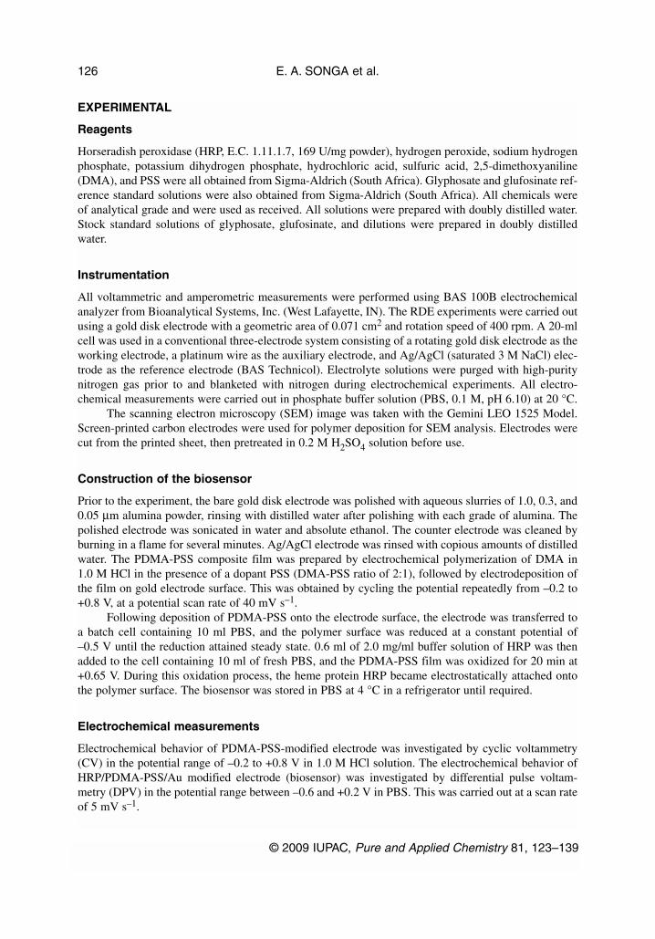
Electrochemical synthesis of PDMA-PSS resulted in a dark-green polymer film on the surface of theelectrode. CV for electrochemical synthesis of PDMA-PSS nanoparticles (Fig. 1a) showed two mainredox processes corresponding to transition from leucoemeraldine to emeraldine and also emeraldine topernigraniline states. Also, another redox process corresponding to incorporation of oligomers into thepolymer matrix or degradation products of the polymer was noticed. The electrochemical behavior ofthe PDMA-PSS/Au electrode was studied in 1.0 M HCl solution by CV, and the results are shown inFig. 1b. The number of electrons taking part in the reaction was calculated to be one. Two pairs of redoxpeaks centered at around 0.20 and 0.56 V corresponding to the transformation of leucoemeraldine baseto emeraldine salt and emeraldine salt to pernigraniline salt [32], respectively, can be observed for themodified electrode. This implies that the redox peak centered at around 0.4 V (Fig. 1a) is due to incor-poration of oligomers into the polymer matrix or degradation products of the polymer.
© 2009 IUPAC, Pure and Applied Chemistry 81, 123–139
Amperometric nanobiosensor for herbicides 127
E. A. SONGA et al.
© 2009 IUPAC, Pure and Applied Chemistry 81, 123–139
128
Fig. 1 CVs for (a) electropolymerization of PDMA-PSS; (b) electrochemical behavior of PDMA-PSS/Auelectrode.
Micrographs of PDMA and PDMA-PSS nanoparticles
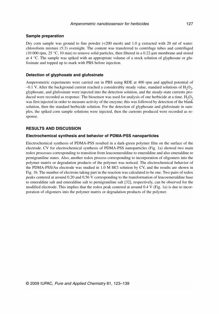
SEM images of the bulk PDMA and PDMA-PSS films electrodeposited on screen-printed electrodesare shown in Fig. 2. The SEM image of PDMA film (Fig. 2a) shows a tubular morphology expected forsubstituted polymers, while that of PDMA-PSS film (Fig. 2b) shows buds or nuclei of polymer domi-nating the surface of the electrode, giving the polymer a “cauliflower-like” appearance. The buds ap-pear to be more orderly and uniformly aligned compared to the tubular structures making up the PDMAfilm. The change in morphology when the dopant PSS was incorporated indicates the presence of PSSin the polymer matrix. It suggests that PSS played a role in aligning the DMA monomers, promoting amore ordered para-linked reaction. The bud sizes are less than 1000 nm, they vary from approximately100 to 1000 nm. The image indicates that the sizes of PDMA-PSS particles are in the nanometer range.Considering a bud size of about 200 nm consisting of a number of small particles (5–10), each particlein the bud would be 20–40 nm. This assists in classifying them as “nanoparticles”. The “nanoparticles”were, therefore, applied as a platform for biosensor construction.
Electrochemical behavior and application of the nanobiosensor
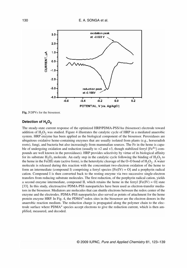
Electrochemical behavior of the nanobiosensor was investigated in PBS by DPV. The DPV results(Fig. 3) showed that the biosensor exhibits a quasi-reversible behavior with one redox couple (reduc-tion peak at –0.106 V and oxidation peak at –0.023 V) and a peak separation of 0.083 V.Characterization of the nanobiosensor at various scan rates (data not shown) indicated that the reduc-tion and oxidation peak currents increased with square root of scan rates (ν1/2) but were not propor-tional to it. The peak potentials shifted negatively with increasing scan rates. The results of these testsconfirmed quasi-reversibility of this system. The peak at –0.106 V is characteristic of the enzyme HRP,and it confirms that HRP was electrostatically attached onto the electrode surface.
© 2009 IUPAC, Pure and Applied Chemistry 81, 123–139
Amperometric nanobiosensor for herbicides 129
Fig. 2 SEM images of (a) PDMA and (b) PDMA-PSS nanoparticles (magnification of 50 000×).
Detection of H2O2
The steady-state current response of the optimized HRP/PDMA-PSS/Au (biosensor) electrode towardaddition of H2O2 was studied. Figure 4 illustrates the catalytic cycle of HRP in a mediated anaerobicsystem. HRP enzyme has been applied as the biological component of the biosensor. Peroxidases areubiquitous oxidative heme-containing enzymes that are usually isolated from plants (e.g., horseradishroots), fungi, and bacteria but also increasingly from mammalian sources. The Fe in the heme is capa-ble of undergoing oxidation and reduction (usually to +2 and +3, though stabilized ferryl [Fe4+] com-pounds are well known in the peroxidases). HRP provides selectivity by virtue of its biological affinityfor its substrate H2O2 molecule. An early step in the catalytic cycle following the binding of H2O2 tothe heme in the Fe(III) state (active form), is the heterolytic cleavage of the O–O bond of H2O2. A watermolecule is released during this reaction with the concomitant two-electron oxidation of the heme toform an intermediate (compound I) comprising a ferryl species [Fe(IV) = O] and a porphyrin radicalcation. Compound I is then converted back to the resting enzyme via two successive single-electrontransfers from reducing substrate molecules. The first reduction, of the porphyrin radical cation, yieldsa second enzyme intermediate, compound II, which retains the heme in the ferryl [Fe(IV) = O] state[33]. In this study, electroactive PDMA-PSS nanoparticles have been used as electron-transfer media-tors in the biosensor. Mediators are molecules that can shuttle electrons between the redox center of theenzyme and the electrodes. PDMA-PSS nanoparticles also served as points of attachment for the hemeprotein enzyme HRP. In Fig. 4, the PDMA0 redox sites in the biosensor are the electron donors in theanaerobic reaction medium. The reduction charge is propagated along the polymer chain to the elec-trode surface where PDMA+ species accept electrons to give the reduction current, which is then am-plified, measured, and decoded.
E. A. SONGA et al.
© 2009 IUPAC, Pure and Applied Chemistry 81, 123–139
130
Fig. 3 DPVs for the biosensor.
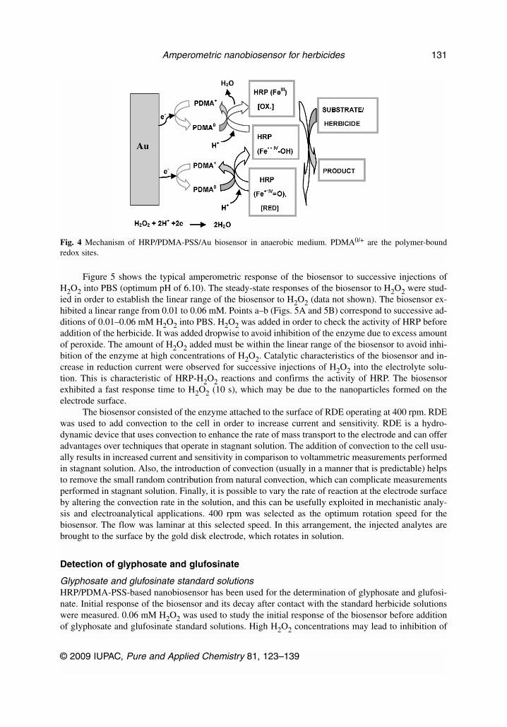
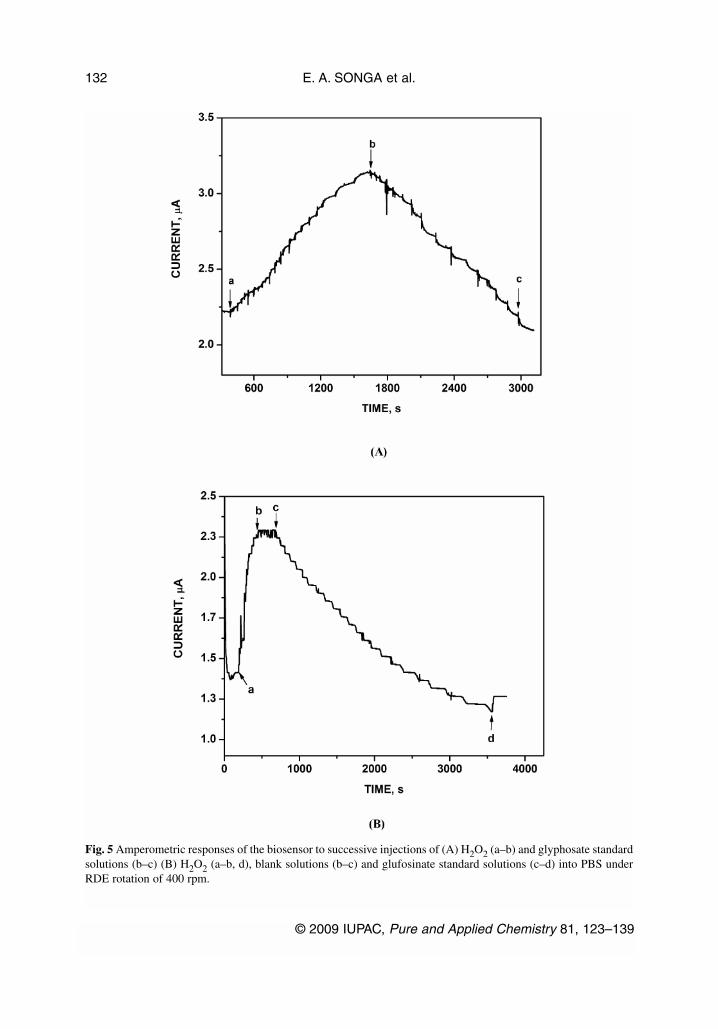
Figure 5 shows the typical amperometric response of the biosensor to successive injections ofH2O2 into PBS (optimum pH of 6.10). The steady-state responses of the biosensor to H2O2 were stud-ied in order to establish the linear range of the biosensor to H2O2 (data not shown). The biosensor ex-hibited a linear range from 0.01 to 0.06 mM. Points a–b (Figs. 5A and 5B) correspond to successive ad-ditions of 0.01–0.06 mM H2O2 into PBS. H2O2 was added in order to check the activity of HRP beforeaddition of the herbicide. It was added dropwise to avoid inhibition of the enzyme due to excess amountof peroxide. The amount of H2O2 added must be within the linear range of the biosensor to avoid inhi-bition of the enzyme at high concentrations of H2O2. Catalytic characteristics of the biosensor and in-crease in reduction current were observed for successive injections of H2O2 into the electrolyte solu-tion. This is characteristic of HRP-H2O2 reactions and confirms the activity of HRP. The biosensorexhibited a fast response time to H2O2 (10 s), which may be due to the nanoparticles formed on theelectrode surface.
The biosensor consisted of the enzyme attached to the surface of RDE operating at 400 rpm. RDEwas used to add convection to the cell in order to increase current and sensitivity. RDE is a hydro-dynamic device that uses convection to enhance the rate of mass transport to the electrode and can offeradvantages over techniques that operate in stagnant solution. The addition of convection to the cell usu-ally results in increased current and sensitivity in comparison to voltammetric measurements performedin stagnant solution. Also, the introduction of convection (usually in a manner that is predictable) helpsto remove the small random contribution from natural convection, which can complicate measurementsperformed in stagnant solution. Finally, it is possible to vary the rate of reaction at the electrode surfaceby altering the convection rate in the solution, and this can be usefully exploited in mechanistic analy-sis and electroanalytical applications. 400 rpm was selected as the optimum rotation speed for thebiosensor. The flow was laminar at this selected speed. In this arrangement, the injected analytes arebrought to the surface by the gold disk electrode, which rotates in solution.
Detection of glyphosate and glufosinate
Glyphosate and glufosinate standard solutionsHRP/PDMA-PSS-based nanobiosensor has been used for the determination of glyphosate and glufosi-nate. Initial response of the biosensor and its decay after contact with the standard herbicide solutionswere measured. 0.06 mM H2O2 was used to study the initial response of the biosensor before additionof glyphosate and glufosinate standard solutions. High H2O2 concentrations may lead to inhibition of
© 2009 IUPAC, Pure and Applied Chemistry 81, 123–139
Amperometric nanobiosensor for herbicides 131
Fig. 4 Mechanism of HRP/PDMA-PSS/Au biosensor in anaerobic medium. PDMA0/+ are the polymer-boundredox sites.
E. A. SONGA et al.
© 2009 IUPAC, Pure and Applied Chemistry 81, 123–139
132
Fig. 5 Amperometric responses of the biosensor to successive injections of (A) H2O2 (a–b) and glyphosate standardsolutions (b–c) (B) H2O2 (a–b, d), blank solutions (b–c) and glufosinate standard solutions (c–d) into PBS underRDE rotation of 400 rpm.
the enzyme and the inability to precisely monitor herbicide concentrations. Typical amperometric re-sponses of the biosensor to successive injections of glyphosate and glufosinate standard solutions, underRDE rotation of 400 rpm, are illustrated in Fig. 5. Points b–c (Fig. 5A) correspond to additions of2.0–78.0 µg L–1 glyphosate standards into PBS, while points c–d (Fig. 5B) correspond to additions of2.0–70.0 µg L–1 glufosinate standards. Points b–c in Fig. 5B correspond to addition of blank solutionsprepared the same way as the standard solutions. No significant change in current was observed whenblank solutions were added. This shows that the decrease in current or the inhibition of HRP was en-tirely due to the herbicides. After addition of H2O2 or H2O2 and blank, different concentrations ofglyphosate and glufosinate standards were injected successively into PBS and their steady-state currentsrecorded as response. It was observed that the biosensor response reduced after injection of the herbi-cide standards into the solution leading to a decrease in signal production. This shows that glyphosateand glufosinate reduced the activity of the enzyme, therefore reducing the response of the biosensor.The active site of HRP contains Fe, which is capable of undergoing oxidation and reduction. Sinceglyphosate has three reactive functional groups (amine, carboxylate, and phosphonate) that can coordi-nate strongly to transition-metal ions and has high affinity for Fe, it could have bound strongly to theFe in the active site of HRP blocking the active sites hence a decrease in biosensor response. The rapidreduction in HRP activity following the glyphosate addition (Fig. 5A) suggests that glyphosate or itsdegradation products may form insoluble stable Fe-complexes or insoluble precipitates that are not use-ful. Glyphosate has been reported to posses a high affinity and chelating capacity for Fe and other met-als, resulting in the formation of poorly soluble glyphosate-metal complexes or insoluble precipitates[30,31]. The report suggests that glyphosate or its degradation product(s) can diminish the availabilityof Fe(III), the active form of HRP, by forming insoluble complexes. Alternatively, glyphosate may in-hibit HRP activity directly through an unknown mechanism. Since HRP was electrostatically attachedto the RDE surface, no visible complexes were observed in the electrolyte solution. Glufosinate has areactive amine functional group similar to glyphosate and may also have a high affinity for Fe in the ac-tive site of HRP. It was observed that glufosinate reacted in a similar way to glyphosate and inhibitedthe activity of HRP.
Glyphosate depressed the HRP activity by 30 % within 22 min after glyphosate addition to thePBS. Glufosinate depressed the HRP activity by 46 % within 50 min. Inhibition of HRP increased asthe concentrations of the herbicides were increased. After contact of the biosensor with glyphosate for22 min, the enzyme electrode was removed from the solution, rinsed with PBS, and used for other setsof measurements. It was observed that the biosensor recovered its activity after rinsing with PBS, thiswas investigated with H2O2. When H2O2 was injected into fresh PBS under RDE rotation of 400 rpm,an increase in reduction current was again observed. After addition of H2O2 to check the activity of theenzyme, the same procedure for addition of glyphosate was repeated as before and measurements weretaken. It was observed that the same biosensor could be used for over 60 measurements. After contactof the biosensor with glufosinate for 50 min, H2O2 was immediately injected into the solution, and sud-denly an increase in current was observed. This is illustrated at point d in Fig. 5B. This shows that notall of the active sites of HRP had been blocked by glufosinate even after contact for 50 min. The sen-sor to sensor reproducibility for glyphosate and glufosinate was very good with relative standard devi-ation (RSD) values less than 10 % (triplicate measurements).
The results from this study indicate that both glyphosate and glufosinate can be classified as re-versible inhibitors. This is because the biosensor was reactivated by transferring from the inhibitor so-lution and rinsing with PBS, and one biosensor could be used for several measurements. Reversible in-hibitors bind to enzymes with noncovalent interactions such as hydrogen bonds, hydrophobicinteractions, and ionic bonds. Multiple weak bonds between the inhibitor and the active site combine toproduce strong and specific binding. Reversible inhibitors generally do not undergo chemical reactionswhen bound to the enzyme and can be easily removed by dilution or dialysis. This explains why no in-soluble precipitates were observed in the electrolyte solutions after contact of the biosensor with theherbicides. The strong and specific binding of the protein-bound Fe with glyphosate explains the high
© 2009 IUPAC, Pure and Applied Chemistry 81, 123–139
Amperometric nanobiosensor for herbicides 133
sensitivity of such Fe-containing enzyme to glyphosate. Glufosinate also bound strongly to the Fe in theprotein, reducing its activity. Glufosinate demonstrated a particular kind of reversible inhibition knownas noncompetitive inhibition. This is a form of inhibition where the binding of the inhibitor to the en-zyme reduces its activity but does not affect the binding of substrate. As a result, the extent of inhibi-tion depends only on the concentration of the inhibitor. The development of this method is a step for-ward in the analysis of glyphosate without formation of insoluble complexes that would causeinterference at sub-µg L–1 analysis.
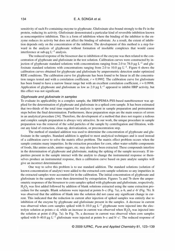
The reduced response of the biosensor due to inhibition of the enzyme was then related to the con-centration of glyphosate and glufosinate in the test solution. Calibration curves were constructed by in-jection of glyphosate standard solutions with concentrations ranging from 2.0 to 78.0 µg L–1 and glu-fosinate standard solutions with concentrations ranging from 2.0 to 10.0 µg L–1. Figure 6 shows thecalibration curves obtained for glyphosate and glufosinate by amperometric detection under optimizedRDE conditions. The calibration curve for glyphosate has been found to be linear in all the concentra-tion ranges tested and with a correlation coefficient, r = 0.9992. The calibration curve for glufosinatehas been found to have a narrow linear range but with an excellent correlation coefficient, r = 0.9998.Application of glyphosate and glufosinate as low as 2.0 µg L–1 appeared to inhibit HRP activity, butthis effect was not significant.
Glyphosate and glufosinate in samplesTo evaluate its applicability in a complex sample, the HRP/PDMA-PSS-based nanobiosensor was ap-plied for the determination of glyphosate and glufosinate in a spiked corn sample. It has been estimatedthat two-thirds of the total time required for analysis is spent in sample preparation and pretreatmentsteps before the final determination. Furthermore, these preparation steps are the main sources of errorsin an analytical procedure [34]. Therefore, the development of a method that does not require a tediousand complex sample preparation is always very attractive. In our work, the unique procedure in samplepreparation was the removal of the solid particles of the sample by centrifugation and filtration, with-out any kind of extraction, clean-up, derivatization, or preconcentration step.
The method of standard addition was used to determine the concentration of glyphosate and glu-fosinate in the samples. Standard addition is applied to most analytical techniques and is used insteadof a calibration curve to solve the matrix effect problem. The matrix effect problem occurs when thesample contains many impurities. In the extraction procedure for corn, other water-soluble componentsof foods, like amino acids, amino sugars, etc. may also have been extracted. These compounds interferein the determination of glyphosate and glufosinate, making the spiking of the sample necessary. If im-purities present in the sample interact with the analyte to change the instrumental response or them-selves produce an instrumental response, then a calibration curve based on pure analyte samples willgive an incorrect determination.
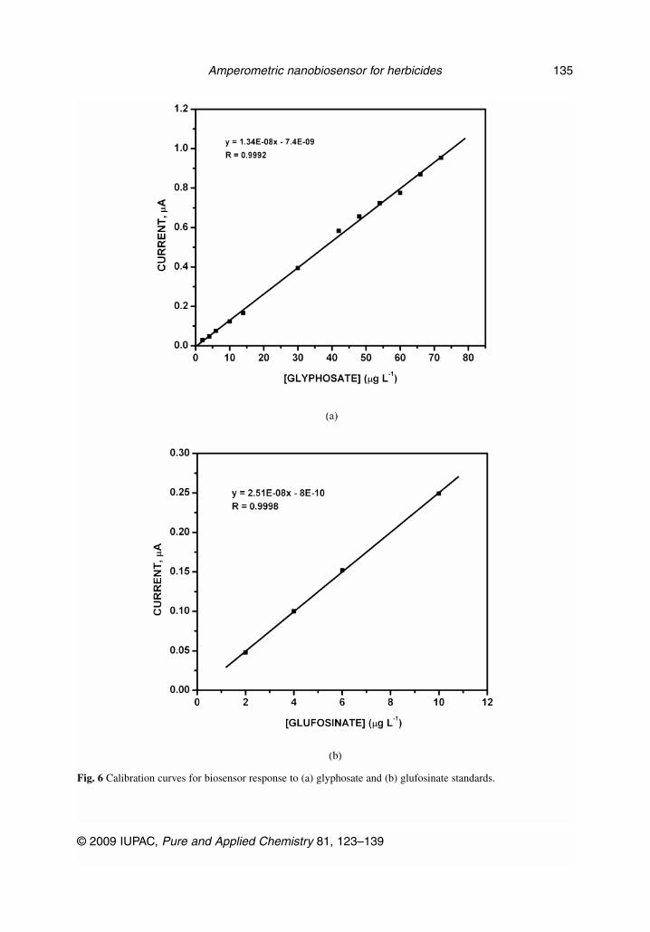
One way to solve this problem is to use standard addition. The standard solutions (solution ofknown concentration of analyte) were added to the extracted corn sample solutions so any impurities inthe extracted samples were accounted for in the calibration. The initial concentration of glyphosate andglufosinate in the samples were then determined by extrapolation. Figures 7a and 7b show the amper-ometric responses of the biosensor to corn samples spiked with glyphosate and glufosinate, respectively.H2O2 was first added followed by addition of blank solutions extracted using the same extraction pro-cedure for the sample. Blank solutions were injected at points b–c (Fig. 7a), a–b, and a'–b' (Fig. 7b). Itwas observed that the addition of blank into the solution did not cause any significant change in cur-rent. This indicated that the reduction in current after injection of spiked samples was entirely due toinhibition of the enzyme by glyphosate and glufosinate present in the samples. A decrease in currentwas observed when corn samples spiked with 0–10.0 µg L–1 glyphosate were injected into the elec-trolyte solution at points c–d, while an increase in current was observed when H2O2 was injected intothe solution at point d (Fig. 7a). In Fig. 7b, a decrease in current was observed when corn samplesspiked with 0–40.0 µg L–1 glufosinate were injected at points b–c and b'–c'. The reduced response of
E. A. SONGA et al.
© 2009 IUPAC, Pure and Applied Chemistry 81, 123–139
134
© 2009 IUPAC, Pure and Applied Chemistry 81, 123–139
Amperometric nanobiosensor for herbicides 135
Fig. 6 Calibration curves for biosensor response to (a) glyphosate and (b) glufosinate standards.
E. A. SONGA et al.
© 2009 IUPAC, Pure and Applied Chemistry 81, 123–139
136
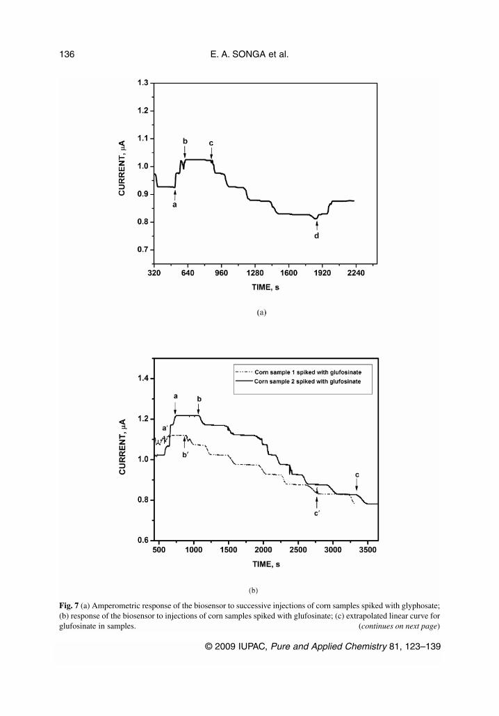
Fig. 7 (a) Amperometric response of the biosensor to successive injections of corn samples spiked with glyphosate;(b) response of the biosensor to injections of corn samples spiked with glufosinate; (c) extrapolated linear curve forglufosinate in samples. (continues on next page)
the biosensor due to inhibition of the enzyme was related to the concentration of glyphosate and glu-fosinate in the test solutions and used to obtain an extrapolated plot shown in Fig. 7c. The extrapolatedcurve was found to be linear within a concentration range of 0–4.8 µg L–1 and with a perfect correla-tion coefficient (r = 0.9999, n = 4).
The detection limits for glyphosate and glufosinate achieved by the procedure described abovewere both 0.1 µg L–1 (three times the standard deviation of replicate analyses of the blank divided bysensitivity of the biosensor toward the analyte). The sensitivity of the biosensor is defined as the slopeof the linear calibration plot. The sensor reproducibility for successive blank measurements was verygood with RSD value of 1 %. The sensitivity for glyphosate and glufosinate of the proposed methodwas much high due to their high affinity for Fe in the protein heme. The concentration of glufosinatein the corn sample (the same sample analyzed twice) analyzed was found to be 1.70 µg L–1. The con-centration of glyphosate in the corn sample was found to be 1.50 µg L–1. The absolute value of thex-intercept is the concentration of herbicide in the sample solutions, in this case 1.70 and 1.50 µg L–1.The point at zero concentration added herbicide is the reading of the unknown in the sample; the otherpoints are the readings after adding increasing amounts (spikes) of standard solution. In Fig. 7c, thereading of the unknown in the sample is 5.0 e–8 A, which corresponds to glufosinate concentration of2.0 µg L–1 in Fig. 6b.
The Environmental Protection Agency (EPA) [35] has set an MRL of glyphosate in fruits and veg-etables in the range of 0.2–5.0 mg kg–1, in soybean at 20 mg kg–1, and in drinking water at 0.7 mg L–1.The FAO has set an MRL of glyphosate in the range of 0.1–5.0 mg kg–1 for fruits and grains and MRLof glufosinate at 0.05 mg kg–1 for grains [6]. It can be seen that all the MRL values of glyphosate andglufosinate in fruits, vegetable, grains, and water are higher than the detection limits obtained in thiswork. The nanobiosensor is, therefore, sufficient in providing high sensitivity for determination ofglyphosate and glufosinate in real samples.
© 2009 IUPAC, Pure and Applied Chemistry 81, 123–139
Amperometric nanobiosensor for herbicides 137
Fig. 7 (Continued).
CONCLUSIONS
In this study, a novel HRP/PDMA-PSS-based nanobiosensor has been successfully developed and eval-uated for the detection of glyphosate and glufosinate in corn sample. The limit of detection found forthe nanobiosensor was 0.1 µg L–1, providing, therefore, sufficient sensitivity for the determination ofglyphosate and glufosinate in real samples without any preconcentration step. This method shows sev-eral advantages over other detection techniques presented in literature, including the optical methodsand ICP/MS. The nanobiosensor demonstrates its fast response time, simplicity, rapidity, sensitivity,and low cost. Our research is presently directed toward analysis of more real samples.
ACKNOWLEDGMENTS
Financial support from Third World Organization for Women in Science (TWOWS), Italy and NationalResearch Foundation (NRF), South Africa is gratefully acknowledged.
REFERENCES
1. H. Kataoka, S. Ryu, N. Sakiyama, M. Makita. J. Chromatogr., A 726, 253 (1996).2. M. G. Cikalo, D. M. Goodall, W. Mathews. J. Chromatogr., A 745, 189 (1996).3. T. Fujii, T. Ohata, M. Horinaka. Proc. Jpn. Acad. B 72, 7 (1996).4. L. P. Walsh, C. McCormick, C. Martin, D. M. Stocco. Environ. Health Perspect. 108, 769 (2000).5. S. Richard, S. Moslemi, S. Herbert, B. Nora, S. Gilles-Eric. Environ. Health Perspect. 113, 716
(2005).6. FAO. Food and Agriculture Organization. Food Standards Programme. <http://www.codexali-
mentarius.net/download/report/655/al29_24e.pdf> (2006).7. C. D. Stalikas, G. A. Pilidis. J. Chromatogr., A 872, 215 (2000).8. M. P. García de Llasera, L. Gómez-Almaraz, L. E. Vera-Avila, A. Peña-Alvarez. J. Chromatogr.,
A 1093, 139 (2005). 9. M. Ibáñez, O. J. Pozo, J. V. Sancho, F. V. J. Lopez, F. Hernández. J. Chromatogr., A 1081, 145
(2005).10. K. Sato, J. Y. Jin, T. Takeuchi, T. Miwa, K. Suenami, Y. Takekoshi, S. Kanno. J. Chromatogr., A
919, 313 (2001).11. S. Y. Chang, C. H. Liao. J. Chromatogr., A 959, 309 (2002).12. M. Molina, M. Silva. Electrophoresis 22, 1175 (2001).13. Z. Guo, Q. Cai, Z. Yang. J. Chromatogr., A 1100, 160 (2005).14. Y. Zhu, F. Zhang, C. Tong, W. Liu. J. Chromatogr., A 850, 297 (1999).15. S. Andreescu, T. Noguer, V. Magearu, J. L. Marty. Talanta 57, 169 (2002).16. G. A. Evtugyn, H. C. Budnikov, E. B. Nikolsyaka. Talanta 46, 465 (1998).17. A. L. Kukla, N. I. Kanjuk, N. F. Starodub, Y. M. Shirshov. Sens. Actuators, B 57, 213 (1999).18. T. Panasyuk-Dealaney, V. M. Mirsky, M. Ulbricht, O. S. Wolfbeis. Anal. Chim. Acta 435, 157
(2001).19. M. F. Yulaev, R. A. Sitdikov, N. M. Dmitrieva, E. V. Yazynina, A. V. Zherdev, B. B. Dzantiev.
Sens. Actuators, B 75, 129 (2001).20. R. W. Keay, C. J. McNeil. Biosens. Bioelectron. 13, 963 (1998).21. J. P. Mastin, C. A. F. Striley, R. E. Biagini, C. J. Hines, R. D. Hull, B. A. MacKenzie, S. K.
Robertson. Anal. Chim. Acta 376, 119 (1998). 22. C. Nakamura, M. Hasegawa, N. Nakamura, J. Miyake. Biosens. Bioelectron. 18, 599 (2003).23. B. Saad, M. Ariffin, M. I. Saleh. Talanta 47, 1231 (1998).24. I. Karube, Y. Nomura, Y. Arikawa. Trends Anal. Chem. 14, 295 (1995).25. I. Karube, K. Hiramoto, M. Kawarai, K. Sode. Membrane 14, 311 (1989).
E. A. SONGA et al.
© 2009 IUPAC, Pure and Applied Chemistry 81, 123–139
138
26. C. F. B. Coutinho, L. H. Mazo. Quim. Nova 28, 1038 (2005).27. P. G. Daniele, C. Stefano, E. Prenesti, S. Sammartano. Talanta 45, 425 (1997).28. R. L. Glass. J. Agric. Food Chem. 32, 1249 (1984).29. E. Morillo, T. Undabeytia, C. Maqueda, L. Madrid, M. Bejarano. Chemosphere 28, 2185 (1994).30. B. C. Barja, J. Herszage, M. dos Santos Afonso. Polyhedron 20, 1821 (2001). 31. K. A. Barrett, M. B. McBride. Environ. Sci. Technol. 39, 9223 (2005).32. A. Morrin, O. Nagmna, A. J. Killard, S. E. Moulton, M. R. Smyth, G. G. Wallace. Electroanalysis
17, 423 (2005).33. E. I. Iwuoha, I. Leister, E. Miland, M. R. Smyth, C. O. Fágáin. Anal. Chem. 69, 1674 (1997).34. C. D. Stalikas, C. N. Konidari. J. Chromatogr., A 907, 1 (2001).35. EPA. Glyphosate; Tolerance for Residues, Environmental Protection Agency, Washington, DC
(2006) <https://www.epa.gov/EPA-PEST/2006/June/Day-07/p8827.htm>.
© 2009 IUPAC, Pure and Applied Chemistry 81, 123–139
Amperometric nanobiosensor for herbicides 139