Embed Size (px)
Citation preview

Short Bowel Syndrome(SBS)
Jibran MohsinSurgical Resident
SIMS/Services Hospital, Lahore

Outline• Anatomy
• Physiology
• Definition
• Pathophysiology
• Classification(including etiology)
• Clinical Presentation
• Workup
• Treatment

Anatomy
Length of small gut* Neonate 250 cm
Adult Average 600 cm (Range 260- 800 cm)
Short Gut Syndrome 200 cm viable (Adult)>50 %-80% lost
Lifelong TPN Dependence(Minimal gut length required for life)
Adult With intact colon 60 cmWithout colon 100 cm
Infant With ileocecal valve 11 cmWithout ileoceal valve 12-25 cm
__________________________________________________________________*As a consequence, the infant and the young child have a favorable long-term prognosis compared to an adult in regards to potential intestinal growth after
intestinal resection

PhysiologyJejunum Ileum
Villi Long ShorterAbsorptive surface area Large LessTight Junction Relatively large
• epithelium more porous to larger molecules
• free and rapid flux of water and electrolytes
Tighter• permitting less flux of
water and electrolytes from the vascular space into the intestinal lumen
Water absorption Less effective More efficientAbsorption Carbohydrates, proteins, fat,
vitamins (iron-duodenum)Bile acids, vitamin B-12
GI hormones that affect intestinal motility
enteroglucagon and peptide YY
Etymology:• Duodenum (Latin: duodēnum Twelve ) (duodenum is 12 fingerbreadth long)• Jejunum (Latin: jejunus fasting )(because it was usually found to be empty after death).• Ileum (Greek: eilein to twist up tightly)

PhysiologyProximal jejunal resection is better tolerated than distal ileum
resection

Definition
Clinically defined by malabsorption, diarrhea, steatorrhea, fluid and electrolyte disturbances, and malnutrition due to ≥ 50 % (viable gut <200 cm) of structural or functional loss of small gut.

Pathophysiology
Underlying Disease
extensive bowel
resection
affects normal intestinal
physiology
alteration of intestinal
digestion and absorption
nutritional, metabolic,
and infectious consequences

Pathophysiology
• Loss of ileocecal valve– transit time is faster, and loss of fluid and nutrients
is greater
– Colonic bacteria colonize the small bowel, worsening diarrhea and nutrient loss.

Pathophysiology
Preservation of the colonMERITS DEMERITS
colonic water absorption could be increased to as much as five times its normal capacity following small bowel resection
increase incidence of urinary calcium oxalate stone formation(Oxalate is normally bound by calcium in the small bowel and thus is insoluble when it reaches the colon. After massive enterectomy, much of this calcium is bound by free intraluminal fats)
Colonic bacteria metabolize undigested carbohydrates(starch & fibres) into short-chain fatty acids, such as butyrate, propionate, and acetate. (up to 500 kcal/day)
small intestinal bacterial overgrowth.( in absence of ileocecal valve)

Natural History( 3 Phases)
• Acute Phase
• Adaption Phase
• Maintenance Phase

Acute Phase
• Starts immediately after bowel resection and lasts 1-4 months
• Ostomy output of greater than 5 L/day (as high as 6-8 L/day)
• Life-threatening dehydration and electrolyte imbalances
• Extremely poor absorption of all nutrients
• Development of hypergastrinemia and hyperbilirubinemia

Adaption Phase• Enterocyte hyperplasia + villous hyperplasia + increased crypt depth
increased surface area;• intestinal dilatation and lengthening increased capacity/Low transit
time(reduction in volume and frequency of bowel movements)_______________________________________________________
• Begins within 48 hours of resection and lasts up to 1-2 years
• ~ 90% of the bowel adaptation takes place during this phase
• Luminal nutrition is essential for adaptation and should be initiated as early as possible; parenteral nutrition is also essential throughout this period

Maintenance Phase
• Absorptive capacity of the intestine is at its maximum

Classification
Congenital versus AcquiredCONGENITAL ACQUIRED
Congenital Short Small Bowel * NEONATAL PERIOD ADULT
Necrotizing enterocolitis Crohn disease
Intestinal atresias radiation enteritis
Intestinal volvulus mesenteric vascular accidents
OLDER INFANTS and CHILDREN
Trauma
Intussusception with ischemic small-intestinal injury
recurrent intestinal obstruction
* Also associated with gastroschisis ,omphalocele and meconium peritonitis.

ClassificationStructural versus functionalSTRUCTURAL FUNCTIONAL
Any insult leading to
< 200 cm or loss of ≥ 50% of viable small bowel
Increased risk of developing SBS ( NOT ALWAYS)
PREDISPOSING FACTORS• premorbid length of small bowel
• segment of intestine that is lost,
• Age of the patient (infant tolerate better)
• Remaining length of small bowel and colon,
• Presence or absence of the ileocecal valve
Length maintained BUT function is lost
For example:
Radiation Enteritis
Cloacal Extrophy

Classification
SBS due to proximal jejunal resection versus SBS due to distal ileum resection

Clinical Presentation
• Diarrhea ± steatorrhea– dehydration and electrolytes disturbances
• Significant weight loss, fatigue, malaise, and lethargy
• Malnutrition– Mineral deficiencies (folate, iron, calcium, magnesium,
zinc)– Vitamins deficiencies (A,D, E, K , B complex esp B12)– Macro-nutrient deficiencies (CHO,Protein,fat)

Clinical Presentation• Recurrent bacterial enteritis
• Stones and related problems– Gall stones due to altered bile metabolism– Renal stones due to high oxalate
• Vomiting, bloating, GERD, gastric ulceration
• Failure to thrive
• Drug toxicities
• Bowel Obstruction (potential complication)

Clinical Presentation
• TPN related issues– Line sepsis and fulminant liver failure
• Enteral Feeding related issues– gastrostomy or nasogastric tube issues

Clinical Presentation• During the physical examination, pay close attention to these clinical signs
– Vitals
– State of hydration
– State of nutrition, as measured by a patient's weight for height and anthropometric measurements
– Signs of sepsis
– Form of nutritional therapy used in the patient (eg, central line access or enteral access)
– Specific clinical signs of nutritional deficiency
– Signs of liver disease

Workup
• Hematological and Biochemical investigations
• Radiological investigations
• Microbiological investigations
• Histopathological investigations
• Miscellaneous

WorkupHematological and Biochemical investigations
CBC • Anemia (MCH, MCHC, MCV)• Thrombocytosis/thrombocytopenia• Hyper segmented neutrophils
Albumin (half life 21 days) • good indicator of hepatic protein synthesis• indicator of overall nutritional status
Prealbumin ( 3-5 days) • indicator of acute nutritional status• monitor the efficacy of nutrition support regimens
AST/ALT • TPN induced liver failure
Electrolytes(Na, K, Cl, Zn, Ca, Mg,Cr, Se, PO4
-3)• TPN monitoring
BUN • Renal reserve• Dehydration(>20:1)• Overfed with protein
Vitamins Level
Coagulation Profile • Deranged Liver function

Workup
Hematological and Biochemical investigations Frequency*
Electrolytes, BUN, creatinine, calcium, magnesium, phosphorous Twice weekly
Comprehensive metabolic panel, CBC, triglycerides, cholesterol Weekly
Folate, vitamin B-12, vitamin E, copper, zinc, selenium Monthly
*both in initial phase and the late period or at the time of presentation for instability

Workup
Radiological investigations
Plain Chest X-ray Post CV line insertion
Plain Abdominal X-ray Suspected bowel obstruction
Barium imaging of the bowel
Abdominal USG fungal balls in the kidney (sepsis)
Renal Stones and related problems
Gall stones and related problemsLiver failure (spleen, ascites, liver texture, Portal vein flow-Duplex)
CT-Abdomen identify persistent sepsisPotential Liver/Bowel transplant
Angiography

Workup
Microbiological investigations
• Blood cultures (both central and peripheral sites)
• Urinalysis and blood culture (specifically to search for fungal infection)
• CV line tip
SOURCE of SEPSIS: • Line Sepsis,
• Gut mucosal atrophy bacterial translocation • Skin flora penetration

Workup
Histopathological investigations
• Liver biopsy(rare)– TPN related liver problems

Workup
Miscellaneous
• Upper GI endoscopy– to assess for peptic ulcer disease and possible signs
of liver disease e.g. esophageal varices, hypertensive gastropathy
• Dual radiographic absorptiometry– Bone density estimation in Metabolic Bone Disease

Treatment(aggressive MULTI DISCPLINARY APPROACH)
MEDICAL CARE• Nutrition
– Early aggressive enteral feeding– Parenteral nutrition
• Aggressive Hydration
• Electrolytes replacement
• Acid reducing agents(PPI, H2 blocker)
• Antibiotics for bacterial overgrowth
• Bile salt chelators (Cholestyramine)
• Psychosocial support
SURGICAL CARE
• Nontransplant surgery
• Small bowel ± liver transplantation

Nutrition
Early aggressive enteral nutrition* as soon as patient can tolerate
most important stimulus for intestinal adaptation
Early discontinuation of parenteral therapy.
Prolonged NPO
Gut MUCOSAL ATROPHY
BACTERIAL TRANSLOCATION
SEPSIS

Enteral Nutritioncrypt cells proliferate, leading to an increase in crypt depth and lengthening of the
intestinal villi.
nutrient receptors
production of trophic intestinal hormone and secretions
production of digestive enzymes
Facilities bile flow prevent cholestasis

Common Concerns with enteral nutrition
Fat Intolerance
(increase in stool output with the appearance of fecal-reducing substances)
• LCFA(high energy density) better tolerated• carbohydrates parenterally and providing fats enterally• Continuous enteral feeds is better tolerated than bolus feeds
High stool volume and frequency
• DON’T STOP or substantially lower the volume and frequency of feeds in response to changes in stool volume
(as long as it does not compromise the child's hydration, acid base balance, and serum electrolyte levels)RATIONALE:Most of fluid and electrolytes disturbances can be corrected by IV formula• Decreasing amount of carbohydrates within the enteral feeds • decreasing the volume and concentration of feeds• Avoid fibers
GERD, Vomiting Decreasing either the volume or rate of feeds

Early Aggressive Enteral Nutrition
Elemental/modular formulas RATIONALE
CHO mixture of monosaccharides and polysaccharides is preferred to disaccharides
in order to limit osmotic load
Fat Medium-chain triglycerides (MCT) readily absorbed in the stomach and proximal small bowel improving fat and total energy absorption
long-chain triglycerides (LCT) • prevent essential fatty acid deficiency• 10% of the patient's energy needs• trophic effect on the intestinal mucosa
Protein Oligopeptide formulas are better absorbed than elemental amino acid formulas
di-tripeptide absorption exceeds that of amino acids.
Concentration
either one-fourth or one-half strength increasing in volume before increasing energy density/conc.

Factors predictive of achieving independence from TPN
• Residual Bowel length
• Intact colon
• Intact ileocecal valve
• Healthy (versus diseased) residual small gut
• Jejunal resection (versus ileal resection)
• Bacterial overgrowth
• Dysmotility
• Teduglutide– enhancement or
restoration of the structural and functional integrity of the remaining intestine

Micronutrients
Supplementation Rationale/Route
Water soluble Vit A, Vit D, and Vit E In case of significant steatorrhea (IV)
Calcium Oral route for bone mineralization and growth(Dual energy x-ray absorptiometry (DEXA) scan)
Vitamin K Not required ( synthesize by enteric bacteria)(monitored by PT time)
deficiency of water-soluble vitamins is rare except Vitamin B12
Vitamin B12 IV on a monthly basis or as a nasal gel
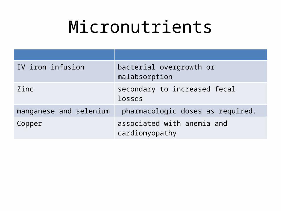
Micronutrients
IV iron infusion bacterial overgrowth or malabsorption
Zinc secondary to increased fecal losses
manganese and selenium pharmacologic doses as required.
Copper associated with anemia and cardiomyopathy

Bacterial overgrowth• Predisposing factor
– Absence of ileocecal valve– Dysmotilty of residual gut
• Manifestations– deconjugation of bile salts and depletion of bile salt stores– Vitamin B12 deficiency Pernicious anemia– carbohydrate malabsorption worsening of osmotic diarrhea– metabolic lactic acidosis CNS disturbances– Dehydration
• Treatment– metronidazole alternating with either kanamycin or oral gentamicin

• Codeine and loperamide– to slow intestinal transit time– DEMERIT: leads to bacterial overgrowth
• Octreotide– Rarely used for concerns of the effect on growth and
worsening cholestatic liver disease
• Glutamine and CCK– No selective advantage

Surgical Care
• Diverting ileostomy– In case of malfunctional colon
• Central Venous line insertion
• PEG (percutaneous endoscopic gastrotomy)
• Reversal of stoma– To capitalize on absorptive capacity of all residual gut

Surgical Care
To slow transit time*• Segmental reversal of small
bowel
• Interposition of segment of colon between segments of small gut
• Construction of small gut valves
• Retrograde electrical pacing of small gut
To increase gut length/area
• LILT ( Longitudinal Intestinal Lengthening and tailoring) or Bianchi procedure
• STEP (Serial transerve enteroplasty procedure)

LILT (Bianchi1980) Procedure
Separation of dual blood supply Longitudinal division Iso-peristaltic end-to-end anastomosis

STEP
Serial applications of an intestinal stapling device, with firings oriented perpendicular to long axis of intestine
By 2013, amongst 111 patients operated 47 % cases had achieved enteral autonomy by 21 months.
1st performed in 2003 on 2-year-old baby who had been born with gastroschisis

Intestinal Transplantation
• Indications– Life threatening complications due to intestinal
failure or long term TPN• Impending or overt liver failure• Thrombosis of major central veins• Frequent episodes of catheter-related sepsis• Frequent episodes of severe dehydration.

Available at surgicalpresentations