Embed Size (px)
Citation preview

Presented by-
Ms. Nidhi Shukla
M.Sc. Nursing 1st year
Prenatal or antenatal development is the process in which embryo or fetus gestates during pregnancy from
fertilization until birth.
It is also known as fetal development or embryology.
Development biology is the study of the sequences of events from the fertilization of a secondary oocyte by a
sperm cell to the formation of an adult organism.

Age of the fetus –
Gestational age is the duration of pregnancy calculated from the first day of last menstrual period (LMP).
It is greater than the post conception (fertilization) age by 2 weeks.
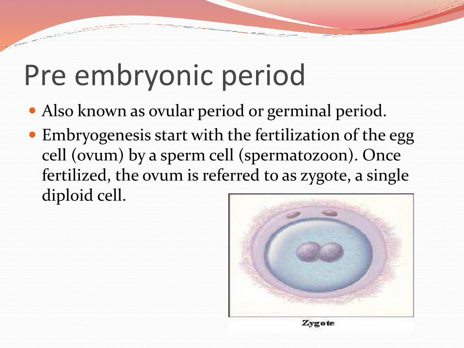
Pre embryonic period Also known as ovular period or germinal period.
Embryogenesis start with the fertilization of the egg cell (ovum) by a sperm cell (spermatozoon). Once fertilized, the ovum is referred to as zygote, a single diploid cell.
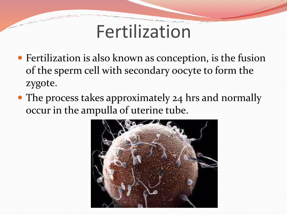
Fertilization Fertilization is also known as conception, is the fusion
of the sperm cell with secondary oocyte to form the zygote.
The process takes approximately 24 hrs and normally occur in the ampulla of uterine tube.
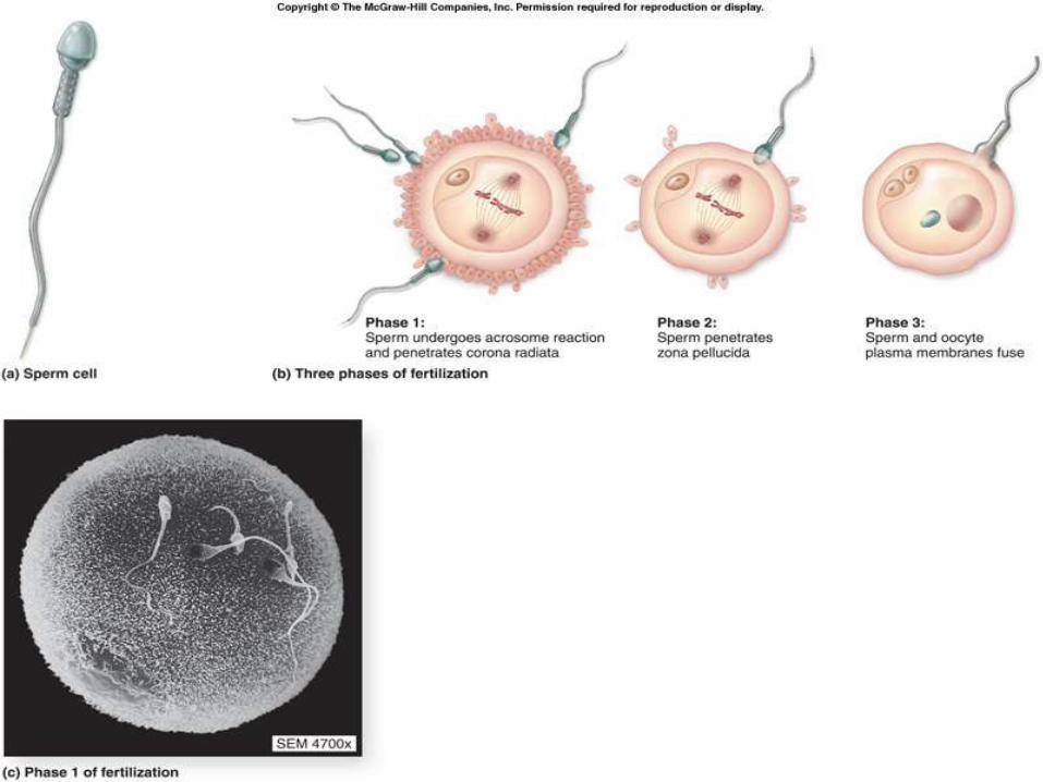
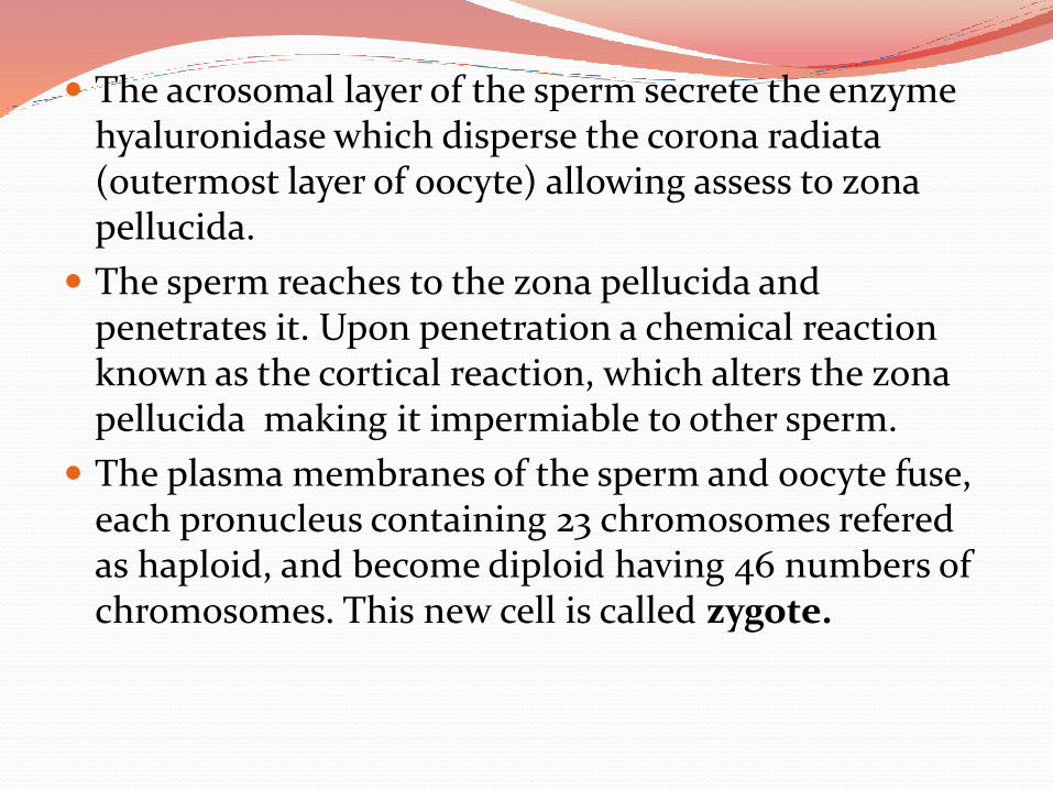
The acrosomal layer of the sperm secrete the enzyme hyaluronidase which disperse the corona radiata(outermost layer of oocyte) allowing assess to zonapellucida.
The sperm reaches to the zona pellucida and penetrates it. Upon penetration a chemical reaction known as the cortical reaction, which alters the zonapellucida making it impermiable to other sperm.
The plasma membranes of the sperm and oocyte fuse, each pronucleus containing 23 chromosomes referedas haploid, and become diploid having 46 numbers of chromosomes. This new cell is called zygote.

During the first week the zygote travels along the uterine tube towards the uterus, the zona pellucidasurrounds the zygote. It nourished by glycogen secreted by globlet cell of uterine tube and later the secretory cells of uterus.
The zygote undergoes mitotic cellular replication refered as cleavage, resulting into the formation of smaller cells known as blastomeres.
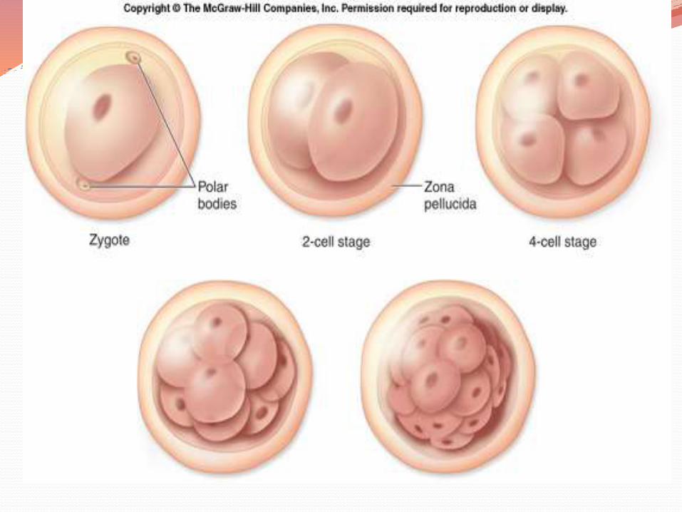
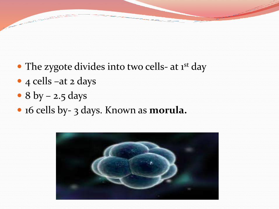
The zygote divides into two cells- at 1st day
4 cells –at 2 days
8 by – 2.5 days
16 cells by- 3 days. Known as morula.
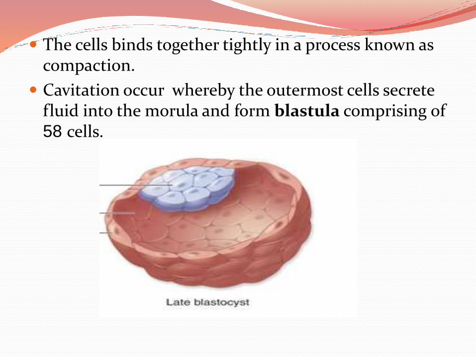
The cells binds together tightly in a process known as compaction.
Cavitation occur whereby the outermost cells secrete fluid into the morula and form blastula comprising of 58 cells.
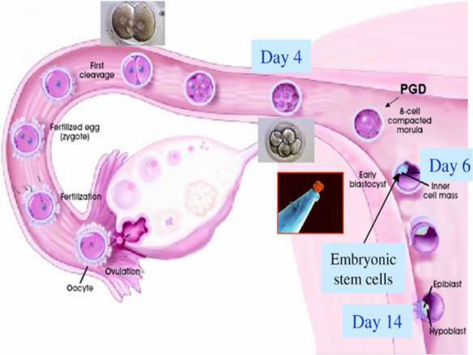
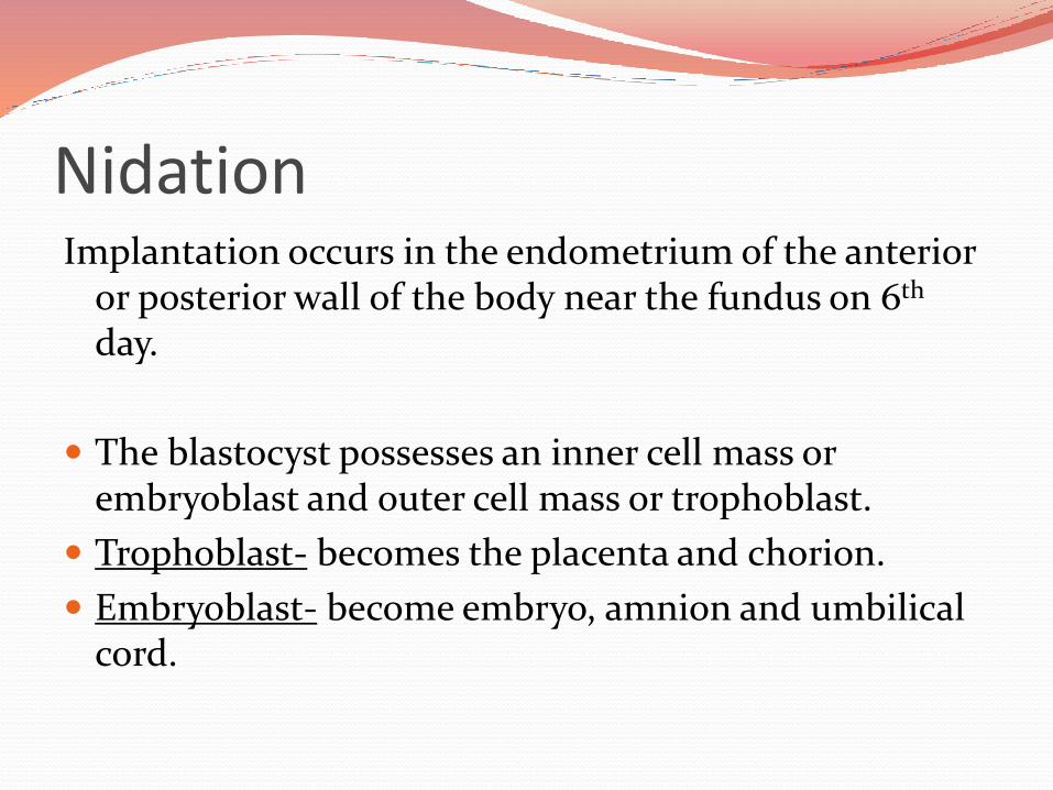
Nidation Implantation occurs in the endometrium of the anterior
or posterior wall of the body near the fundus on 6th
day.
The blastocyst possesses an inner cell mass or embryoblast and outer cell mass or trophoblast.
Trophoblast- becomes the placenta and chorion.
Embryoblast- become embryo, amnion and umbilical cord.
Trophoblast
Differentiate into two layers
syncytiotrophoblast
cytotrophoblast
EmbryoblastDevelops the embryo, and differentiate into two types of
cells-
Epiblast- epiblast have three layers, which forms the particular parts of the embryo. The first appearance of these layers, collectively known as the primitivestreak is around 15 days.
Hypoblast- the hypoblast cell migrate along with inner cytotrophoblastic lining secreting extracellular tissue which becomes the yolk sac.
Changes or development by weeks of gestation
1. Pre embryonic period-Week 1-2 – no developments occurs since fertilization
hasn’t actually occurred.
Week 3- from 15-21 days, embryonic 5-7 days.
Fertilization occur and form zygote.
The embryo hatches from its protein shell and perform implantation (5-6 days).
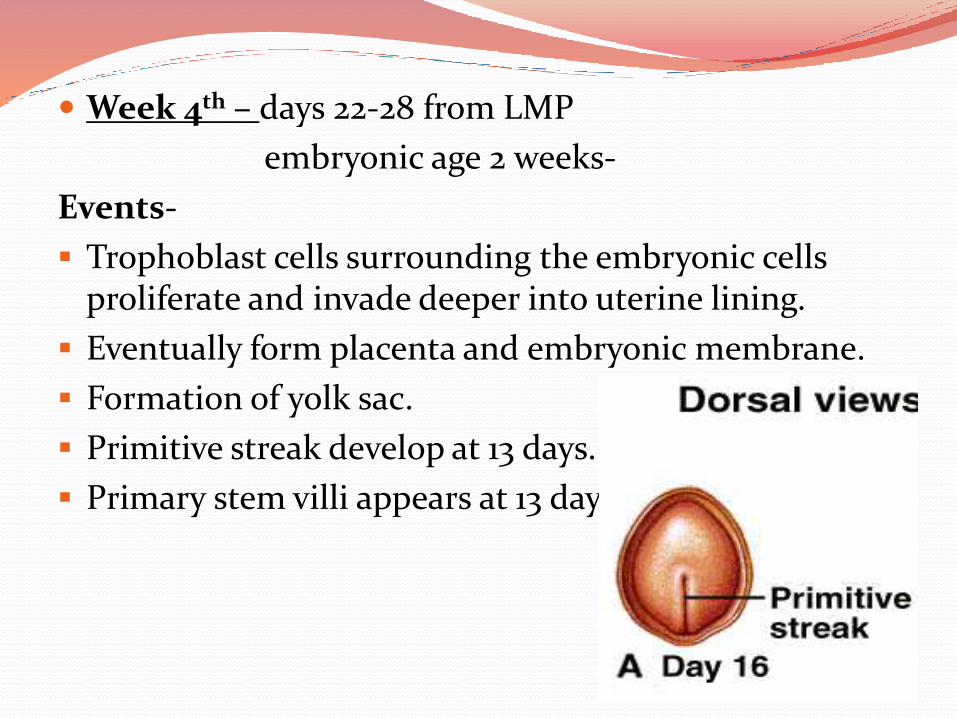
Week 4th – days 22-28 from LMP
embryonic age 2 weeks-
Events-
Trophoblast cells surrounding the embryonic cells proliferate and invade deeper into uterine lining.
Eventually form placenta and embryonic membrane.
Formation of yolk sac.
Primitive streak develop at 13 days.
Primary stem villi appears at 13 days
Week 5
Gestational age: 4 weeks
Embryonic age: Week no. 3
A notochord forms in the center of the embryonic disk. (day 16 of fert.
gastrulation commences. (day 16 of fert.)
A neural groove (future spinal cord)forms over the notochord with a brain bulge at one end. Neuromeres appear. (day 18 of fert.)
Somites, the divisions of the future vertebra, form. (day 20 of fert.)
Primitive heart tube is forming. Vasculature begins to develop in embryonic disc. (day 20 of fert.


Week 6- gestational age-5 week
embryonic age 4 weeks
Events –
Embryo measures 4 mm
The heart bulge, n begins to beat in a regular rhythem.
The neural tube closes.
Arm buds and tail are visible.
Pulmonary primordium appear
Hepatic plate appear
Buccopharyngeal membrane ruptures. This form the future mouth.
Anterior and posterior horns differentiate in the spinal cord.

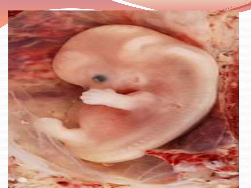
Week 7-
embryonic age 5 week
Events-
Length is 9 mm
Lens pits and optic cups develops
Nasal pits form
Brain divides into 5 vessicles including the early telencephalon.
Leg buds form.
The metanephros, precursor of kidney start to develop.
Stomach differentiation begins.
Week 8-
The embryo measures 13 mm (1/2 inch) in length.
Lungs begin to form.
The brain continues to develop.
Arms and legs have lengthened with foot and hand areas distinguishable.
The hands and feet have digits, but may still be webbed.
The gonadal ridge begins to be perceptible.
The lymphatic system begins to develop.
Main development of external genitalia starts.
Week 9-
The embryo measures 18 mm (3/4 inch) in length.
Fetal heart tone (the sound of the heart beat) can be heard using doppler
Nipples and hair follicles begin to form.
Location of the elbows and toes are visible.
Spontaneous limb movements may be detected by ultrasound.
All essential organs have at least begun.
The vitelline duct normally closes
Fetal development From the 10 weeks of gestation (8th week of
embryogenic) the developing organism is called fetus.
All the major structures are already formed in the fetus but they continue to grow.
Week 10 -12 Embryo measures 30–80 mm (1.2–3.2 inches) in length. Intestines rotate. Facial features continue to develop. The eyelids are more developed. The external features of the ear begin to take their final
shape. The head comprises nearly half of the fetus' size. The face is well formed The eyelids close and will not reopen until about the 28th
week. Tooth buds appear. The fetus can make a fist with its fingers. Genitals appear well differentiated. Red blood cells are produced in the liver
Week 13-16-
The fetus reaches a length of about 15 cm (6 inches).
A fine hair called lanugo develops on the head.
Fetal skin is almost transparent.
More muscle tissue and bones have developed, and the bones become harder.
Sucking motions are made with the mouth.
Meconium is made in the intestinal tract.
The liver and pancreas produce fluid secretions.
From week 13, sex prediction
At week 15, main development of external genitalia is finished
Abdominal wall closes.
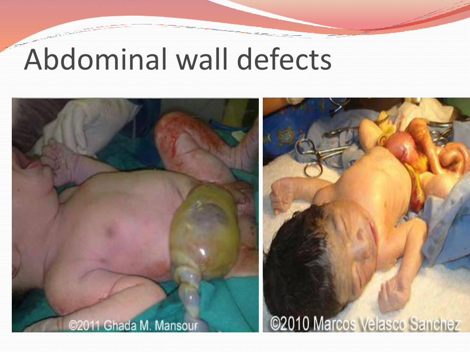
Abdominal wall defects
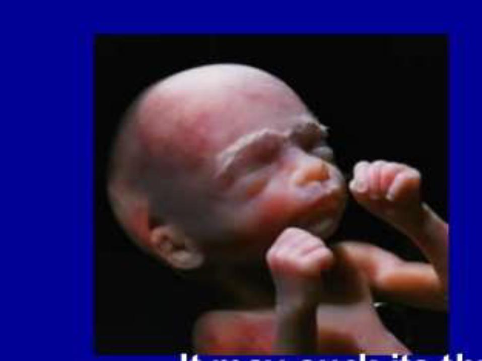
Week 17-21 The fetus reaches a length of 20 cm (8 inches).
Lanugo covers the entire body.
Eyebrows and eyelashes appear.
Nails appear on fingers and toes.
The fetus is more active with increased muscle development.
"Quickening" usually occurs (the mother and others can feel the fetus moving).
The fetal heartbeat can be heard with a stethoscope.
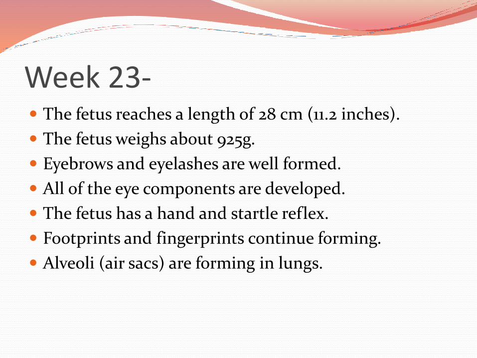
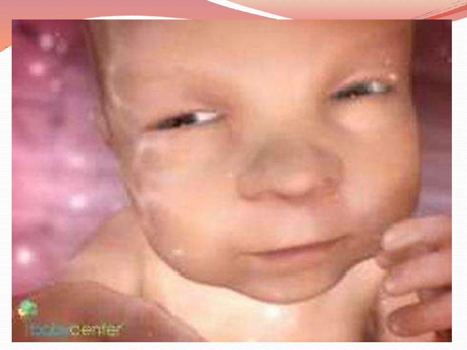
Week 23- The fetus reaches a length of 28 cm (11.2 inches).
The fetus weighs about 925g.
Eyebrows and eyelashes are well formed.
All of the eye components are developed.
The fetus has a hand and startle reflex.
Footprints and fingerprints continue forming.
Alveoli (air sacs) are forming in lungs.

Week 27 The fetus reaches a length of 38 cm (15 inches).
The fetus weighs about 1.2 kg.
The brain develops rapidly.
The nervous system develops enough to control some body functions.
The eyelids open and close.
The respiratory system, while immature, has developed to the point where gas exchange is possible.
Week 31 The fetus reaches a length of about 38–43 cm (15–
17 inches).
The fetus weighs about 1.5 kg (3 lb 0 oz).
The amount of body fat rapidly increases.
Rhythmic breathing movements occur, but lungs are not fully mature.
Thalamic brain connections, which mediate sensory input, form.
Bones are fully developed, but are still soft and pliable.
The fetus begins storing a lot of iron, calcium and phosphorus
Week 35 The fetus reaches a length of about 40–48 cm (16–
19 inches).
The fetus weighs about 2.5 to 3 kg (5 lb 12 oz to 6 lb 12 oz).
Lanugo begins to disappear.
Body fat increases.
Fingernails reach the end of the fingertips.
A baby born at 36 weeks has a high chance of survival, but may require medical interventions
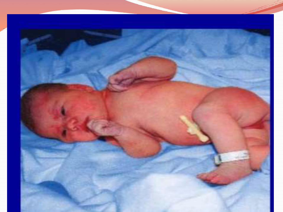
Week 36-40 The fetus is considered full-term at the end of the 39th
week of gestational age.
It may be 48 to 53 cm (19 to 21 inches) in length.
The lanugo is gone except on the upper arms and shoulders.
Fingernails extend beyond fingertips.
Small breast buds are present on both sexes.
Head hair is now coarse and thickest

Thank you