Embed Size (px)
Citation preview
DENTALEMERGENCIES OF ROOT CANAL THERAPY
BY A.Selva Arockiam 3rd year BDS CSICDSR MADURAI
Endodontic mishaps or procedural accidents are those unfortunate accidents that happen during treatment.
some owing to inattention to detail.
others being totally unpredictable.
Access related
Treating the wrong tooth
Missed canals
Damage to existing restoration
Access cavity perforations
Crown fractures
Instrument related
Ledge formation
Cervical canal perforations
Midroot perforations
Apical perforations
Separated instruments and foreign objects
Canal blockage
Obturation related
Over- or underextended root canal fillings
Nerve paresthesia
Vertical root fractures
Miscellaneous
Post space perforations
Irrigant related mishaps
Tissue emphysema
Instrument aspiration and ingestion

ACCESS-RELATED MISHAPS
Treatment of the Wrong Tooth
Treatment of the wrong tooth can be so easily prevented.
One should make sure through inquiry, testing, examining, and radiography that
one has confirmed which tooth requires treatment and then mark it with a felt-tip pen.
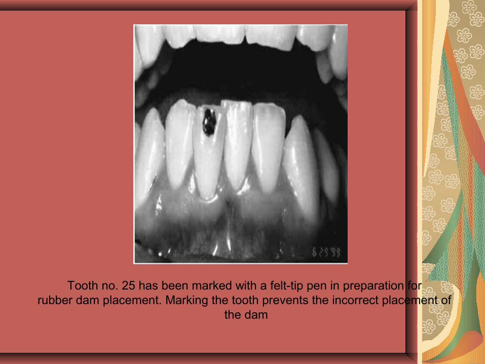
Tooth no. 25 has been marked with a felt-tip pen in preparation for rubber dam placement. Marking the tooth prevents the incorrect placement of
the dam
MISSED CANAL
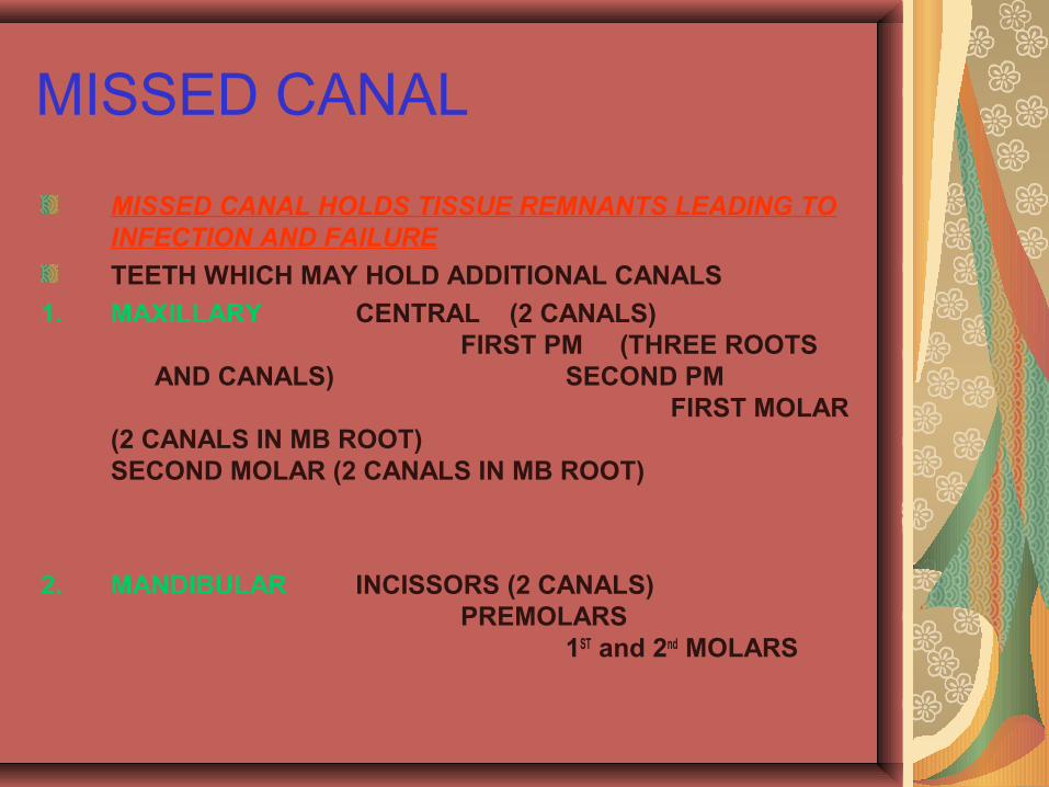
MISSED CANAL HOLDS TISSUE REMNANTS LEADING TO INFECTION AND FAILURE
TEETH WHICH MAY HOLD ADDITIONAL CANALS
1. MAXILLARY CENTRAL (2 CANALS) FIRST PM (THREE ROOTS
AND CANALS) SECOND PM FIRST MOLAR
(2 CANALS IN MB ROOT)SECOND MOLAR (2 CANALS IN MB ROOT)
2. MANDIBULAR INCISSORS (2 CANALS)PREMOLARS
1ST and 2nd MOLARS
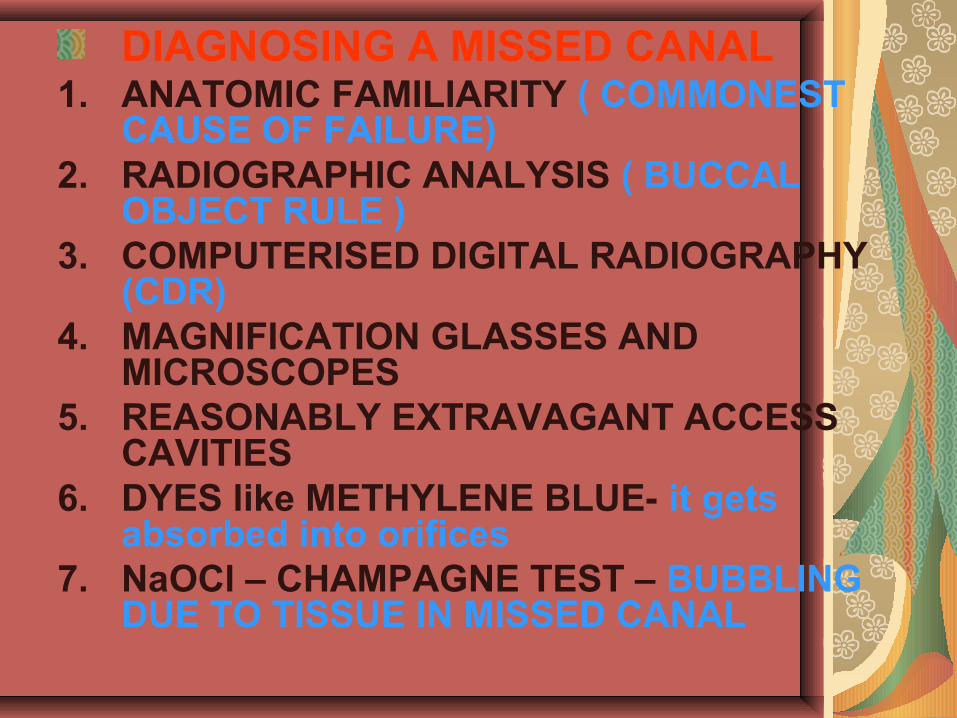
DIAGNOSING A MISSED CANAL1. ANATOMIC FAMILIARITY ( COMMONEST
CAUSE OF FAILURE)2. RADIOGRAPHIC ANALYSIS ( BUCCAL
OBJECT RULE )3. COMPUTERISED DIGITAL RADIOGRAPHY
(CDR)4. MAGNIFICATION GLASSES AND
MICROSCOPES5. REASONABLY EXTRAVAGANT ACCESS
CAVITIES6. DYES like METHYLENE BLUE- it gets
absorbed into orifices7. NaOCl – CHAMPAGNE TEST – BUBBLING
DUE TO TISSUE IN MISSED CANAL
Missed Canals
One must be diligent and prepare adequate occlusal access!
Always expect there will be an extra canal.
Magnification with either telescopic lenses or a surgical microscope is indispensable.
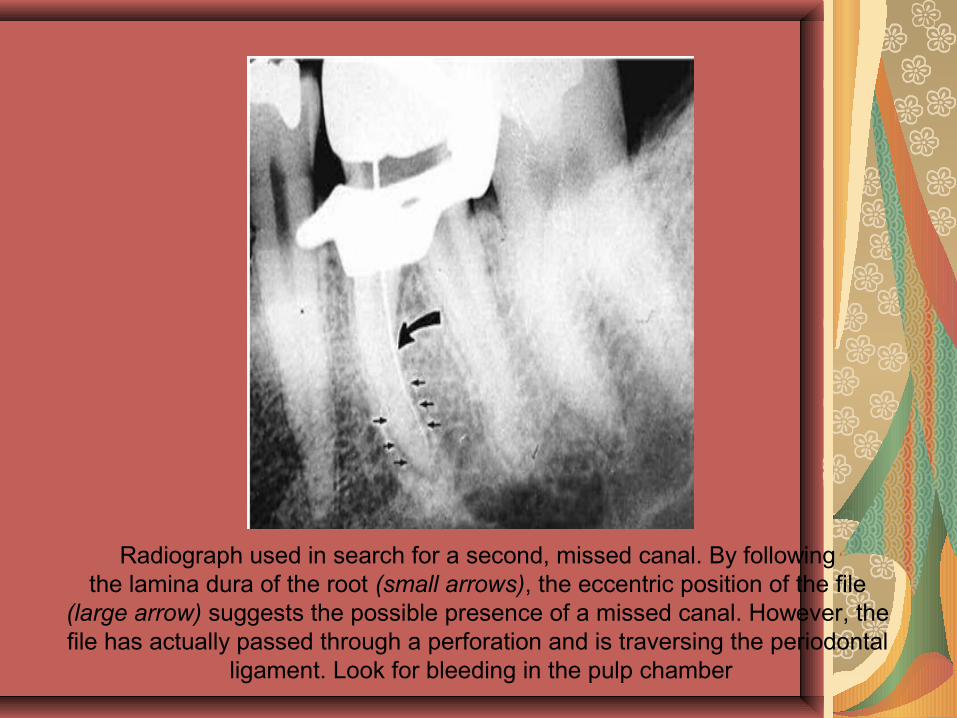
Radiograph used in search for a second, missed canal. By following the lamina dura of the root (small arrows), the eccentric position of the file
(large arrow) suggests the possible presence of a missed canal. However, the file has actually passed through a perforation and is traversing the periodontal
ligament. Look for bleeding in the pulp chamber
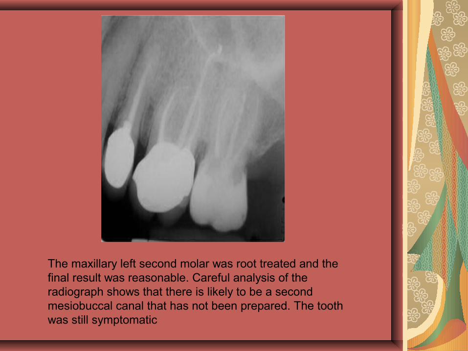
The maxillary left second molar was root treated and the final result was reasonable. Careful analysis of the radiograph shows that there is likely to be a second mesiobuccal canal that has not been prepared. The tooth was still symptomatic
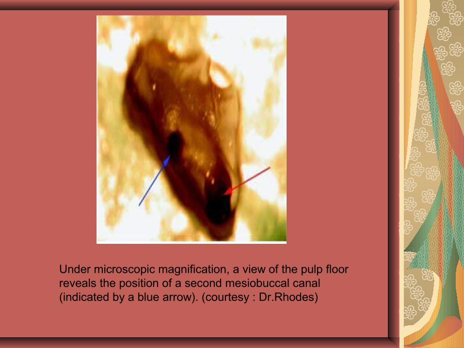
Under microscopic magnification, a view of the pulp floor reveals the position of a second mesiobuccal canal (indicated by a blue arrow). (courtesy : Dr.Rhodes)
Damage to an Existing Restoration
Porcelain crowns are the most susceptible to chipping and fracture.When one is present, use a water-cooled, smooth diamond point and do not force the stone. Let it cut its own way. Also, donot place a rubber dam clamp on the gingiva of any porcelain or porcelain-faced crown.
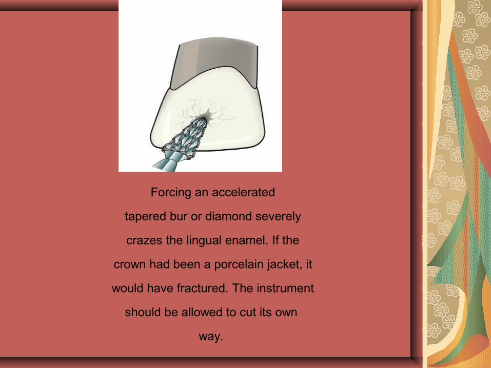
Forcing an accelerated
tapered bur or diamond severely
crazes the lingual enamel. If the
crown had been a porcelain jacket, it
would have fractured. The instrument
should be allowed to cut its own
way.
Access Cavity Perforations
Sign of an access cavity perforation is blood in the cavity or the patient complaining of a taste of NaOCl.
This most frequently happens in the floor of molar preparations when one is searching for a third or fourth canal.
Access Cavity Perforations
The site of the perforation must be found, the floor of the preparation cleansed, the bleeding stopped, and mineral trioxide aggregate (MTA) applied to the perforation .
Because it takes MTA more than 3 hours to set, it should be covered with a fast-setting cement.
The other canal orifices should be protected by placing paper points or an instrument in the canals to prevent blockage.
In the event MTA cannot be immediately applied, it is best to stop the bleeding, place calcium hydroxide over the “wound,” place a good temporary filling, and set an appointment with the patient.
The perforation area will be dry at the next appointment.
Then MTA can be applied and treatment continued.
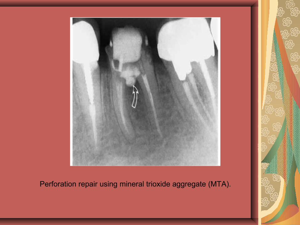
Perforation repair using mineral trioxide aggregate (MTA).
Crown Fractures
Preparing an endodontic access cavity in a tooth, par-ticularly a molar or premolar with a large restoration, materially weakens the crown. Infrequently the crown fractures, either during preparation or at a subsequent appointment.One of the frequent causes is failure to relieve the occlusion.
If the fracture is chisel shaped and a cusp breaks off down to the periodontal ligament, the tooth can usually be salvaged.
If the fracture extends through the pulp chamber and down into the root, however, the case is hopeless and the tooth should be extracted.
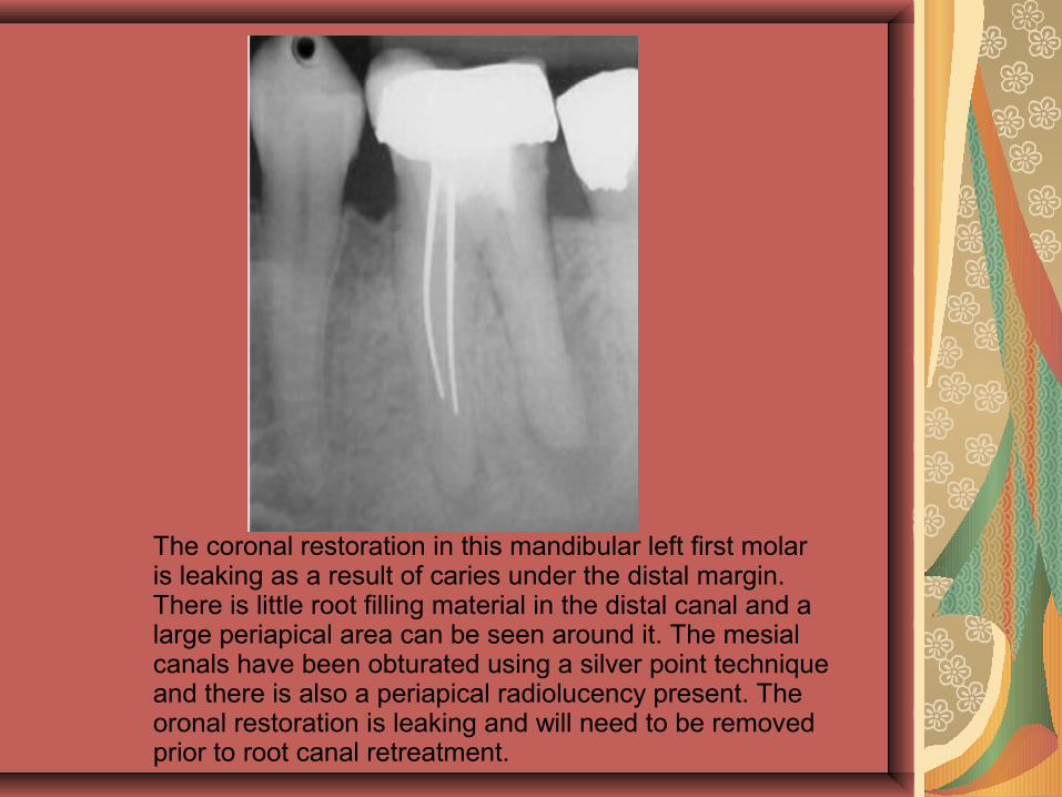
The coronal restoration in this mandibular left first molar is leaking as a result of caries under the distal margin. There is little root filling material in the distal canal and a large periapical area can be seen around it. The mesial canals have been obturated using a silver point technique and there is also a periapical radiolucency present. The oronal restoration is leaking and will need to be removed prior to root canal retreatment.
Improper healing: Curved or narrow canals that were not treated during the initial treatmentComplicated canals went undetected during the initial treatmentThe crown or restoration did not prevent saliva from contaminating the inside of the tooth.
New problems in a successfully treated toothNew decay can expose a root canal filling material, causing infection.A cracked or loose filling or crown can expose the tooth to new infection.
INSTRUMENTATION-RELATED MISHAPS
INSTRUMENTATION-RELATED MISHAPS
Overzealous cleaning and shaping have lead to many mishaps, zip-ping and perforations among them
Many of these iatral accidents begin with ledging during canal preparation.
Ledge Formation
A LEDGE IS AN INTERNAL TRANSPORATATION OF THE CANAL FORMED WHEN WE WORK SHORT OF THE WORKING LENGTH
Ledge Formation
Many ledges are caused from inadequate access.The operator does not retain complete control over the direction of the tip of the instrument and it gouges into the wall of the canal starting a ledge . Newer instruments with noncutting tips have materially reduced this problem. The rounded tip does not cut into the wall but slips by it.
Curved roots are another impediment in canal preparation.
This is especially true near the apex of maxillary lateral incisors and the palatal root of maxillary first molars.
Small, flexible instruments with noncutting tips negotiate these curves, but larger, stiffer instruments start a ledge that can develop into a perforation.
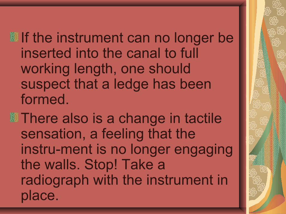
If the instrument can no longer be inserted into the canal to full working length, one should suspect that a ledge has been formed.There also is a change in tactile sensation, a feeling that the instru-ment is no longer engaging the walls. Stop! Take a radiograph with the instrument in place.
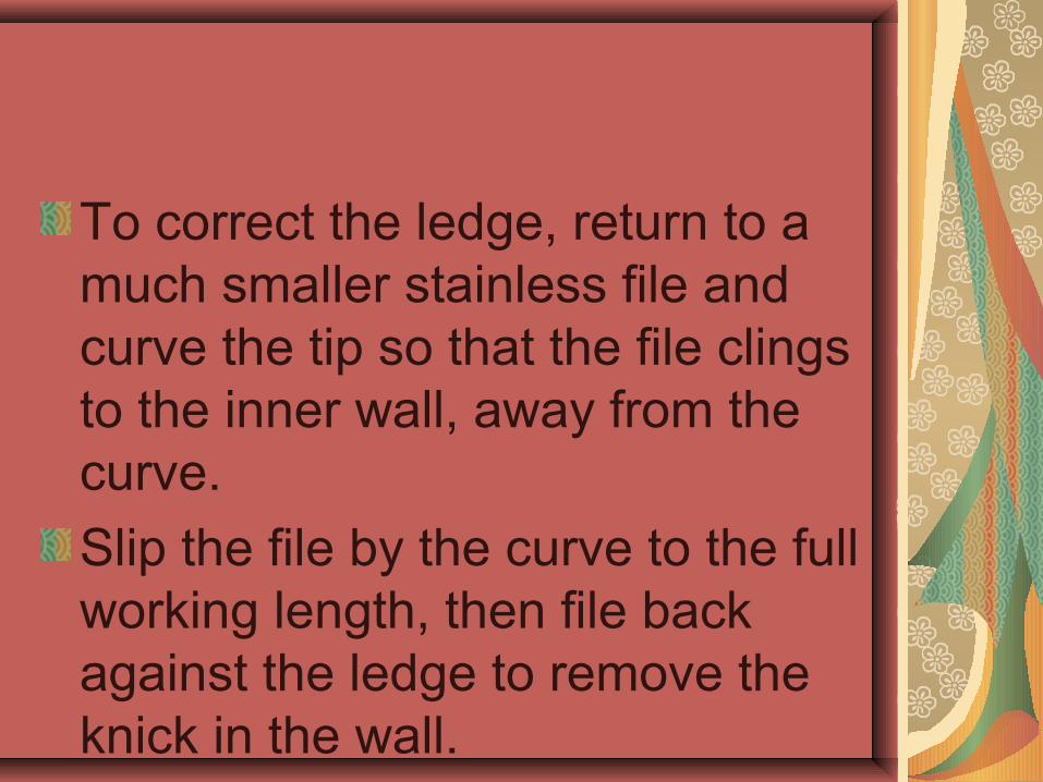
To correct the ledge, return to a much smaller stainless file and curve the tip so that the file clings to the inner wall, away from the curve.
Slip the file by the curve to the full working length, then file back against the ledge to remove the knick in the wall.
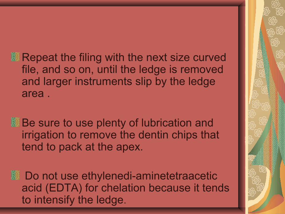
Repeat the filing with the next size curved file, and so on, until the ledge is removed and larger instruments slip by the ledge area .
Be sure to use plenty of lubrication and irrigation to remove the dentin chips that tend to pack at the apex.
Do not use ethylenedi-aminetetraacetic acid (EDTA) for chelation because it tends to intensify the ledge.
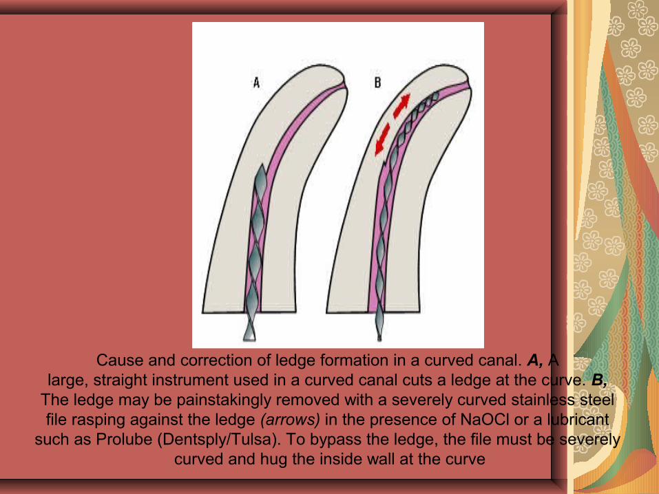
Cause and correction of ledge formation in a curved canal. A, A large, straight instrument used in a curved canal cuts a ledge at the curve. B,
The ledge may be painstakingly removed with a severely curved stainless steel file rasping against the ledge (arrows) in the presence of NaOCl or a lubricant
such as Prolube (Dentsply/Tulsa). To bypass the ledge, the file must be severely curved and hug the inside wall at the curve
Incomplete Canal Debridement
Failure to clean the entire root canal system will often result in failure. Incomplete treatment will leave pulp residue that can serve as a reservoir for bacteria that can initiate or perpetuate periradicular lesions.
Silver Points
Failures of silver points are usually associated with leakage and corrosion.The inability of silver points to seal irregular shaped canals allows leakage of the tissue fluids into the canal. Contact of these fluids with the silver point's result in the formation of corrosive products such as silver sulfates and silver carbonates, which can damage the periradicular tissues
Silver PointsSILVER POINTS FAIL DUE TO CHRONIC LEAKAGE DENTIST SHOULD KEEP IN MIND THAT THE APICAL 2-3mm IS PREPARED PARALLEL AND THE REST OF THE CANAL IS FLARED.STEPS AND TECHNIQUES:
1. ACCESS : PROPER ACCESS IS MADE BY REMOVING THE CORE.
2. PLIERS REMOVAL : STIEGLITZ PLIERS ARE USED USING “FULCRUM MECHANICS”
3. ULTRASONIC REMOVAL : CAN BE USED BOTH DIRECTLY AND INDIRECTLY
4. SOLVENTS & CHELATORS: MAY BE USED WITH SMALL SIZED INSTRUMENTS
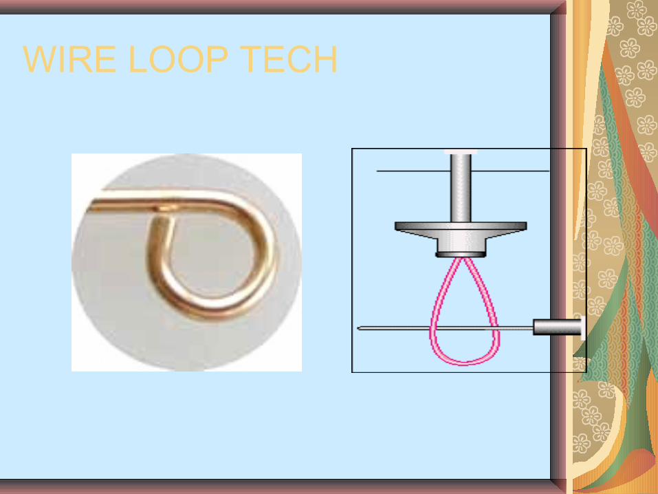
5. BY PASS THE INSTRUMENT &THEN REMOVE WITH H FILE.6. ULTRA SONIC + H FILE7. WIRE LOOP TECHNIQUE : 26 GAUGE NEEDLE WITH INBUILT
WIRE LOOP USED TO ENGAGE & REMOVE SILVER POINT.
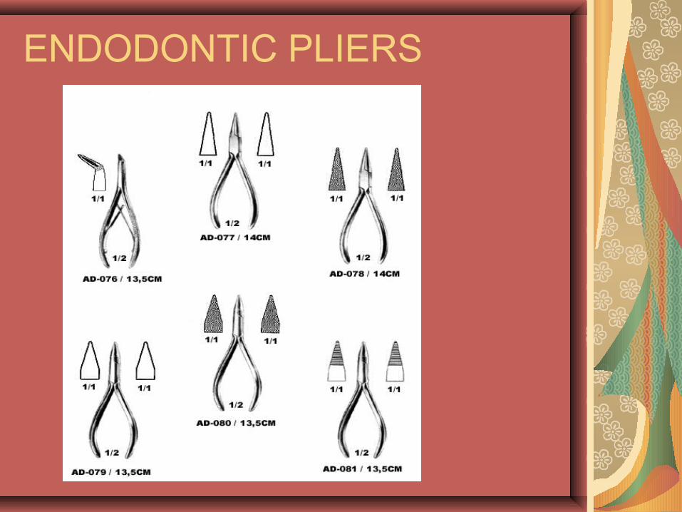
ENDODONTIC PLIERS
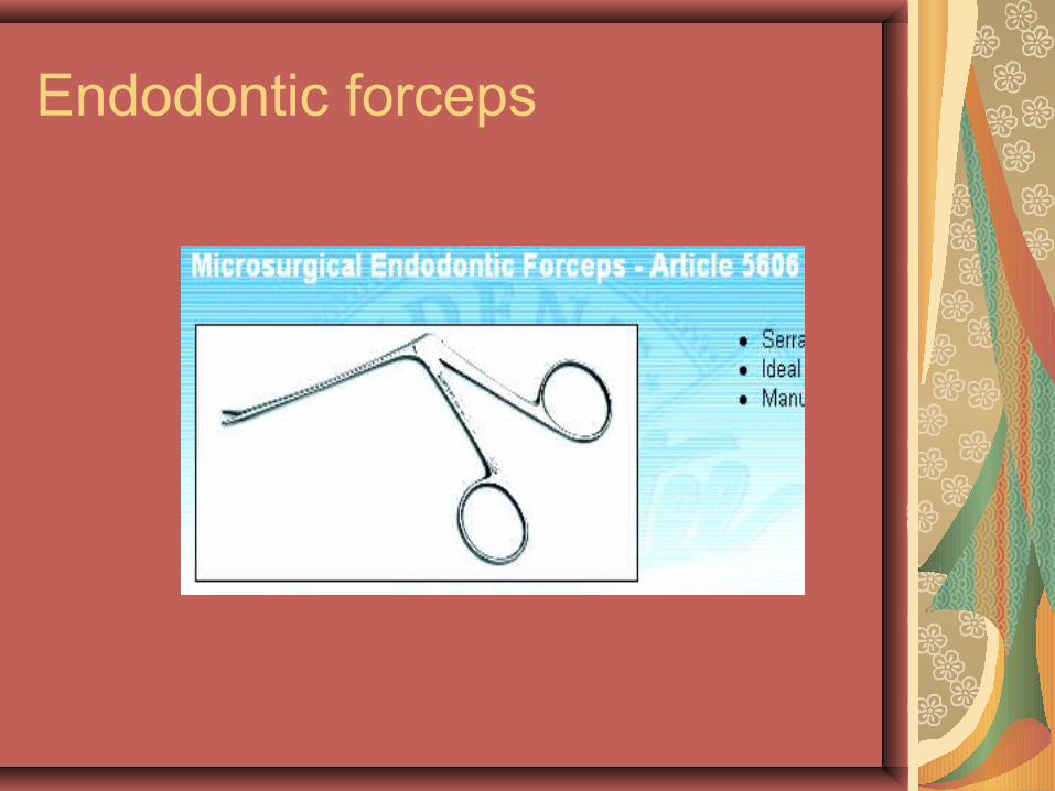
Endodontic forceps
WIRE LOOP TECH
apical transportations
AN APICAL TRANSPORTATION IS MOVING THE CANAL’S PHYSIOLOGICAL TERMINUS TO A DIFFERENT POSITION.
FORAMINAL ZIPS,RIPS OR TEARS ARE CAUSED BY CARRYING PROGRESSIVELY LARGER AND STIFFER IN LENGTH.
MAY RESULT IN OVER EXTENSION OF GP AND FAILURE
TYPES AND MANAGEMENT:TYPE-1 : MINOR TRANSPORTATION: MAY WEAKEN THE ROOT.
“WAIT AND WATCH”TYPE-2 : MODERATE DISTANCE TRANSPORTATION.
MINERAL TRIOXIDE AGGRETATETYPE-3 : SEVERE DISTANCE TRANSPORTATION.
CORRECTIVE SURGERY.
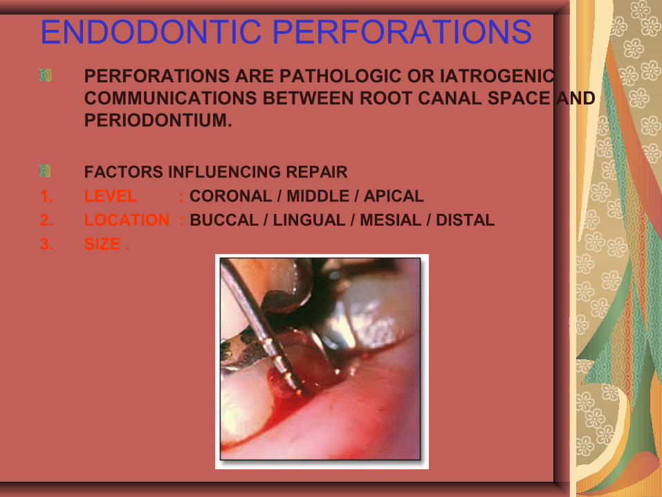
ENDODONTIC PERFORATIONSPERFORATIONS ARE PATHOLOGIC OR IATROGENIC COMMUNICATIONS BETWEEN ROOT CANAL SPACE AND PERIODONTIUM.
FACTORS INFLUENCING REPAIR
1. LEVEL : CORONAL / MIDDLE / APICAL
2. LOCATION : BUCCAL / LINGUAL / MESIAL / DISTAL
3. SIZE .
Cervical perforations usually occur when too large an instrument is used to widen canal access, frequently a Gates-Glidden or Peeso drill. The first indication is the appearance of blood in the cavity. Midroot perforations are usually caused by zipping, frequently in the distal wall of a curved mesial root of a mandibular molar.Apical perforationsare usually due to overeager instrumentation, just plain drilling out through the apical orifice. Again, this can usually be determined with paper points—they appear bloody at the tip If a canal curves at or near the apex, using larger and larger instruments will cause zipping that hollows out this area and leads to perforation.
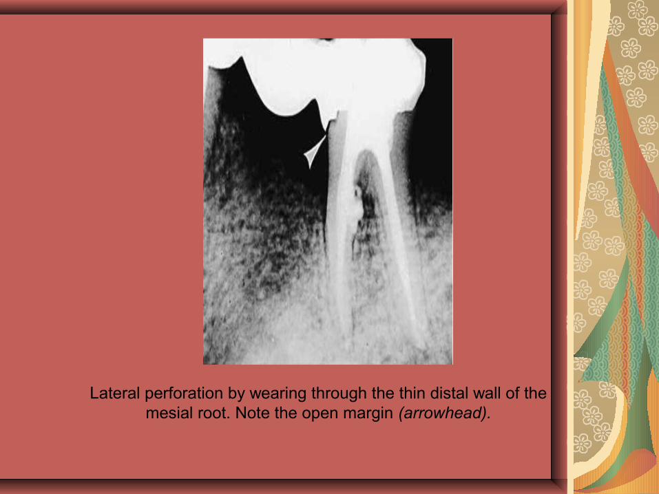
Lateral perforation by wearing through the thin distal wall of the mesial root. Note the open margin (arrowhead).
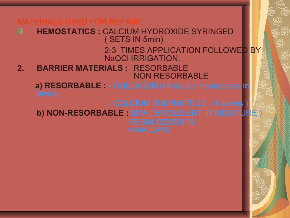
MATERIALS USED FOR REPAIR:HEMOSTATICS : CALCIUM HYDROXIDE SYRINGED
( SETS IN 5min)2-3 TIMES APPLICATION FOLLOWED BY NaOCl IRRIGATION.
2. BARRIER MATERIALS : RESORBABLENON RESORBABLE
a) RESORBABLE : - COLLAGEN (14 days ) hemostasis in 5mins
- CALCIUM SULPHATE ( 2 - 4 weeks )b) NON-RESORBABLE : MTA ( EXCELLENT IN MOISTURE )
RESIN CEMENTS AMALGAM
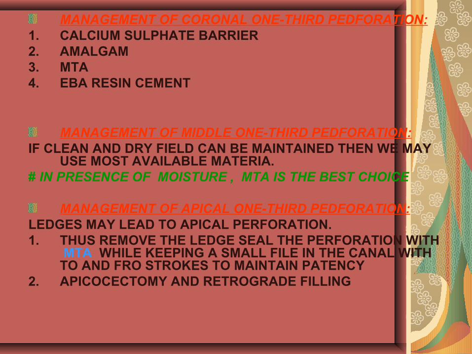
MANAGEMENT OF CORONAL ONE-THIRD PEDFORATION: 1. CALCIUM SULPHATE BARRIER2. AMALGAM3. MTA 4. EBA RESIN CEMENT
MANAGEMENT OF MIDDLE ONE-THIRD PEDFORATION:IF CLEAN AND DRY FIELD CAN BE MAINTAINED THEN WE MAY
USE MOST AVAILABLE MATERIA.# IN PRESENCE OF MOISTURE , MTA IS THE BEST CHOICE
MANAGEMENT OF APICAL ONE-THIRD PEDFORATION:LEDGES MAY LEAD TO APICAL PERFORATION.1. THUS REMOVE THE LEDGE SEAL THE PERFORATION WITH
MTA WHILE KEEPING A SMALL FILE IN THE CANAL WITH TO AND FRO STROKES TO MAINTAIN PATENCY
2. APICOCECTOMY AND RETROGRADE FILLING
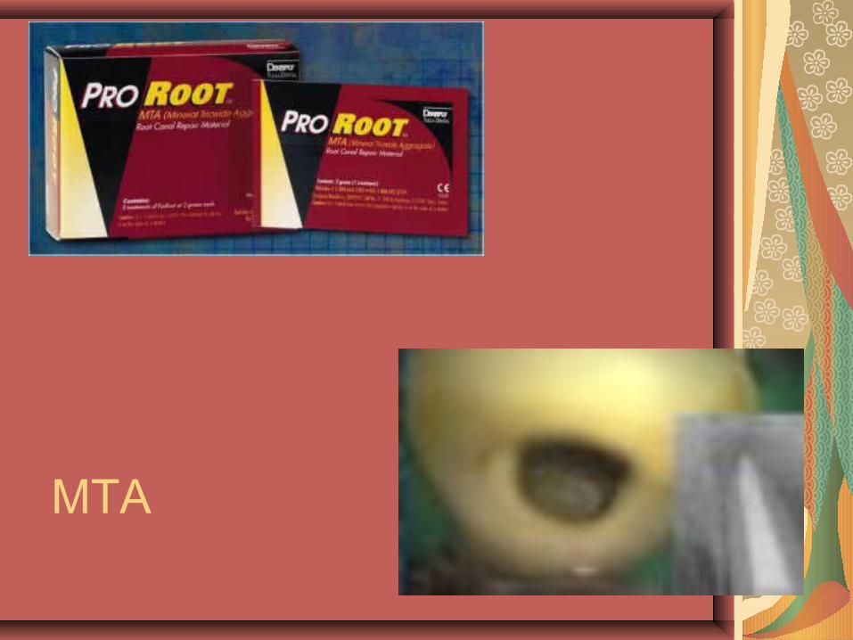
MTA
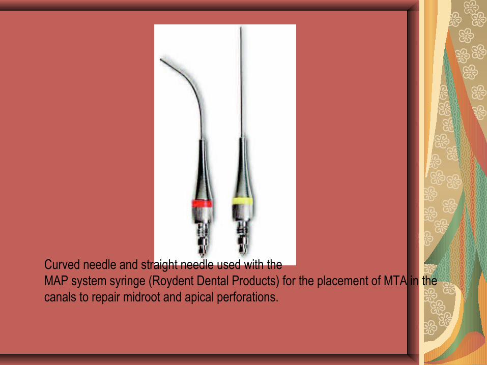
Curved needle and straight needle used with the MAP system syringe (Roydent Dental Products) for the placement of MTA in the canals to repair midroot and apical perforations.
Broken Instruments and Foreign Objects
Many objects have been reported to break or separate in the root canal—amalgam, files and reamers, lentulo spirals, Gates-Glidden drills, and burs.
The first caveat is prevention. Do not force any endodontic instrument, especially in a twisting or rotary motion.
Instruments should be inspected after their use to determine whether they have been stressed .
Broken Instruments and Foreign Objects
REMOVAL INFLUENCED BY 1. LENGTH2. CROSS SECTIONAL DIAMETER 3. CURVATURE OF THE CANAL4. INSTRUMENT MATERIAL: SS
INSTRUMENTS DON’T FRACTURE LIKE NiTi INSTRUMENTS WHICH FRACTURE DUE TO HEAT FROM ULTRASONICS
If a broken instrument can be grasped, it may be removed with Stieglitz forceps. Failing that, using fine ultrasonic instruments to loosen and “float” out the broken piece has proven very successful.If all retrieval fails, broken instruments have been successfully bypassed and a successful root filling placed around them. If a broken instrument extends out the apex into medullary bone, it should prob-ably be removed surgically and a retrofilling placed .
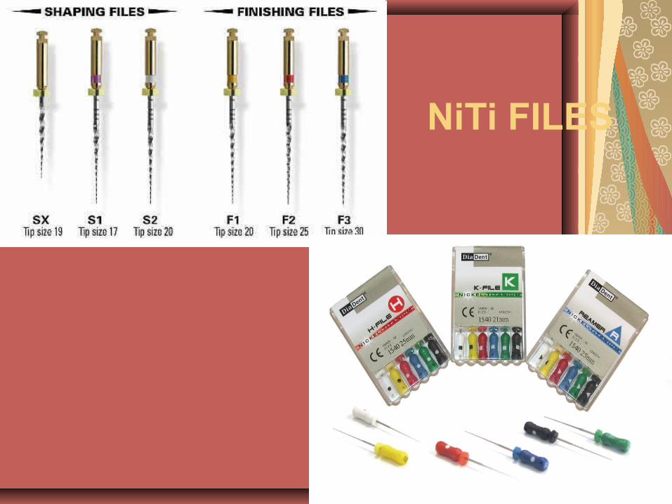
NiTi FILES
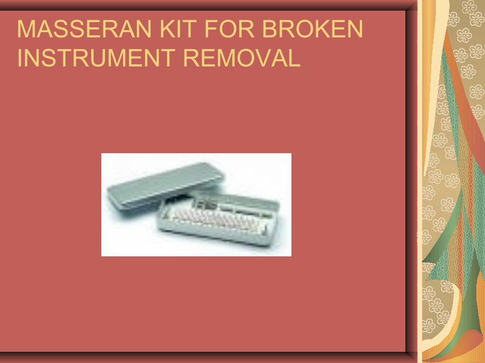
MASSERAN KIT FOR BROKEN INSTRUMENT REMOVAL
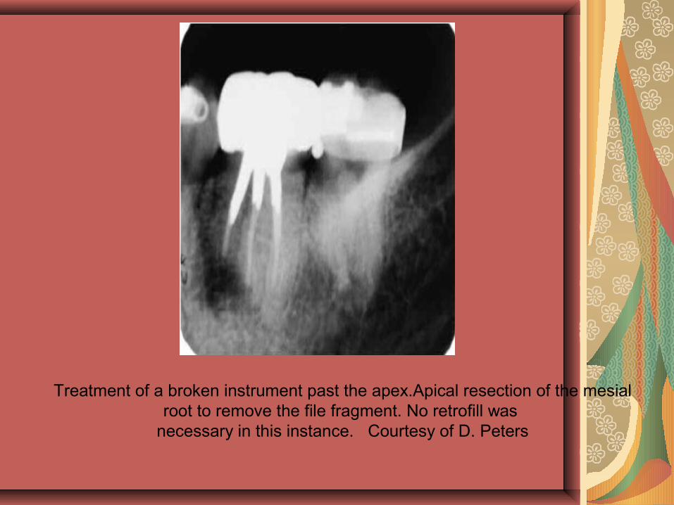
Treatment of a broken instrument past the apex.Apical resection of the mesial root to remove the file fragment. No retrofill was
necessary in this instance. Courtesy of D. Peters
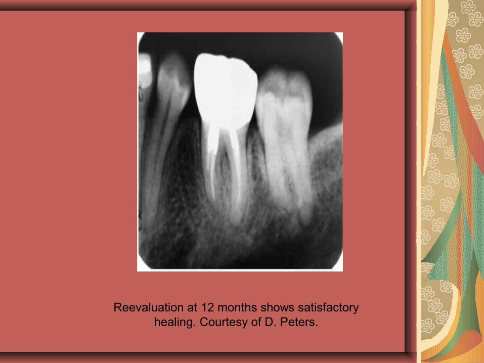
Reevaluation at 12 months shows satisfactory healing. Courtesy of D. Peters.
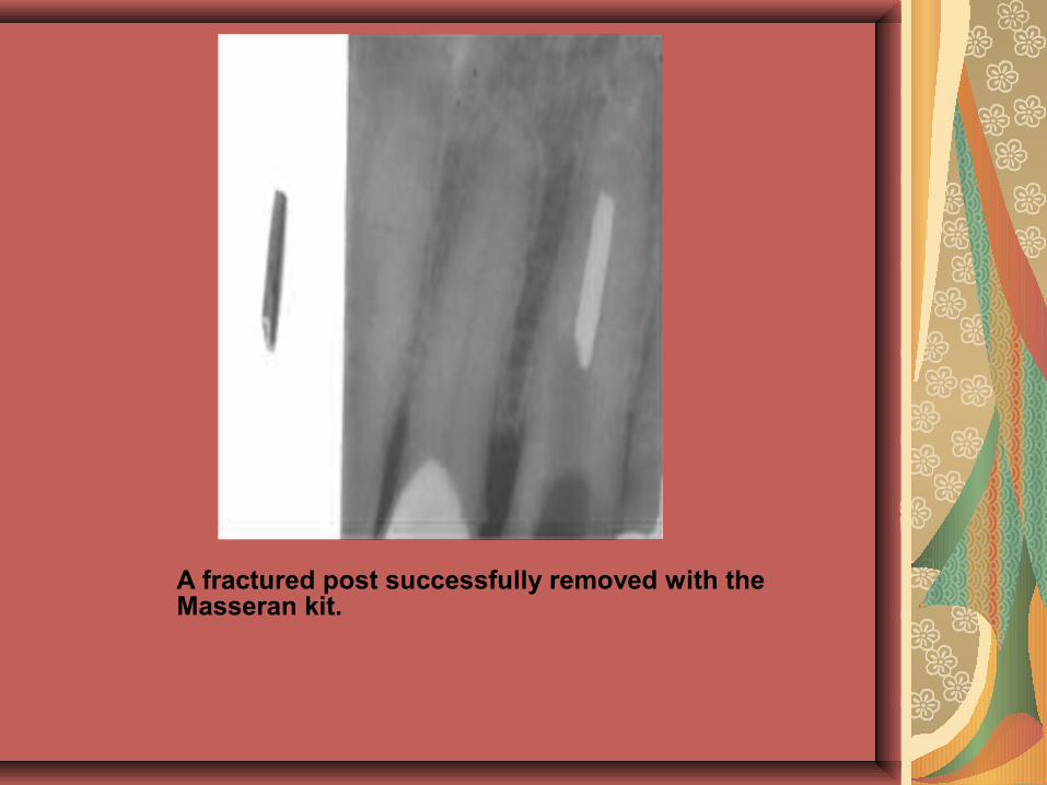
A fractured post successfully removed with the Masseran kit.
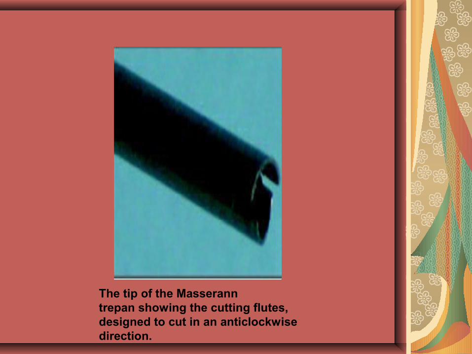
The tip of the Masserann trepan showing the cutting flutes, designed to cut in an anticlockwise direction.
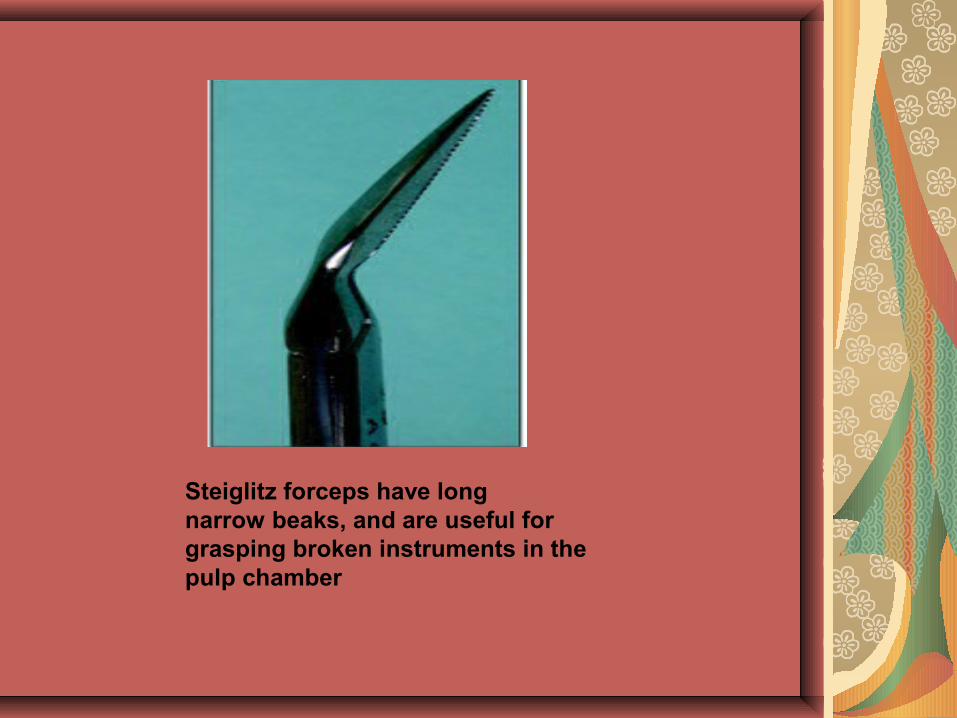
Steiglitz forceps have long narrow beaks, and are useful for grasping broken instruments in the pulp chamber
Blocked canal
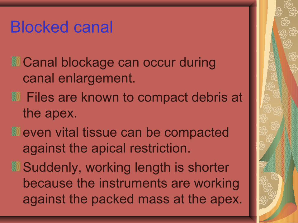
Canal blockage can occur during canal enlargement.
Files are known to compact debris at the apex.
even vital tissue can be compacted against the apical restriction.
Suddenly, working length is shorter because the instruments are working against the packed mass at the apex.
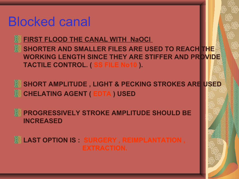
Blocked canalFIRST FLOOD THE CANAL WITH NaOCl
SHORTER AND SMALLER FILES ARE USED TO REACH THE WORKING LENGTH SINCE THEY ARE STIFFER AND PROVIDE TACTILE CONTROL. ( SS FILE No10 ).
SHORT AMPLITUDE , LIGHT & PECKING STROKES ARE USED
CHELATING AGENT ( EDTA ) USED
PROGRESSIVELY STROKE AMPLITUDE SHOULD BE INCREASED
LAST OPTION IS : SURGERY , REIMPLANTATION , EXTRACTION.
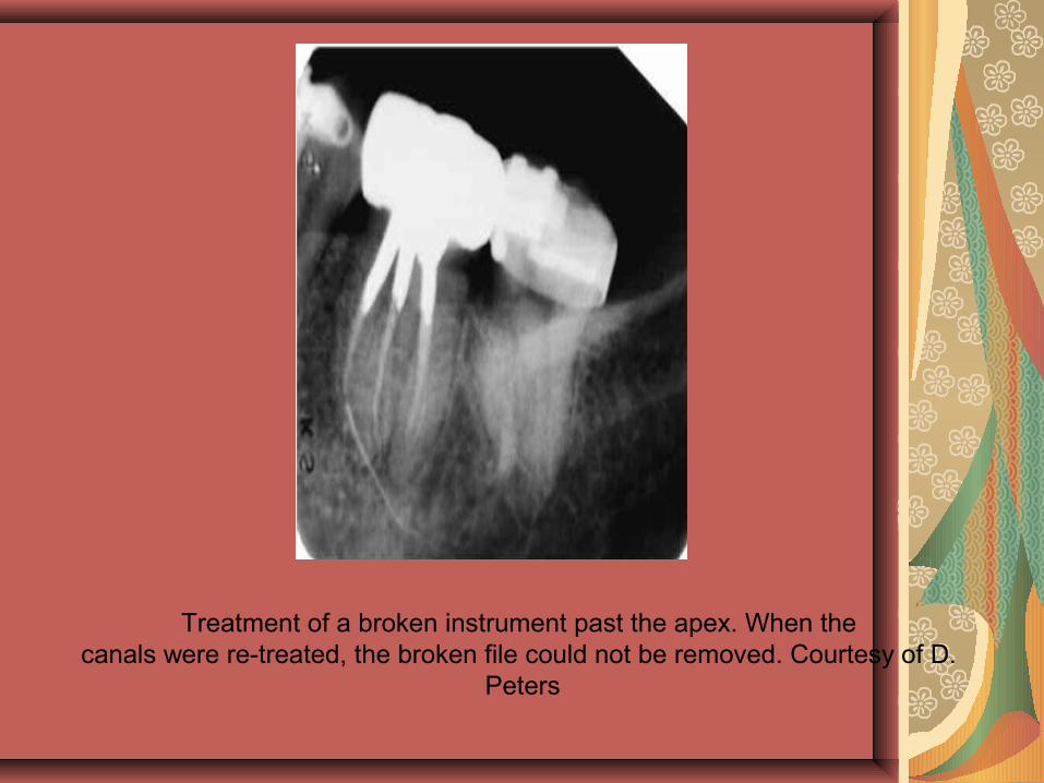
Treatment of a broken instrument past the apex. When the canals were re-treated, the broken file could not be removed. Courtesy of D.
Peters
OBTURATION-RELATED MISHAPS
Inadequate Gutta Percha Obturation
Inadequate gutta percha fills can be under extended (too short), under filled (too thin), or overextended (too long). leads to an inadequate seal, and incomplete debridement of the canals.
Organic solvents, hedstrom files or rotary devices such as the GPX gutta removal kit can be used.
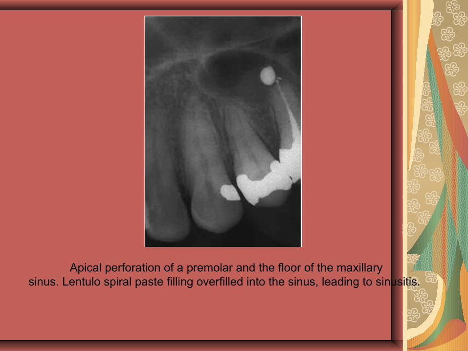
Apical perforation of a premolar and the floor of the maxillary sinus. Lentulo spiral paste filling overfilled into the sinus, leading to sinusitis.
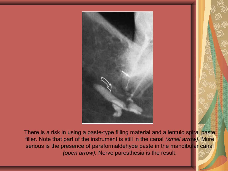
There is a risk in using a paste-type filling material and a lentulo spiral paste filler. Note that part of the instrument is still in the canal (small arrow). More serious is the presence of paraformaldehyde paste in the mandibular canal
(open arrow). Nerve paresthesia is the result.
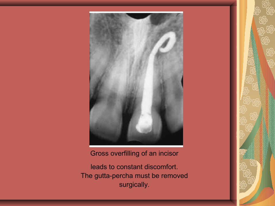
Gross overfilling of an incisor
leads to constant discomfort. The gutta-percha must be removed
surgically.
Vertical Root Fractures
A sudden crunching sound (often referred to as “crepitus”) during obturation is a clear indication that the root has fractured.
This may take place during compaction, more often during lateral than vertical compaction.
It is suspected that stresses built up during compaction are relieved later, following additional stress from mastication or clenching.
Seating of posts, especially tapered posts, is another cause of ver-tical fractures.
when post preparations have removed so much of the root structure that the tooth is materially weakened, the tooth is easily subject to fracture. There is no treatment if the fracture extends down the root. Prevention, then, is the only alternative. Not overpreparing the canals to accompany large filling instruments or posts is of the first order. Then, avoiding excess pressure during compaction of gutta-percha fillings or the cementation of posts is necessary.
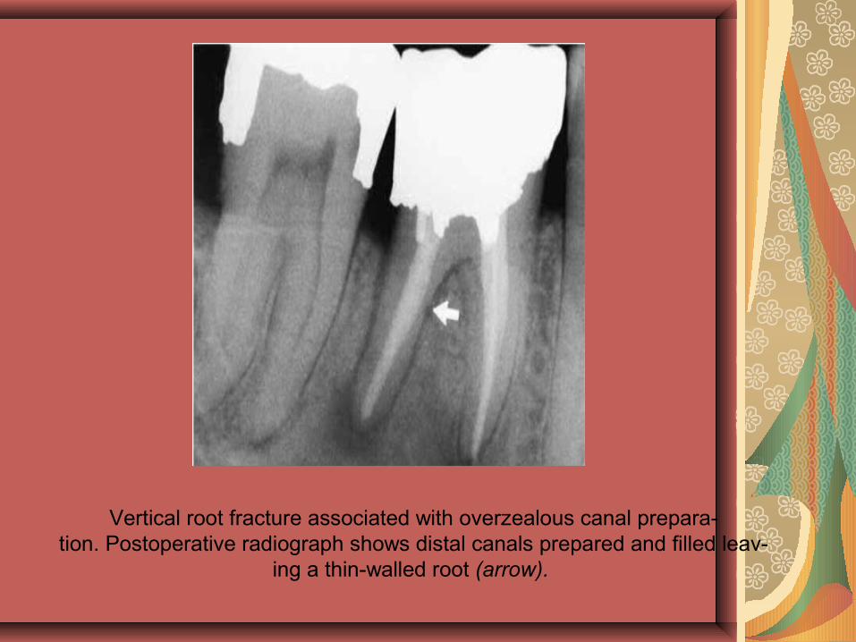
Vertical root fracture associated with overzealous canal prepara-tion. Postoperative radiograph shows distal canals prepared and filled leav-
ing a thin-walled root (arrow).
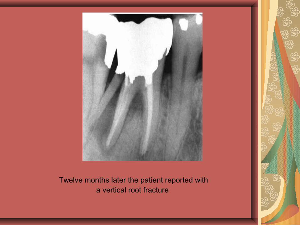
Twelve months later the patient reported with
a vertical root fracture

MISCELLANEOUS MISHAPS
Irrigant-Related Mishaps
The fear of the toxicity of sodium hypochlorite as an irritant of peri-radicular tissue has tended to deter its use. Forcibly injecting NaOCl or any other irrigating solution into the apical tissue can be disastrous. The same can be said for hydrogen peroxide . The patient may immediately complain of severe pain. Swelling can be violent and alarming. The concentration of the irrigant will be a major factor, for example, 5.25% versus 1.3% NaOCl. Antihistamines, ice packs, intramuscular steroids, even hospitalization and surgical interven-tion may be needed.Paresthesia, scarring, and muscle weakness may follow.
Prevention, of course, is the only solution! Inadvertent extrusion of irrigants past the apex can be avoided by using passive placement of a modified needle.
The needle must not be wedged in the canal.
The blunt-nosed, side-orifice ProRinse needle(Dentsply/Tulsa Dental) is the one recommended.
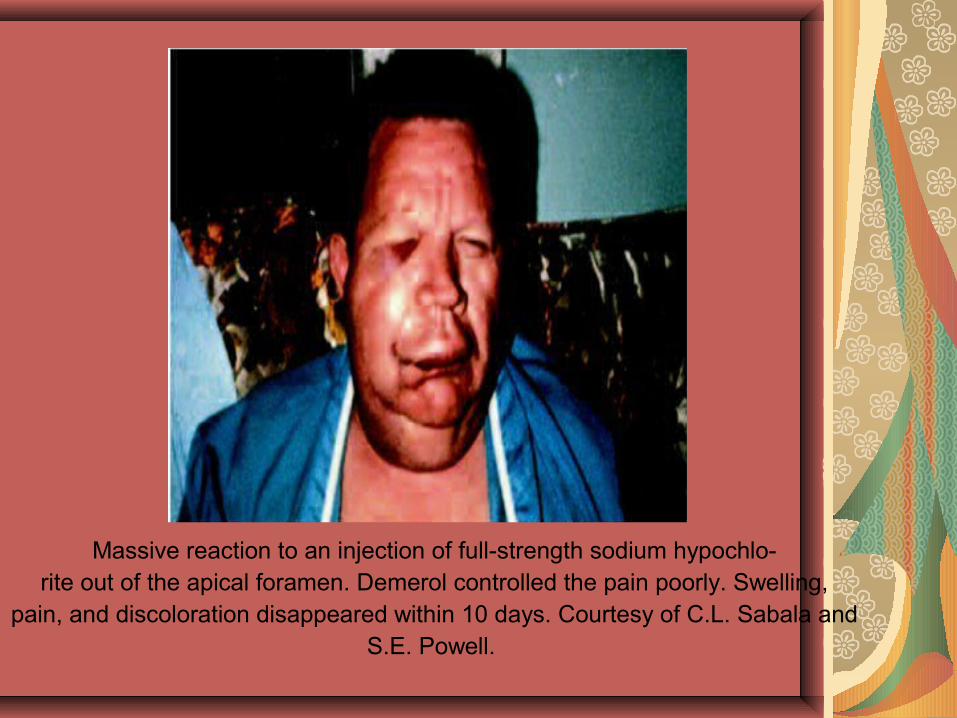
Massive reaction to an injection of full-strength sodium hypochlo-rite out of the apical foramen. Demerol controlled the pain poorly. Swelling,
pain, and discoloration disappeared within 10 days. Courtesy of C.L. Sabala and
S.E. Powell.
Tissue Emphysema
Tissue emphysema, although relatively uncommon, should not be overlooked. Two actions may cause this to happen: a blast of air to dry a canal and exhaust air from a high-speed drill directed toward the tissue and not evacuated to the rear of the handpiece during apical surgery. Emphysema from a blast of air down the canal is more likely to happen with youngsters, in whom the canals in anterior teeth are relatively large.
The usual sequence of events is rapid swelling,erythema, and crepitus, the latter being pathognomonic of erythe-ma. If the air pocket breaks through into the neck region, there is a sudden swelling of the neck. The voice sounds brassy, and the patient has difficulty breathing. If it breaks through into the mediastinum,a crunching noise is heard on auscultation. Death can follow! Although the problem should not be treated lightly, the majority of reported cases have followed a benign course to total recovery.
Prevention is simple: use paper points.
Do not blow air directly down an open canal.
Use the “Venturi effect”:
blow air across the canal opening to aid drying, and employ a handpiece that exhausts the spent air out the back of the handpiece rather than into the operating field.
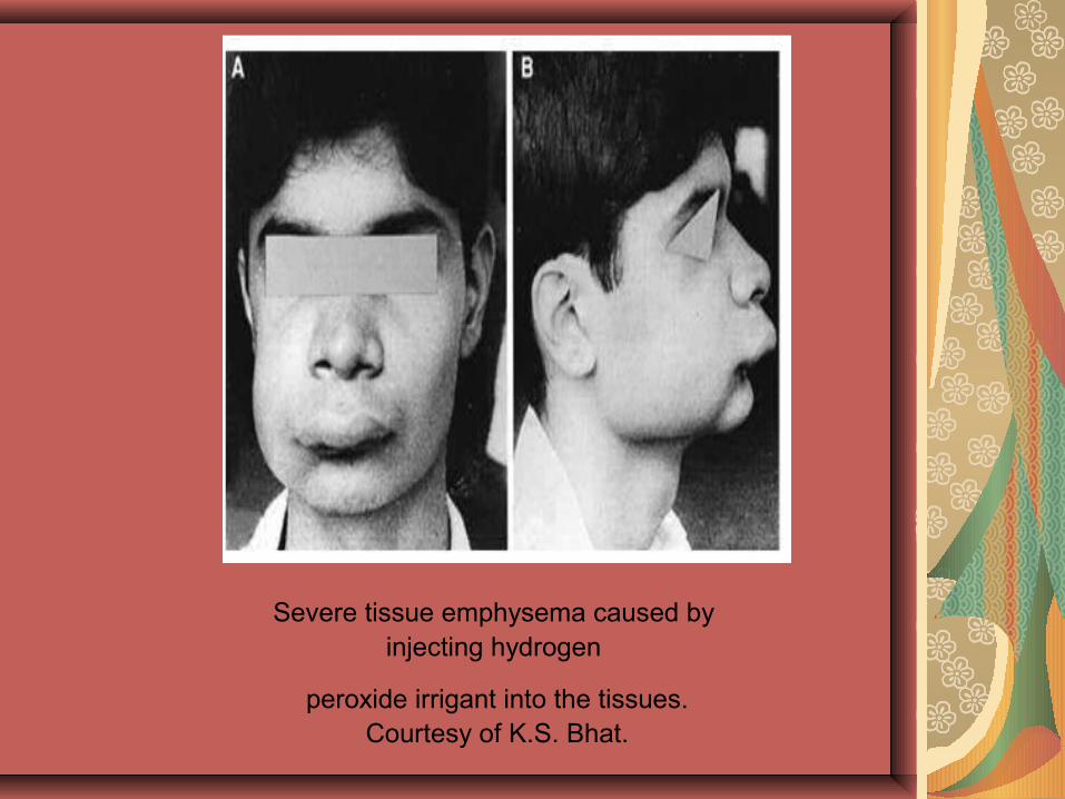
Severe tissue emphysema caused by injecting hydrogen
peroxide irrigant into the tissues. Courtesy of K.S. Bhat.
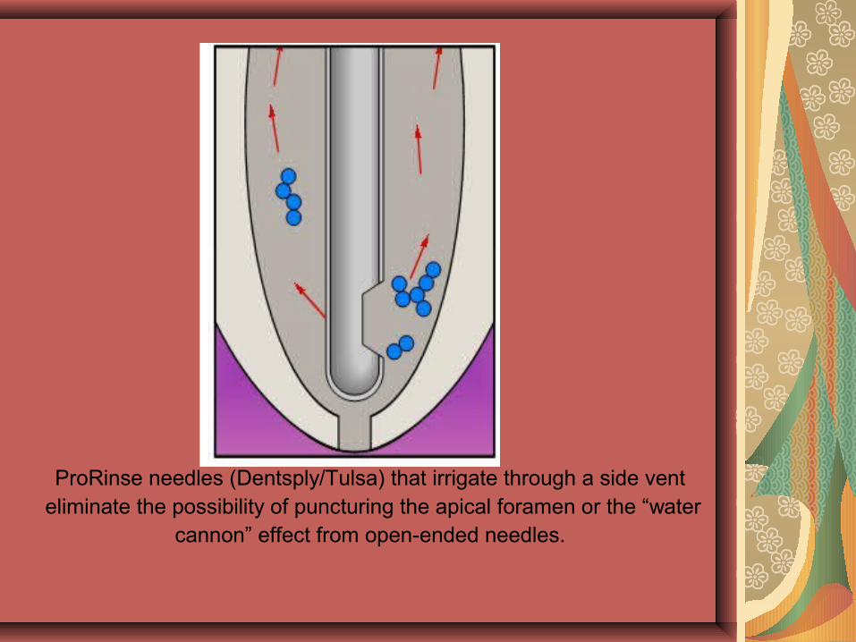
ProRinse needles (Dentsply/Tulsa) that irrigate through a side vent eliminate the possibility of puncturing the apical foramen or the “water
cannon” effect from open-ended needles.
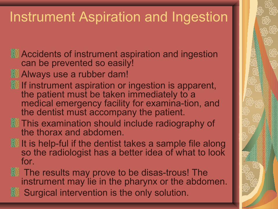
Instrument Aspiration and Ingestion
Accidents of instrument aspiration and ingestion can be prevented so easily!Always use a rubber dam! If instrument aspiration or ingestion is apparent, the patient must be taken immediately to a medical emergency facility for examina-tion, and the dentist must accompany the patient. This examination should include radiography of the thorax and abdomen. It is help-ful if the dentist takes a sample file along so the radiologist has a better idea of what to look for. The results may prove to be disas-trous! The instrument may lie in the pharynx or the abdomen. Surgical intervention is the only solution.
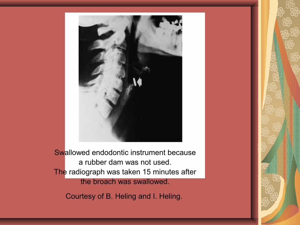
Swallowed endodontic instrument because a rubber dam was not used.
The radiograph was taken 15 minutes after the broach was swallowed.
Courtesy of B. Heling and I. Heling.
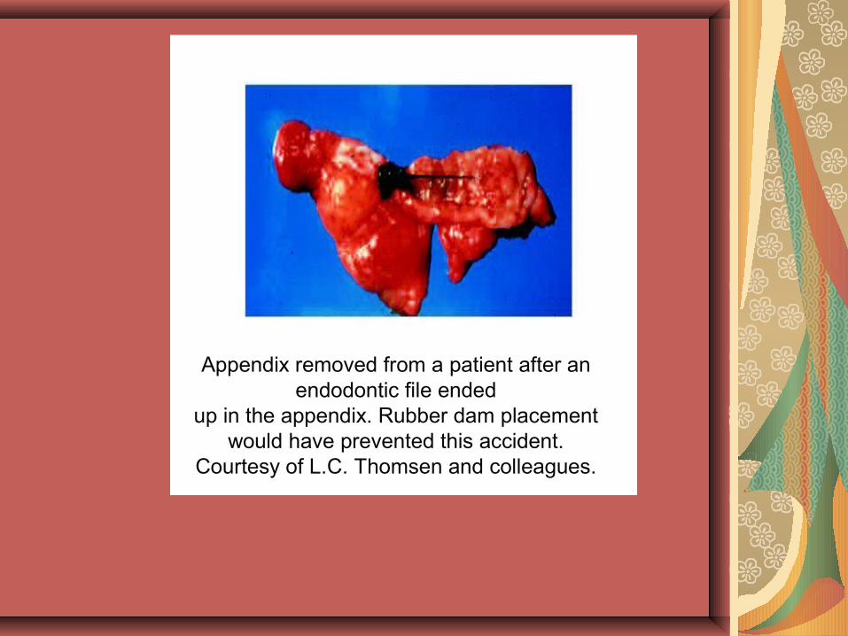
Appendix removed from a patient after an endodontic file ended
up in the appendix. Rubber dam placement would have prevented this accident.
Courtesy of L.C. Thomsen and colleagues.

THANK YOU