Embed Size (px)
Citation preview
1541THORACIC EMERGENCIES
Catherine A. Young, MD, JD • Christine O. Menias, MD • Sanjeev Bhalla, MD • Srinivasa R. Prasad, MD
Esophageal emergencies—primarily, perforation and conditions with the potential to progress to perforation—result in significant morbid-ity and mortality if they are not recognized and treated promptly. The spectrum of esophageal emergencies includes esophagitis, for-eign body impaction, and traumatic esophageal injury. Because there is considerable variability in the clinical manifestations of emergent esophageal conditions, computed tomography (CT) may play both primary and complementary roles in their diagnosis and evaluation. An awareness of the CT findings associated with the spectrum of acute esophageal disease facilitates the accurate and prompt diagnosis of esophageal emergencies and thereby contributes to a more successful outcome.©RSNA, 2008 • radiographics.rsnajnls.org
CT Features of Esoph-ageal Emergencies1
LEARNING OBJECTIVES FOR TEST 1After reading this article and taking the test, the reader
will be able to:
Identify the esopha- ■
geal conditions that may manifest emer-gently.
Describe the clini- ■
cal and CT manifes-tations of common emergent esopha-geal conditions.
Discuss the util- ■
ity of CT in the evaluation of acute esophageal disease.
RadioGraphics 2008; 28:1541–1553 • Published online 10.1148/rg.286085520 • Content Codes: 1From the Mallinckrodt Institute of Radiology, Washington University School of Medicine, Barnes Jewish Hospital, 510 S Kingshighway Blvd, c/o Staff Library, Campus Box 81, St. Louis, MO 63110 (C.A.Y., C.O.M., S.B.); and Department of Radiology, University of Texas Health Science Center, San Antonio, Tex (S.R.P.). Recipient of a Certificate of Merit award for an education exhibit at the 2007 RSNA Annual Meeting. Received March 12, 2008; revision requested April 10; final revision received June 16; accepted June 18. All authors have no financial relationships to disclose. Address correspondence to C.A.Y. (e-mail: [email protected]).
©RSNA, 2008
CME FEATURESee accompanying
test at http://www.rsna.org
/education/rg_cme.html
See last page
TEACHING POINTS
Note: This copy is for your personal non-commercial use only. To order presentation-ready copies for distribution to your colleagues or clients, contact us at www.rsna.org/rsnarights.

1542 October Special Issue 2008 RG ■ Volume 28 • Number 6
IntroductionEsophageal processes such as esophagitis, for-eign body impaction, traumatic esophageal injury, and complications of perforation often present emergently. The increasing use of cross-sectional imaging in emergent care settings, the availability and ease of computed tomography (CT), and the often nonspecific manifestations of acute esophageal conditions all ensure a role for CT in the initial detection and diagnosis of these pathologic processes. In addition, CT is a useful adjunct to conventional esophagography and direct visualization, helping delineate the location and extent of disease, assess complica-tions, and exclude alternative diagnoses. Delays in diagnosis account for most of the morbidity and mortality associated with esophageal emer-gencies; accurate diagnosis and early initiation of an appropriate management strategy (conser-vative, endoscopic, or surgical) are integral to successful outcomes. The purpose of this article is to acquaint radiologists with characteristic CT findings of some commonly encountered emer-
gent esophageal conditions and their associated complications.
EsophagitisGastroesophageal reflux disease is a common cause of noncardiac chest pain (1). Esophagitis related to the ingestion of caustic substances, irradiation, medication, or infection also may result in acute chest pain. In severe esophagitis, full-thickness esophageal necrosis may lead to per-foration with associated complications. Contrast material–enhanced esophagography and endos-copy remain the reference standards for the evalu-ation of esophagitis. CT may be performed when the diagnosis is unclear or when a complication is suspected. Whatever the cause of severe esophagi-tis, its CT appearance is predominantly character-ized by diffuse esophageal thickening, submucosal edema, and mucosal enhancement (Figs 1, 2).
Foreign Body ImpactionAlthough foreign body ingestion is seen rela-tively often in the emergency department, most ingested objects pass spontaneously, without intervention (2,3). However, the thin esopha-geal wall, the lack of a supporting adventitia,
Figures 1, 2. Esophagitis. (1) Axial contrast-enhanced CT image, obtained in a middle-aged man with a new onset of chest pain during chemotherapy with a plat-inum-based agent, shows mucosal enhancement and diffuse submucosal edema (arrowhead), findings suggestive of chemotherapy-related esophagitis. (2) Axial contrast-enhanced CT image, obtained to determine whether esophageal perfora-tion was present in an elderly man who drank a caustic drain cleaning solution containing lye, shows a diffusely thickened esophageal wall (arrowheads).

RG ■ Volume 28 • Number 6 Young et al 1543
and the relatively poor blood supply leave the esophagus vulnerable to perforation and pres-sure necrosis from foreign bodies that become lodged in it. Between 10% and 20% of ingested foreign bodies, including those retained within the esophagus, therefore require endoscopic removal, and surgery is needed in about 1% of cases (2,4,5). Sharp or pointed foreign bod-ies, button batteries, and objects that cause obstruction require emergent removal; less ur-gent intervention is indicated if a foreign body fails to spontaneously clear the esophagus in a timely manner (4–6). An argument has been made that vinyl gloves, which tend to harden, should be removed surgically rather than endo-scopically (7).
Foreign body ingestion and impaction occur across all age groups, with a median age of about 40 years (2,3,5). The clinical manifestations of esophageal foreign body impaction are variable: Symptoms may include dysphagia, odynophagia, foreign body sensation, and food refusal. Re-gurgitation and an inability to swallow saliva are suggestive of esophageal obstruction. When a his-tory of foreign body ingestion is elicited, a radio-graphic evaluation is performed, generally with conventional radiography of the neck, chest, and abdomen (2). Barium studies are discouraged because they may hinder subsequent attempts at endoscopic examination and retrieval (2,4). CT
may be useful in select cases when a more defini-tive diagnosis or localization is desired before endoscopic intervention, or when perforation or another complication is suspected (2). In patients who do not provide a history of foreign body ingestion and who report nonspecific symptoms such as chest pain, CT often is used to distin-guish between or exclude potential causes during the initial evaluation (8).
The CT appearance of foreign body impaction is variable, depending on the item ingested, the site of impaction, and the presence of an underly-ing pathologic esophageal process or associated complication. Foreign body ingestion is most commonly seen in children (Fig 3), people with psychiatric disorders, and prisoners. In children, coins are the most frequently ingested foreign bodies (3,9).Vinyl glove ingestion has been re-ported among people with cognitive deficits and pica (7) (Fig 4). Food boluses (often meat) ac-count for most cases of foreign body impaction
Figure 3. Food impaction in the esophagus of a toddler with a 1-day history of food refusal. (a) Sagittal reformatted image from contrast-enhanced CT of the neck shows a tubular object with attenuation close to that of fat, lodged in the cervical esophagus (*). (b) Esopha-gram helps confirm the presence of an obstructing foreign body (*). A hot-dog end was removed from the normal esophagus at endoscopy.
TeachingPoint
TeachingPoint
1544 October Special Issue 2008 RG ■ Volume 28 • Number 6
tinguishable from those produced by a neoplastic process (Fig 7).
TraumaTraumatic injury to the esophagus may result from both extraluminal and intraluminal processes. In-juries that are proved due to extraluminal blunt or penetrating trauma typically involve the cervical and upper thoracic parts of the esophagus but are rare overall, probably because of the small size of the esophagus, its relatively protected position, and the likelihood of serious concomitant vascular, tra-cheal, or spinal cord injury (12,13). Intraluminal processes such as instrumentation, foreign body impaction, barotrauma, and erosive esophagitis, among others, give rise to a spectrum of injuries that may be characterized according to the degree of resultant esophageal perforation. At one end of the spectrum are injuries such as mucosal lac-erations, intramural dissection, and hematoma, which are relatively innocuous; and, at the other end, transmural perforation, which is potentially
seen in adults (2,3) (Fig 5). In one series, an underlying pathologic esophageal process—most commonly, stricture—was found in close to one-third of adult patients treated endoscopically for foreign body impaction (2); the number may be higher among those with a food impaction (10). Bones from fish and chicken constitute the second most common foreign body in both pe-diatric and adult populations (5) and are more likely to become lodged in the hypopharynx or cervical esophagus, where they may be difficult to visualize endoscopically (2); CT may be es-pecially useful in such cases (Fig 6). After the more commonly ingested items, there is great variety among the types of foreign bodies seen in esophageal impaction (3). In recent years, inad-vertently swallowed medication blister packs have been implicated in gastrointestinal perforation and hemorrhage as well as esophageal impaction (3,11). Chronic impaction of a foreign body in the esophagus may produce erosion, fistula for-mation, and inflammatory reaction that are indis-
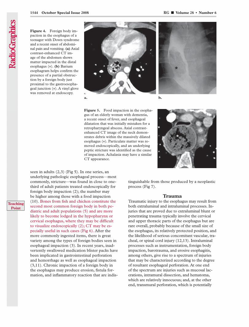
Figure 5. Food impaction in the esopha-gus of an elderly woman with dementia, a recent onset of fever, and esophageal dilatation that was initially mistaken for a retropharyngeal abscess. Axial contrast-enhanced CT image of the neck demon-strates debris within the massively dilated esophagus (*). Particulate matter was re-moved endoscopically, and an underlying peptic stricture was identified as the cause of impaction. Achalasia may have a similar CT appearance.
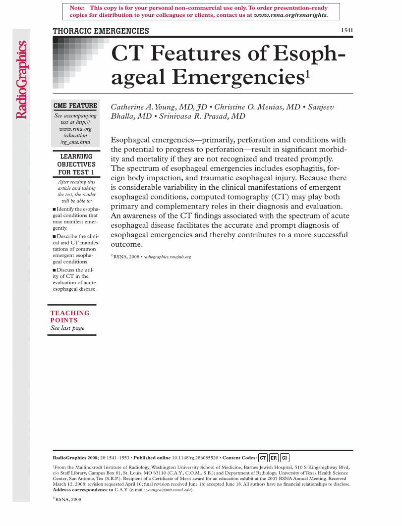
Figure 4. Foreign body im-paction in the esophagus of a teenager with Down syndrome and a recent onset of abdomi-nal pain and vomiting. (a) Axial contrast-enhanced CT im-age of the abdomen shows matter impacted in the distal esophagus (*). (b) Barium esophagram helps confirm the presence of a partial obstruc-tion by a foreign body just proximal to the gastroesopha-geal junction (*). A vinyl glove was removed at endoscopy.
TeachingPoint
RG ■ Volume 28 • Number 6 Young et al 1545
intrathoracic perforation is generally considered a surgical emergency (13,14).
catastrophic. Mucosal lacerations, most intramural perforations, and some cervical and contained per-forations of the esophagus may be managed con-servatively; acute free (uncontained) transmural
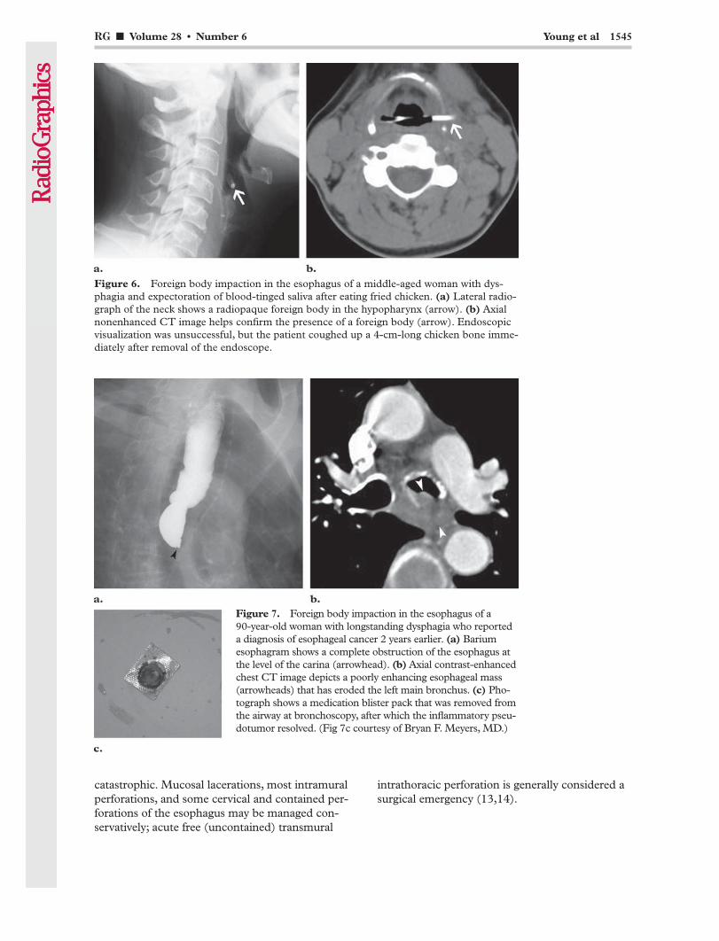
Figure 6. Foreign body impaction in the esophagus of a middle-aged woman with dys-phagia and expectoration of blood-tinged saliva after eating fried chicken. (a) Lateral radio-graph of the neck shows a radiopaque foreign body in the hypopharynx (arrow). (b) Axial nonenhanced CT image helps confirm the presence of a foreign body (arrow). Endoscopic visualization was unsuccessful, but the patient coughed up a 4-cm-long chicken bone imme-diately after removal of the endoscope.
Figure 7. Foreign body impaction in the esophagus of a 90-year-old woman with longstanding dysphagia who reported a diagnosis of esophageal cancer 2 years earlier. (a) Barium esophagram shows a complete obstruction of the esophagus at the level of the carina (arrowhead). (b) Axial contrast-enhanced chest CT image depicts a poorly enhancing esophageal mass (arrowheads) that has eroded the left main bronchus. (c) Pho-tograph shows a medication blister pack that was removed from the airway at bronchoscopy, after which the inflammatory pseu-dotumor resolved. (Fig 7c courtesy of Bryan F. Meyers, MD.)
1546 October Special Issue 2008 RG ■ Volume 28 • Number 6
collection, mediastinal inflammation, focal esopha-geal wall defect, and pleural effusion. Inasmuch as some findings are nonspecific and may be subtle, the diagnosis of esophageal injury requires a high index of suspicion in appropriate clinical settings.
Mallory-Weiss Tear and Other Mucosal LacerationsA Mallory-Weiss tear is a longitudinal mucosal laceration observed in the distal esophagus or across the gastroesophageal junction. Its patho-genesis is similar to that of Boerhaave syndrome: Both occur in the setting of retching or vomiting, frequently after excessive alcohol consumption; they also may occur as a complication of endos-copy (16). Some degree of hematemesis is invari-ably present and is an indication for upper endos-copy. Similar linear mucosal lacerations occurring elsewhere in the esophagus as a result of forceful swallowing of an impacted foreign body or food bolus may pose a diagnostic dilemma. A mucosal laceration without transmural perforation is likely to be radiographically occult. However, to the attentive observer, CT images of the esophagus in patients with chest pain occasionally show evi-dence of hemorrhage or foci of extraluminal gas at a site of mucosal injury (Fig 8). Unless bleed-ing persists, the treatment of a Mallory-Weiss tear, like that of other mucosal lacerations, is sup-portive (16).
The symptoms of esophageal injury are often nonspecific. Pain is the most common symptom, and it is often severe. Dysphagia or odynophagia also is strongly suggestive of an esophageal abnor-mality. When esophageal injury (usually, perfora-tion) is suspected, the imaging evaluation should generally commence with esophagography while the patient swallows a water-soluble contrast ma-terial. If no extra-esophageal leakage is observed, esophagography should be repeated during the oral administration of barium, which has a greater sensitivity for the detection of small perfora-tions (15). In patients with penetrating trauma to the neck or chest, CT should be performed before contrast-enhanced esophagography, given the significant risk of damage to critical vascular structures. CT may be useful also in patients who are too ill to cooperate in esophagography or as a complement to contrast-enhanced luminal stud-ies, to further delineate the extent of disease, as-sess complications, and guide therapy (13,15,16). In patients with acute chest pain, CT is used to exclude serious conditions such as aortic dissec- tion and pulmonary embolism (8). CT findings of esophageal injury include esophageal wall thickening, periesophageal gas and fluid collections, contrast material extravasation, mediastinal fluid
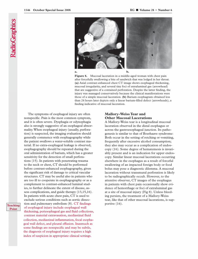
Figure 8. Mucosal laceration in a middle-aged woman with chest pain after forcefully swallowing a bite of sandwich that was lodged in her throat. (a) Axial contrast-enhanced chest CT image shows esophageal thickening, mucosal irregularity, and several tiny foci of extraluminal gas (arrowhead) that are suggestive of a contained perforation. Despite the latter finding, the injury was managed conservatively because the clinical manifestations were those of a simple mucosal laceration. (b) Barium esophagram obtained less than 24 hours later depicts only a linear barium-filled defect (arrowheads), a finding indicative of mucosal laceration.
TeachingPoint
RG ■ Volume 28 • Number 6 Young et al 1547
risk factor. Other contributing events are foreign body impaction and forceful vomiting. Spontane-ous intramural hematoma of the esophagus also has been reported, most often in patients who are undergoing anticoagulant drug therapy or who have inherent coagulopathy (16,17).
CT findings of dissection correlate with those seen at esophagography: a mucosal flap with sub-mucosal distribution of gas or contrast material, giving the esophagus the classic double-barreled appearance (Figs 9, 10). Dissection tends to oc-cur posterior to the true lumen of the esophagus,
Intramural Dissec- tion and HematomaOften referred to collectively as submucosal dis-section or intramural rupture, intramural dissec-tion and intramural hematoma of the esophagus are rare. By contrast to mucosal laceration and transmural perforation, they may be considered intermediate forms of esophageal injury. Symp-toms include an abrupt onset of retrosternal chest pain, dysphagia or odynophagia, and hematemesis, with most patients experiencing at least two of the three, and with hematemesis generally occurring later in the clinical course (17). A history of recent instrumentation is probably the most important
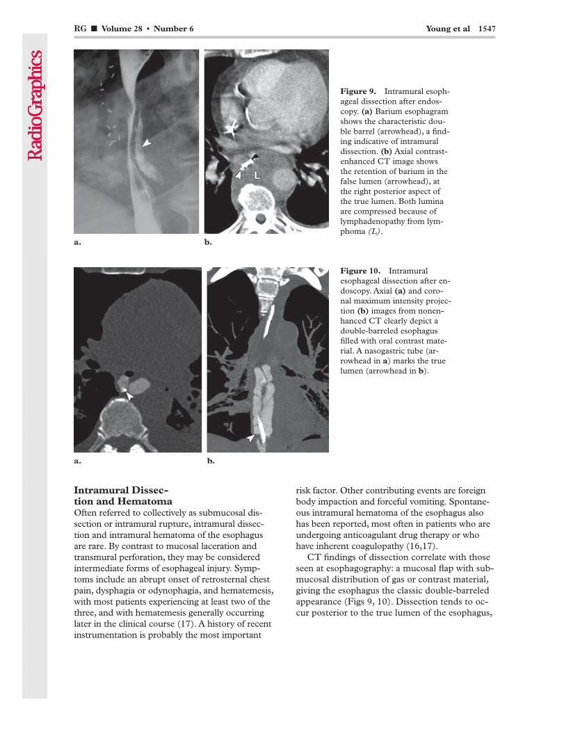
Figure 9. Intramural esoph-ageal dissection after endos-copy. (a) Barium esophagram shows the characteristic dou-ble barrel (arrowhead), a find-ing indicative of intramural dissection. (b) Axial contrast-enhanced CT image shows the retention of barium in the false lumen (arrowhead), at the right posterior aspect of the true lumen. Both lumina are compressed because of lymphadenopathy from lym-phoma (L).
Figure 10. Intramural esophageal dissection after en-doscopy. Axial (a) and coro- nal maximum intensity projec-tion (b) images from nonen-hanced CT clearly depict a double-barreled esophagus filled with oral contrast mate-rial. A nasogastric tube (ar-rowhead in a) marks the true lumen (arrowhead in b).
1548 October Special Issue 2008 RG ■ Volume 28 • Number 6
symptoms may mimic those of acute myocardial infarction or aortic dissection. CT is invaluable for its ability to help differentiate intramural he-matoma of the esophagus from acute cardiovas-cular disease (16,18). The distinction is integral to proper treatment, because anticoagulation therapy is understandably contraindicated in the presence of an intramural hematoma of the esophagus. With conservative management, intra-mural esophageal dissection and hematoma are expected to resolve within a few days or weeks (16,17).
and its full extent may be best appreciated on sag-ittal or coronal reformatted images (Figs 9–11).
An intramural hematoma of the esophagus may occur spontaneously or in association with traumatic esophageal dissection. At CT, it ap-pears as an eccentric hyperattenuating mass within the wall of the esophagus (Fig 12). The clinical diagnosis of a spontaneous intramu-ral hematoma may be challenging because its
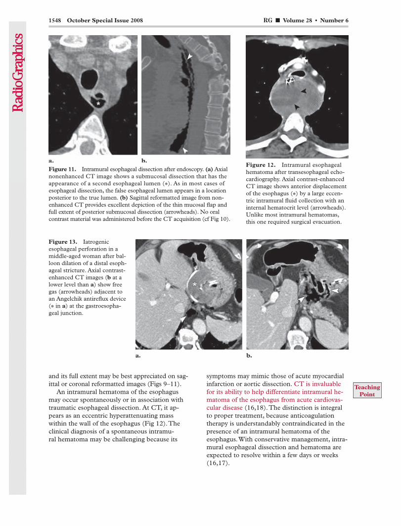
Figure 12. Intramural esophageal hematoma after transesophageal echo-cardiography. Axial contrast-enhanced CT image shows anterior displacement of the esophagus (*) by a large eccen-tric intramural fluid collection with an internal hematocrit level (arrowheads). Unlike most intramural hematomas, this one required surgical evacuation.
Figure 13. Iatrogenic esophageal perforation in a middle-aged woman after bal-loon dilation of a distal esoph-ageal stricture. Axial contrast-enhanced CT images (b at a lower level than a) show free gas (arrowheads) adjacent to an Angelchik antireflux device (* in a) at the gastroesopha-geal junction.
Figure 11. Intramural esophageal dissection after endoscopy. (a) Axial nonenhanced CT image shows a submucosal dissection that has the appearance of a second esophageal lumen (*). As in most cases of esophageal dissection, the false esophageal lumen appears in a location posterior to the true lumen. (b) Sagittal reformatted image from non-enhanced CT provides excellent depiction of the thin mucosal flap and full extent of posterior submucosal dissection (arrowheads). No oral contrast material was administered before the CT acquisition (cf Fig 10).
TeachingPoint
RG ■ Volume 28 • Number 6 Young et al 1549
of left atrial radiofrequency ablation, may lead to full-thickness necrosis and perforation (20) (Fig 15). In addition, esophageal rupture may occur spontaneously, as in Boerhaave syndrome, in which incomplete cricopharyngeal relaxation during vomiting results in abruptly increased intraluminal pressure sufficient to rupture the esophagus. The distal left posterior wall is the most common site of spontaneous rupture, which classically results in pneumomediastinum and left pleural effusion (Figs 16, 17). Perforation of the cervical esophagus should be considered in the presence of cervical subcutaneous emphysema or
Transmural PerforationTransmural perforation may occur in a variety of settings. Its clinical presentation and CT ap-pearance are similarly variable, depending on the mechanism of injury, the site and size of perfo-ration, and the time elapsed since the onset of symptoms (13,15). Iatrogenic perforation of the esophagus is increasingly common, with thera-peutic endoscopic procedures such as stricture dilation (Fig 13) and stent placement (Fig 14) being the leading causes (16). Perforation also oc-curs, albeit infrequently, as a complication of sur-gical procedures such as gastric fundoplication, esophageal myotomy, thyroidectomy, and anterior cervical diskectomy (16,19). Thermal injury to the anterior wall of the esophagus, a complication
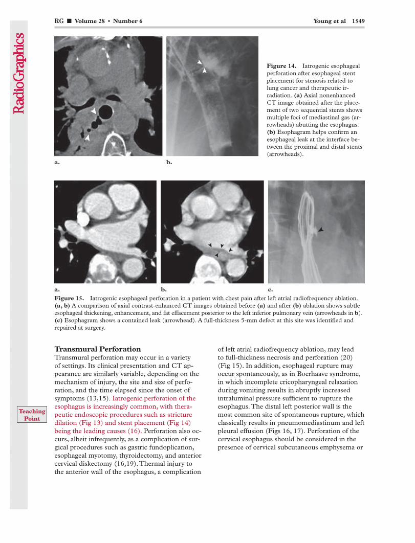
Figure 14. Iatrogenic esophageal perforation after esophageal stent placement for stenosis related to lung cancer and therapeutic ir-radiation. (a) Axial nonenhanced CT image obtained after the place-ment of two sequential stents shows multiple foci of mediastinal gas (ar-rowheads) abutting the esophagus. (b) Esophagram helps confirm an esophageal leak at the interface be-tween the proximal and distal stents (arrowheads).
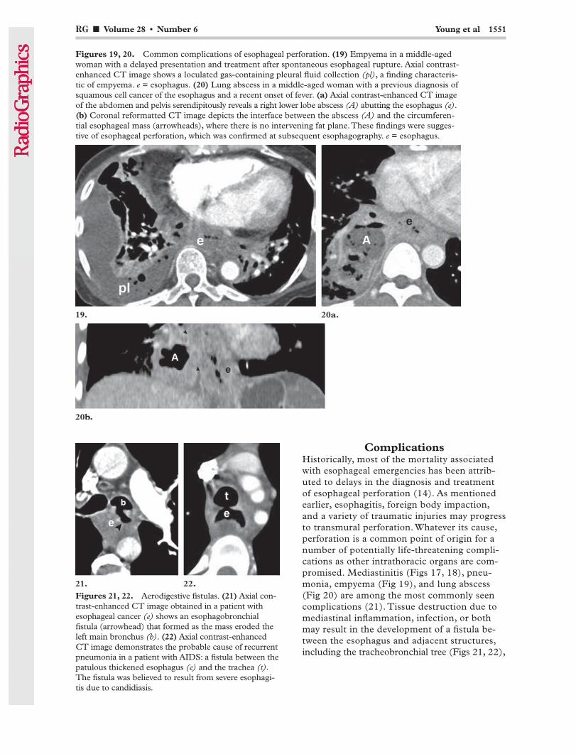
Figure 15. Iatrogenic esophageal perforation in a patient with chest pain after left atrial radiofrequency ablation. (a, b) A comparison of axial contrast-enhanced CT images obtained before (a) and after (b) ablation shows subtle esophageal thickening, enhancement, and fat effacement posterior to the left inferior pulmonary vein (arrowheads in b). (c) Esophagram shows a contained leak (arrowhead). A full-thickness 5-mm defect at this site was identified and repaired at surgery.
TeachingPoint

1550 October Special Issue 2008 RG ■ Volume 28 • Number 6
esophagitis, Barrett syndrome, esophageal can-cer, and aortic rupture. The optimal treatment of esophageal perforation depends on a host of considerations. Treatment methods range from nonsurgical management to esophagectomy or surgical exclusion and diversion; however, with an early diagnosis of uncontained perforation, surgery remains the mainstay of therapy (13,14).
a superior mediastinal fluid collection (Fig 18). Patients with cervical esophageal perforation typi-cally present with crepitus and neck pain instead of chest or epigastric pain. Other possible causes of noniatrogenic esophageal perforation include foreign body impaction, caustic and infectious
Figures 16, 17. Emetogenic esophageal perforation (Boerhaave syndrome) in two late-middle-aged men. (16) Axial contrast-enhanced CT image demonstrates leakage of oral contrast material adjacent to the distal esophagus, with associated pneumomediastinum and left pleural effusion. Diffuse irregularity is visible in the mucosa lining the esophageal lumen (arrowhead). (17) Axial nonenhanced CT image shows oral contrast material, gas, and debris outside the esophagus (ar-rowheads) but confined within the mediastinum (arrows). The presence of an esophageal rupture was confirmed at surgery.
Figure 18. Emetogenic esophageal perforation. (a) Esophagram obtained in a young man with schizo-phrenia and bulimia shows leakage of barium from the cervical esophagus (*). (b) Axial contrast-enhanced CT image of the neck depicts a defect in the left posterior wall of the esophagus (arrowheads), through which gas and pooling barium (arrow) have escaped. Rupture of the cervical esophagus is typi-cally associated with subcutaneous emphysema.
RG ■ Volume 28 • Number 6 Young et al 1551
ComplicationsHistorically, most of the mortality associated with esophageal emergencies has been attrib-uted to delays in the diagnosis and treatment of esophageal perforation (14). As mentioned earlier, esophagitis, foreign body impaction, and a variety of traumatic injuries may progress to transmural perforation. Whatever its cause, perforation is a common point of origin for a number of potentially life-threatening compli-cations as other intrathoracic organs are com-promised. Mediastinitis (Figs 17, 18), pneu- monia, empyema (Fig 19), and lung abscess (Fig 20) are among the most commonly seen complications (21). Tissue destruction due to mediastinal inflammation, infection, or both may result in the development of a fistula be-tween the esophagus and adjacent structures, including the tracheobronchial tree (Figs 21, 22),
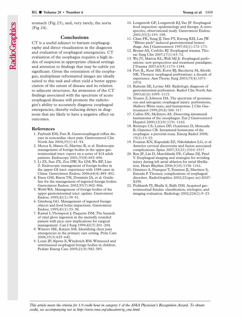
Figures 19, 20. Common complications of esophageal perforation. (19) Empyema in a middle-aged woman with a delayed presentation and treatment after spontaneous esophageal rupture. Axial contrast-enhanced CT image shows a loculated gas-containing pleural fluid collection (pl), a finding characteris-tic of empyema. e = esophagus. (20) Lung abscess in a middle-aged woman with a previous diagnosis of squamous cell cancer of the esophagus and a recent onset of fever. (a) Axial contrast-enhanced CT image of the abdomen and pelvis serendipitously reveals a right lower lobe abscess (A) abutting the esophagus (e). (b) Coronal reformatted CT image depicts the interface between the abscess (A) and the circumferen-tial esophageal mass (arrowheads), where there is no intervening fat plane. These findings were sugges-tive of esophageal perforation, which was confirmed at subsequent esophagography. e = esophagus.
Figures 21, 22. Aerodigestive fistulas. (21) Axial con-trast-enhanced CT image obtained in a patient with esophageal cancer (e) shows an esophagobronchial fistula (arrowhead) that formed as the mass eroded the left main bronchus (b). (22) Axial contrast-enhanced CT image demonstrates the probable cause of recurrent pneumonia in a patient with AIDS: a fistula between the patulous thickened esophagus (e) and the trachea (t). The fistula was believed to result from severe esophagi-tis due to candidiasis.

1552 October Special Issue 2008 RG ■ Volume 28 • Number 6
Figure 23. Esophagogastric fistula in a patient with persistent pain after gastric fundoplication com-plicated by esophageal perforation. (a) Axial nonenhanced CT image demonstrates an intraperitoneal gas collection (*) extending from the gastroesophageal junction to the anterior gastric fundus (s), a finding suggestive of an esophagogastric fis-tula. (b) Barium esophagram helps confirm the presence of an esoph-agogastric fistula (arrowheads).
Figure 24. Aortoesophageal fistula in a middle-aged man with a history of erosive esophagitis and chest pain initially thought to have a cardiac origin. (a) Axial contrast-enhanced CT image obtained with an aortic dissection protocol shows mediastinitis and a large gas-containing abscess (A) directly abutting the aorta. The wall of the proximal descending aorta is irregular, giving rise to small projections of contrast material from the esophagus (ar-rowheads), findings suggestive of an aortoesophageal fistula. (b) Coronal reformatted CT image from the same study as a nicely depicts the long interface between the two lumina, with total obliteration of the intervening fat plane (arrowheads). These findings and the history of hematemesis are indicative of an aortoesophageal fistula (22). The diagnosis was confirmed at endovascular treatment and subsequent surgery.
RG ■ Volume 28 • Number 6 Young et al 1553
10. Longstreth GF, Longstreth KJ, Yao JF. Esophageal food impaction: epidemiology and therapy. A retro-spective, observational study. Gastrointest Endosc 2001;53(2):193–198.
11. Chan FK, Sung JJ, Tam PY, Kwong KH, Lau JW. “Blister pack”-induced gastrointestinal hemor-rhage. Am J Gastroenterol 1997;92(1):172–173.
12. Bryant AS, Cerfolio RJ. Esophageal trauma. Tho-rac Surg Clin 2007;17(1):63–72.
13. Wu JT, Mattox KL, Wall MJ Jr. Esophageal perfo-rations: new perspectives and treatment paradigms. J Trauma 2007;63(5):1173–1184.
14. Port JL, Kent MS, Korst RJ, Bacchetta M, Altorki NK. Thoracic esophageal perforations: a decade of experience. Ann Thorac Surg 2003;75(4):1071–1074.
15. Rubesin SE, Levine MS. Radiologic diagnosis of gastrointestinal perforation. Radiol Clin North Am 2003;41(6):1095–1115.
16. Younes Z, Johnson DA. The spectrum of spontane-ous and iatrogenic esophageal injury: perforations, Mallory-Weiss tears, and hematomas. J Clin Gas-troenterol 1999;29(4):306–317.
17. Cullen SN, McIntyre AS. Dissecting intramural haematoma of the oesophagus. Eur J Gastroenterol Hepatol 2000;12(10):1151–1162.
18. Restrepo CS, Lemos DF, Ocazionez D, Moncada R, Gimenez CR. Intramural hematoma of the esophagus: a pictorial essay. Emerg Radiol 2008; 15(1):13–22.
19. Fountas KN, Kapsalaki EZ, Nikolakakos LG, et al. Anterior cervical discectomy and fusion associated complications. Spine 2007;32(21):2310–2317.
20. Ren JF, Lin D, Marchlinski FE, Callans DJ, Patel V. Esophageal imaging and strategies for avoiding injury during left atrial ablation for atrial fibrilla-tion. Heart Rhythm 2006;3(10):1156–1161.
21. Giménez A, Franquet T, Erasmus JJ, Martínez S, Estrada P. Thoracic complications of esophageal disorders. RadioGraphics 2002;22(spec no):S247– S258.
22. Pickhardt PJ, Bhalla S, Balfe DM. Acquired gas-trointestinal fistulas: classification, etiologies, and imaging evaluation. Radiology 2002;224(1):9–23.
stomach (Fig 23), and, very rarely, the aorta (Fig 24).
ConclusionsCT is a useful adjunct to barium esophagog-raphy and direct visualization in the diagnosis and evaluation of esophageal emergencies. CT evaluation of the esophagus requires a high in-dex of suspicion in appropriate clinical settings and attention to findings that may be subtle yet significant. Given the orientation of the esopha-gus, multiplanar reformatted images are ideally suited to this task and often yield a better appre-ciation of the extent of disease and its relation to adjacent structures. An awareness of the CT findings associated with the spectrum of acute esophageal disease will promote the radiolo-gist’s ability to accurately diagnose esophageal emergencies, thereby reducing delays in diag-nosis that are likely to have a negative effect on outcomes.
References 1. Faybush EM, Fass R. Gastroesophageal reflux dis-
ease in noncardiac chest pain. Gastroenterol Clin North Am 2004;33(1):41–54.
2. Mosca S, Manes G, Martino R, et al. Endoscopic management of foreign bodies in the upper gas-trointestinal tract: report on a series of 414 adult patients. Endoscopy 2001;33(8):692–696.
3. Li ZS, Sun ZX, Zou DW, Xu GM, Wu RP, Liao Z. Endoscopic management of foreign bodies in the upper-GI tract: experience with 1088 cases in China. Gastrointest Endosc 2006;64(4):485–492.
4. Eisen GM, Baron TH, Dominitz JA, et al. Guide-line for the management of ingested foreign bodies. Gastrointest Endosc 2002;55(7):802–806.
5. Webb WA. Management of foreign bodies of the upper gastrointestinal tract: update. Gastrointest Endosc 1995;41(1):39–51.
6. Ginsberg GG. Management of ingested foreign objects and food bolus impactions. Gastrointest Endosc 1995;41(1):33–38.
7. Kamal I, Thompson J, Paquette DM. The hazards of vinyl glove ingestion in the mentally retarded patient with pica: new implications for surgical management. Can J Surg 1999;42(3):201–204.
8. Winters ME, Katzen SM. Identifying chest pain emergencies in the primary care setting. Prim Care 2006;33(3):625–642.
9. Louie JP, Alpern E, Windreich RM. Witnessed and unwitnessed esophageal foreign bodies in children. Pediatr Emerg Care 2005;21(9):582–585.
This article meets the criteria for 1.0 credit hour in category 1 of the AMA Physician’s Recognition Award. To obtaincredit, see accompanying test at http://www.rsna.org/education/rg_cme.html.
RG Volume 28 • Volume 6 • October 2008 Young et al
CT Features of Esophageal Emergencies Catherine A. Young, MD, JD, et al
Page 1543 When a history of foreign body ingestion is elicited, a radiographic evaluation is performed, generally with conventional radiography of the neck, chest, and abdomen. Barium studies are discouraged because they may hinder subsequent attempts at endoscopic examination and retrieval. Page 1544 Bones from fish and chicken constitute the second most common foreign body in both pediatric and adult populations and are more likely to become lodged in the hypopharynx or cervical esophagus, where they may be difficult to visualize endoscopically; CT may be especially useful in such cases. Page 1546 CT findings of esophageal injury include esophageal wall thickening, periesophageal gas or fluid collections, contrast material extravasation, mediastinal fluid collection, mediastinal inflammation, focal esophageal wall defect, and pleural effusion. Inasmuch as some findings are nonspecific and may be subtle, the diagnosis of esophageal injury requires a high index of suspicion in appropriate clinical settings. Page 1548 CT is invaluable for its ability to help differentiate intramural hematoma of the esophagus from acute cardiovascular disease. Page 1549 Iatrogenic perforation of the esophagus is increasingly common, with therapeutic endoscopic procedures such as stricture dilation and stent placement being the leading causes.
RadioGraphics 2008; 28:1541–1553 • Published online 10.1148/rg.286085520 • Content Codes: