Embed Size (px)
Citation preview
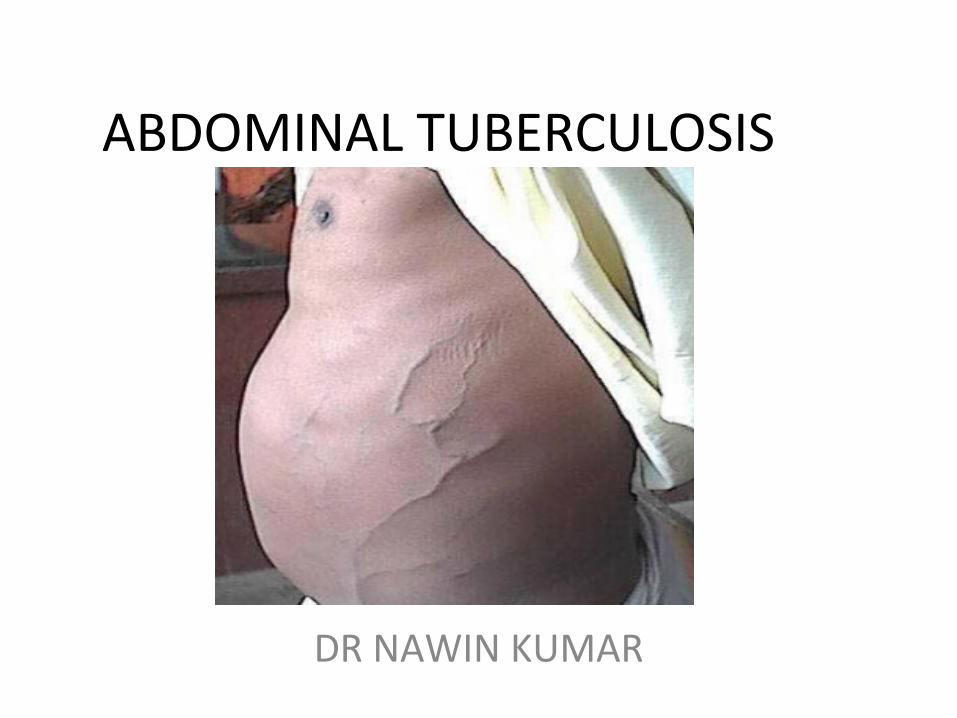
ABDOMINAL TUBERCULOSIS
DR NAWIN KUMAR
ABDOMINAL TUBERCULOSIS
TUBERCULOUS INFECTION OF ABDOMEN INCLUDING
• GASTRO INTESTINAL TRACT,
• PERITONEUM,
• OMENTUM,
• MESENTRY AND ITS NODES
• AND OTHER SOLID INTRA- ABDOMINAL ORGANS LIKE LIVER, SPLEEN,
PANCREAS.
IT IS ONE OF MOST COMMON FORM OF EXTRA PULMONARY
TUBERCULOSIS
CLASSIFICATION OF ABDOMINAL TUBERCULOSIS
1. GASTROINTESTINAL TUBERCULOSIS
2. TUBERCULOSIS OF THE MESENTERY
3. PERITONEAL TUBERCULOSIS
4. TUBERCULOSIS OF SOLID VISCERA
5. MISCELLANEOUS
GASTROINTESTINAL TUBERCULOSIS• ULCERATIVE• HYPERTROPHIC/HYPERPLASTIC• SCLEROTIC/ FIBROUS• DIFFUSE COLITIS
TUBERCULOSIS OF THE MESENTERY• MESENTRIC ADENITIS• MESENTRIC CYST • MESENTRIC ABCESS
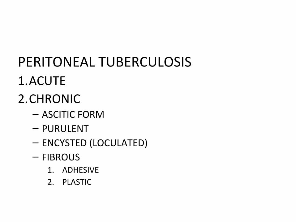
PERITONEAL TUBERCULOSIS1.ACUTE2.CHRONIC– ASCITIC FORM– PURULENT– ENCYSTED (LOCULATED)– FIBROUS
1. ADHESIVE2. PLASTIC
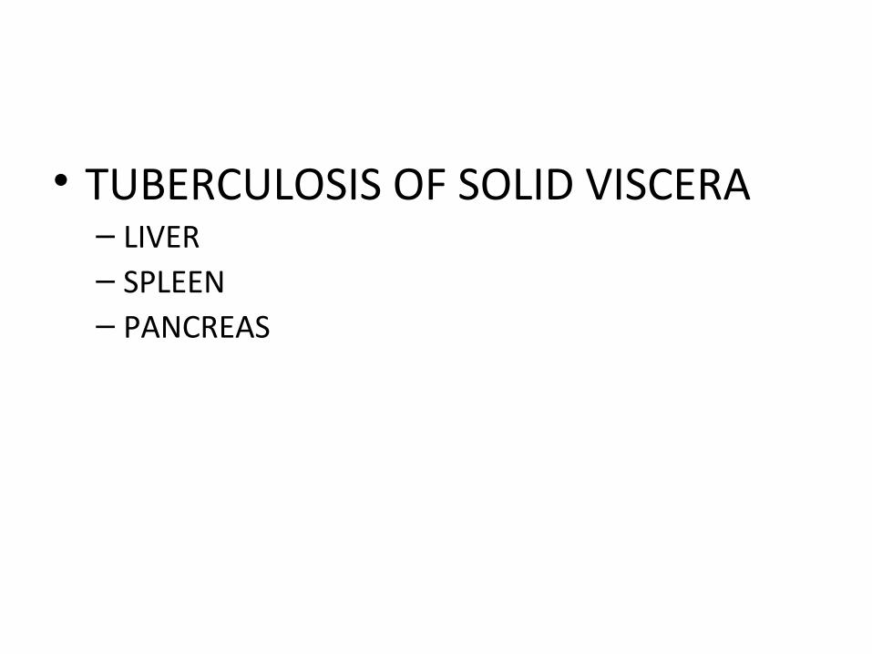
• TUBERCULOSIS OF SOLID VISCERA– LIVER – SPLEEN – PANCREAS
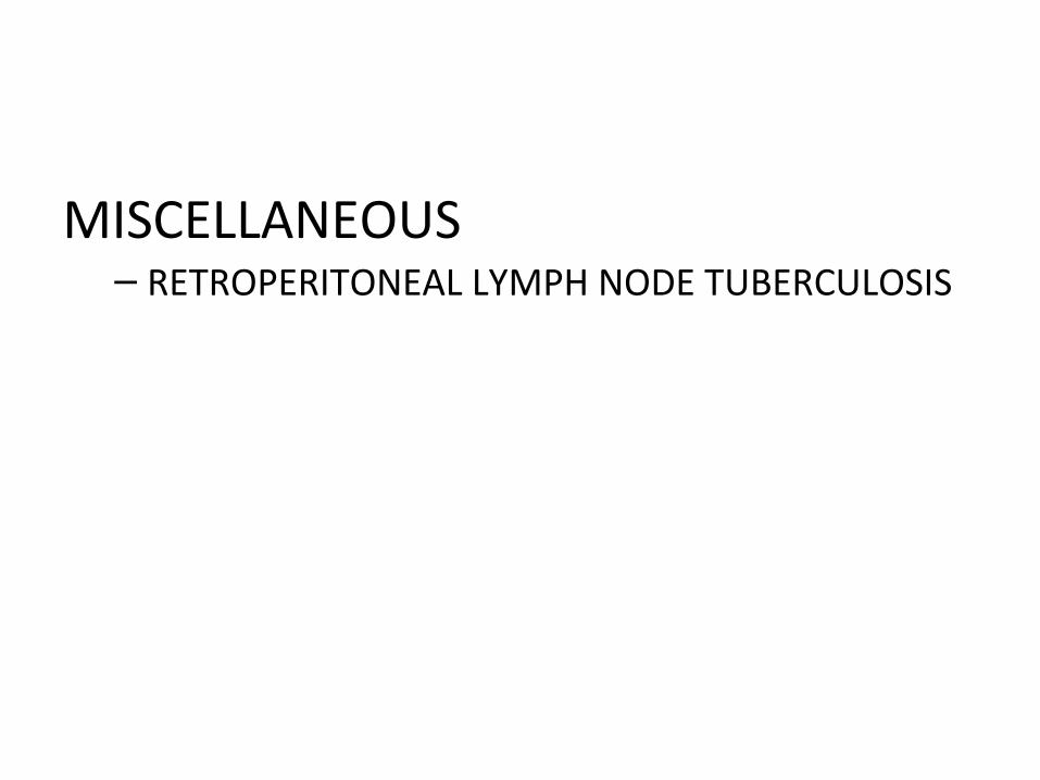
MISCELLANEOUS– RETROPERITONEAL LYMPH NODE TUBERCULOSIS
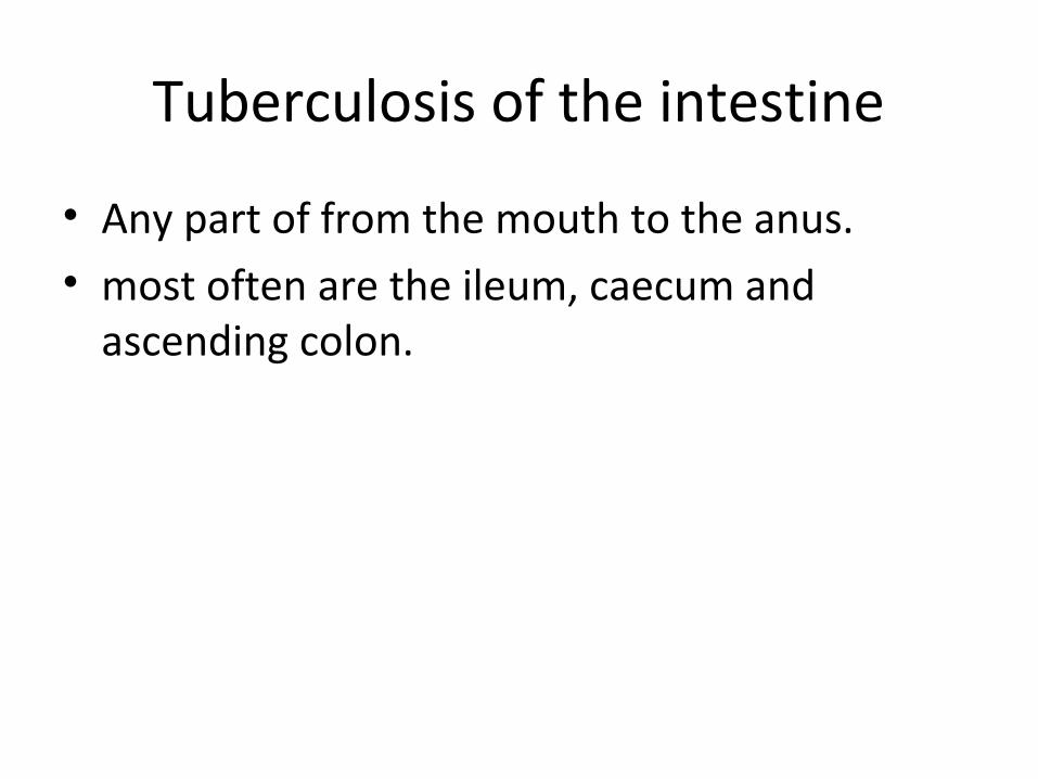
Tuberculosis of the intestine
• Any part of from the mouth to the anus. • most often are the ileum, caecum and
ascending colon.
Ulcerative tuberculosis
• secondary to pulmonary tuberculosis • as a result of swallowing tubercle bacilli. • multiple ulcers in the terminal ileum, lying
transversely, and the overlying serosa is thickened, reddened and covered in tubercles.
• Clinical features• Diarrhea and weight loss are the predominant
symptoms
• Radiology• barium meal and follow-through or small bowel enema• absence of filling of the lower ileum, caecum and most• of the ascending colon– Narrowing – Hypermotility of the ulcerated segment
• Treatment• MDT• operation – perforation – intestinal obstruction
Hyperplastic tuberculosis
• usually occurs in the ileocaecal region, although solitary and multiple lesions in the lower ileum are sometimes seen.
• ingestion of Mycobacterium tuberculosis • high resistance to the organism.
• The infection establishes itself in lymphoid follicles
• chronic inflammation • thickening of the intestinal wall • narrowing of the lumen• There is early involvement of the regional lymph
nodes, which may caseate.• Unlike CD, with which it shares many similarities,
abscess and fistula formation is rare.
Clinical features• abdominal pain with intermittent diarrhea • The ileum above the partial obstruction is distended,
and the stasis and consequent infection lead to steatorrhoea, anemia and loss of weight.
• mass in the right iliac fossa– appendix mass– carcinoma of the caecum– CD– tuberculosis – actinomycosis of the caecum.
• Radiology• barium follow-through or small bowel enema • long narrow filling defect in the terminal ileum• Treatment• Treatment• MDT• operation – perforation – intestinal obstruction

PERITONEAL TUBERCULOSIS1.ACUTE2.CHRONIC– ASCITIC FORM– PURULENT– ENCYSTED (LOCULATED)– FIBROUS
1. ADHESIVE2. PLASTIC
ACUTE
• very rare type• mimics acute peritonitis• when laparotomy is done straw colored fluid
escapes• tubercles are seen over peritoneum and
greater omentum
ACUTE
• early phases tubercles are grayish and translucent.
• undergo caseation and appear yellowish or white• D/D-– carcinoma.– patchy fat necrosis
• diagnosis is done by omental biopsy• Fluid - for bacteriological studies• wound closed without drainage.
CHRONIC
• has considerably declined with the practice of pasteurizing milk and availability of vaccination and newer anti tuberculosis chemotherapy
• making a return due to A. I.D.S.
CHRONIC
• common symptoms-– pain abdomen– fever– loss of weight– night sweats– mass abdomen
CHRONIC
Infection originates from:• tuberculous mesenteric lymph nodes;• tuberculosis of the ileocaecal region;• a tuberculous pyosalpinx;• blood-borne infection from pulmonary
tuberculosis, usually the ‘miliary’ but occasionally the ‘cavitating’ form
ASCITIC TYPE
• pathology– peritoneum is studded with tubercles– pale straw colored fluid. – insidious onset
ASCITIC TYPE
• loss of appetite and loss of weight• pallor• abdominal distension- chronic• constipation• diarrhea
ASCITIC TYPE
• abdominal wall has dilated veins• flanks are dull and this dullness can be shifted• mass may can palpated – rolled up omentum
studded with tubercles
ASCITIC TYPE
• mantoux test is positive• laparoscopy• areas of caseation are biopsied• chest x-ray• ascitic tap- high specific gravity, can be
cultured and guinea pig inoculation
ENCYSTED TYPE
• limited and loculated to one quadrant • encapsulated collection of fluid• differential diagnosis- ovarian cyst,
mesenteric cyst• can cause intestinal obstruction• investigation is similar to ascitic tye
FIBROUS FORM
• multiple adhesions• blind loop syndrome- diarrhea,
steatorrhea, weight loss , vit B deficiency, intestinal obstruction• multiple palpable swellings• treatment is surgical adhesionolysis,
resection of blind loop
PURULENT FORM
• from fallopian tubes• caseation of mesenteric lymph nodes• cold abscess or abscesses• can cause obstruction• can burst into bowel• faecal fistula can occur
Actinomycosis of the ileocaecal region
• Rare• narrowing of the lumen of the intestine does not
occur • mesenteric nodes do not become involved• a local abscess spreads to the retroperitoneal tissues
and the adjacent abdominal wall,• multiple indurated discharging sinuses• liver may become involved via the portal vein.
• Clinical features• Usually post-appendicectomy - 3 weeks after surgery, • a mass is palpable• wound begins to dis-charge• At first, the discharge is thin and watery- later thicker and malodorous• secondary faecal fistula • Pus -bacteriological examination- characteristic sulphur granules.• Treatment• Penicillin or cotrimoxazole treatment
– prolonged– high dosage.


![Follow Sipi cantpancreatitis · tuberculous]Tuberculous 38. 2010167550 lymphaderioPathy [lymph Fallow Up: 4 Korea Republ.. 09-Sep- node 11. tuberculosis]Tuberculous Pleural effusion](https://img.pdfslide.us/doc/110x75/5f7d6a51d573d133e30b0217/follow-sipi-tuberculoustuberculous-38-2010167550-lymphaderiopathy-lymph-fallow.jpg)