Embed Size (px)
Citation preview
How ApoE4 Interacts with
Estrogen to Cause
Alzheimer’s Disease
Presenter: Nicholas Smith
Eastern Illinois University
Honors Biology Department

Alzheimer’s DiseaseAn estimated 5.2 million Americans have Alzheimer’s Disease
(AD) (5).
Women are 1.5 times more likely to develop AD than men (3,7).
Alzheimer’s Disease
The precise biological cause is unknown yet many biological
markers are found within patients with the disease:
ApoE4, a lipid transporting protein, is a major risk factor for
AD.
This disease is more common in patients over the age of 65.
Symptoms include: memory loss, disorientation, difficulty with
abstract thought, behavioral and psychological changes and poor
judgment amongst other symptoms.
Relationship to Estrogen
A sharp decline in production of sex hormones, in particular
estrogen, by the ovaries following menopause is thought to trigger
AD (4).
Consistently, several studies showed that estrogen treatment in
postmenopausal women decreased AD risk.
In contrast, several other studies showed that estrogen therapy
increased AD risk in women.
It is unclear why estrogen protects some women from AD, yet
increases AD risk in others.
Apolipoprotein E (apoE)
ApoE, a lipid transporting protein, acts as a master switch to
determine if estrogen therapy is beneficial or harmful in women.
Humans have three major forms of apoE (apoE2, apoE3 and
apoE4) (6).
Estrogen treatment significantly prevents AD only in women with
apoE3 and increases the risk of AD in women with apoE4 (1,2).
Research Goal/Hypothesis
The mechanism whereby estrogen prevents AD in the presence of
apoE3, but not apoE4, is unclear.
Goal: To examine if apoE isoforms differentially alter the
production and localization of estrogen receptor (ER) subtypes in
brain areas that are susceptible to AD.
Hypothesis: The presence of apoE3 and apoE4 will differentially
alter the production and location of estrogen receptor (ER)
subtype ERα in the brain.
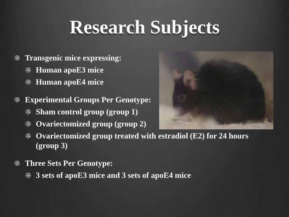
Research Subjects
Transgenic mice expressing:
Human apoE3 mice
Human apoE4 mice
Experimental Groups Per Genotype:
Sham control group (group 1)
Ovariectomized group (group 2)
Ovariectomized group treated with estradiol (E2) for 24 hours
(group 3)
Three Sets Per Genotype:
3 sets of apoE3 mice and 3 sets of apoE4 mice
Research Methods Outline
Animal Research:
Sham Surgery
Ovariectomy Surgery
Two week recovery period for mice
Group 1 & 2 mice brain tissue extraction on Day 14
Estradiol injection in group 3 mice on Day 14
Group 3 mice brain tissue extraction 24 hours later
The olfactory bulb, right cerebral cortex, and brainstem brain
tissue are isolated
Brain tissue is stored at -20 degrees Celsius in RNAlater
solution

Research Methods Outline
Basic Science Research:
Tissue Homogenization and RNA Extraction
RNA Concentration Quantification
RNA Standardization (1000 ng/microliter)
Reverse Transcription (RNA cDNA)
Polymerase Chain Reaction (PCR) Set Up
Real-Time Reverse Transcriptase Quantitative PCR (RT-
qPCR)

Ovariectomy
Ovariectomy Surgery: Dorsal, bilateral surgical removal of both
ovaries.
Sham Surgery: Ovaries are identified but not removed. This serves
as an experimental control.
Following surgery, mice recover under a lamp light for several
hours and then under normal conditions for 14 days.
Brain Tissue Isolation
Procedure: Isolate both olfactory bulbs, the right medial cerebral
cortex and brainstem.
When: 14 days following ovariectomy/sham surgery.
On day 14, the group 3 mice are treated with estradiol (10
ng/microliter) for 24 hours and then the same brain regions
described above are isolated.
Brain tissue is stored in RNAlater solution at -20 degrees Celsius.
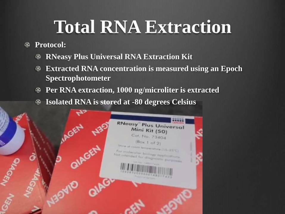
Total RNA ExtractionProtocol:
RNeasy Plus Universal RNA Extraction Kit
Extracted RNA concentration is measured using an Epoch
Spectrophotometer
Per RNA extraction, 1000 ng/microliter is extracted
Isolated RNA is stored at -80 degrees Celsius

Reverse Transcriptase
Protocol:
Invitrogen SuperScript III Reverse Transcriptase procedure
cDNA is stored at -20 degrees Celsius
RT-qPCR
Instrument: Applied Biosystems RT-qPCR Instrument
Total Reaction Volume: 20 microliters
SYBR Green Master Mix, cDNA, primer, and nuclease-free
water
Annealing Temperature: 60 degrees Celsius
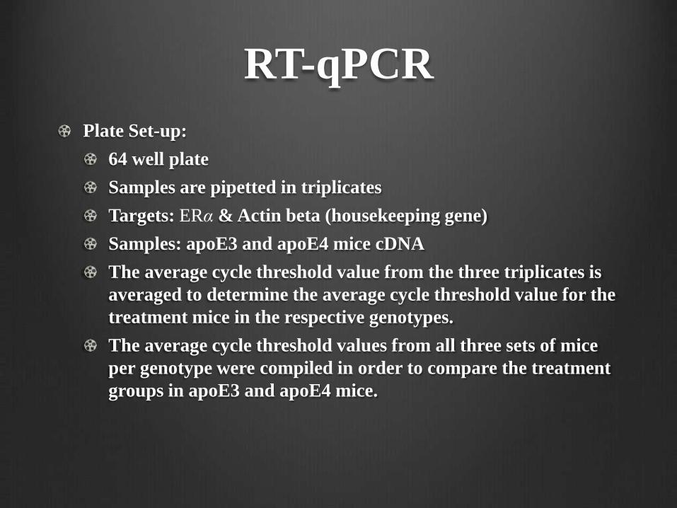
RT-qPCR
Plate Set-up:
64 well plate
Samples are pipetted in triplicates
Targets: ERα & Actin beta (housekeeping gene)
Samples: apoE3 and apoE4 mice cDNA
The average cycle threshold value from the three triplicates is
averaged to determine the average cycle threshold value for the
treatment mice in the respective genotypes.
The average cycle threshold values from all three sets of mice
per genotype were compiled in order to compare the treatment
groups in apoE3 and apoE4 mice.
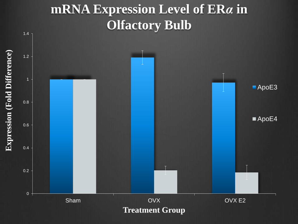
mRNA Expression Level of ERα in
Olfactory Bulb
0
0.2
0.4
0.6
0.8
1
1.2
1.4
Sham OVX OVX E2
Ex
pre
ssio
n (
Fo
ld D
iffe
ren
ce)
Treatment Group
ApoE3
ApoE4
Data Analysis
Delta-delta-cycle-threshold analysis
2^∆test/2^∆control
The expression (fold difference) of mRNA ERα in the olfactory
bulb of apoE3 and apoE4 mice was measured.
The expression fold difference was normalized to the expression of
the housekeeping gene, actin, as well as to the apoE3 and apoE4
sham control group mice (n = 3).
Conclusion
The ERα levels did not differ in apoE3 mice that were
ovariectomized and treated with either estradiol or vehicle as
compared to sham-operated apoE3 mice.
In contrast, ERα levels decreased five-fold in apoE4 mice that were
ovariectomized as compared to sham-operated apoE4 mice.
Conclusion
Estradiol treatment in ovariectomized apoE4 mice did not rescue
the drop induced by ovariectomy in apoE4 mice.
The data suggests that estrogen deficiency induced by ovariectomy
has differential effects on apoE3 and apoE4 mice.
The results suggest that estrogen replacement therapy may be
beneficial to apoE3-carriers, but not apoE4-carriers.
References(1) Burkhardt MS, Foster JK, Laws SM, Baker LD, Craft S, Gandy SE, Stuckey BG, Clarnette
R, Nolan D, Hewson-Bower B, Martins RN. (2004) Oestrogen replacement therapy may
improve memory functioning in the absence of APOE epsilon4. J Alzheimers Dis 6: 221-228.
(2) Corder EH, Saunders AM, Strittmatter WJ, Schmechel DE, Gaskell PC, Small GW, Roses
AD, Haines JL, Pericak-Vance MA. (1993) Gene dose of apoE type 4 allele and the risk of
Alzheimer's disease in late onset families. Science 261: 921-923.
(3) Gao S, Hendrie HC, Hui S. (1998) The relationships between age, sex, and the incidence of
dementia and Alzheimer disease: a meta-analysis. Arch Gen Psychiatry 55: 809-815.
(4) Henderson VW. (2004) Hormone therapy and Alzheimer's disease: benefit or harm? Expert
Opin Pharmacother 5: 389-406.
(5) Thies W., Bleiler L. (2013). 2013 Alzheimer's disease facts and figures. Alzheimers Dement.
9, 208–245.10.1016/j.jalz.2013.02.003
(6) Weisgraber KH. (1994) ApoE: structure-function relationships. Adv Pro Chem 45: 249-302.
(7) Whitehouse PJ. (1997) Genesis of Alzheimer's disease. Neurology 48: S2-7.

AcknowledgementsI would like to thank Dr. Nathan for his guidance and mentorship
throughout the duration of this research project and my
undergraduate career.
I would also like to recognize and thank Eastern Illinois University
and the Biological Sciences Department for providing the tools and
resources necessary to pursue this research project.