Embed Size (px)
Citation preview
V i k a s h B h a t t a r a i ( I n t e r n )F o r S u r g e r y D e p a r t m e n t
C M S - T H B h a r a t p u r
SHOCK

Introduction
Shock is a clinical syndrome that results from inadequate tissue perfusion leading to imbalance between delivery and requirement for oxygen and substrates which causes cellular ischemia and dysfunction.
The cellular ischemia which occurs induces the production and release of inflammatory mediators that further compromises the perfusion.
It is the most common so, important cause of deaths among surgical patients. So every surgeons must know…..

Pathophysiology of shock
Cellular level: Micro-vascular level:Systemic:
Cardiovascular Respiratory Renal Endocine
Ischemia-perfusion syndrome:
perfusion to the tissues is reduced
cells are deprived of oxygen
switch from aerobic to anaerobic metabolism.
Accumulation of lactic acid in the blood systemic metabolic acidosis.
glucose within cells is exhausted, anaerobic respirationceases
failure of sodium/potassium pumps
Intracellular lysosomes release autodigestive enzymes cell lysis
Intracellular contents, including potassium are released into the blood stream.
tissue ischaemia progresses
activation of the immune and coagulation systems
Hypoxia and acidosis activate complement and prime neutrophils
generation of oxygen free radicals and cytokine
injury of the capillary endothelial cells
further activate the immune and coagulation systems
Damaged endothelium becomes ‘leaky’
tissue edema, exacerbating cellular hypoxia
As preload and afterload decrease
Compensatory baroreceptor response
increased sympathetic activity
release of catecholamines into the circulation
tachycardia and systemic vasoconstriction (except in sepsis)
metabolic acidosis and increased sympathetic response
increased respiratory rate
increased minute ventilation
excretion of carbon dioxide
compensatory respiratory alkalosis
Decreased perfusion to kidney
Reduced filtration at the glomerulus
renin–angiotensin–aldosterone axis is stimulated
further vasoconstriction
increased sodium and water reabsorption
decreased urine output
decreased preload
activation of the adrenal and renin–angiotensin systems
vasopressin (antidiuretic hormone)- hypothalamus
Cortisol – adrenal cortex
vasoconstriction and resorption of water
decreased urine output
During systemic hypoperfusion
cellular and organ damage due to tissue hypoxia andlocal activation of inflammation.
Further injury occurs once normal circulation is restored
The acid and potassium load
direct myocardial depression, vascular dilatation and further hypotension
cellular and humoral elements activated by the hypoxia (complement,neutrophils, microvascular thrombi)
flushed back into circulation
further endothelial injury to organs such as the lungs and the kidneys acute lung injuryacute renal injury multiple organ failure and death.
Reperfusion injury can currently only be attenuated by reducingthe extent and duration of tissue hypoperfusion.
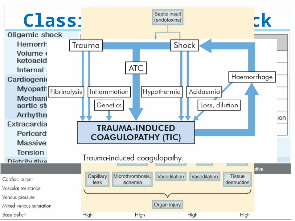
Classification of Shock

Clinical manifestations
• Hypotension (mean arterial BP <60 mmHg) • Tachycardia• Tachypnea• Pallor• Restlessness• Altered sensorium• Signs of intense peripheral vasoconstriction, weak pulses and coldclammy extremities. •In distributive (e.g., septic) shock, vasodilatation
predominates and extremities are warm. Sepsis Fever
• Oliguria (<400 mL/day) and metabolic acidosis common• If not controlled, anuria (<100 mL/day)

Severity Of Shock
CompensatedDecompensationMild ModerateSevere

Pitfalls
• The classic cardiovascular responses are not elicited in every patient.• The patient in shock may not be recognized.• Tachycardia:
Patients on beta-blockers, pacemakers, penetrating injury with less tissue injury
• Blood pressure: one should recognise it as one of the last signs of shock
Children and young fit patients, elderly who are normally hypertensive
• Capillary refill:Distributive shock, patients are not cold, clammy but have brisk
capillary refill

Possible consequences
Unresuscitable Shock:profound shock for prolonged perioddelayed, inadequate or inappropriate resuscitationdeath is inevitable
Multiple organ failure:defined as 2 or more failed organ systemsprolonged ongoing systemic ischemia (Occult hypoperfusion)
Ischemia reperfusion syndrome……..Mgt?

Resuscitation
Immediate – Ensure airway and adequate oxygenation and ventilation
Then attention is directed to Cardiovascular resuscitation
NEVER DELAY resuscitation for definitive DxRapid and careful clinical examination – clues to make appropriate
initial determination
People actively bleeding high volume therapy is counterproductive immediate operation for control of bleeding with parallel resuscitation should be done
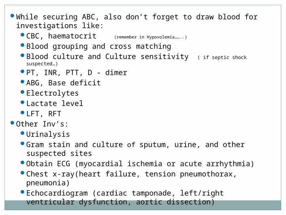
While securing ABC, also don’t forget to draw blood for investigations like:CBC, haematocrit (remember in Hypovolemia……..)
Blood grouping and cross matchingBlood culture and Culture sensitivity ( if septic shock suspected…)
PT, INR, PTT, D - dimerABG, Base deficitElectrolytesLactate levelLFT, RFT
Other Inv’s:UrinalysisGram stain and culture of sputum, urine, and other suspected sitesObtain ECG (myocardial ischemia or acute arrhythmia)Chest x-ray(heart failure, tension pneumothorax, pneumonia)Echocardiogram (cardiac tamponade, left/right ventricular dysfunction,
aortic dissection)

In all cases of shock, FIRST LINE THERAPY SHOULD ALWAYS BE iv FLUIDS to correct hypovolemia and inadequate pre-load
Otherwise, EMPTY heart will rapidly and permanently deplete myocardium of oxygen store becomes unresponsive to resuscitation
For initial resuscitation wide short bore catheters better than long narrow line Central venous catheter, which is more appropriate for monitoring than Fluid replacement

Types of fluids
There is no ideal Res. Fluid and is matter of debateStudies show no overt differences b/n crystalloids and colloidsRather colloids are more expensive and have worse side-effect
profilesMOST IMPtly Both have ZERO oxygen carrying capacity, and if
blood is lost ideal replacement is blood but they help buying time while awaiting blood products
Don’t use hypotonic solutions like Dextrose Poor plasma volume expanders (exception, if free water loss as in DI, and sodium overload in cirrhosis.)

Dynamic Fluid response
One of the methods to determine Shock status of patient250-500 mL fluid given over 5-10 mins and Cardiovascular
response observed (HR, BP, CVP)
Responders: not actively losing fluid but require filling
Transient responders: moderate ongoing fluid loss
Non-responders: severely volume depleted

After fluid challenge, There should be raise in CVP of 2-5 cmH2O which gradually drifts back over 10-20 mins
Those not showing change further fluid resuscitation
Vasopressors/Inotropic therapy may be required
Once HR, BP, CVP, UO restored patient is kept on maintenance fluid therapy.4-2-1 mL/kg/hr for 1st 10 kg, 2nd 10kg and rest respectivelyor,100-50-20 mL/kg/day for 1st 10 kg, 2nd 10kg and rest respectively

Vasopressors and inotropic support

Monitoring
CO, SVR and preload monitoring (real time monitoring of cardiovascular response) Invasive: Pulmonary Artery Catheter Non-invasive: Doppler Ultrasound, Pulse waveform analysis and Indicator dilution method
Mixed Venous Oxygen saturation (N- 50-70%, Sepsis > 70%, Others - <50%)
Level of consciousness adequate marker of cerebral perfusion
New methods: monitoring regional tissue perfusion Muscular tissue oxygen probes Near infrared spectroscopy Sub lingual capnometry
ICU setup is appropriate
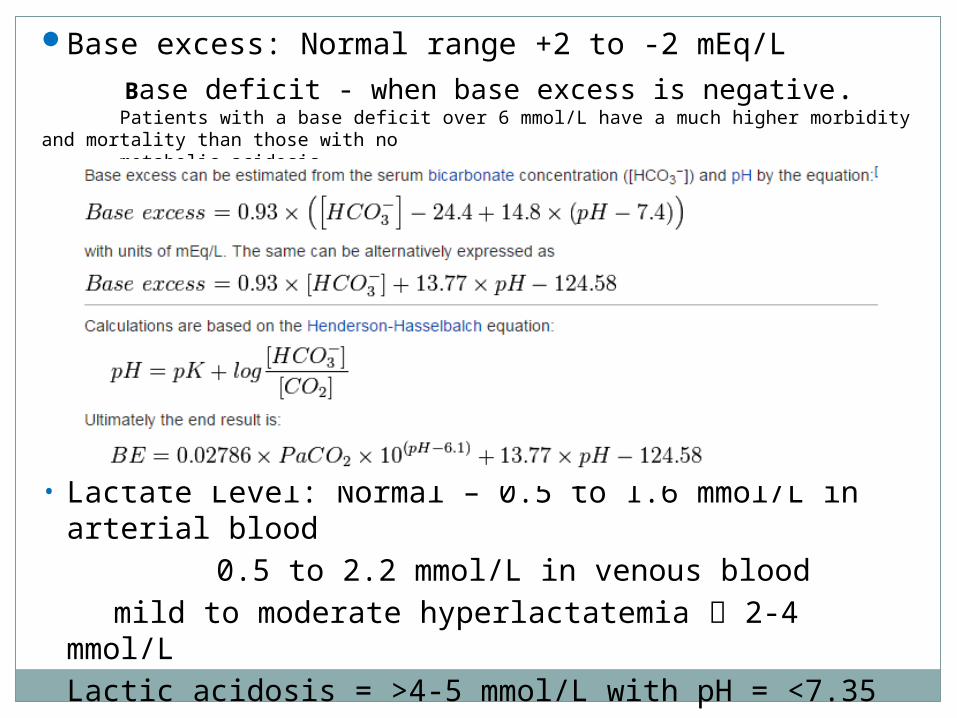
Base excess: Normal range +2 to -2 mEq/L Base deficit - when base excess is negative. Patients with a base deficit over 6 mmol/L have a much higher morbidity and mortality than those with no metabolic acidosis
• Lactate Level: Normal – 0.5 to 1.6 mmol/L in arterial blood 0.5 to 2.2 mmol/L in venous blood
mild to moderate hyperlactatemia 2-4 mmol/LLactic acidosis = >4-5 mmol/L with pH = <7.35

Endpoints of resuscitation
Easy to know WHEN TO START, hard to know WHEN TO STOP
Traditionally patients resuscitated until vitals and U.O. normalizedWhich is WRONG, as some tissues may still be in OH, Later MOF
New concept, resuscitation not stopped until sensitive parameters like Base deficit, Lactate level and mixed venous oxygen saturation are normalised.

Antimicrobial Regimens (IV Therapy)• Immunocompetent adult(1) piperacillin-tazobactam (3.375 g q4–6h)(2) imipenem-cilastatin (0.5 g q6h) or meropenem (1 g q8h); or (3) cefepime (2 g q12h). If the pt is allergic to β- lactam agents, use ciprofloxacin(400 mg q12h) orlevofloxacin (500–750 mg q12h)plus clindamycin (600 mg q8h). Vancomycin(15 mg/kg q12h) should be added to each of the above regimens.• Neutropenia (<500 neutrophils/μL)(1) imipenem-cilastatin (0.5 g q6h) or meropenem (1 g q8h) or cefepime(2 g q8h)(2) piperacillin-tazobactam (3.375 g q4h)plus tobramycin (5–7 mg/kg q24h). Vancomycin(15 mg/kg q12h) should be added if the pt has an indwelling vascular catheter, has received quinolone prophylaxis, or has received intensive chemotherapy that produces mucosal damage

• Splenectomy Cefotaxime (2 g q6–8h) or ceftriaxone (2 g q12h)should be used. If the
local prevalence of cephalosporin-resistant pneumococci is high, add vancomycin.
If the pt is allergic to β- lactam drugs, vancomycin (15 mg/kg q12h) plus either moxifloxacin (400 mg q24h) or levofloxacin (750 mg q24h) or aztreonam (2 g q8h) should be used.
• IV drug user Vancomycin (15 mg/kg q12h)
• AIDS Cefepime (2 g q8h) or piperacillin-tazobactam (3.375 g q4h) plus
tobramycin (5–7 mg/kg q24h) should be used.
If the pt is allergic to β- lactam drugs, ciprofloxacin (400 mg q12h) or levofloxacin 750 mg q12h) plus vancomycin (15 mg/kg q12h) plus tobramycin
should be used.

Management of Hemorrhagic shock
Every effort directed towards rapidly identifying and stopping Haemorrhage
Identify haemorrhage
Immediate resuscitative manoeuvres Identify the site of haemorrhage
Haemorrhage controlThe bleeding, shocked patient must be moved rapidly to a placeof haemorrhage control.

Summary

Thank You!


![SHOCK[1] - Hypovolemic Shock](https://img.pdfslide.us/doc/110x75/58edc1bc1a28abae538b4711/shock1-hypovolemic-shock.jpg)