Embed Size (px)
Citation preview


A 63 year old buissinessman Mr.Basheer,
from Pudunagaram, came to surgery OPD
on 2/1/2013 with H/O right sided
abdominal pain of 3 days duration .
There was no H/O nausea, vomiting,
anorexia, fever or weight loss.
Bowel & bladder was normal
He is neither a smoker nor an alcoholic

1. He is a known hypertensive on treatment
2. He has H/O tuberculosis 30 years back completed Antituberculartreatment under category 1
3. Morbid obesity +

On examination
Vitals stable, afebrile
abdomen was distended ,
umbilical hernia + ,
dialated veins + on the right flank, flow from below
upwards.
Diffuse tenderness + involving the right hypochondrium & RIF
No organomegaly
No free fluid
Bowel sounds heard
External genitalia appeared normal
Hernial orifices free except for umbilical hernia
Clinical diagnosis of Subhepatic appendicitis was suspected & was worked up.
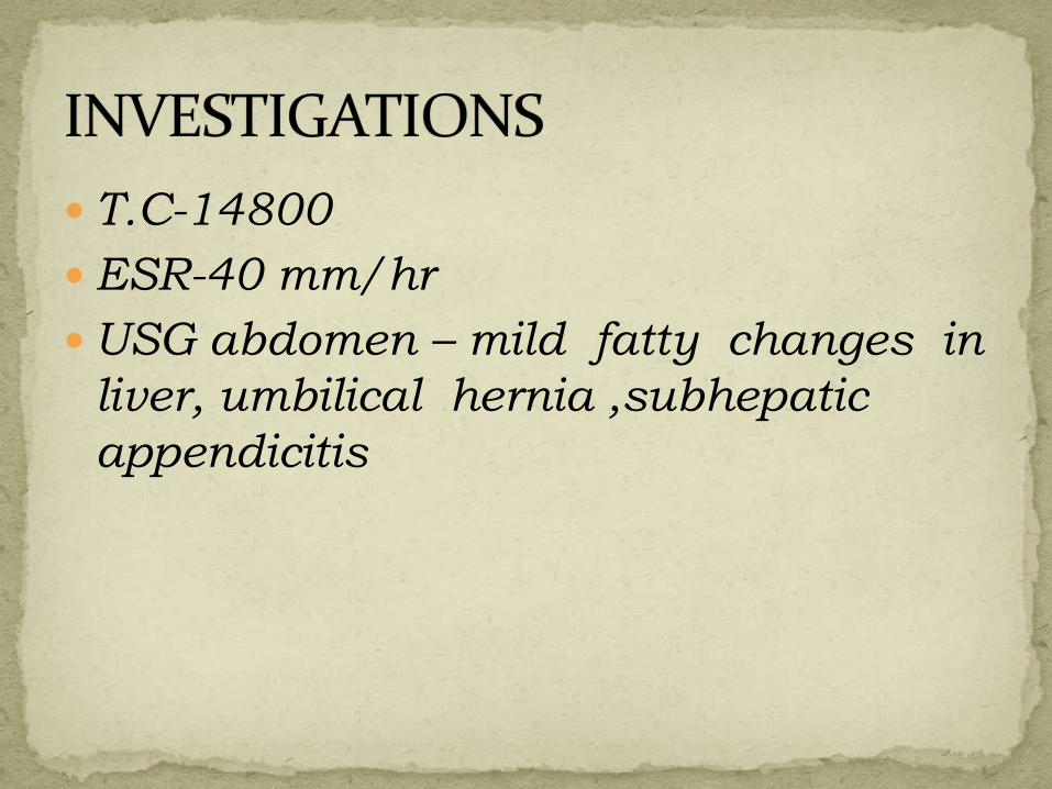
T.C-14800
ESR-40 mm/hr
USG abdomen – mild fatty changes in
liver, umbilical hernia ,subhepatic
appendicitis

His Alvarado [MANTRELS] score was 4
Tenderness in the RIF 2
Leucocytosis 2
He was advised conservative management with oral antibiotics & analgesics and was sent home.
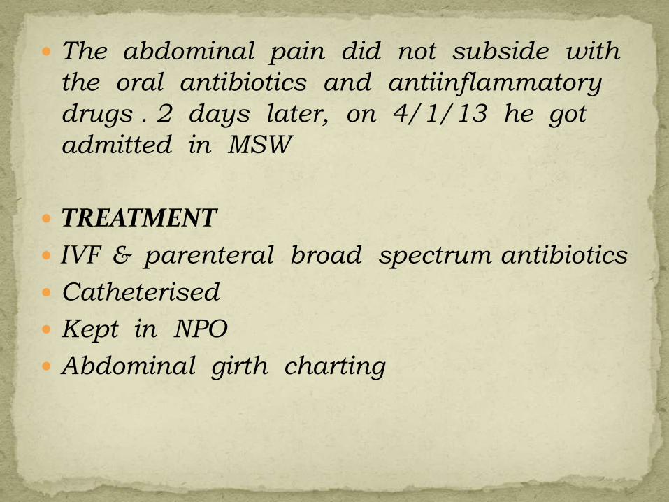
The abdominal pain did not subside with
the oral antibiotics and antiinflammatory
drugs . 2 days later, on 4/1/13 he got
admitted in MSW
TREATMENT
IVF & parenteral broad spectrum antibiotics
Catheterised
Kept in NPO
Abdominal girth charting
On the next day on 5/2/13 patient developed
difficulty in breathing
O/E basal crepts heard bilaterally
Chest X ray –bilateral basal pneumonia
Patient was given inhalational & intravenous
bronchodilators and steroids.
Sputum AFB & gramstaining was not done
since the patient could not produce sputum

2 days later, on 7/1/13 , a swelling was observed in the right inguinal region, cord was thickened .
USG scrotum- bilateral acute epididymitis R > L & bilateral minimal hydrocele
On 8/1/13 ,His breathing difficulty persisted ,O/E coarse inspiratory crepts heard in the left infrascapular area,
Chest X-ray - nonhomogenous opacity on left lower zones
Diagnosis – left lower lobe pneumonia
He was given I.V antibiotics .

C.T. ABDOMEN - liver normal size with fatty
changes and calcified focus,umbilical hernia ,
minimal fluid collection in RIF,right cord
appear thickened with minimal surrounding
fluid,minimal gaseous distention of small bowel
loops.
C.T.THORAX – consolidation seen in posterior
segment of left upper lobe,trace of left pleural
effusion seen
WORKING DIAGNOSIS –
1) Left pneumonia
2) Right epididymitis
3) Subhepatic appendicitis
4) Chronic liver disease

His total count & abdominal girth was
progressively increasing over 8 days.
On 12/1/13 screening USG showed focal
collection in RIF & USG guided aspiration
was done which yielded 5ml of frank pus &
was sent for culture & sensitivity.
D/D 1.R epididymitis
2.appendicular abscess
Patient was planned for exploratory
laparotomy.

EXPLORATORY LAPAROTOMY WITH RIGHT HIGH INGUINAL ORCHIDECTOMY was done on 12/1/13 under epidural anaesthesiathrough low right paramedian incision.
FINDINGS – Dilatation of bowel + RIF explored ,PUS COLLECTION + ,APPENDIX NORMAL, ASCENDING INFLAMMATION OF CORD STRUCTURES WITH NECROSIS & ABSCESS FORMATION found
PROCEDURE - RIF explored , Pus evacuated , peritoneal wash given ,R orchidectomy done

Pus c/s-organism isolated was E.coli
sensitive to amikacin, ceftazidime, ofloxacin
& pipiracillin/tazobactum
HPE reports –
1) Epididymis – Non specific epididymitis with
duct obstruction ,Epididymal cyst
2) Testes – No significant pathology
3) Spermatic cord - funiculitis

ASCENDING INFECTION OF RIGHT CORD STRUCTURES RESULTING IN RIF ABSCESS

Post operative period was uneventful except for oozing from drain tube site
Pus c/s from drain tube site : organism isolated was E.coli sensitive to amikacin,ampicillin,sulbactum,ofloxacin,pipiracillin,tazobactum,imipenam
On 28/1/13 USG Abdomen showed 8 *2.1 cm , 25 cc collection seen in the abdominal wall in the right lower abdomen, a diagnosis of anterior abdominal wall abscess was made for which USG guided wound debridement done under L/A on 9/2/13 &
the patient was discharged on the 30 th post -operative day on 11/2/13 after check USG.

Inflammation confined to epididymis -EPIDIDYMITIS
Infection spreading to testes - EPIDIDYMO-ORCHITIS

Mode of infection
1. Primary infection of urethra, prostate or seminal vesicles → via vas → epididymis
2. In men with BOO – high pressure in the prostatic urethra→reflux of infected urine up the vasa
3. Young men – STD –Chlamydia & gonococci – asso . With urethritis
4. Bloodborne - if E.coli,streptococci,proteus without evidence of urinary infection

Acute epididymitis can follow any form of urethral instrumentation or catheterisation
Acute tuberculous epididymitis - if vas is thickened & there is little response to antibiotics
Mumps – at any age
Infections with other enteroviruses, brucellosis,lymphogranuloma venerum also can cause epididymitis

initial symptoms of urinary infection
Ache in groin
Fever
Epididymis &testes swell and become painful
Scrotal wall – first red edematous & shiny & may become adherent to epididymis
Occasionally an abscess can form & discharge pus through scrotal skin
Resolution may take 6-8 weeks

Urine analysis & urine culture & sensitivity should be obtained in all cases
Need aggressive treatment with parenteralantibiotics
Doxycycline – DOC- for chlamydial infection
Broad spectrum antibiotic against urinary tract pathogens
Analgesics
Plenty of oral fluids
SCROTAL ELEVATION
If suppuration + - drainage is necessary
reactive hydrocoele,
ABSCESS FORMATION
infarction of the testicle,
testicular atrophy,
reduced fertility.

Due to retrograde infection from a tuberculous focus in seminal vesicles –so lower pole of epididymis is involved first
Clinical features – fiirm discrete swelling of lower pole of epididymis which aches a little ,the disease progresses until the whole epididymis is firm & craggy behind a normal feeling testes
Lax secondary hydrocele Beading of vas Seminal vesicles indurated & swollen Cold abscess formation & discharge
Investigations – examination of urine & semen for tubercle bacilli ,IVU & chest X ray

TREATMENT
ATT
If no resolution within 2 months - epididymectomyor orchidectomy