Embed Size (px)
Citation preview
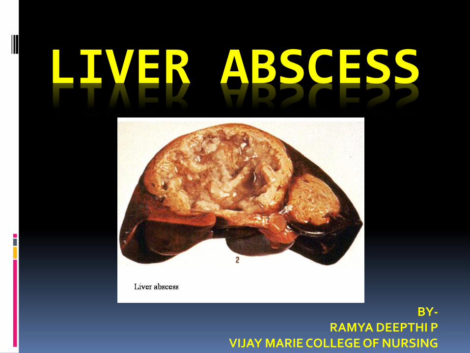
LIVER ABSCESS
BY-RAMYA DEEPTHI P
VIJAY MARIE COLLEGE OF NURSING
INTRODUCTION Liver abscess are more common in developing
countries.
Majority of them are due to parasitic infestations-amoebic, echinococcosis.
It is rarely seen in developed countries.
Etiology I. Bacterial infectionsa. Pyogenci liver abscessb. Pyophlebitis abscessc. Cholangitic abscessII. Parasitic infestationsA. Protozoal disease
a. Amoebiasisb. Malariac. Kala azar
B. Helmenthic diseasea. Ascariasisb. Liver flukesc. Echinococcosis (Hydatid disease)
C. LeptospirosisD. Syphilis
PYOGENIC LIVER ABSCESS Most liver abscess re of bacterial in origin.
Infecting organisms are-
1. Garm negative bacteria- E. Coli, Pseudomonas Klebsiella, enetrobacter
Route of entry to liver are-
a. Portal vein
b. Arterial supply
c. Ascending infection from biliary tract
d. Direct invasion of liver from nearby source
e. Penetrating injury
f. Cryptogenic (unknown)
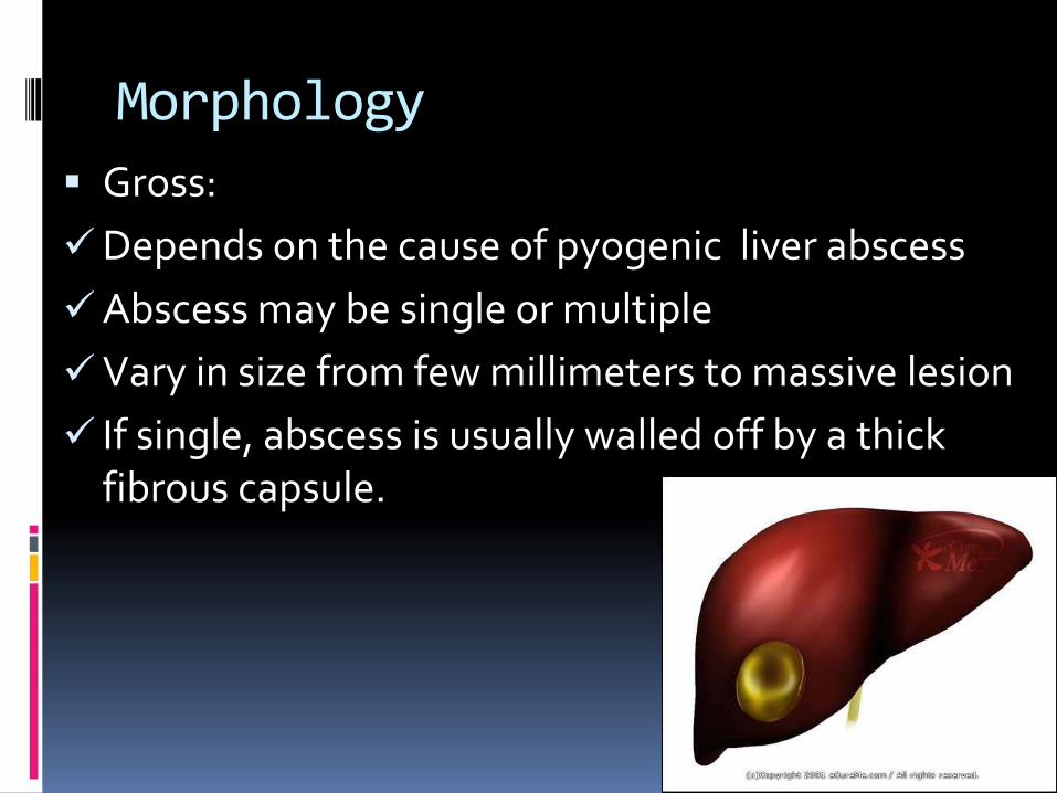
Morphology
Gross:
Depends on the cause of pyogenic liver abscess
Abscess may be single or multiple
Vary in size from few millimeters to massive lesion
If single, abscess is usually walled off by a thick fibrous capsule.
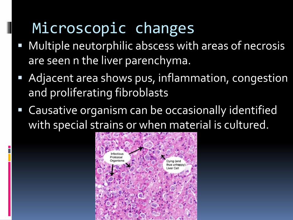
Microscopic changes Multiple neutorphilic abscess with areas of necrosis
are seen n the liver parenchyma.
Adjacent area shows pus, inflammation, congestion and proliferating fibroblasts
Causative organism can be occasionally identified with special strains or when material is cultured.
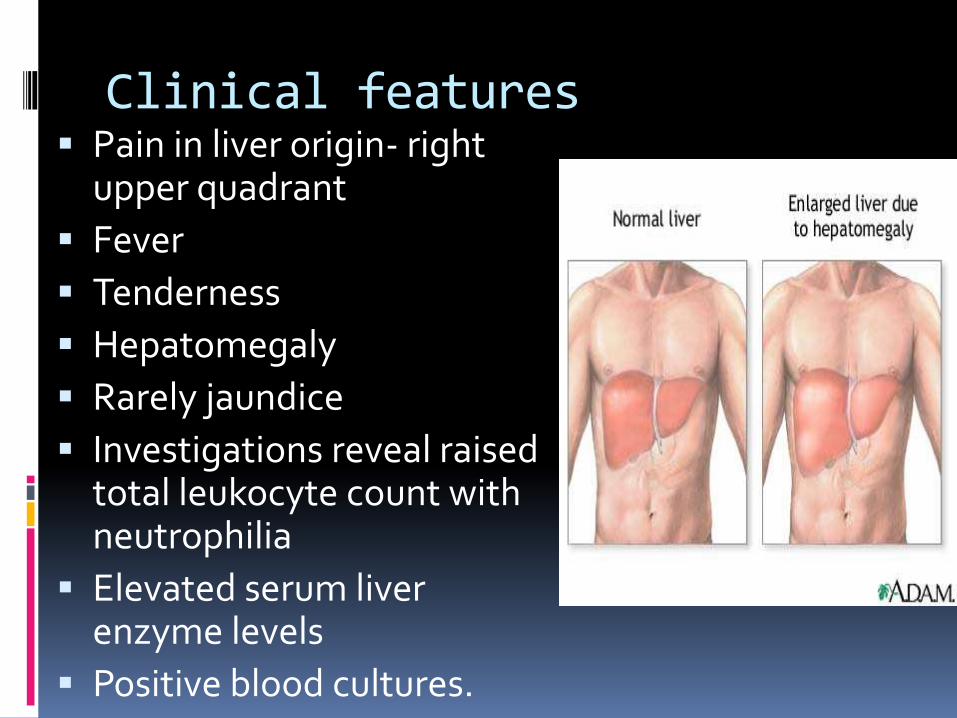
Clinical features Pain in liver origin- right
upper quadrant
Fever
Tenderness
Hepatomegaly
Rarely jaundice
Investigations reveal raised total leukocyte count with neutrophilia
Elevated serum liver enzyme levels
Positive blood cultures.
II. AMOEBIC LIVER ABSCESS AMOEBIC LIVER ABSCESS IS MORE COMMON IN
DEVELOPING COUNTRIES.
However it is not as common as pyogenic abscess.
Pathogenesis is caused by Entamoeba Histolytica.
It spreads form intestinal lesions
Parasite occurs in 2 forms- Trophozoite and cystic form.
Cysts are more infective stages of the parasite and are found in contaminated water and food.
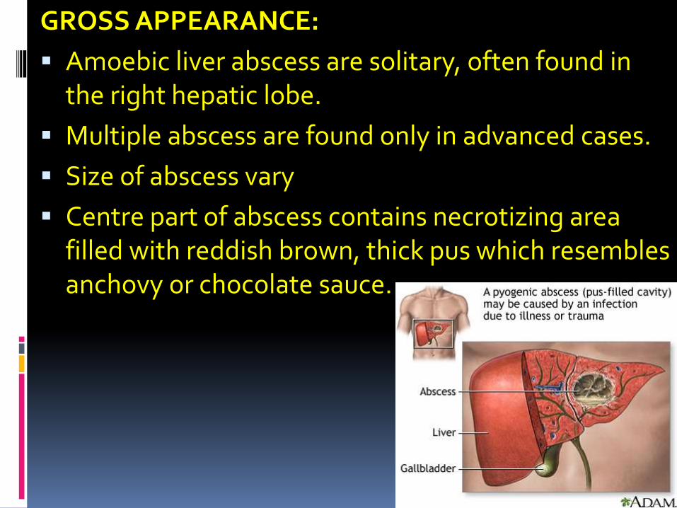
GROSS APPEARANCE:
Amoebic liver abscess are solitary, often found in the right hepatic lobe.
Multiple abscess are found only in advanced cases.
Size of abscess vary
Centre part of abscess contains necrotizing area filled with reddish brown, thick pus which resembles anchovy or chocolate sauce.
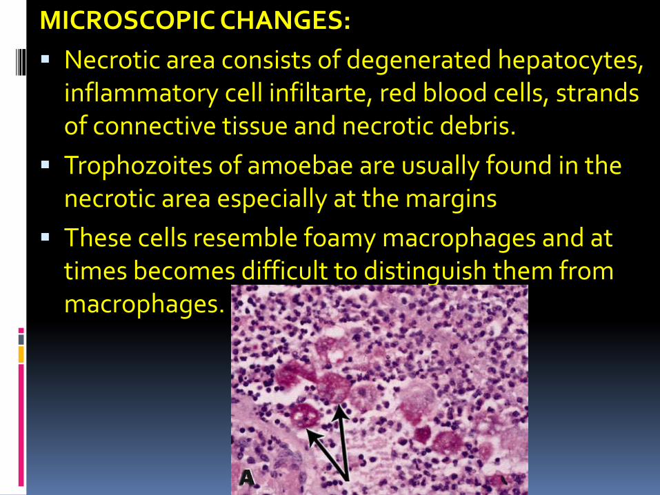
MICROSCOPIC CHANGES:
Necrotic area consists of degenerated hepatocytes, inflammatory cell infiltarte, red blood cells, strands of connective tissue and necrotic debris.
Trophozoites of amoebae are usually found in the necrotic area especially at the margins
These cells resemble foamy macrophages and at times becomes difficult to distinguish them from macrophages.
Complications of amoebic abscess Large abscess may rupture, penetrate the
diaphragm and enter into lung
Rupture into pleural cavity , peritoneal cavity or pericardia sac.
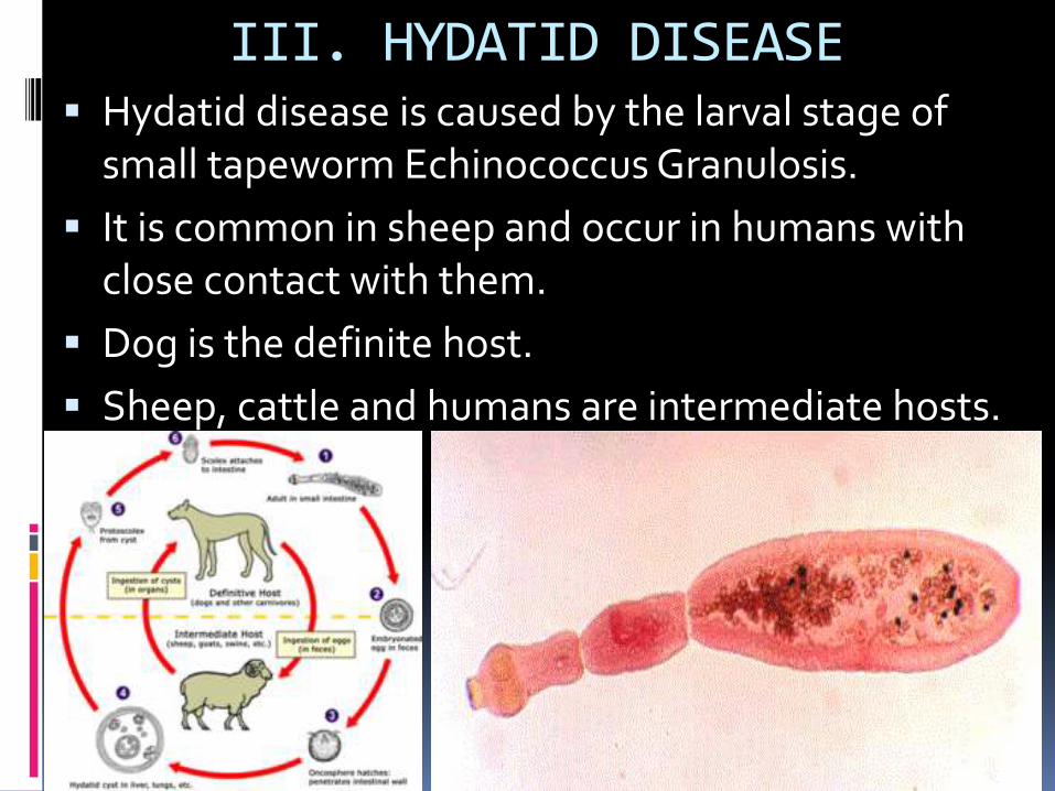
III. HYDATID DISEASE Hydatid disease is caused by the larval stage of
small tapeworm Echinococcus Granulosis.
It is common in sheep and occur in humans with close contact with them.
Dog is the definite host.
Sheep, cattle and humans are intermediate hosts.
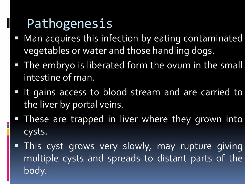
Pathogenesis Man acquires this infection by eating contaminated
vegetables or water and those handling dogs.
The embryo is liberated form the ovum in the smallintestine of man.
It gains access to blood stream and are carried tothe liver by portal veins.
These are trapped in liver where they grown intocysts.
This cyst grows very slowly, may rupture givingmultiple cysts and spreads to distant parts of thebody.
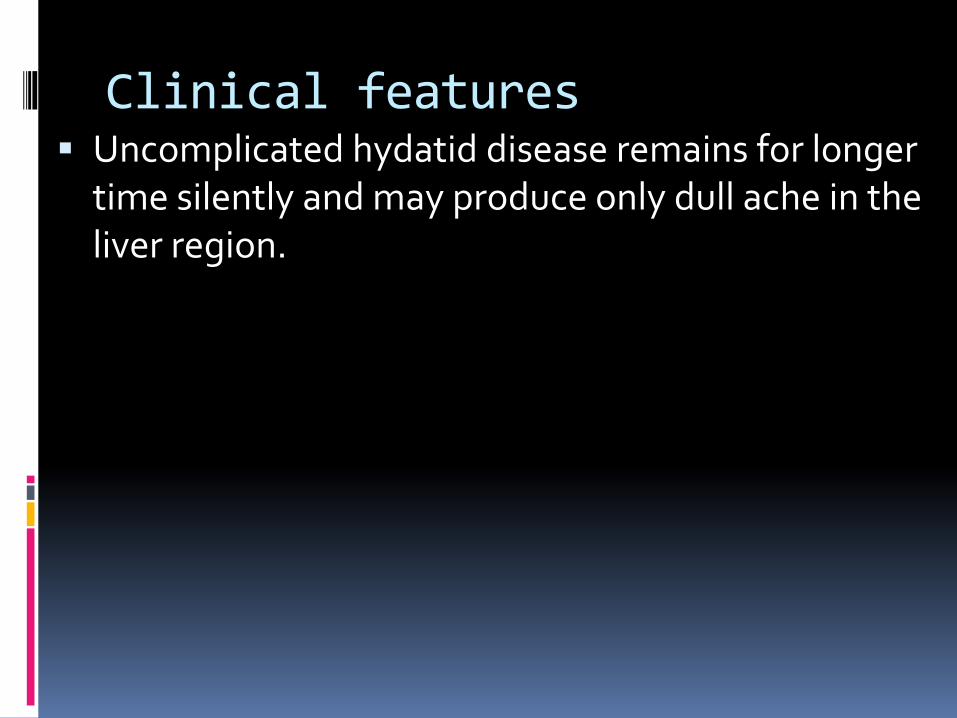
Clinical features Uncomplicated hydatid disease remains for longer
time silently and may produce only dull ache in the liver region.
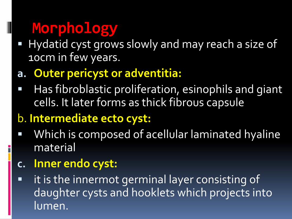
Morphology Hydatid cyst grows slowly and may reach a size of
10cm in few years.
a. Outer pericyst or adventitia:
Has fibroblastic proliferation, esinophils and giant cells. It later forms as thick fibrous capsule
b. Intermediate ecto cyst:
Which is composed of acellular laminated hyaline material
c. Inner endo cyst:
it is the innermot germinal layer consisting of daughter cysts and hooklets which projects into lumen.
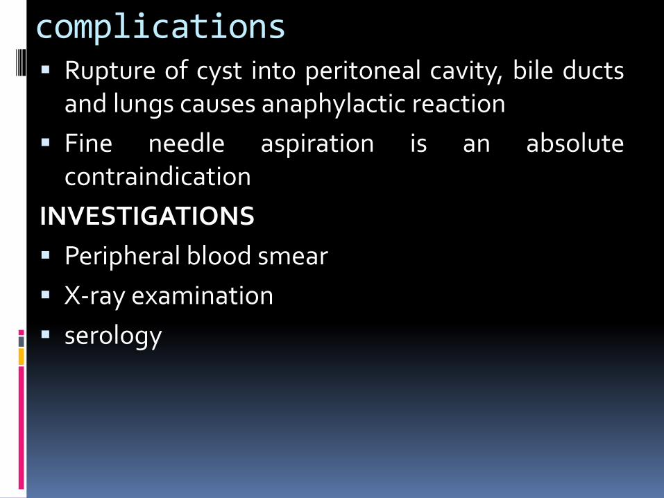
complications Rupture of cyst into peritoneal cavity, bile ducts
and lungs causes anaphylactic reaction
Fine needle aspiration is an absolutecontraindication
INVESTIGATIONS
Peripheral blood smear
X-ray examination
serology

Thank you…


![Erasmus 2012 benign liver [Alleen-lezen] · B. Secondary benign liver lesions 1. Abscess C. Hepatic Pseudolesions 1. Focal Steatosis, Focal spared Area in Fatty Liver 2. ... • Incidental](https://img.pdfslide.us/doc/110x75/5b0382477f8b9a2e228c816d/erasmus-2012-benign-liver-alleen-lezen-secondary-benign-liver-lesions-1-abscess.jpg)


![Review Article Klebsiella pneumoniae Liver Abscess · 2019-04-04 · Diabetes mellitus (DM) is believed to be one of the risk factors of K. pneumoniae liver abscess [13]. Compared](https://img.pdfslide.us/doc/110x75/5ee27cb6ad6a402d666cec7f/review-article-klebsiella-pneumoniae-liver-abscess-2019-04-04-diabetes-mellitus.jpg)