Embed Size (px)
Citation preview
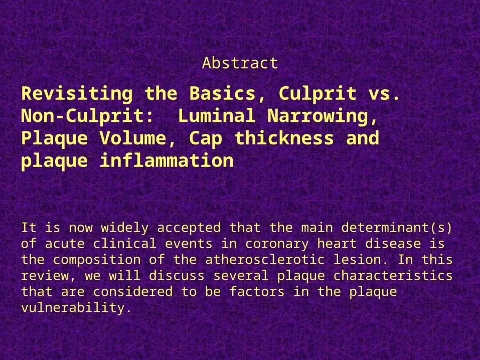
Revisiting the Basics, Culprit vs. Non-Culprit: Luminal Narrowing, Plaque Volume, Cap thickness and plaque inflammation
It is now widely accepted that the main determinant(s) of acute clinical events in coronary heart disease is the composition of the atherosclerotic lesion. In this review, we will discuss several plaque characteristics that are considered to be factors in the plaque vulnerability.
Abstract

Abstract (con’t)Luminal narrowing.
In a classic paper, Ambrose et al, reported that acute myocardial infarctions frequently developed in lesions that were not considered stenotic a few months before the ischemic event. Shortly afterwards, Little et al confirmed these findings. Moreover, in their series, 19 out of 29 patients had an occluded vessel responsible for their new myocardial infarction that was less than 50% stenotic in their previous angiogram, and 28 out of 29 patients had less than 70% narrowing in their culprit vessel on the first angiogram. In some biomechanical models, increase of stenosis leads to decrease of peak stress in the plaque, especially in lipid-rich plaques. It should be remembered, however, that plaque burden is a strong predictor of vascular events as demonstrated by a high EBCT score. The plaque burden, however, is predictive of the patient’s prognosis, not of a particular lesion progression. Also, a prospective five-year angiographic follow-up of factors associated with progression of coronary artery disease in the Coronary Artery Surgery Study showed that initial lesion severity was predictive of late segment occlusion.
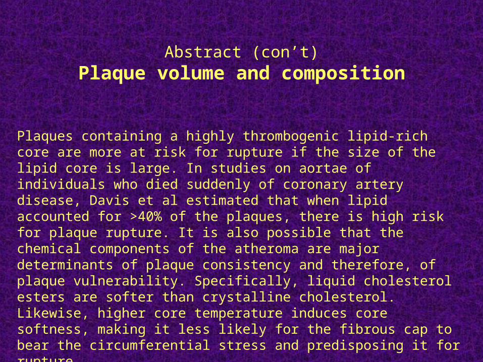
Plaques containing a highly thrombogenic lipid-rich core are more at risk for rupture if the size of the lipid core is large. In studies on aortae of individuals who died suddenly of coronary artery disease, Davis et al estimated that when lipid accounted for >40% of the plaques, there is high risk for plaque rupture. It is also possible that the chemical components of the atheroma are major determinants of plaque consistency and therefore, of plaque vulnerability. Specifically, liquid cholesterol esters are softer than crystalline cholesterol. Likewise, higher core temperature induces core softness, making it less likely for the fibrous cap to bear the circumferential stress and predisposing it for rupture.
.Abstract (con’t)
Plaque volume and composition
Abstract (con’t)
Fibrous cap thickness.
• Extracellular collagen-rich matrix produced by smooth muscle cells underlie the cap thickness and strength. The peak circumferential stress is inversely related to the cap thickness. An important determinant of cap thickness and composition is the presence or absence of inflammatory cells, mainly macrophages.

Abstract (con’t)
Plaque inflammation (mainly cap and vicinity).
Disruption of the fibrous cap is usually associated with heavy infiltration by macrophages and not uncommonly, T-lymphocytes as well. Macrophages especially may release several matrix-degrading proteases (MMPs): MMP-1 (collagenases), MMP-2 and 9 (gelatinases) and MMP-3 (stromelysin). Their main role is to degrade the fibrillar collagen that underlies the skeleton of the fibrous cap. A word of caution is well advised since Pasterkamp et al showed significant inflammation of the caps and shoulders of plaques in the femoral and coronary arteries. Clearly, inflammation is only one of many parameters, many yet to be reported, that determine plaque vulnerability.

Abstract (con’t)
Summary
In summary, size and composition of the lipid core, thickness and composition of the fibrous cap, and inflammation within or in the vicinity of the fibrous cap are well-established predictors of plaque rupture. Predictors of other forms of lesions underlying luminal thrombosis (e.g. erosion) are not yet well characterized.
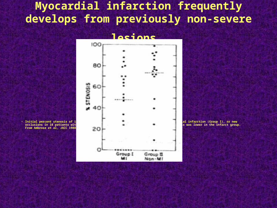
Myocardial infarction frequently develops from
previously non-severe lesions
• Initial percent stenosis of infarct-related artery at restudy of 23 patients with myocardial infarction (Group I), or new occlusions in 18 patients without myocardial infarctions (Group II). The degree of stenosis was lower in the infarct group. From Ambrose et al, JACC 1988;12:56-62
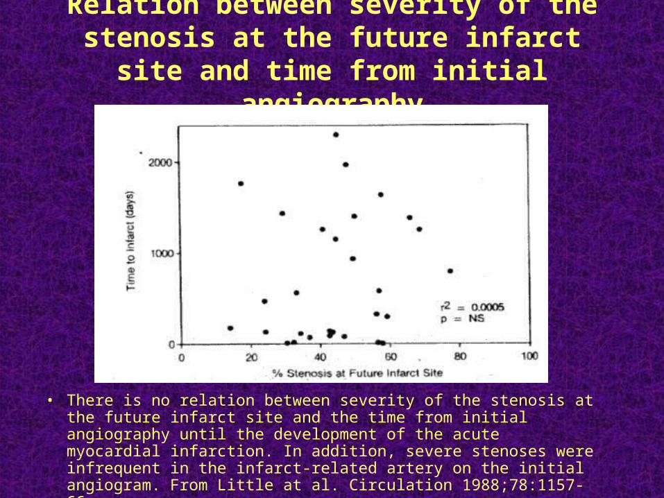
Relation between severity of the stenosis at the future infarct site and time from initial
angiography
• There is no relation between severity of the stenosis at the future infarct site and the time from initial angiography until the development of the acute myocardial infarction. In addition, severe stenoses were infrequent in the infarct-related artery on the initial angiogram. From Little at al. Circulation 1988;78:1157-66
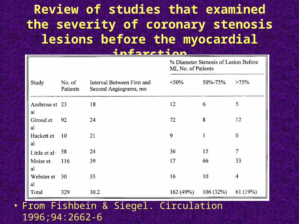
Review of studies that examined the severity of coronary stenosis lesions before the
myocardial infarction
• From Fishbein & Siegel. Circulation 1996;94:2662-6
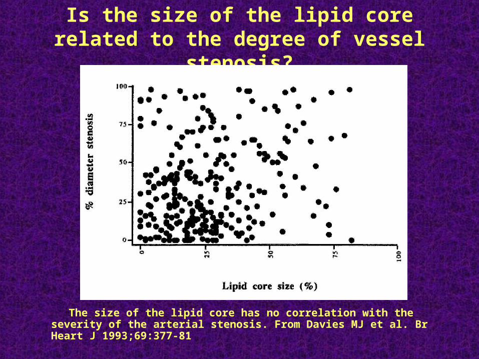
Is the size of the lipid core related to the degree of vessel stenosis?
The size of the lipid core has no correlation with the severity of the
arterial stenosis. From Davies MJ et al. Br Heart J 1993;69:377-81
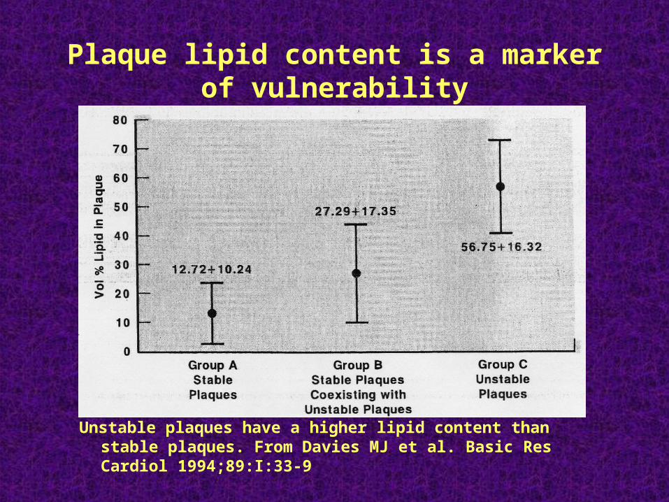
Plaque lipid content is a marker of vulnerability
Unstable plaques have a higher lipid content than stable plaques. From Davies MJ et al. Basic Res Cardiol 1994;89:I:33-9
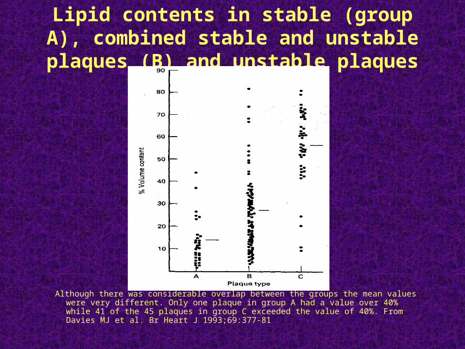
Lipid contents in stable (group A), combined stable and unstable plaques (B) and unstable
plaques (C).
Although there was considerable overlap between the groups the mean values were very different. Only one plaque in group A had a value over 40% while 41 of the 45 plaques in group C exceeded the value of 40%. From Davies MJ et al. Br Heart J 1993;69:377-81
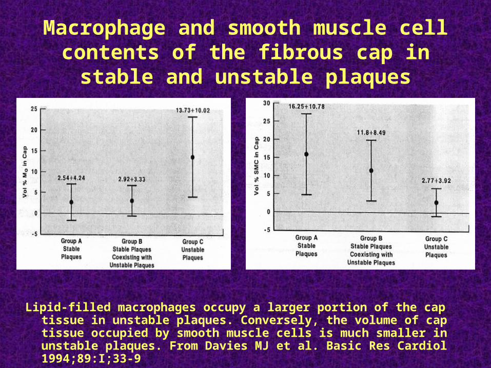
Macrophage and smooth muscle cell contents of the fibrous cap in stable and unstable
plaques
Lipid-filled macrophages occupy a larger portion of the cap tissue in unstable plaques. Conversely, the volume of cap tissue occupied by smooth muscle cells is much smaller in unstable plaques. From Davies MJ et al. Basic Res Cardiol 1994;89:I;33-9
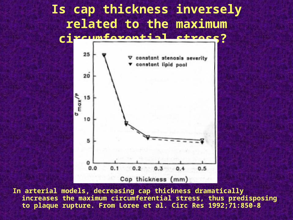
Is cap thickness inversely related to the maximum circumferential stress?
In arterial models, decreasing cap thickness dramatically increases the maximum circumferential stress, thus predisposing to plaque rupture. From Loree et al. Circ Res 1992;71:850-8
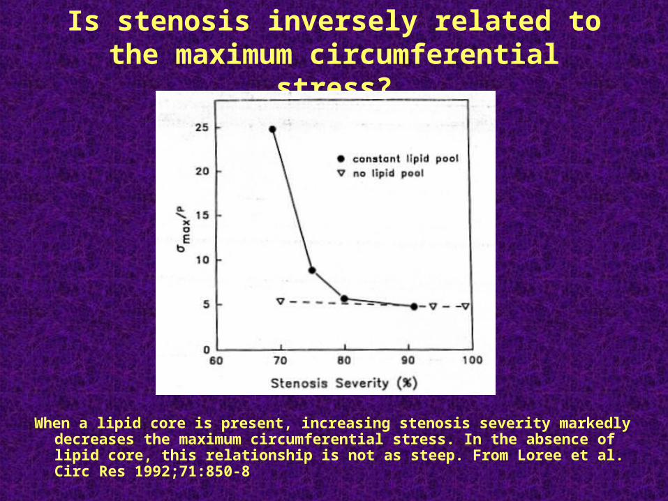
Is stenosis inversely related to the maximum circumferential stress?
When a lipid core is present, increasing stenosis severity markedly decreases the maximum circumferential stress. In the absence of lipid core, this relationship is not as steep. From Loree et al. Circ Res 1992;71:850-8
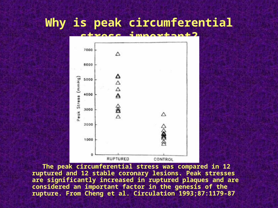
Why is peak circumferential stress important?
The peak circumferential stress was compared in 12 ruptured and 12 stable coronary lesions. Peak stresses are significantly increased in ruptured plaques and are considered an important factor in the genesis of the rupture. From Cheng et al. Circulation 1993;87:1179-87
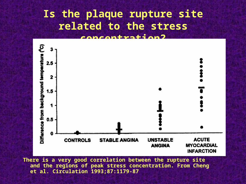
Is the plaque rupture site related to the stress concentration?
There is a very good correlation between the rupture site and the regions of peak stress concentration. From Cheng et al. Circulation 1993;87:1179-87
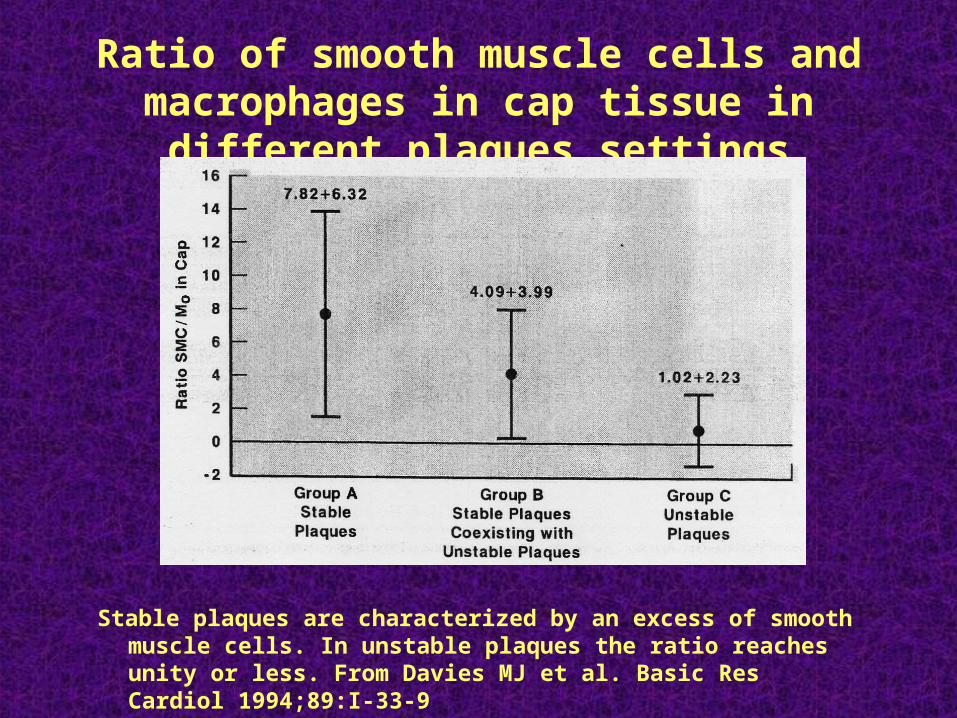
Ratio of smooth muscle cells and macrophages in cap tissue in different plaques settings
Stable plaques are characterized by an excess of smooth muscle cells. In unstable plaques the ratio reaches unity or less. From Davies MJ et al. Basic Res Cardiol 1994;89:I-33-9
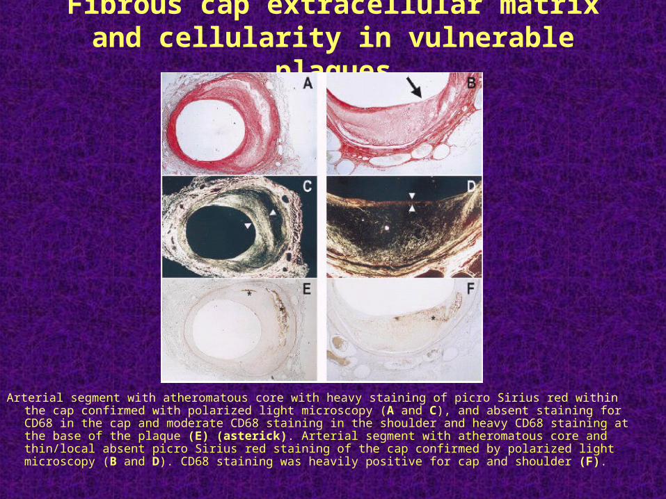
Fibrous cap extracellular matrix and cellularity in vulnerable plaques
Arterial segment with atheromatous core with heavy staining of picro Sirius red within the cap confirmed with polarized light microscopy (A and C), and absent staining for CD68 in the cap and moderate CD68 staining in the shoulder and heavy CD68 staining at the base of the plaque (E) (asterick). Arterial segment with atheromatous core and thin/local absent picro Sirius red staining of the cap confirmed by polarized light microscopy (B and D). CD68 staining was heavily positive for cap and shoulder (F).
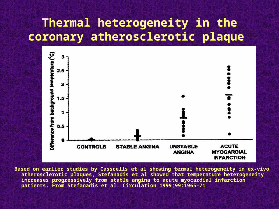
Thermal heterogeneity in the coronary atherosclerotic plaque
Based on earlier studies by Casscells et al showing termal heterogeneity in ex-vivo atherosclerotic plaques, Stefanadis et al showed that temperature heterogeneity increases progressively from stable angina to acute myocardial infarction patients. From Stefanadis et al. Circulation 1999;99:1965-71
CONCLUSIONS
• Size and composition of lipid core, thickness and composition of fibrous cap, and inflammation within or in the vicinity of the fibrous cap are well-established predictors of plaque rupture.
• Predictors of other forms of lesions underlying luminal thrombosis (e.g. erosion) are not as well characterized.