Embed Size (px)
Citation preview
RESPIRATORY SYSTEM ANATOMY
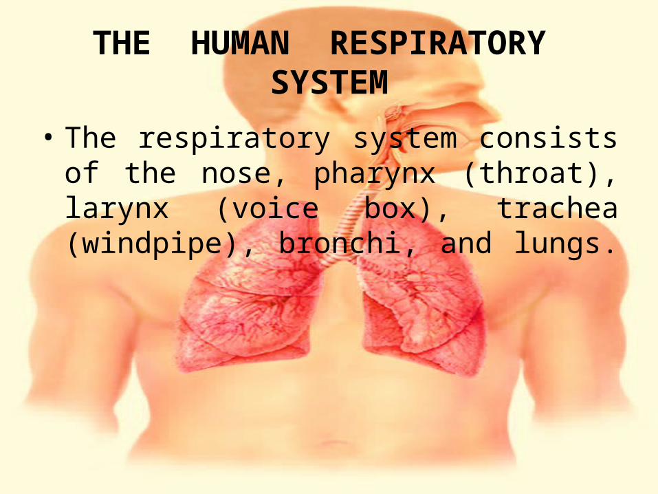
THE HUMAN RESPIRATORY SYSTEM
• The respiratory system consists of the nose, pharynx (throat), larynx (voice box), trachea (windpipe), bronchi, and lungs.
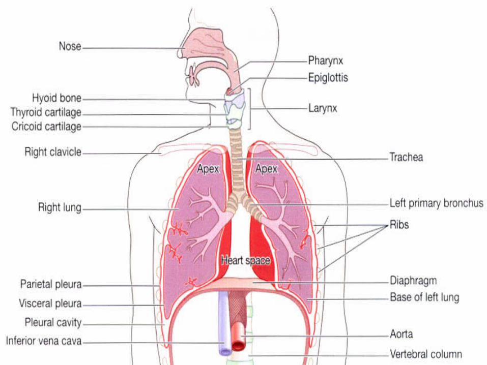
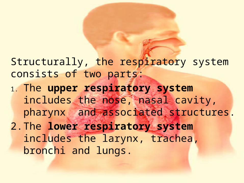
Structurally, the respiratory system consists of two parts: 1. The upper respiratory system includes the
nose, nasal cavity, pharynx and associated structures.
2. The lower respiratory system includes the larynx, trachea, bronchi and lungs.
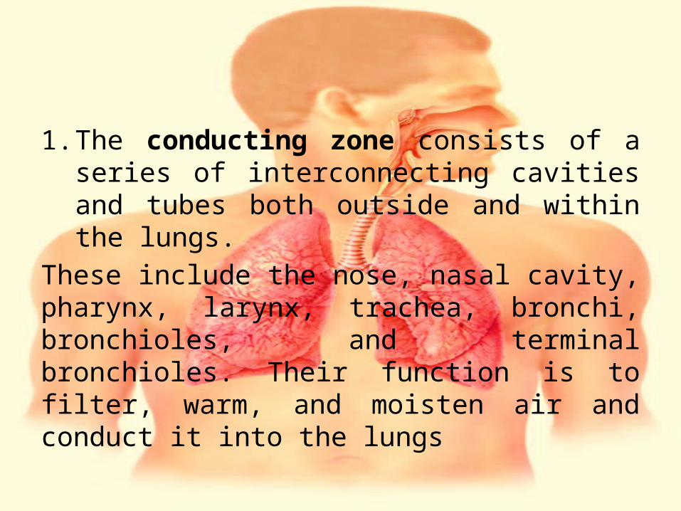
1. The conducting zone consists of a series of interconnecting cavities and tubes both outside and within the lungs.
These include the nose, nasal cavity, pharynx, larynx, trachea, bronchi, bronchioles, and terminal bronchioles. Their function is to filter, warm, and moisten air and conduct it into the lungs
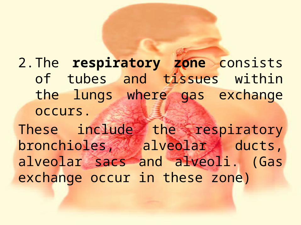
2. The respiratory zone consists of tubes and tissues within the lungs where gas exchange occurs.
These include the respiratory bronchioles, alveolar ducts, alveolar sacs and alveoli. (Gas exchange occur in these zone)
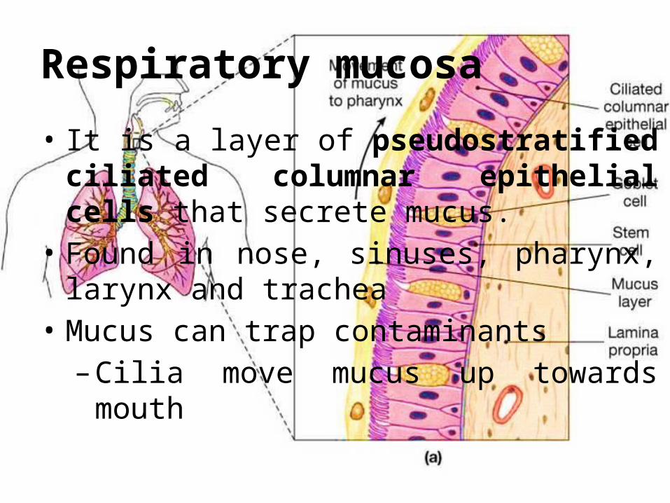
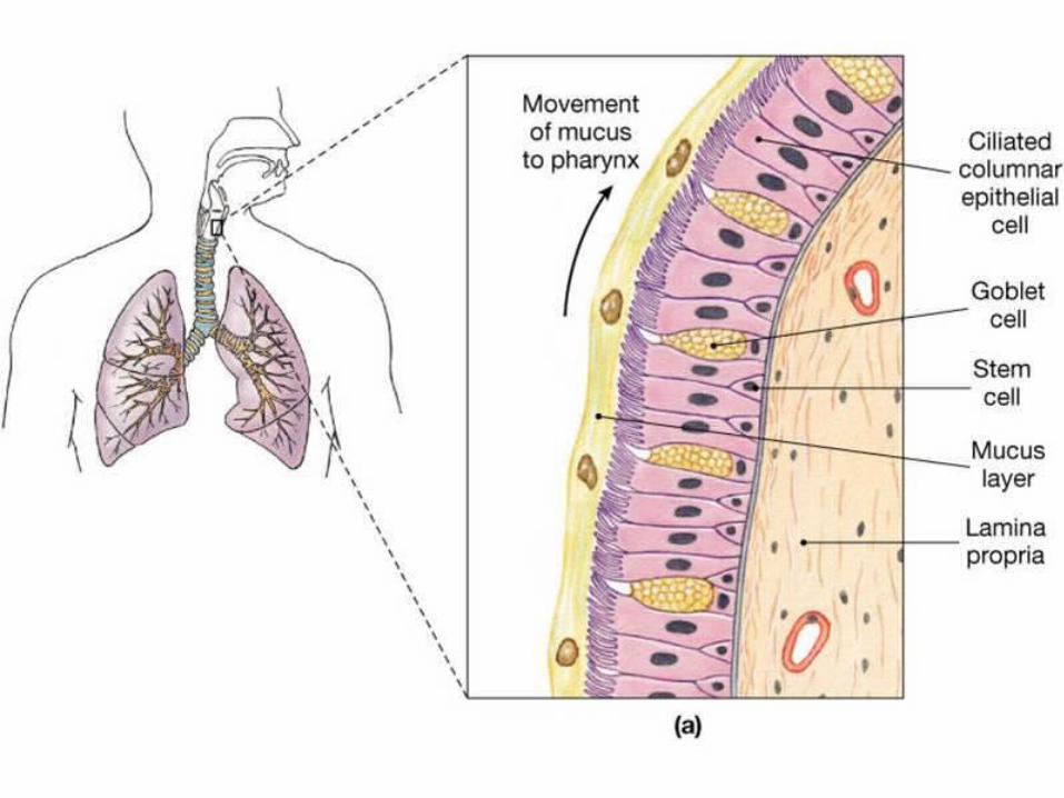
Respiratory mucosa
• It is a layer of pseudostratified ciliated columnar epithelial cells that secrete mucus.
• Found in nose, sinuses, pharynx, larynx and trachea
• Mucus can trap contaminants–Cilia move mucus up towards mouth
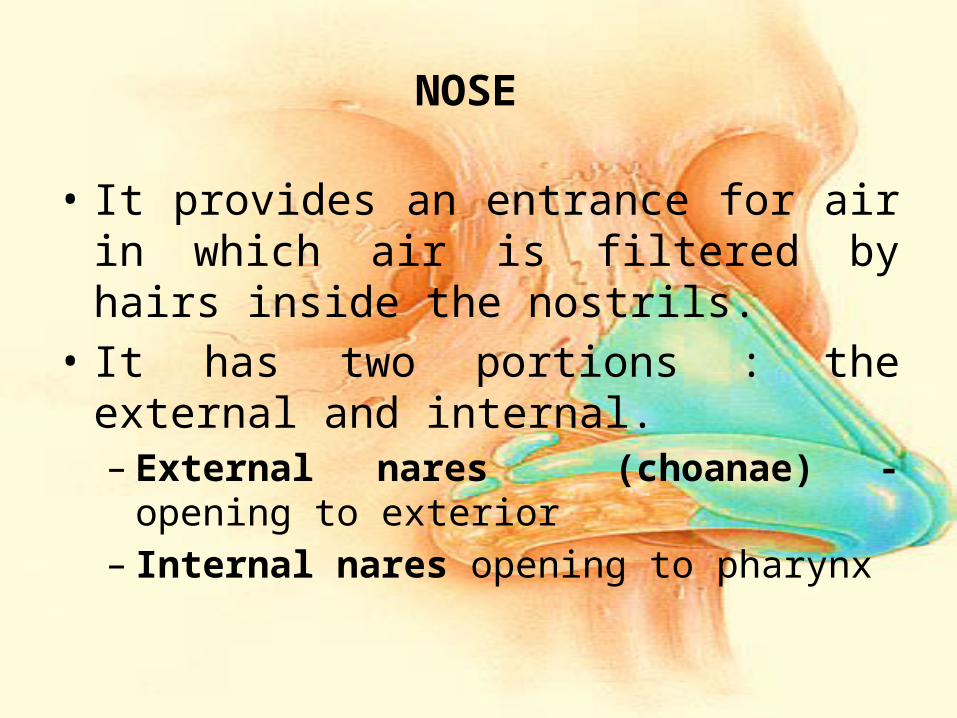
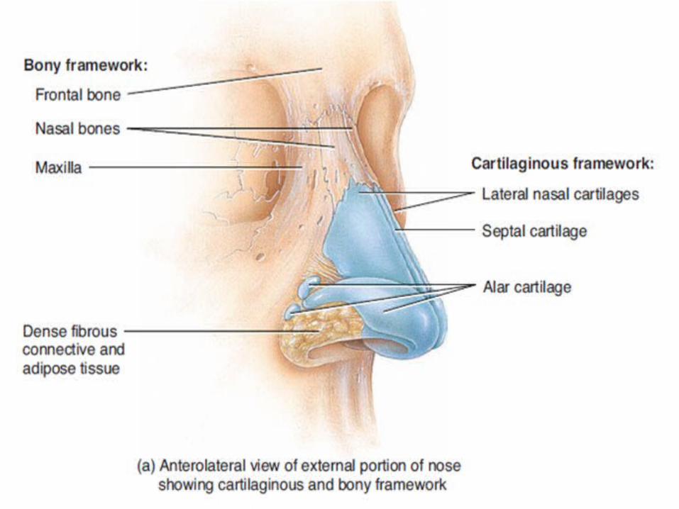
NOSE
• It provides an entrance for air in which air is filtered by hairs inside the nostrils.
• It has two portions : the external and internal.– External nares (choanae) - opening to exterior– Internal nares opening to pharynx
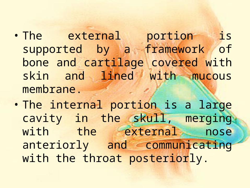
• The external portion is supported by a framework of bone and cartilage covered with skin and lined with mucous membrane.
• The internal portion is a large cavity in the skull, merging with the external nose anteriorly and communicating with the throat posteriorly.
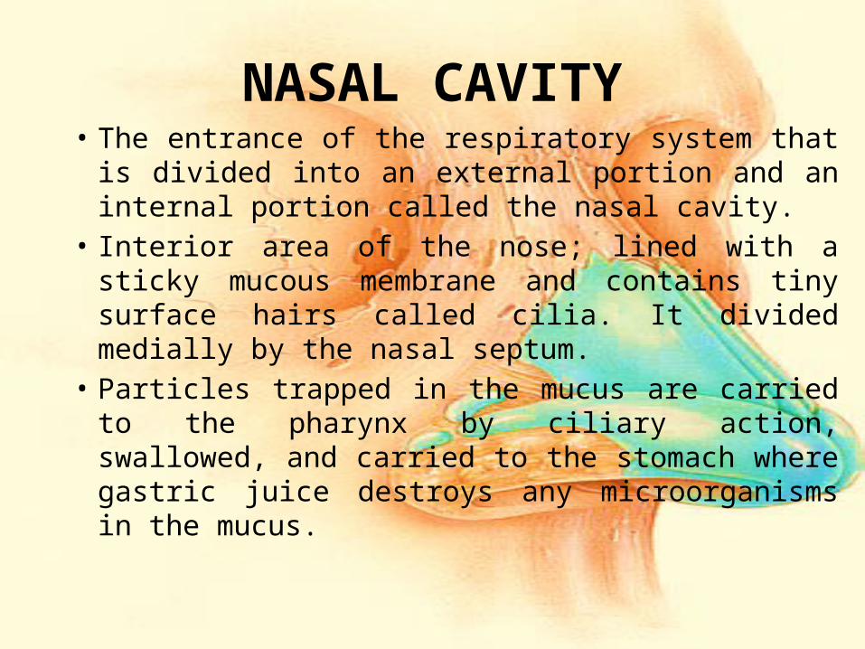
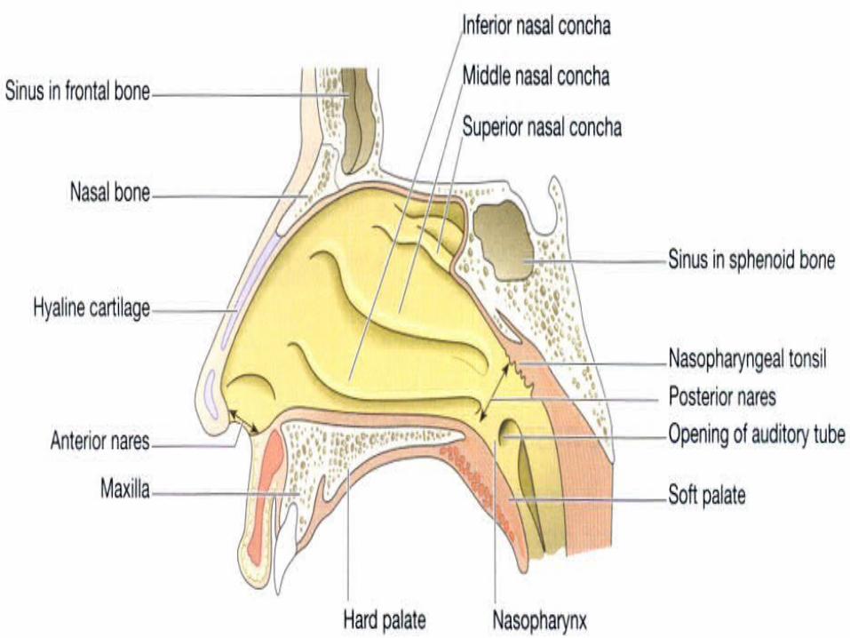
NASAL CAVITY• The entrance of the respiratory system that is
divided into an external portion and an internal portion called the nasal cavity.
• Interior area of the nose; lined with a sticky mucous membrane and contains tiny surface hairs called cilia. It divided medially by the nasal septum.
• Particles trapped in the mucus are carried to the pharynx by ciliary action, swallowed, and carried to the stomach where gastric juice destroys any microorganisms in the mucus.
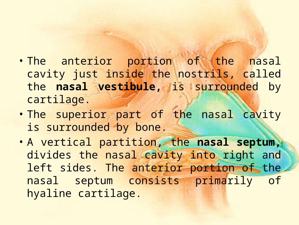
• The anterior portion of the nasal cavity just inside the nostrils, called the nasal vestibule, is surrounded by cartilage.
• The superior part of the nasal cavity is surrounded by bone.
• A vertical partition, the nasal septum, divides the nasal cavity into right and left sides. The anterior portion of the nasal septum consists primarily of hyaline cartilage.
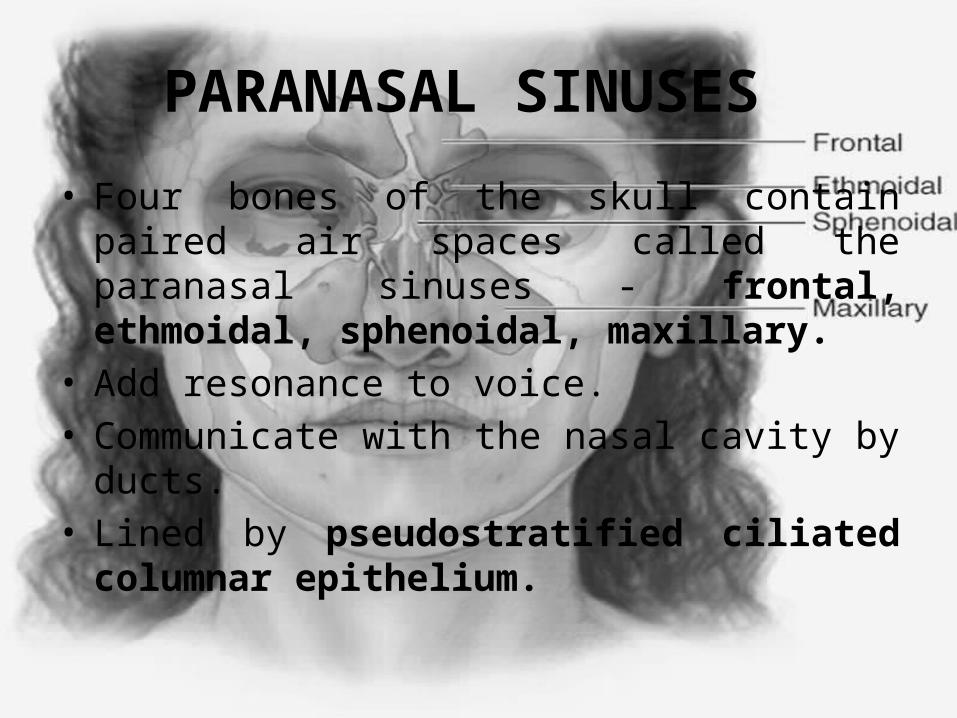
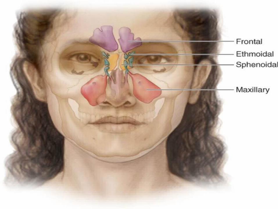
PARANASAL SINUSES
• Four bones of the skull contain paired air spaces called the paranasal sinuses - frontal, ethmoidal, sphenoidal, maxillary.
• Add resonance to voice.• Communicate with the nasal cavity by ducts.• Lined by pseudostratified ciliated columnar
epithelium.
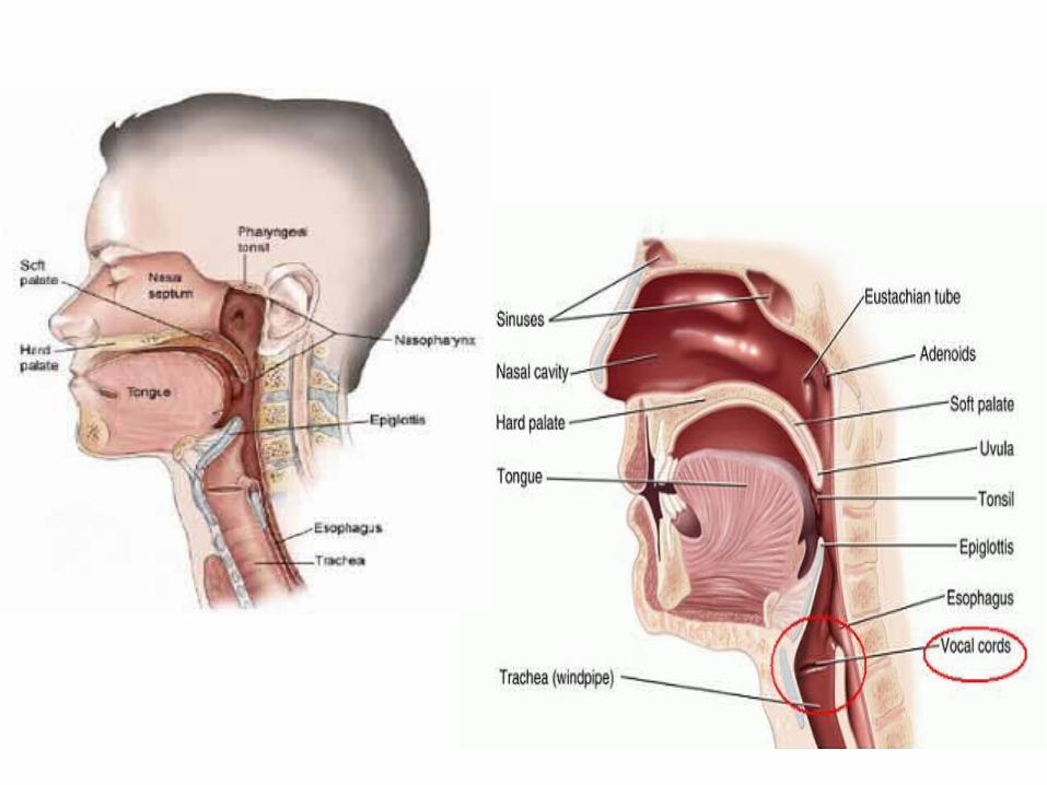
PHARYNX
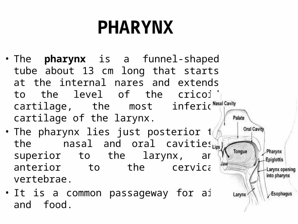
• The pharynx is a funnel-shaped tube about 13 cm long that starts at the internal nares and extends to the level of the cricoid cartilage, the most inferior cartilage of the larynx.
• The pharynx lies just posterior to the nasal and oral cavities, superior to the larynx, and anterior to the cervical vertebrae.
• It is a common passageway for air and food.
• Its wall is composed of skeletal muscles and is lined with a mucous membrane.
• The muscles of the entire pharynx are arranged in two layers, an outer circular layer and an inner longitudinal layer.
• Relaxed skeletal muscles help keep the pharynx patent and contraction of the skeletal muscles assists in deglutition (swallowing).
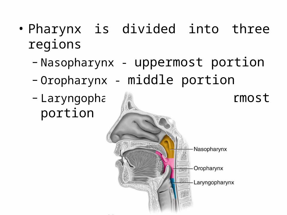
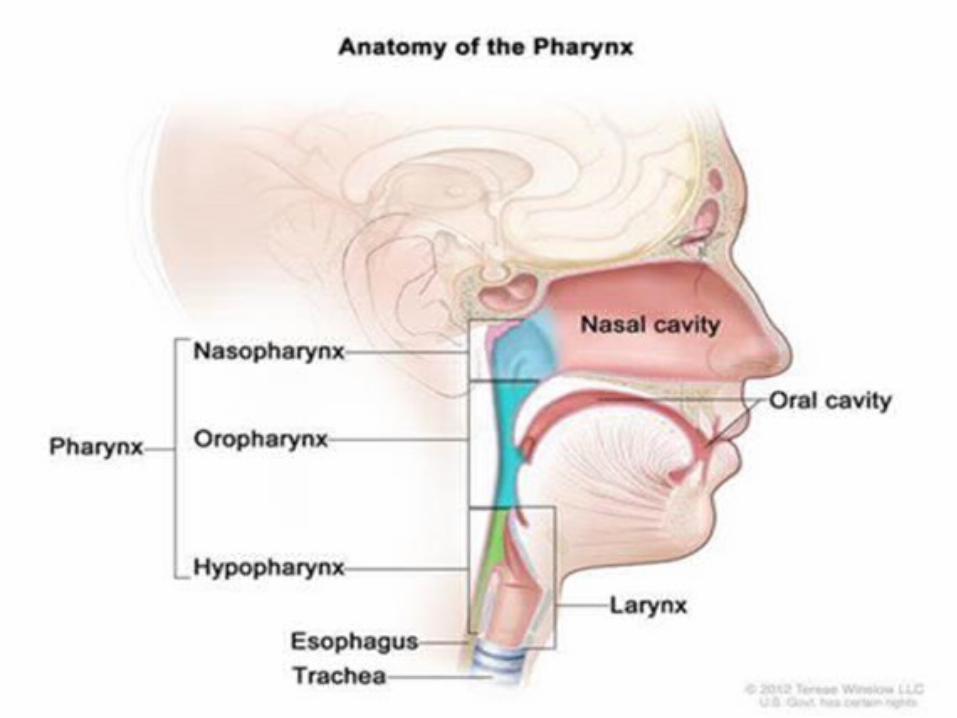
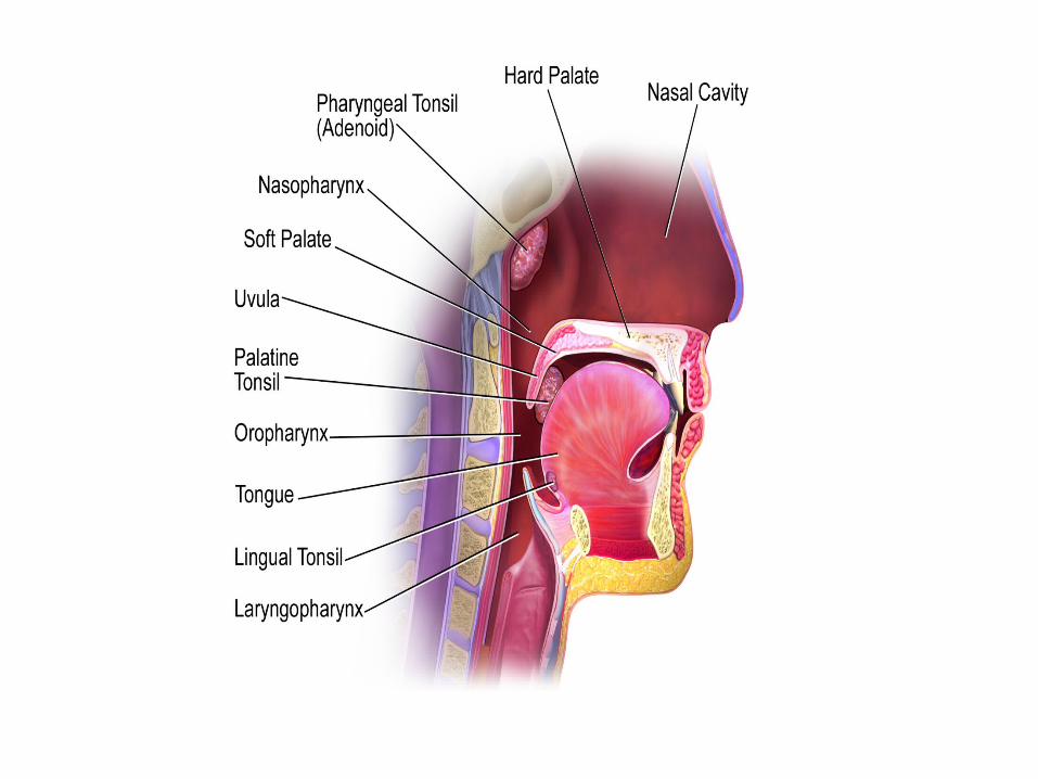
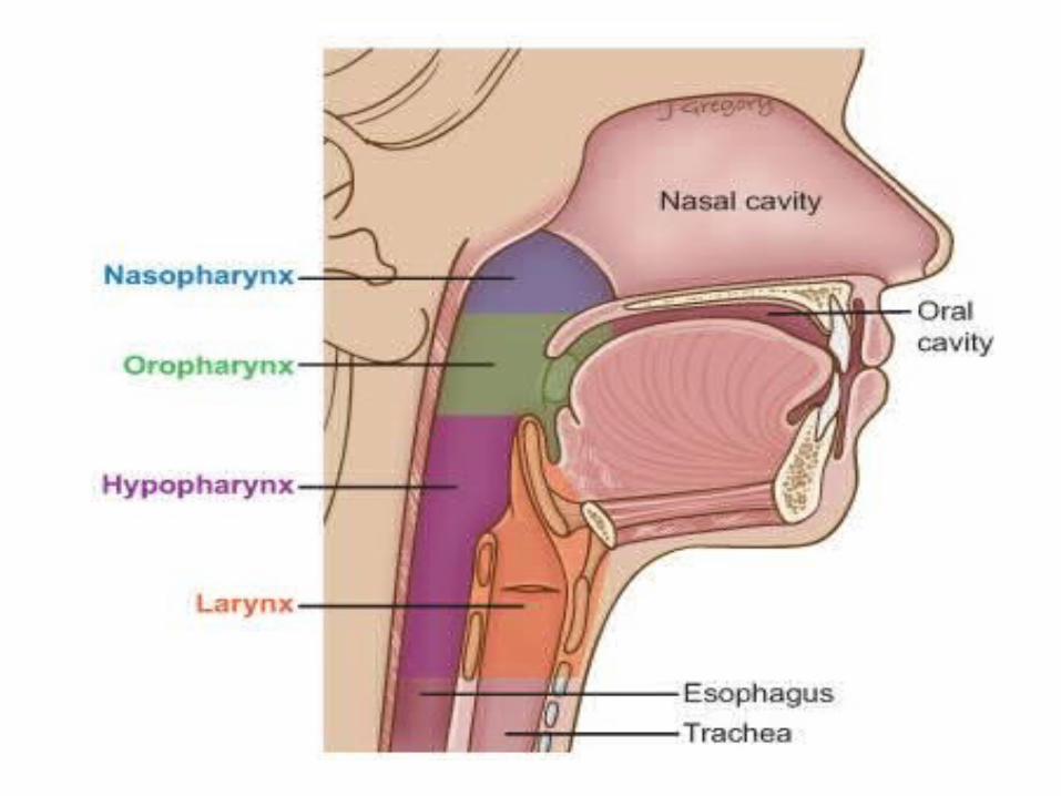
• Pharynx is divided into three regions– Nasopharynx - uppermost portion– Oropharynx - middle portion– Laryngopharynx - lowermost portion
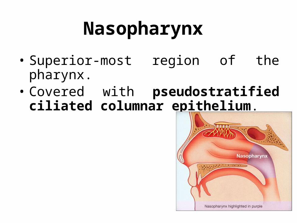
Nasopharynx
• Superior-most region of the pharynx. • Covered with pseudostratified ciliated
columnar epithelium.
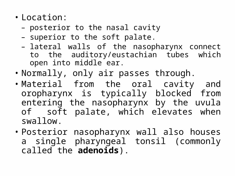
• Location:– posterior to the nasal cavity– superior to the soft palate.– lateral walls of the nasopharynx connect to the
auditory/eustachian tubes which open into middle ear.• Normally, only air passes through. • Material from the oral cavity and oropharynx is
typically blocked from entering the nasopharynx by the uvula of soft palate, which elevates when swallow.
• Posterior nasopharynx wall also houses a single pharyngeal tonsil (commonly called the adenoids).
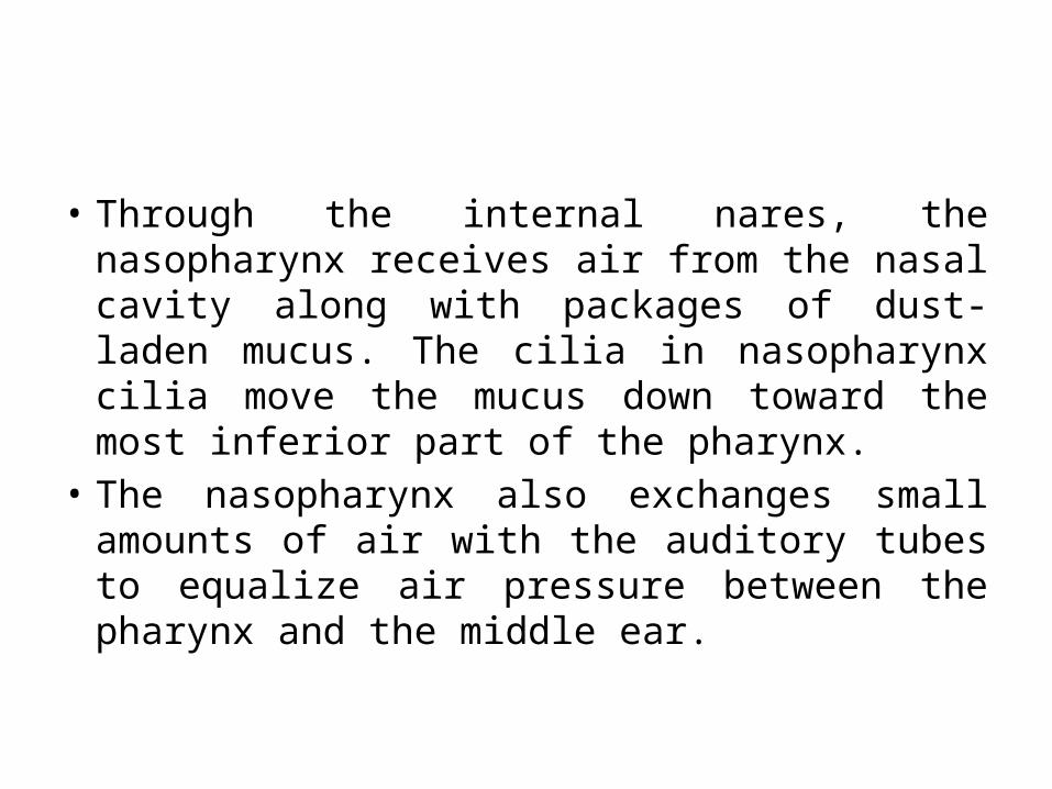
• Through the internal nares, the nasopharynx receives air from the nasal cavity along with packages of dust-laden mucus. The cilia in nasopharynx cilia move the mucus down toward the most inferior part of the pharynx.
• The nasopharynx also exchanges small amounts of air with the auditory tubes to equalize air pressure between the pharynx and the middle ear.
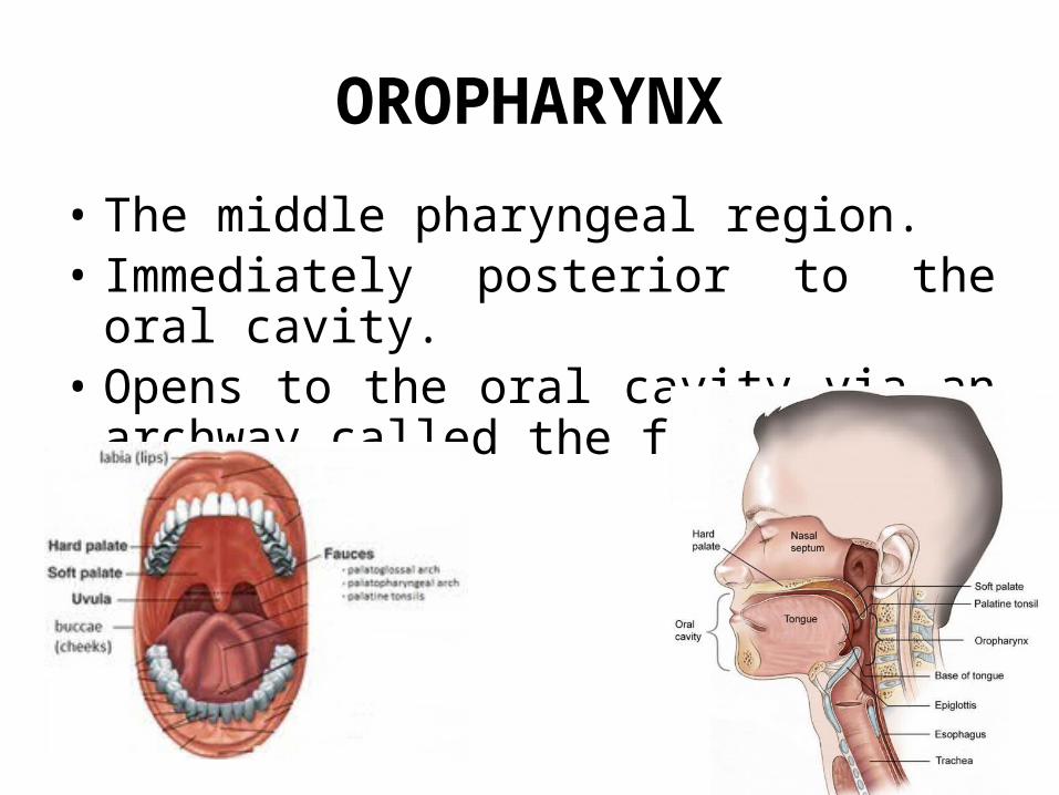
OROPHARYNX
• The middle pharyngeal region. • Immediately posterior to the oral cavity. • Opens to the oral cavity via an archway called
the fauces.
• Boundaries:– superiorly edge of the soft palate.– inferiorly the hyoid bone.
• Common respiratory and digestive pathway through which both air and swallowed food and drink pass.
• Contains nonkeratinized stratified squamous epithelim.
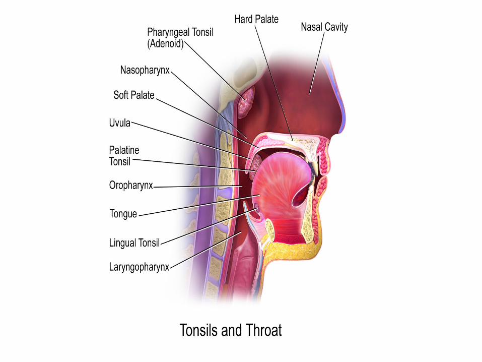
• Lymphatic organs provide the first line of defense against ingested or inhaled foreign materials.
• Two pairs of tonsils, the palatine tonsils and lingual tonsils are found in the oropharynx.
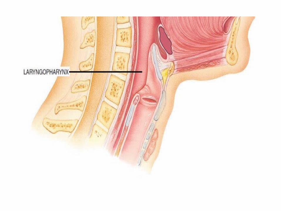
LARYNGOPHARYNX
• Laryngopharynx or hypopharynx• Inferior, narrowed region of the pharynx. • Boundaries– Superiorly hyoid bone
• Inferior end it opens into the esophagus (food tube) posteriorly and the larynx (voice box) anteriorly.
• Lined with a nonkeratinized stratified squamous epithelium.
• Permits passage of both food and air.
Lower Respiratory Tract
It includes conducting airway and respiratory portion Composed of trachea, bronchial tree, lungs,
alveolus and alveoli. Alveoli is the functional unit of lungs.• Conducting airways (pharynx, trachea,
bronchi, up to terminal bronchioles). • Respiratory portion of the respiratory system
(respiratory bronchioles, alveolar ducts, and alveoli).

Larynx
• Larynx or Voice box is a short, cylindrical airway ends in the trachea. It is about 5 cm long.
• Boundaries:– Superiorly it attaches to hyoid bone and opens into th laryngopharynx– Inferiorly trachea. – Posteriorly esophagus
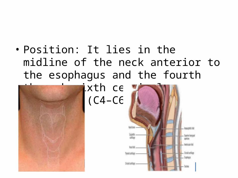
• Position: It lies in the midline of the neck anterior to the esophagus and the fourth through sixth cervical vertebrae (C4–C6).
• It conducts air into the lower respiratory tract.• Produces sounds. • It is composed of a framework of nine pieces of
cartilage (three individual pieces and three cartilage pairs) that are held in place by ligaments and muscles.
• Except for the epiglottis, all laryngeal cartilages are hyaline cartilages.
• Muscles of larynx: extrinsic muscles and intrinsic muscles.
• The extrinsic muscles of the larynx connect the cartilages to other structures in the throat.
• The intrinsic muscles connect the cartilages to one another.
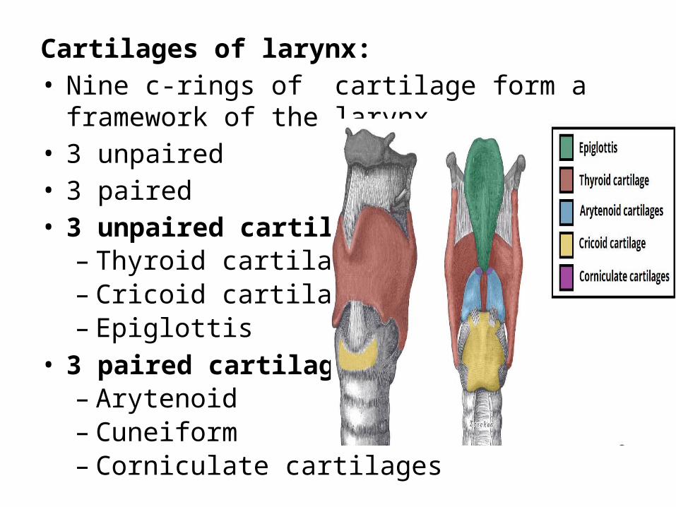
Cartilages of larynx:• Nine c-rings of cartilage form a framework of the
larynx.• 3 unpaired • 3 paired• 3 unpaired cartilages– Thyroid cartilage – Cricoid cartilage – Epiglottis
• 3 paired cartilages– Arytenoid– Cuneiform– Corniculate cartilages
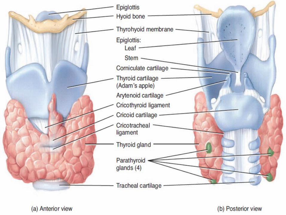
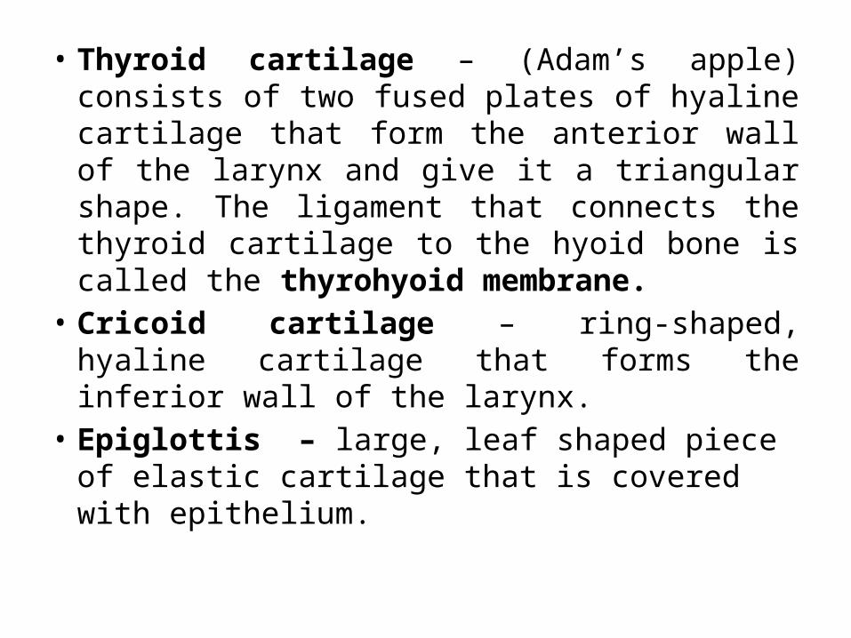
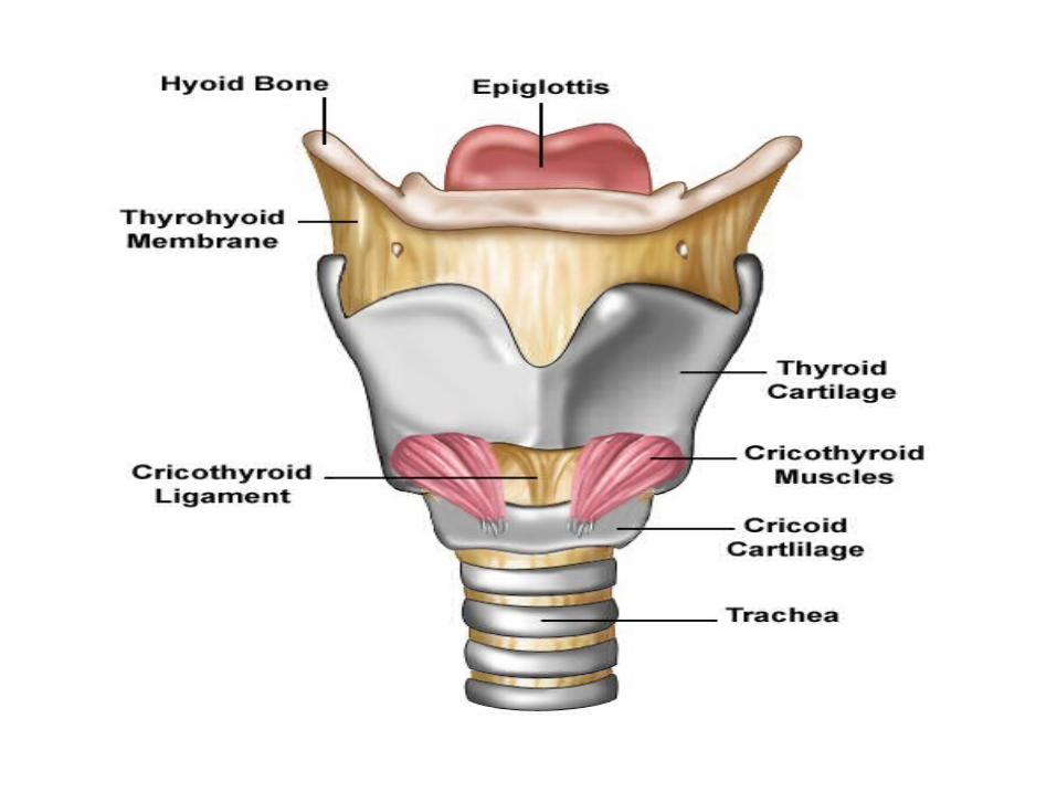
• Thyroid cartilage – (Adam’s apple) consists of two fused plates of hyaline cartilage that form the anterior wall of the larynx and give it a triangular shape. The ligament that connects the thyroid cartilage to the hyoid bone is called the thyrohyoid membrane.
• Cricoid cartilage – ring-shaped, hyaline cartilage that forms the inferior wall of the larynx.
• Epiglottis – large, leaf shaped piece of elastic cartilage that is covered with epithelium.
• Arytenoid cartilages – are triangular pieces of mostly hyaline cartilage located at the posterior, superior border of the cricoid cartilage. They form synovial joints with the cricoid cartilage and have a wide range of mobility.
• Cuneiform cartilages - club-shaped elastic cartilages anterior to the corniculate cartilages, support the vocal folds and lateral aspects of the epiglottis.
• Corniculate cartilages - horn-shaped pieces of elastic cartilage, are located at the top of each arytenoid cartilage
Epithelium of Larynx• The lining of the larynx superior to the vocal
folds is non keratinized stratified squamous epithelium. The lining of the larynx inferior to the vocal folds is pseudostratified ciliated columnar epithelium consisting of ciliated columnar cells, goblet cells, and basal cells.
• The mucus produced by the goblet cells helps to trap dust which is not removed in the upper passages.
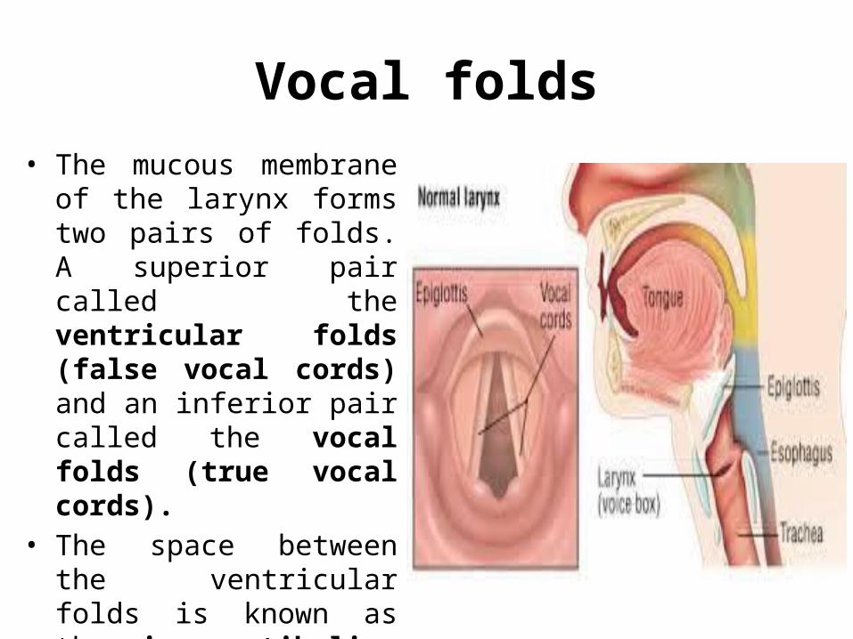
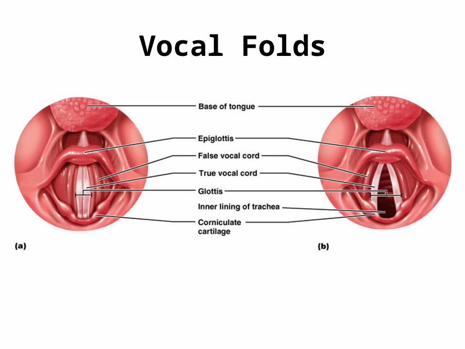
Vocal folds• The mucous membrane of
the larynx forms two pairs of folds. A superior pair called the ventricular folds (false vocal cords) and an inferior pair called the vocal folds (true vocal cords).
• The space between the ventricular folds is known as the rima vestibuli.
Vocal Folds
• Vocal cords contain elastic fibers and are responsible for vocal sounds, which are created when air is forced between these folds, causing them to vibrate from side to side. This action generates sound waves, which can be formed into words by changing the shapes of the pharynx and oral cavity and by using tongue and lips.
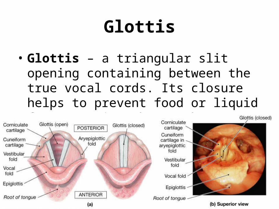
• Glottis – a triangular slit opening containing between the true vocal cords. Its closure helps to prevent food or liquid from entering the trachea.
Glottis
Blood supply of larynx• Blood is supplied to the larynx by the superior
and inferior laryngeal arteries and drained by the thyroid veins, which join the internal jugular vein.
Nerve supply of larynx• The parasympathetic nerve supply is from the
superior laryngeal and recurrent laryngeal nerves, which are branches of the vagus nerves, and the sympathetic nerves are from the superior cervical ganglia, one on each side
Epiglottis • The epiglottis is a large, leaf shaped piece of
elastic cartilage that is covered with epithelium• The “stem” of the epiglottis is the tapered
inferior portion that is attached to the anterior rim of the thyroid cartilage and hyoid bone.
• The broad superior “leaf” portion of the epiglottis is unattached and is free to move up and down like a trap door. Prevents food and drink from entering airway when swallowing
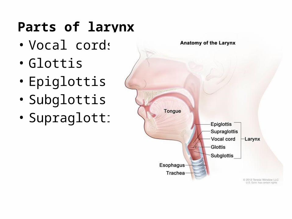
Parts of larynx• Vocal cords• Glottis• Epiglottis• Subglottis • Supraglottis
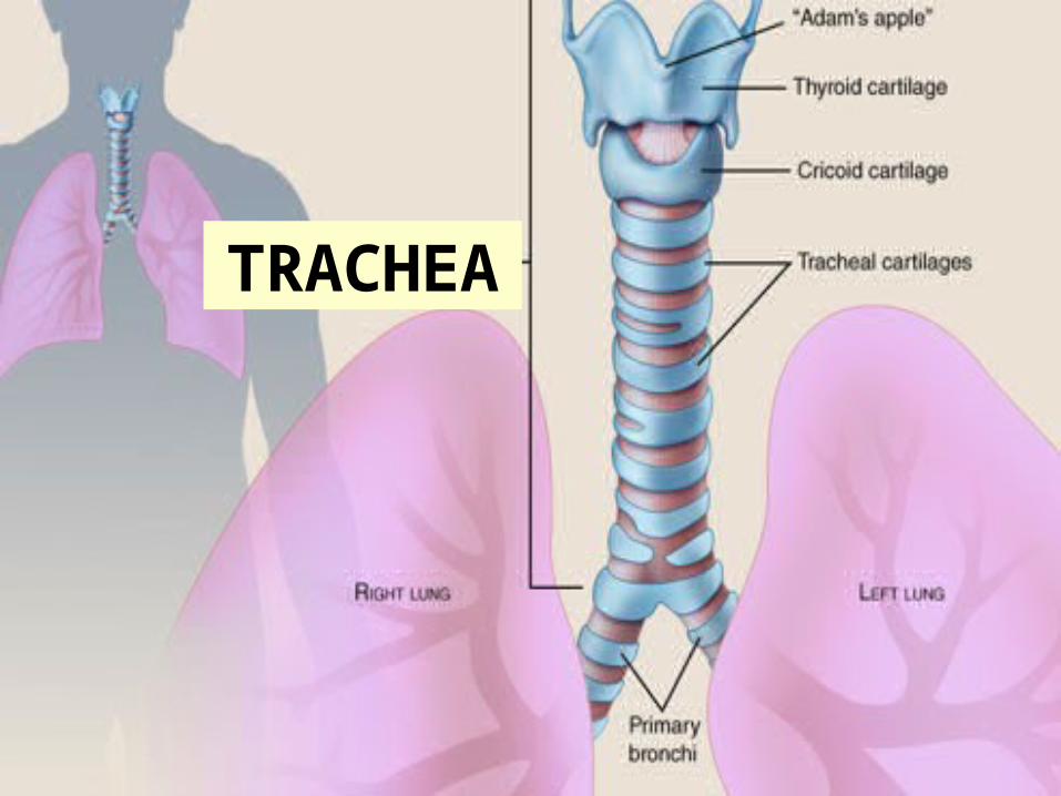
TRACHEA
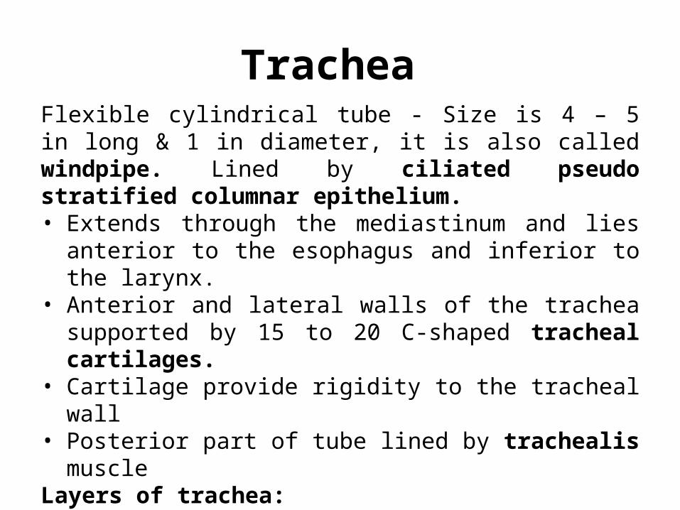
Trachea Flexible cylindrical tube - Size is 4 – 5 in long & 1 in diameter, it is also called windpipe. Lined by ciliated pseudo stratified columnar epithelium. • Extends through the mediastinum and lies anterior to the
esophagus and inferior to the larynx. • Anterior and lateral walls of the trachea supported by 15 to
20 C-shaped tracheal cartilages. • Cartilage provide rigidity to the tracheal wall• Posterior part of tube lined by trachealis muscleLayers of trachea:
– Innermost layer (mucosa) = pseudostratified columnar with cilia & goblet cells
– outer layer (submucosa) = loose connective tissue & mucous glands
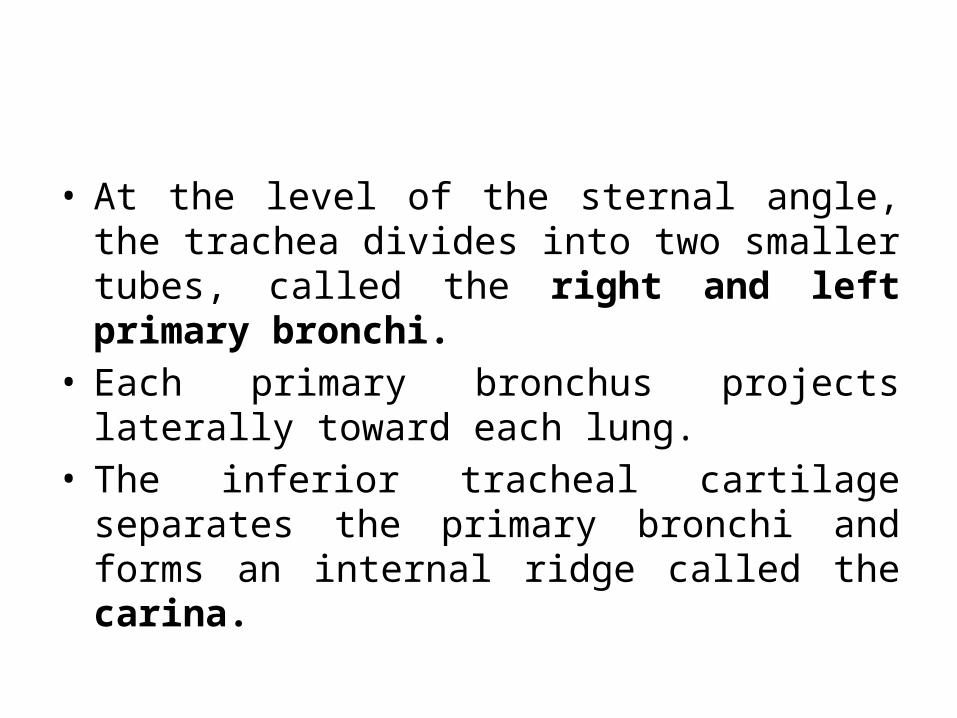
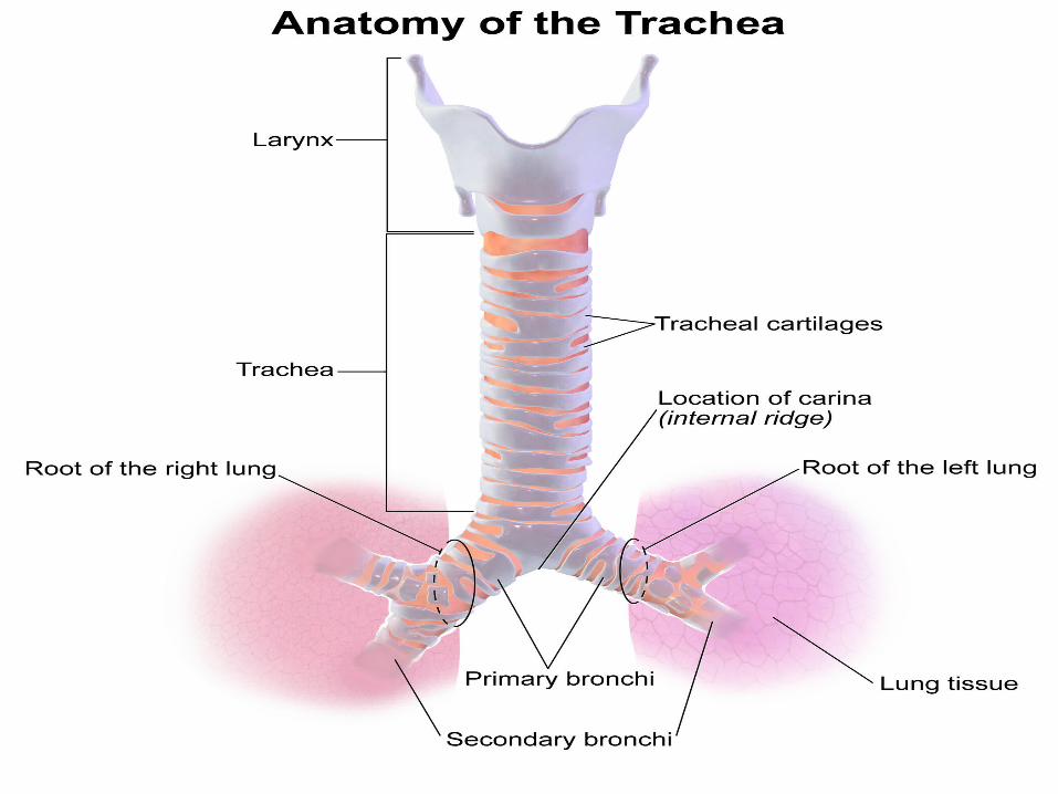
• At the level of the sternal angle, the trachea divides into two smaller tubes, called the right and left primary bronchi.
• Each primary bronchus projects laterally toward each lung.
• The inferior tracheal cartilage separates the primary bronchi and forms an internal ridge called the carina.
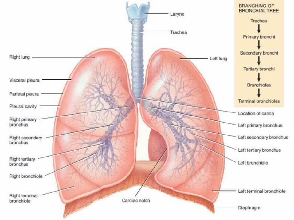
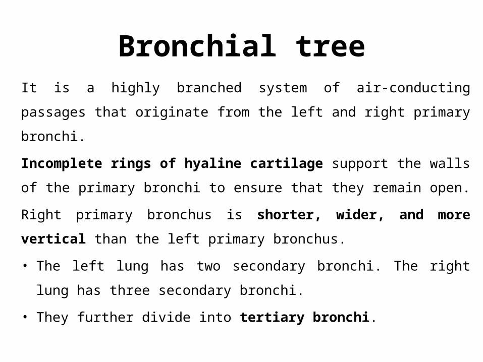
Bronchial treeIt is a highly branched system of air-conducting passages that
originate from the left and right primary bronchi.
Incomplete rings of hyaline cartilage support the walls of the
primary bronchi to ensure that they remain open.
Right primary bronchus is shorter, wider, and more vertical than
the left primary bronchus.
• The left lung has two secondary bronchi. The right lung has three
secondary bronchi.
• They further divide into tertiary bronchi.
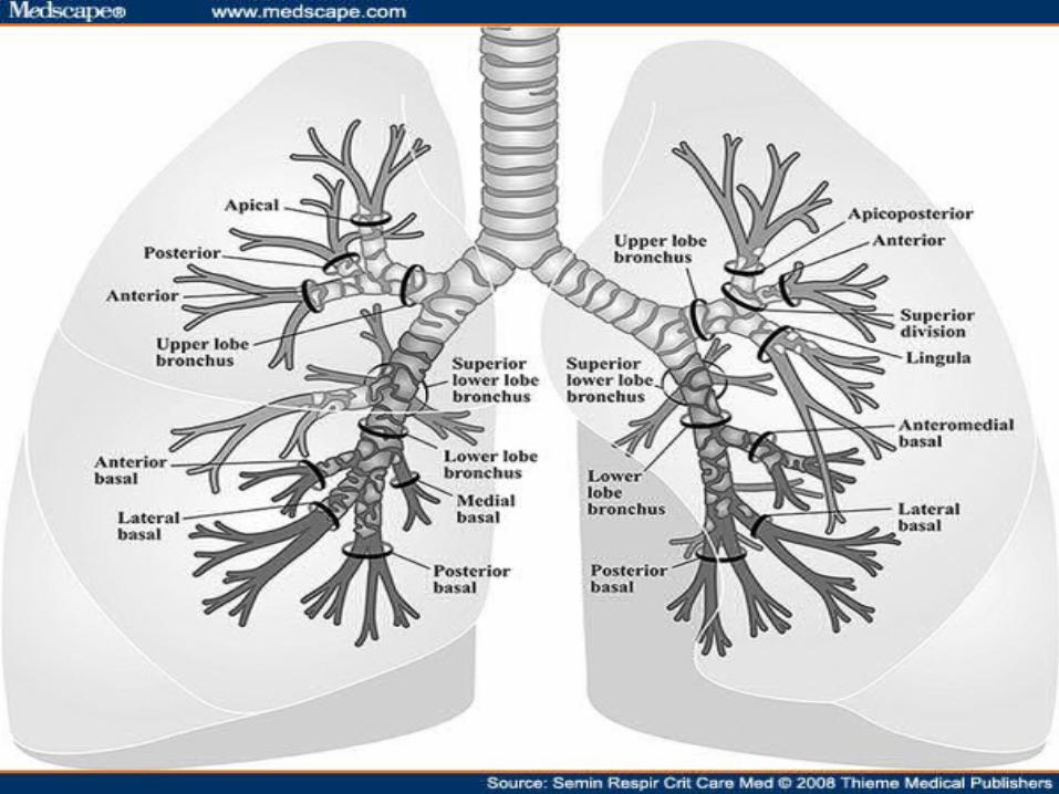
• Primary bronchi supply each lung• Secondary bronchi supply each lobe of the lungs (3
right + 2 left)• Tertiary bronchi splits into successive sets of
Intralobular bronchioles that supply each bronchopulmonary segment ( right = 10, left = 8)
• Bronchioles split into Terminal bronchioles and these split into Respiratory Bronchioles
• Respiratory Bronchioles splits into multiple Alveolar ducts which end in an Alveolar sac
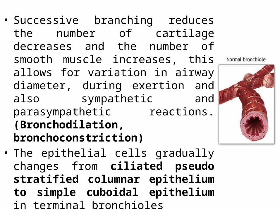
• Successive branching reduces the number of cartilage decreases and the number of smooth muscle increases, this allows for variation in airway diameter, during exertion and also sympathetic and parasympathetic reactions. (Bronchodilation, bronchoconstriction)
• The epithelial cells gradually changes from ciliated pseudo stratified columnar epithelium to simple cuboidal epithelium in terminal bronchioles
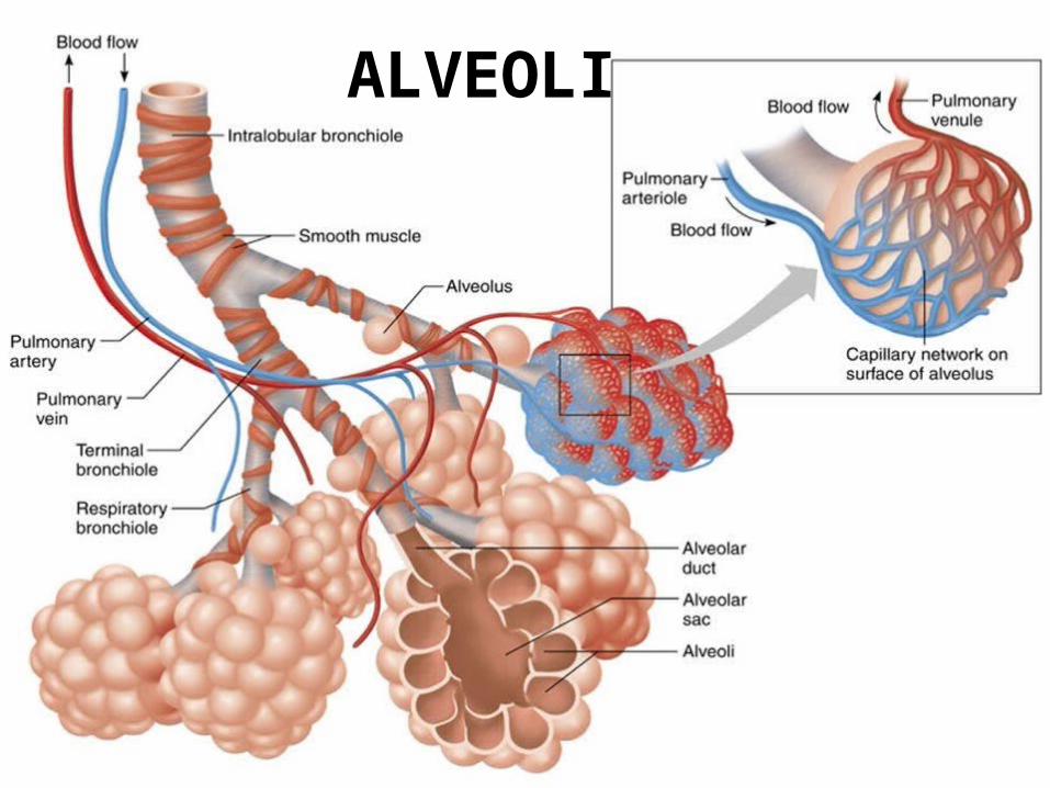
ALVEOLI
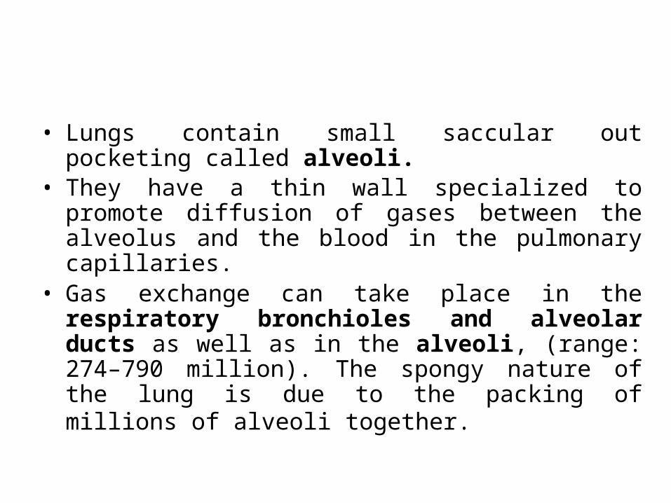
• Lungs contain small saccular out pocketing called alveoli. • They have a thin wall specialized to promote diffusion of
gases between the alveolus and the blood in the pulmonary capillaries.
• Gas exchange can take place in the respiratory bronchioles and alveolar ducts as well as in the alveoli, (range: 274–790 million). The spongy nature of the lung is due to the packing of millions of alveoli together.
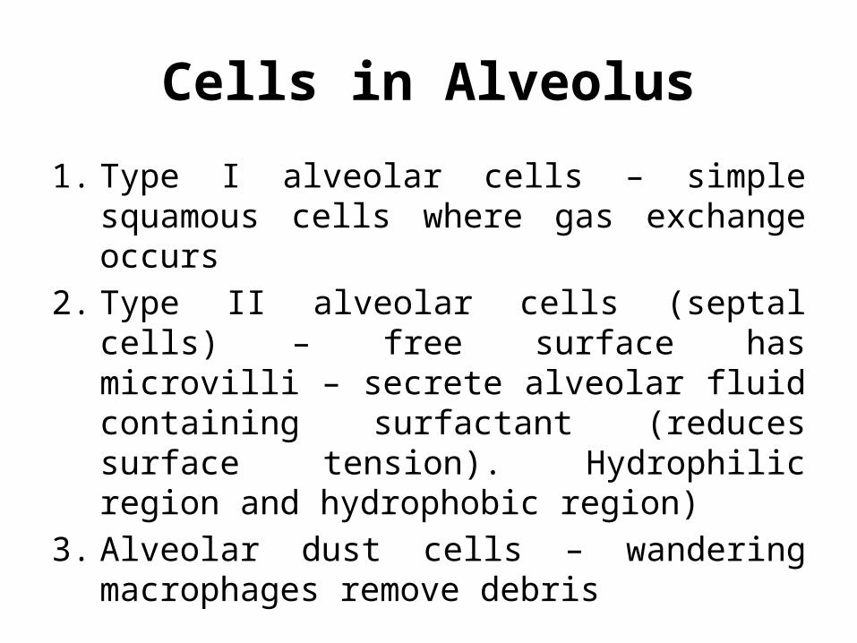
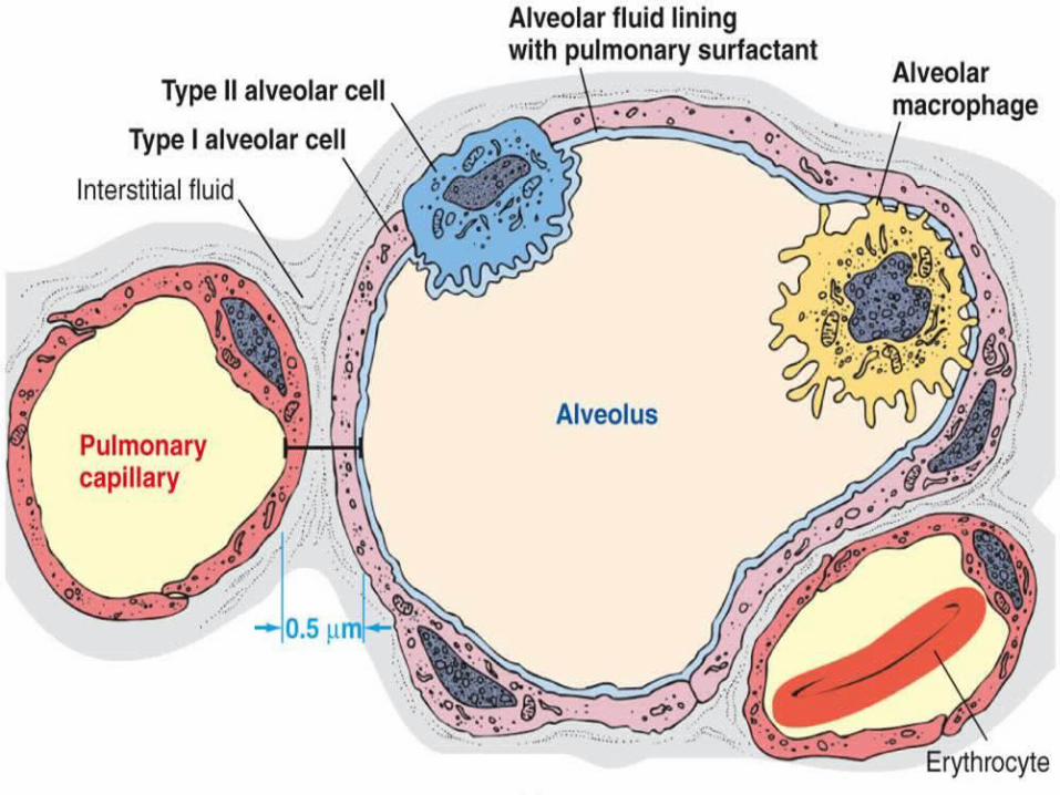
Cells in Alveolus
1. Type I alveolar cells – simple squamous cells where gas exchange occurs
2. Type II alveolar cells (septal cells) – free surface has microvilli – secrete alveolar fluid containing surfactant (reduces surface tension). Hydrophilic region and hydrophobic region)
3. Alveolar dust cells – wandering macrophages remove debris
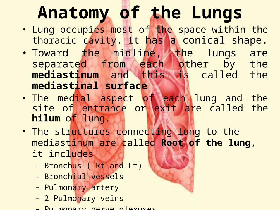
Anatomy of the Lungs• Lung occupies most of the space within the thoracic cavity.
It has a conical shape. • Toward the midline, the lungs are separated from each
other by the mediastinum and this is called the mediastinal surface
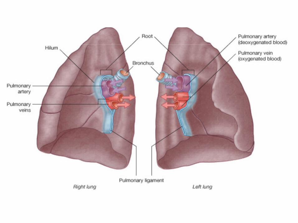
• The medial aspect of each lung and the site of entrance or exit are called the hilum of lung.
• The structures connecting lung to the mediastinum are called Root of the lung, it includes– Bronchus ( Rt and Lt)– Bronchial vessels– Pulmonary artery– 2 Pulmonary veins– Pulmonary nerve plexuses– Bronchopulmonary lymph nodes and lymphatics.
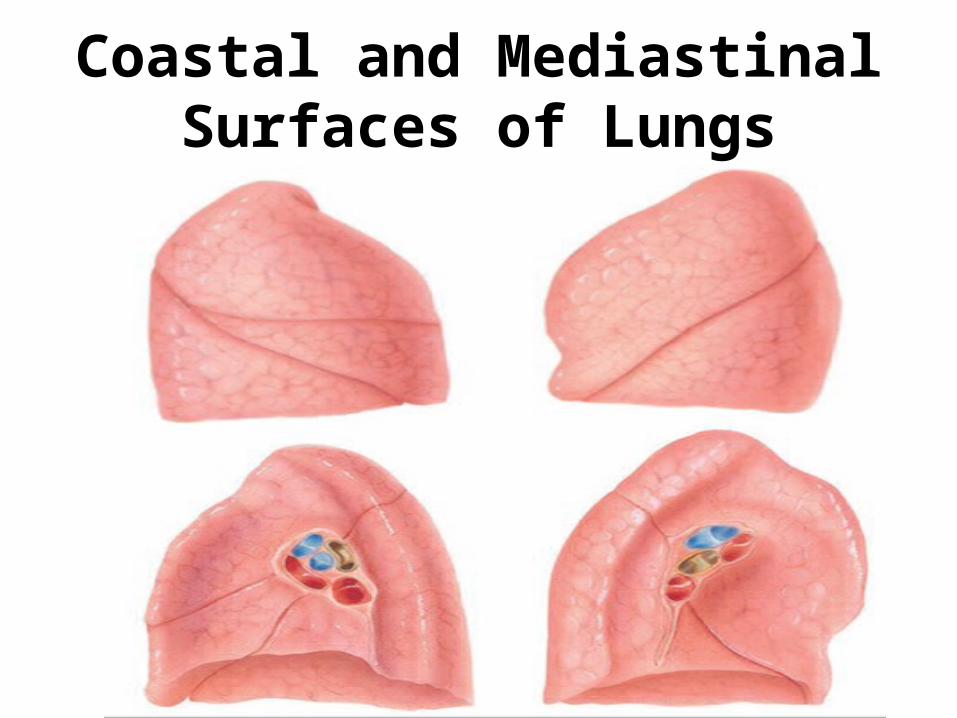
Coastal and Mediastinal Surfaces of Lungs
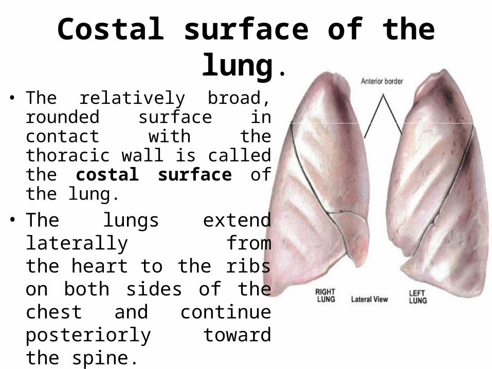
Costal surface of the lung.
• The relatively broad, rounded surface in contact with the thoracic wall is called the costal surface of the lung.
• The lungs extend laterally from the heart to the ribs on both sides of the chest and continue posteriorly toward the spine.
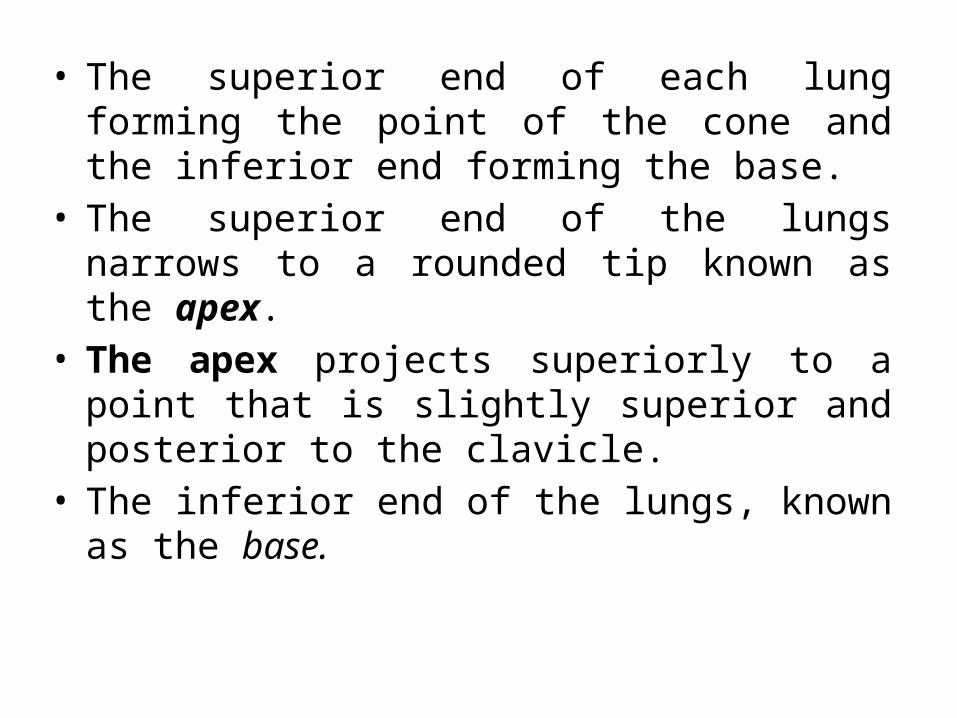
• The superior end of each lung forming the point of the cone and the inferior end forming the base.
• The superior end of the lungs narrows to a rounded tip known as the apex.
• The apex projects superiorly to a point that is slightly superior and posterior to the clavicle.
• The inferior end of the lungs, known as the base.
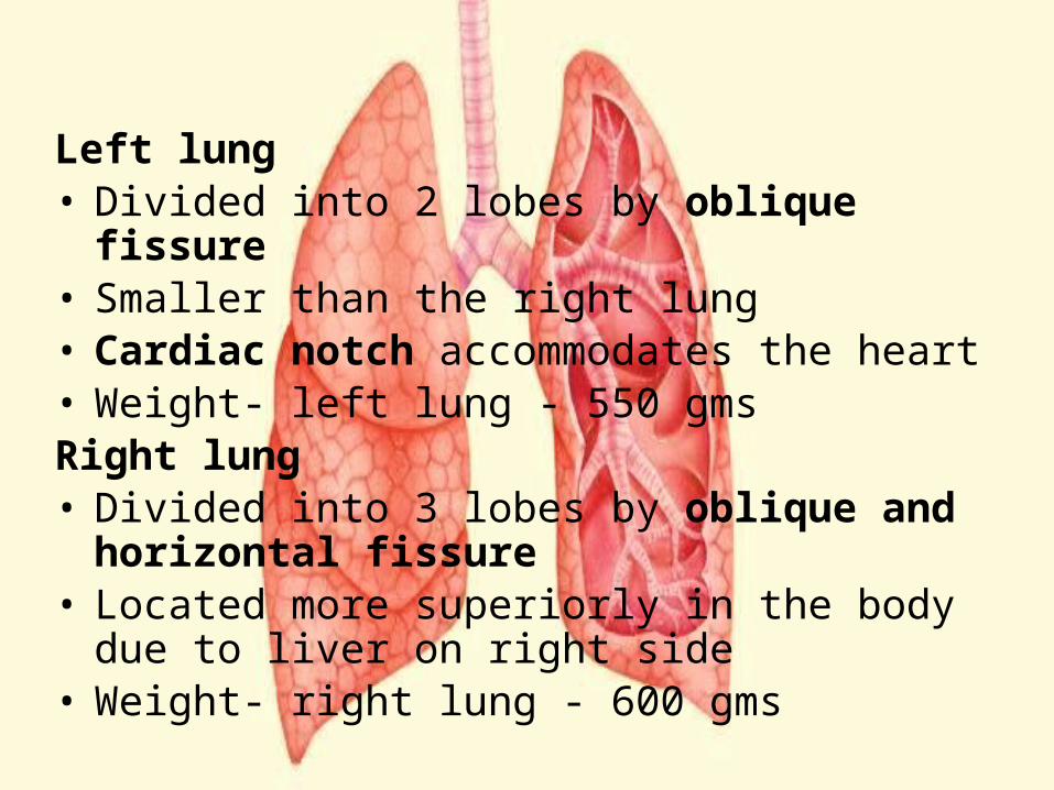
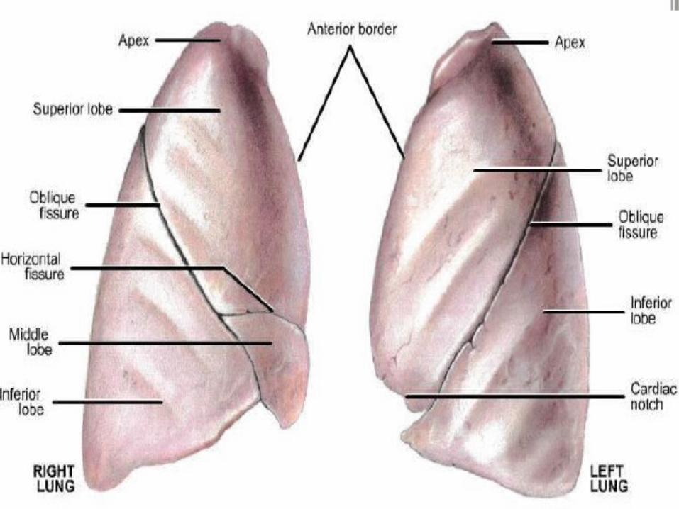
Left lung• Divided into 2 lobes by oblique fissure• Smaller than the right lung • Cardiac notch accommodates the heart • Weight- left lung - 550 gmsRight lung• Divided into 3 lobes by oblique and horizontal
fissure• Located more superiorly in the body due to liver
on right side• Weight- right lung - 600 gms
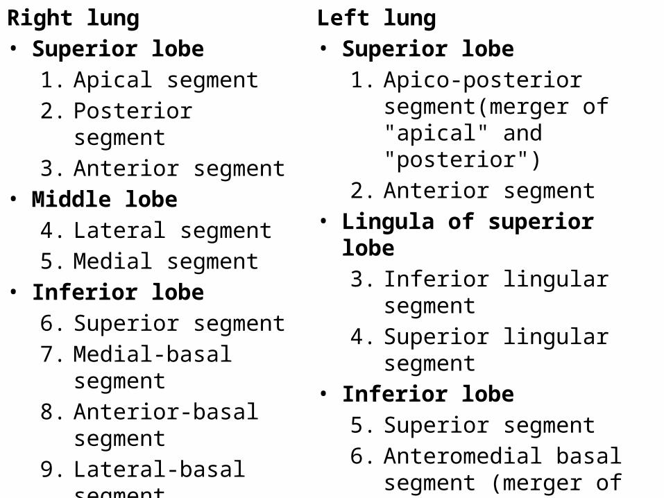
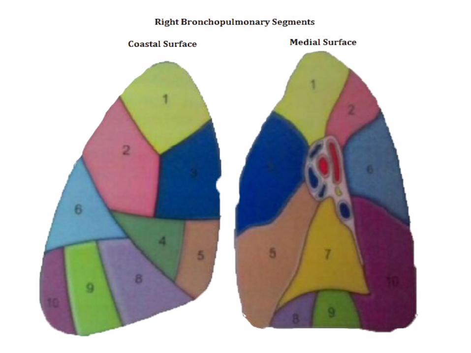
Right lung• Superior lobe
1. Apical segment2. Posterior segment3. Anterior segment
• Middle lobe4. Lateral segment5. Medial segment
• Inferior lobe6. Superior segment7. Medial-basal segment8. Anterior-basal segment9. Lateral-basal segment10. Posterior-basal
segment
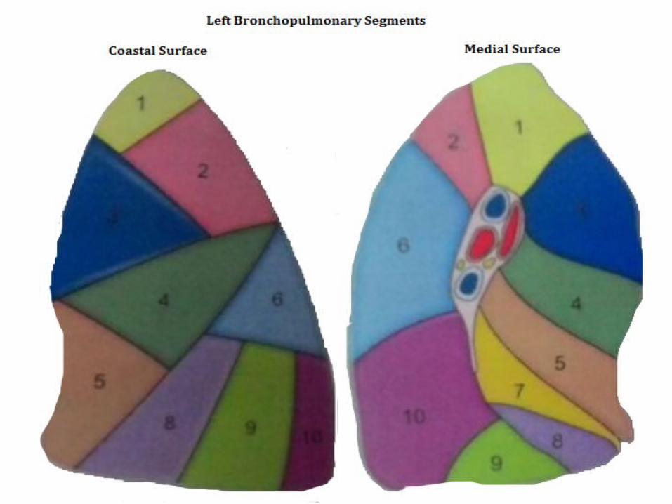
Left lung• Superior lobe
1. Apico-posterior segment(merger of "apical" and "posterior")
2. Anterior segment• Lingula of superior lobe
3. Inferior lingular segment4. Superior lingular segment
• Inferior lobe5. Superior segment6. Anteromedial basal segment
(merger of "anterior basal" and "medial basal")
7. Posterior basal segment8. Lateral basal segment
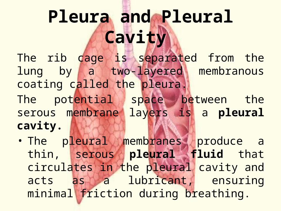
Pleura and Pleural Cavity The rib cage is separated from the lung by a two-layered membranous coating called the pleura.The potential space between the serous membrane layers is a pleural cavity. • The pleural membranes produce a thin, serous
pleural fluid that circulates in the pleural cavity and acts as a lubricant, ensuring minimal friction during breathing.
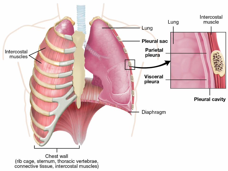
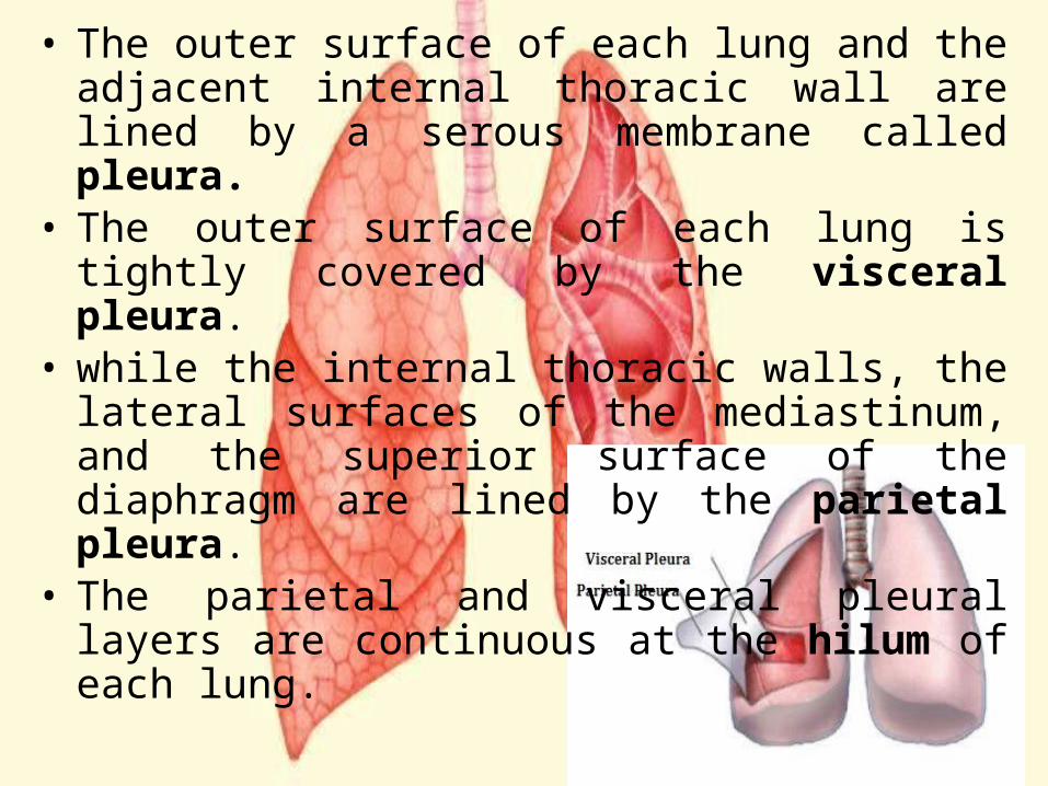
• The outer surface of each lung and the adjacent internal thoracic wall are lined by a serous membrane called pleura.
• The outer surface of each lung is tightly covered by the visceral pleura.
• while the internal thoracic walls, the lateral surfaces of the mediastinum, and the superior surface of the diaphragm are lined by the parietal pleura.
• The parietal and visceral pleural layers are continuous at the hilum of each lung.
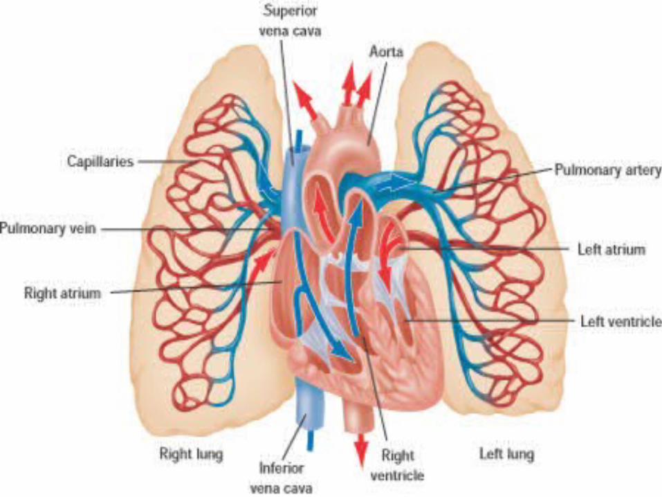
Blood supply of Lungs• Pulmonary circulation – Supplies deoxygenated blood
pumped from the right ventricle and it is carried by pulmonary arteries. When blood passes through the capillaries the alveoli becomes oxygenated.
• Bronchial circulation – Supplies oxygenated blood pumped from the left ventricle and it is carried by bronchial arteries. This circulation is otherwise called systemic circulation. The bronchial arteries supply blood to the bronchi and connective tissue of the lungs. They travel with and branch with the bronchi, ending about at the level of the respiratory bronchioles. They anastomose with the branches of the pulmonary arteries
Pulmonary arteries, the bronchial arteries supply nutrition to the lungs.
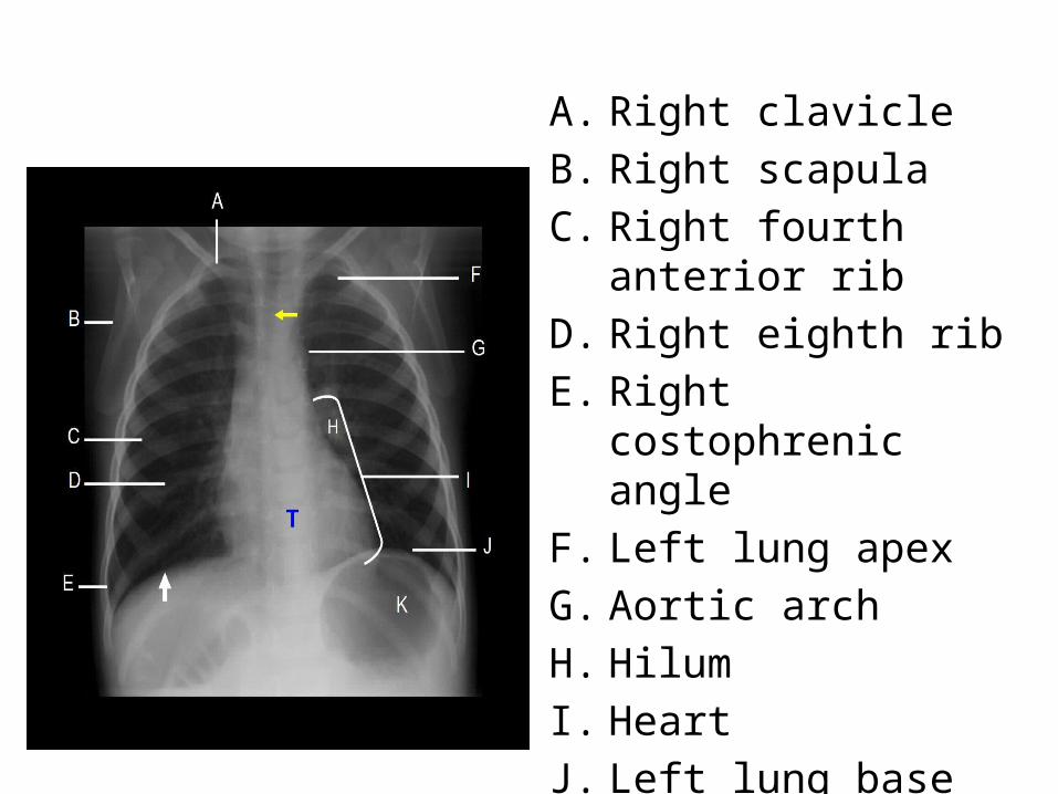
A. Right clavicleB. Right scapulaC. Right fourth anterior ribD. Right eighth rib E. Right costophrenic angle F. Left lung apexG. Aortic archH. Hilum I. Heart J. Left lung base K. Right hemidiaphragm
(white arrow).
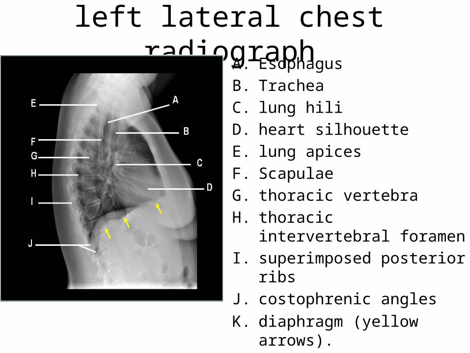
left lateral chest radiographA. EsophagusB. TracheaC. lung hili D. heart silhouetteE. lung apicesF. ScapulaeG. thoracic vertebraH. thoracic intervertebral
foramenI. superimposed posterior ribsJ. costophrenic anglesK. diaphragm (yellow arrows).

Thank you


![Anatomy and Physiology Respiratory System [Tab 2] Respiratory System](https://img.pdfslide.us/doc/110x75/56649ebd5503460f94bc631f/anatomy-and-physiology-respiratory-system-tab-2-respiratory-system.jpg)