Embed Size (px)
Citation preview
Pneumonia
SAMIR EL ANSARY
Pneumonia
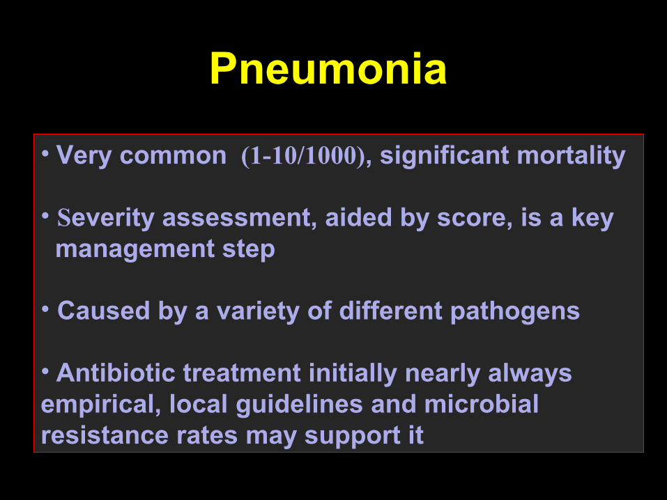
• Very common (1-10/1000), significant mortality
• Severity assessment, aided by score, is a key management step
• Caused by a variety of different pathogens
• Antibiotic treatment initially nearly always empirical, local guidelines and microbial resistance rates may support it
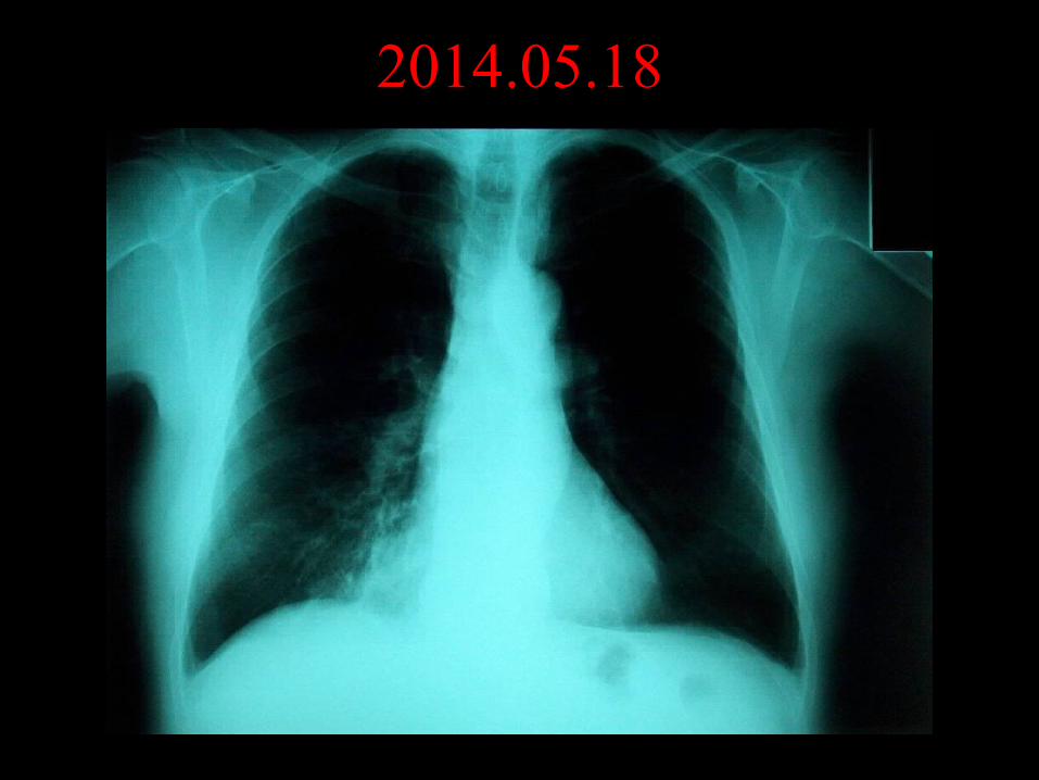
2014.05.18
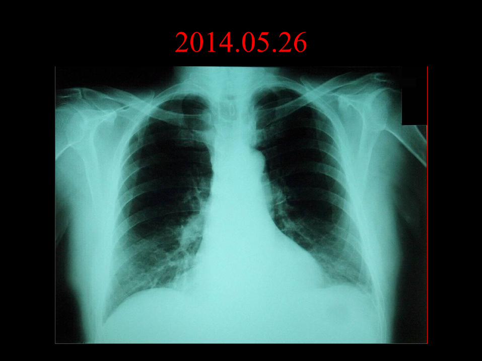
2014.05.26
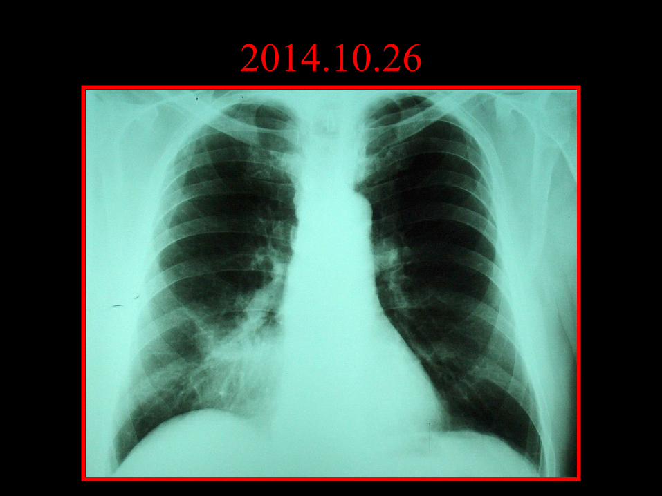
2014.10.26
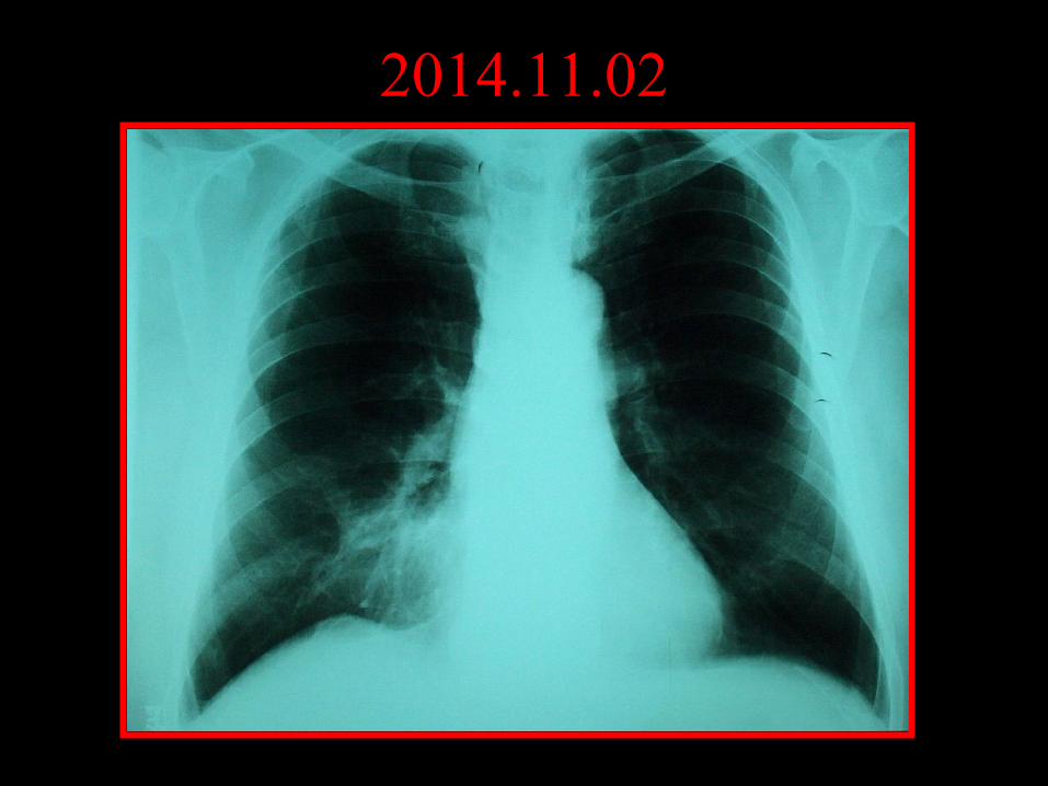
2014.11.02
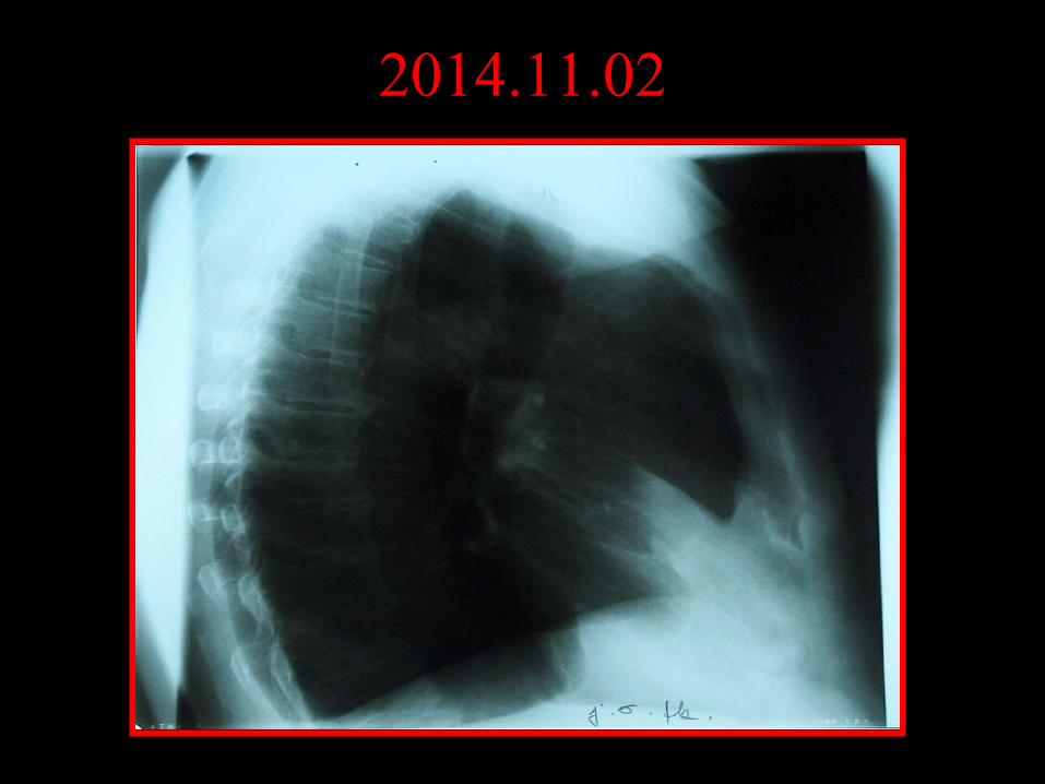
2014.11.02
Definition
Acute, infectious inflammation of the lower respiratory tract parenchyma (distal to bronchiolus terminalis).
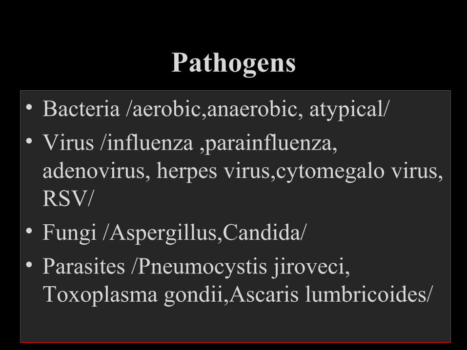
Pathogens
• Bacteria /aerobic,anaerobic, atypical/
• Virus /influenza ,parainfluenza, adenovirus, herpes virus,cytomegalo virus, RSV/
• Fungi /Aspergillus,Candida/
• Parasites /Pneumocystis jiroveci, Toxoplasma gondii,Ascaris lumbricoides/
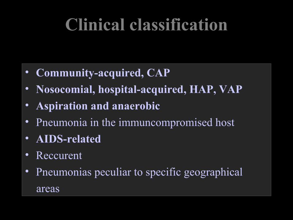
Clinical classification
• Community-acquired, CAP• Nosocomial, hospital-acquired, HAP, VAP• Aspiration and anaerobic• Pneumonia in the immuncompromised host• AIDS-related• Reccurent• Pneumonias peculiar to specific geographical
areas
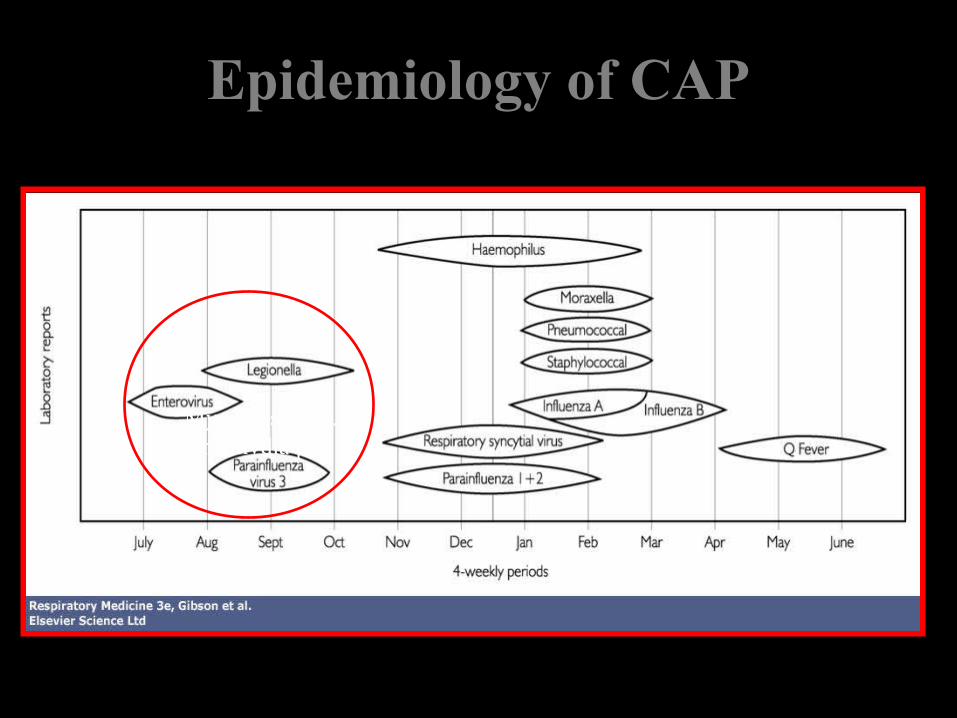
Epidemiology of CAP
Mycoplaspa pn.Chlamydia pn.
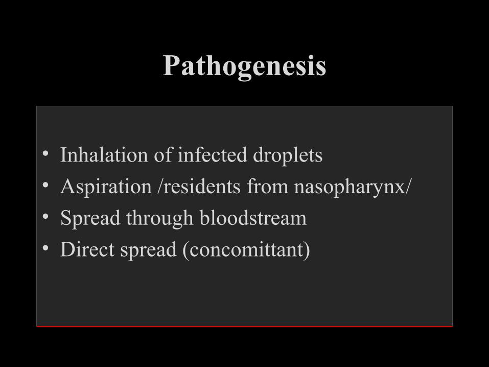
Pathogenesis
• Inhalation of infected droplets
• Aspiration /residents from nasopharynx/
• Spread through bloodstream
• Direct spread (concomittant)
Risk factors
• Prolonged supine position
• Antibiotics, antacids
• Patient contact
• Decreased defense mechanisms
• Infected health care materials
Etiology
• 1. Streptococcus pneumoniae 40-60%
• 2. Mycoplasma pneumoniae 10-20%
• 3. Haemophilus influenzae 6-10%
• 4. Influenza A 5-8%
Clinical features I.
• General symptoms– malaise, anorexia– sweating, rigors– myalgia, arthralgia– headache– fast (bacteremia) vs. slow (Mycoplasma)
progression– marked confusion (Legionella, psittacosis)– acute abdominal or urinary problem (lower lobe,
age!)
• Respiratory symptoms - cough, dsypnea, pleural pain - purulent sputum, hemoptysis• Physical signs - high fever and rigor (Pneumococus) - little or no fever (elderly, seriously ill) - herpes labialis (Pneumococcus) - dullness, inspiratory crackles, bronchial breathing - upper abd. tenderness (lower lobe) - rash (antibiotic, mycoplasma, psittacosis)
Clinical features II.
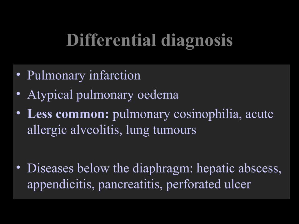
Differential diagnosis
• Pulmonary infarction
• Atypical pulmonary oedema
• Less common: pulmonary eosinophilia, acute allergic alveolitis, lung tumours
• Diseases below the diaphragm: hepatic abscess, appendicitis, pancreatitis, perforated ulcer
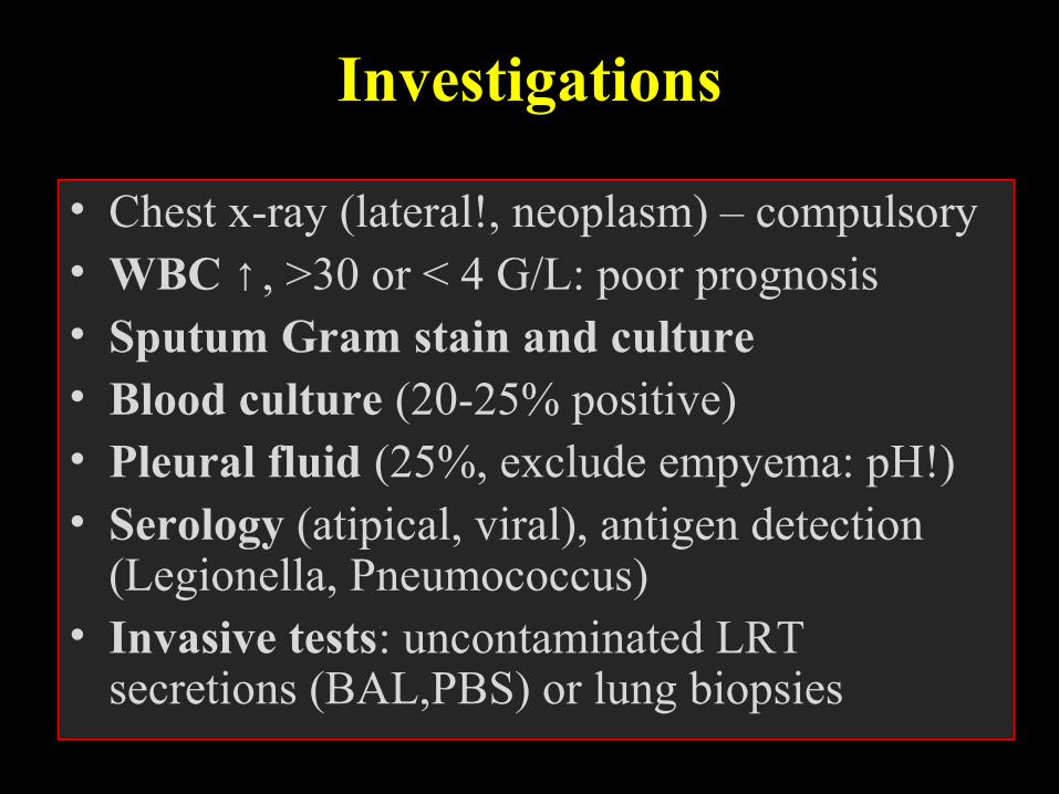
Investigations
• Chest x-ray (lateral!, neoplasm) – compulsory• WBC ↑, >30 or < 4 G/L: poor prognosis• Sputum Gram stain and culture• Blood culture (20-25% positive)• Pleural fluid (25%, exclude empyema: pH!)• Serology (atipical, viral), antigen detection
(Legionella, Pneumococcus)• Invasive tests: uncontaminated LRT
secretions (BAL,PBS) or lung biopsies
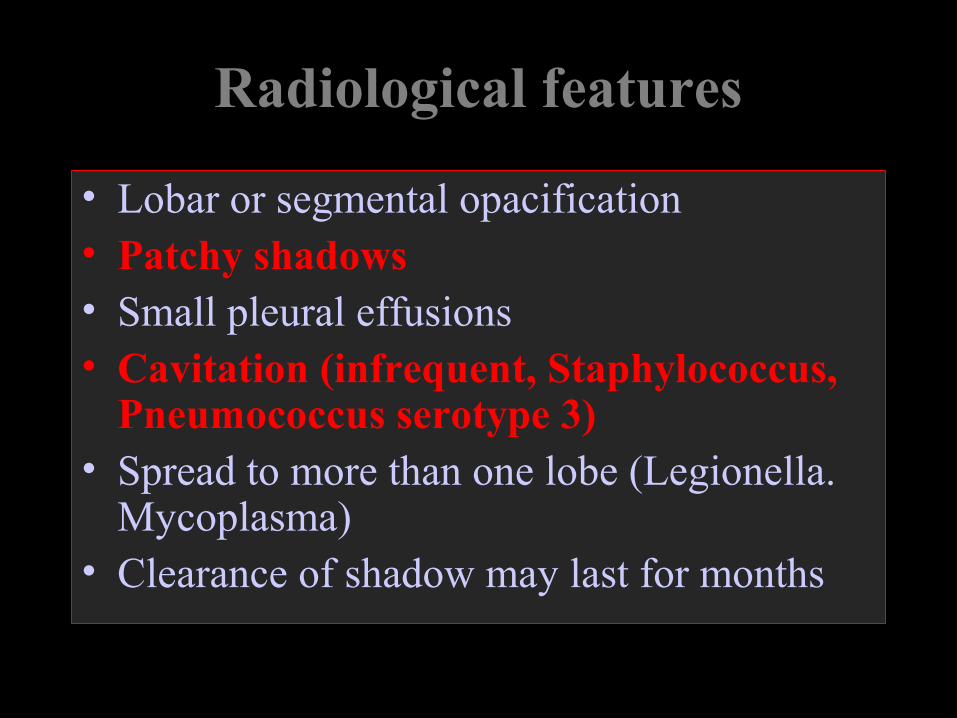
Radiological features
• Lobar or segmental opacification• Patchy shadows• Small pleural effusions• Cavitation (infrequent, Staphylococcus,
Pneumococcus serotype 3)• Spread to more than one lobe (Legionella.
Mycoplasma)• Clearance of shadow may last for months
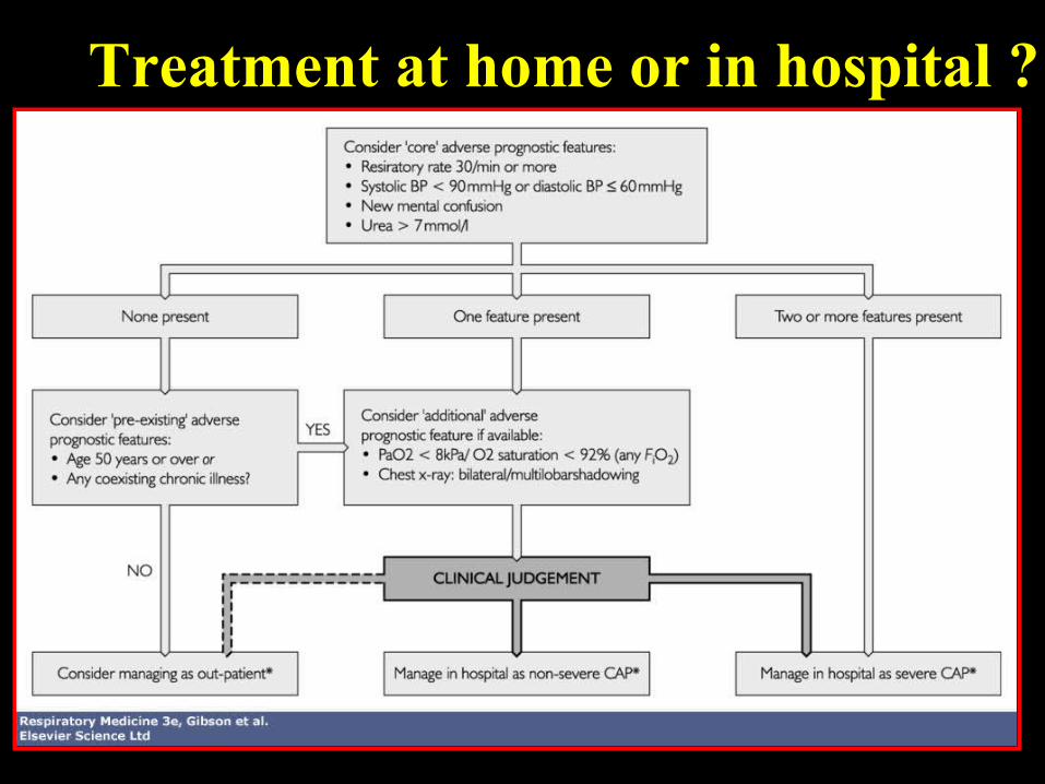
Treatment at home or in hospital ?
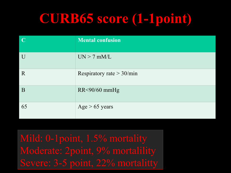
CURB65 score (1-1point)C Mental confusion
U UN > 7 mM/L
R Respiratory rate > 30/min
B RR<90/60 mmHg
65 Age > 65 years
Mild: 0-1point, 1.5% mortalityModerate: 2point, 9% mortalilitySevere: 3-5 point, 22% mortalitty
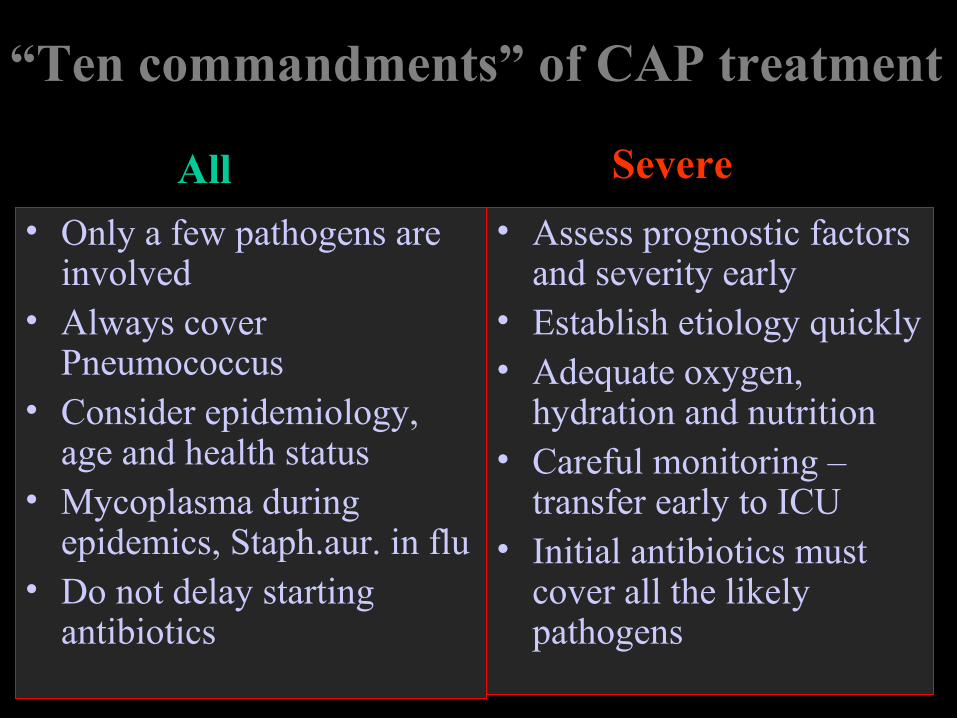
“Ten commandments” of CAP treatment
• Only a few pathogens are involved
• Always cover Pneumococcus
• Consider epidemiology, age and health status
• Mycoplasma during epidemics, Staph.aur. in flu
• Do not delay starting antibiotics
• Assess prognostic factors and severity early
• Establish etiology quickly• Adequate oxygen,
hydration and nutrition• Careful monitoring –
transfer early to ICU• Initial antibiotics must
cover all the likely pathogens
All Severe
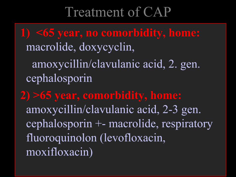
Treatment of CAP 1) <65 year, no comorbidity, home:
macrolide, doxycyclin,
amoxycillin/clavulanic acid, 2. gen. cephalosporin
2) >65 year, comorbidity, home: amoxycillin/clavulanic acid, 2-3 gen. cephalosporin +- macrolide, respiratory fluoroquinolon (levofloxacin, moxifloxacin)
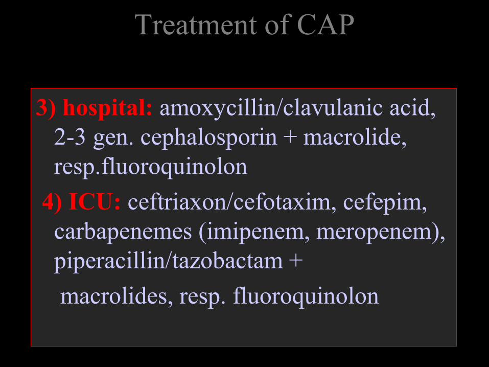
Treatment of CAP
3) hospital: amoxycillin/clavulanic acid, 2-3 gen. cephalosporin + macrolide, resp.fluoroquinolon
4) ICU: ceftriaxon/cefotaxim, cefepim, carbapenemes (imipenem, meropenem), piperacillin/tazobactam +
macrolides, resp. fluoroquinolon
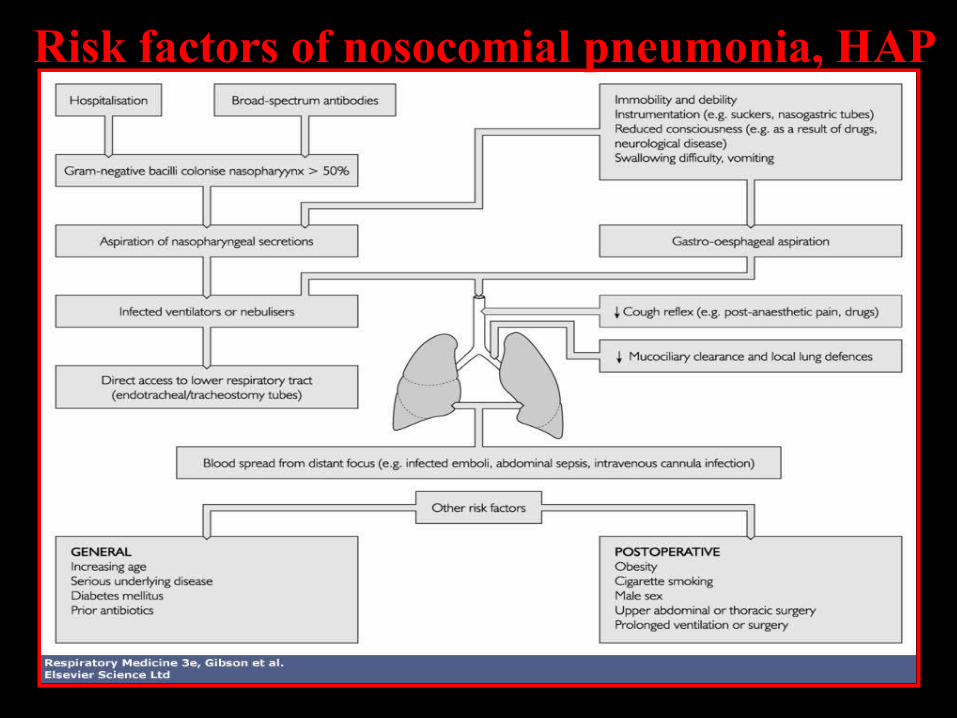
Risk factors of nosocomial pneumonia, HAP
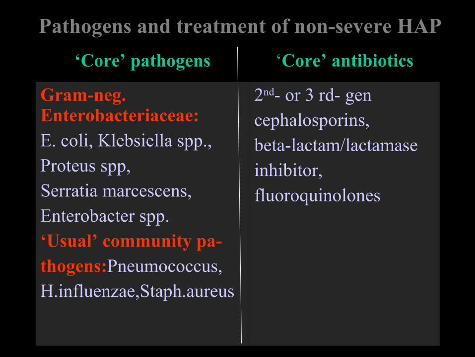
Pathogens and treatment of non-severe HAP
‘Core’ pathogens ‘Core’ antibiotics
Gram-neg. Enterobacteriaceae:E. coli, Klebsiella spp.,Proteus spp,Serratia marcescens,Enterobacter spp.‘Usual’ community pa-thogens:Pneumococcus,H.influenzae,Staph.aureus
2nd- or 3 rd- gencephalosporins,beta-lactam/lactamase inhibitor,fluoroquinolones
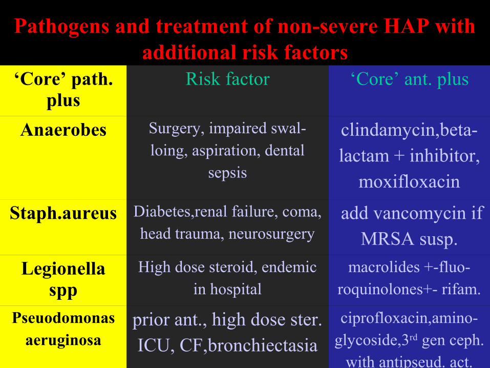
Pathogens and treatment of non-severe HAP with additional risk factors
‘Core’ path. plus
Risk factor ‘Core’ ant. plus
Anaerobes Surgery, impaired swal-loing, aspiration, dental
sepsis
clindamycin,beta-lactam + inhibitor,
moxifloxacin
Staph.aureus Diabetes,renal failure, coma,head trauma, neurosurgery
add vancomycin ifMRSA susp.
Legionella spp
High dose steroid, endemicin hospital
macrolides +-fluo-roquinolones+- rifam.
Pseuodomonasaeruginosa
prior ant., high dose ster.ICU, CF,bronchiectasia
ciprofloxacin,amino-glycoside,3rd gen ceph.
with antipseud. act.
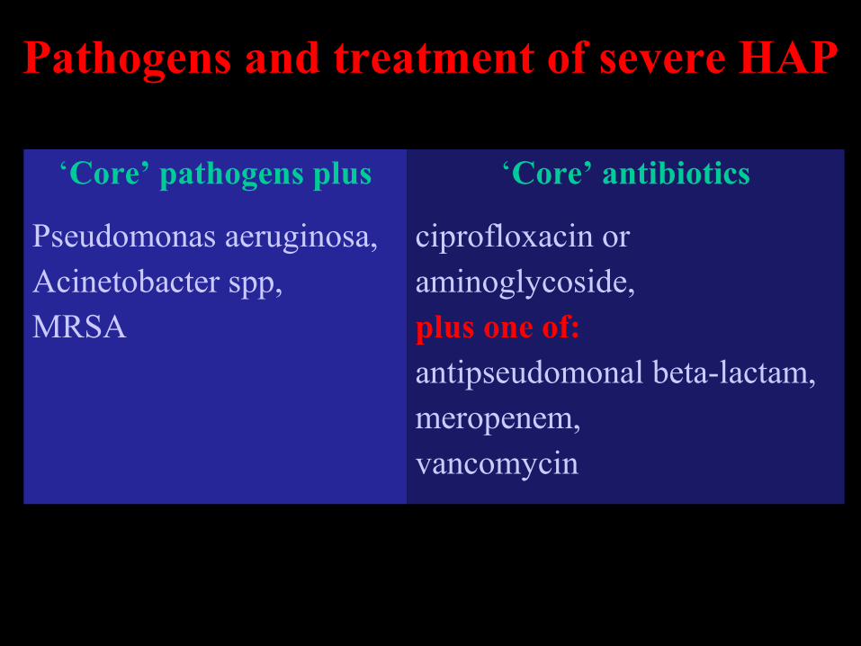
Pathogens and treatment of severe HAP
‘Core’ pathogens plus ‘Core’ antibiotics
Pseudomonas aeruginosa,Acinetobacter spp,MRSA
ciprofloxacin oraminoglycoside,plus one of:antipseudomonal beta-lactam,meropenem,vancomycin
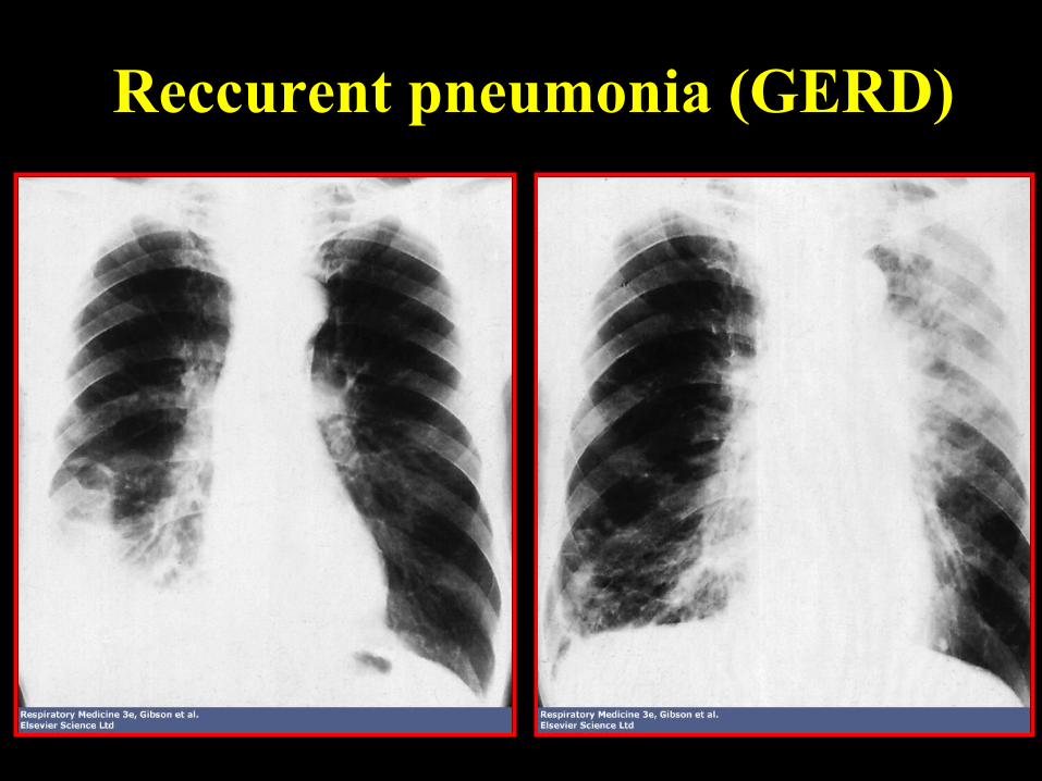
Reccurent pneumonia (GERD)
Streptococcus pneumoniae
• Most common bacterium in adults• Significant morbidity and mortality
• Polysaccharide capsule impairs phagocytosis → need of opsonization → risk population:
lymphoma, hyposplenia, hypogammaglobulinaemia
• Abrupt onset, cough, rigors, high fever, tachycardia, tachypnea, sticky pink sputum, focal crackles.
Streptococcus pneumoniae
• Sputum Gram stain: diplococcus, blood culture (20% pos.)
• Good sputum sample: LRT: > 25 PMN, < 10 EC (low power field)
• X-ray: homogenouos consolidation
• Complications: pleura, pericardium, meninges, joints, endocardium, Type 3: abscess, lung scarring
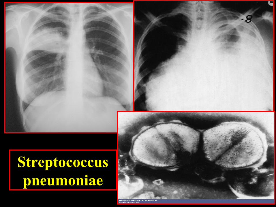
Streptococcuspneumoniae
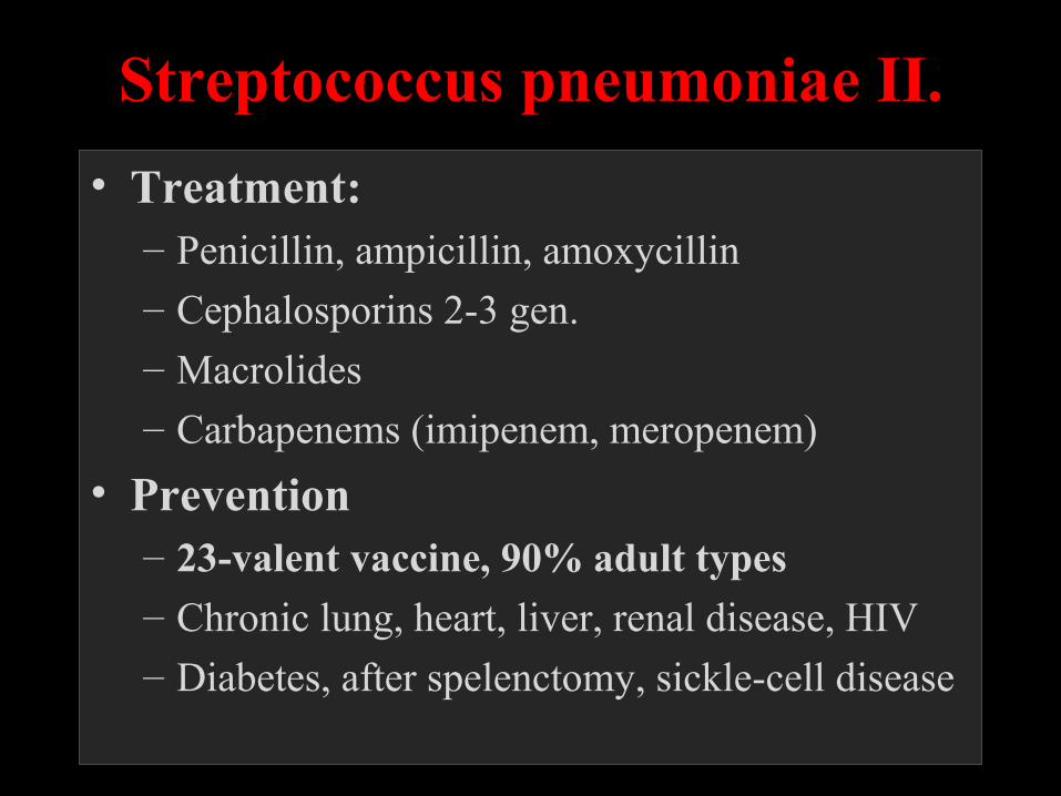
Streptococcus pneumoniae II.
• Treatment: – Penicillin, ampicillin, amoxycillin– Cephalosporins 2-3 gen.– Macrolides– Carbapenems (imipenem, meropenem)
• Prevention– 23-valent vaccine, 90% adult types – Chronic lung, heart, liver, renal disease, HIV
– Diabetes, after spelenctomy, sickle-cell disease
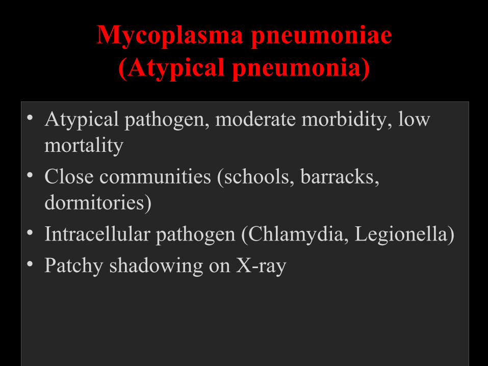
Mycoplasma pneumoniae(Atypical pneumonia)
• Atypical pathogen, moderate morbidity, low mortality
• Close communities (schools, barracks, dormitories)
• Intracellular pathogen (Chlamydia, Legionella)
• Patchy shadowing on X-ray
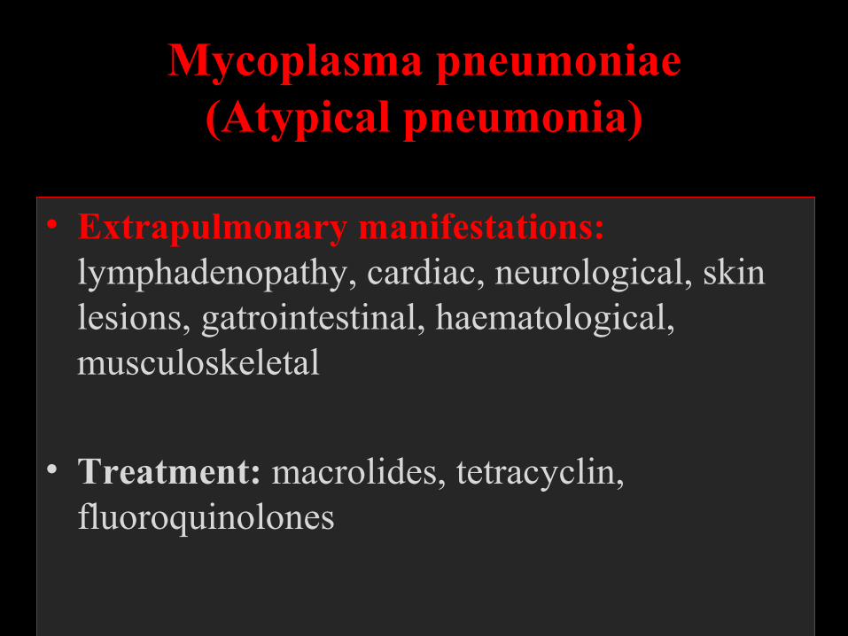
Mycoplasma pneumoniae(Atypical pneumonia)
• Extrapulmonary manifestations: lymphadenopathy, cardiac, neurological, skin lesions, gatrointestinal, haematological, musculoskeletal
• Treatment: macrolides, tetracyclin, fluoroquinolones
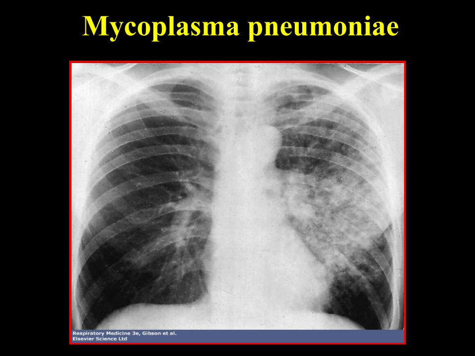
Mycoplasma pneumoniae
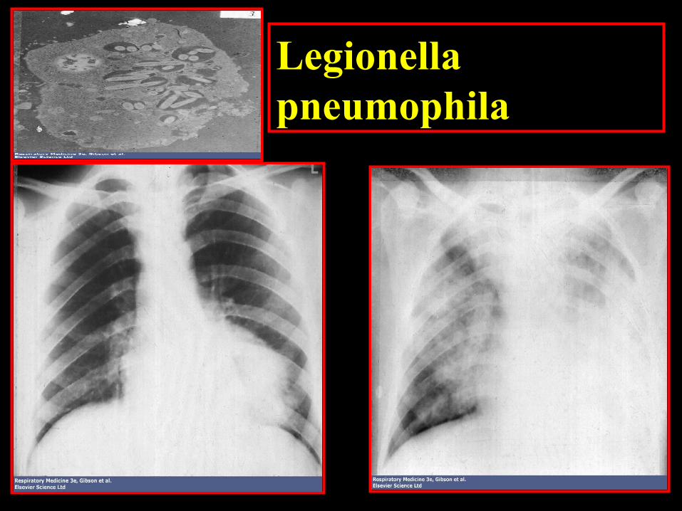
Legionella pneumophila
Staphylococcus aureus• High morbidity and mortality (30-70% in
bacterae-mia)
• 30% of adults carry in the anterior nares
• Intravascular tubes (catheters, cannules)
• Usually follows influenza infections
• Toxins → tissue necrosis → abscess
• Treatment: beta-lactamase resistant penicillins (oxacillin), cephalosporins, MRSA: vancomycin
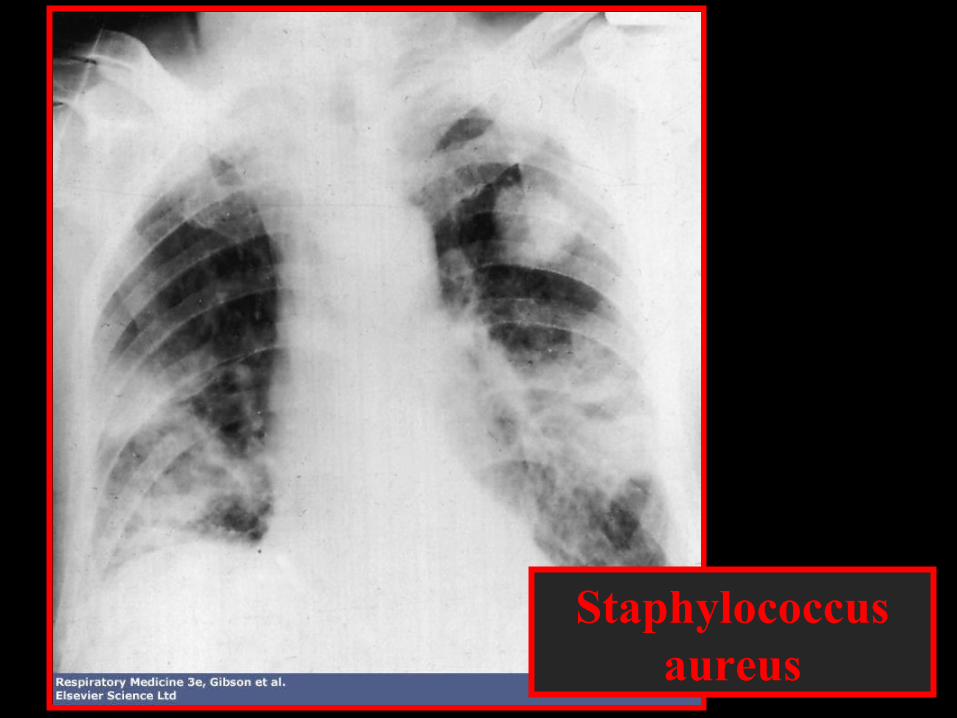
Staphylococcusaureus
Lung abscess
• many other cavitating lesions than abscess• careful review of chest x-ray to distinguish from
empyema• most are secondary to aspiration of oropharyngeal
secretions• exclude malignancy or other cause, bronchoscopy!• a single microbe is unusual unless abscesses developed
after bacterial pneumonia. • More commonly, there is a mixed growth, including
anaerobes
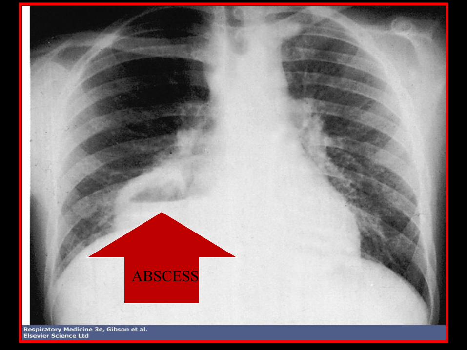
ABSCESS
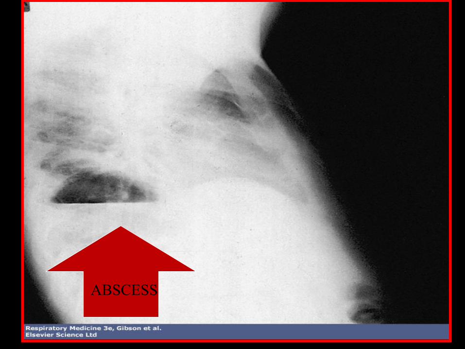
ABSCESS
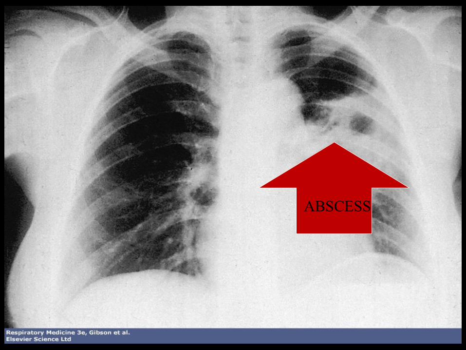
ABSCESS
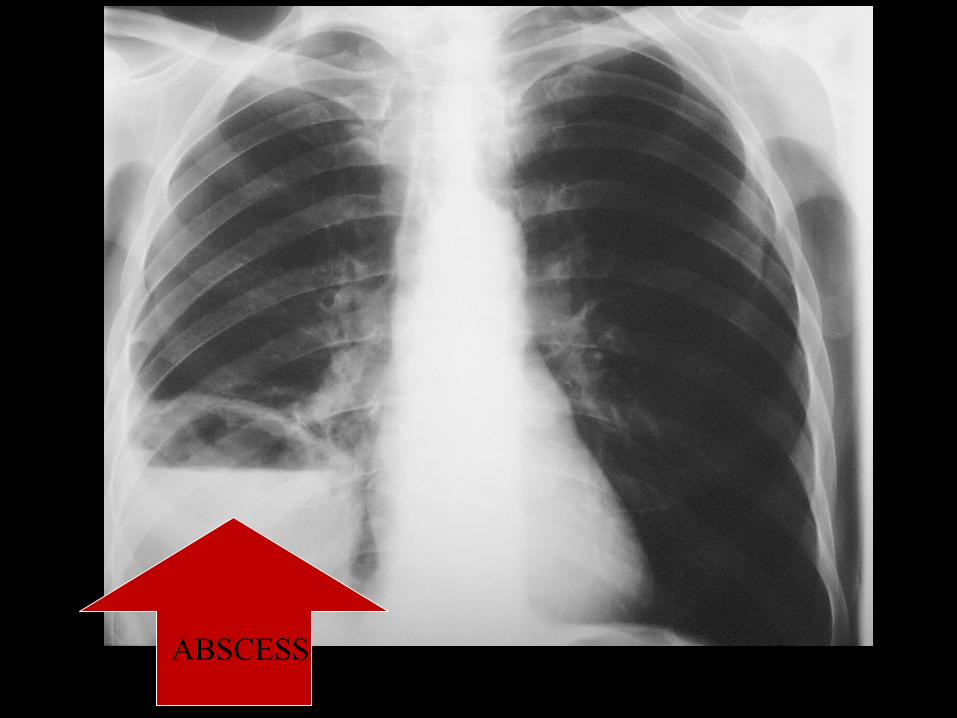
ABSCESS

ABSCESS
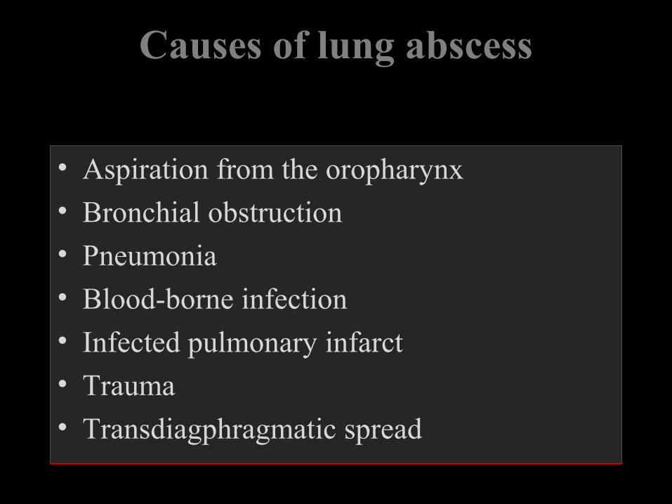
Causes of lung abscess
• Aspiration from the oropharynx
• Bronchial obstruction
• Pneumonia
• Blood-borne infection
• Infected pulmonary infarct
• Trauma
• Transdiagphragmatic spread
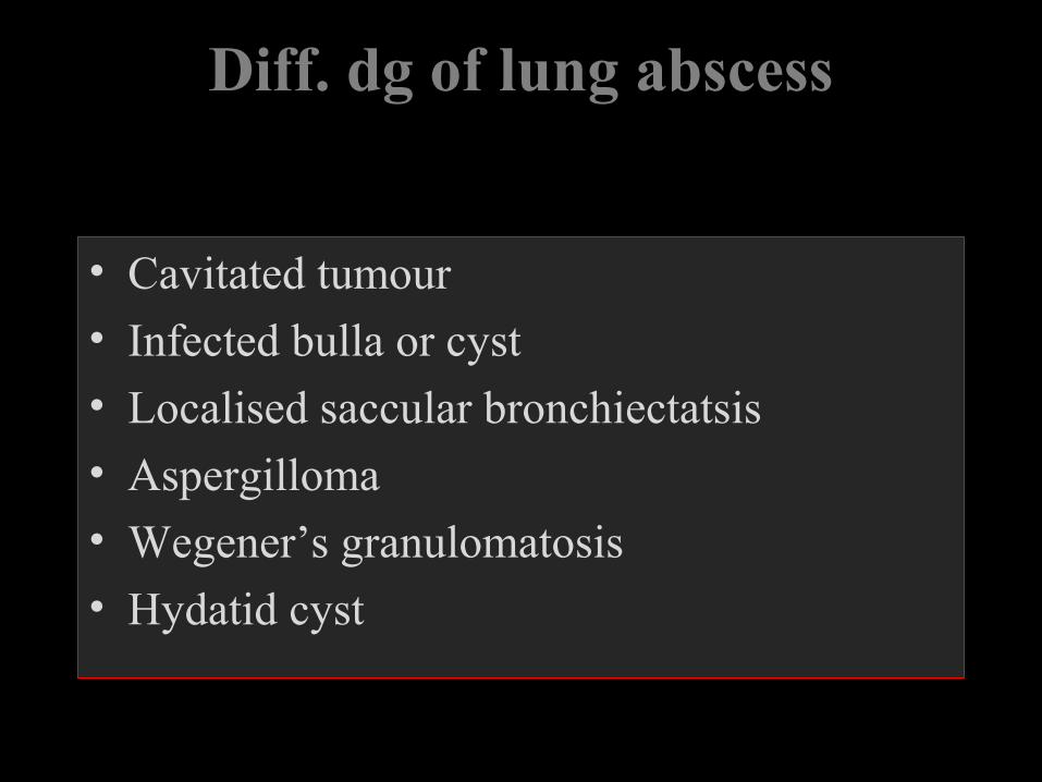
Diff. dg of lung abscess
• Cavitated tumour
• Infected bulla or cyst
• Localised saccular bronchiectatsis
• Aspergilloma
• Wegener’s granulomatosis
• Hydatid cyst
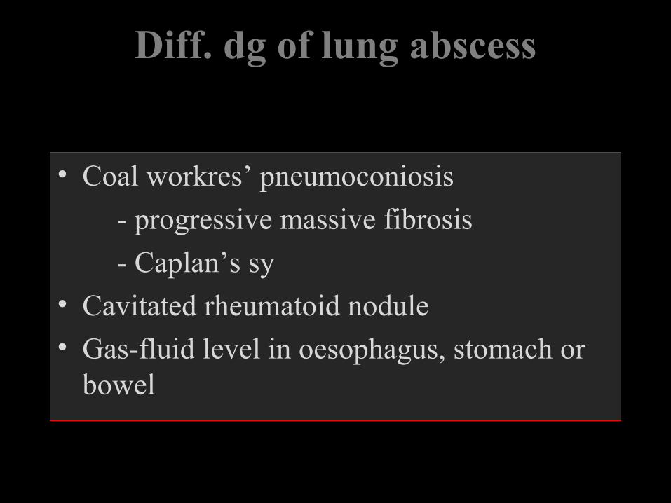
Diff. dg of lung abscess
• Coal workres’ pneumoconiosis
- progressive massive fibrosis
- Caplan’s sy
• Cavitated rheumatoid nodule
• Gas-fluid level in oesophagus, stomach or bowel
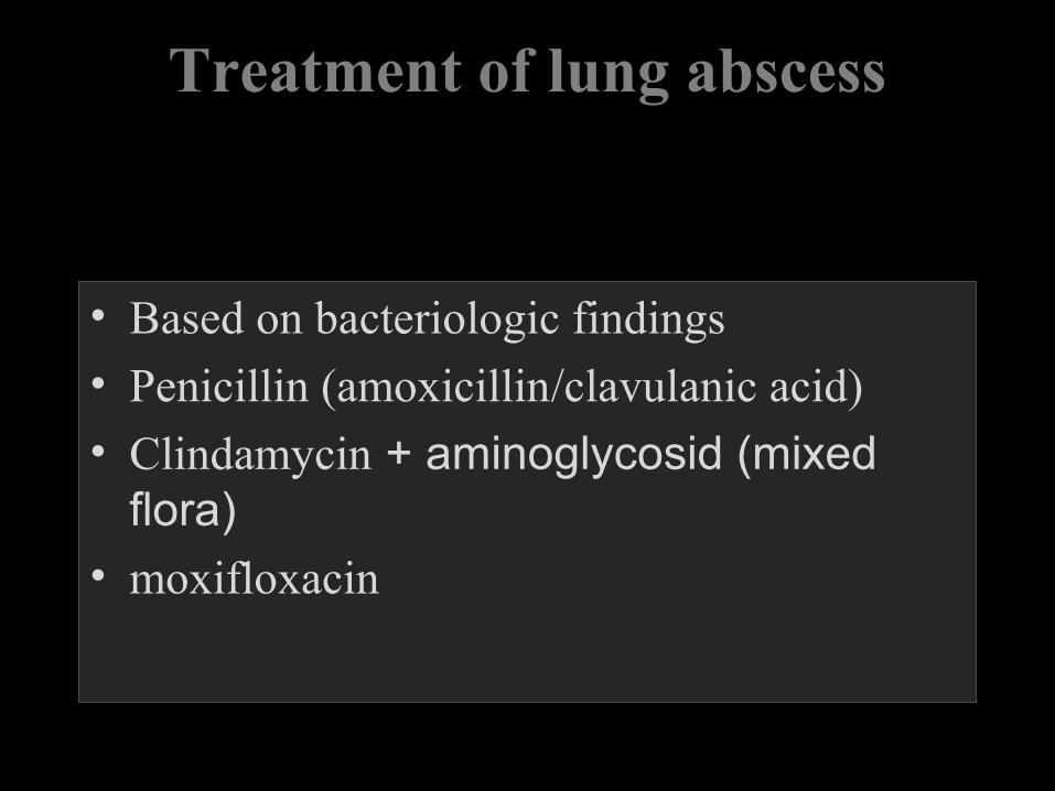
Treatment of lung abscess
• Based on bacteriologic findings
• Penicillin (amoxicillin/clavulanic acid)
• Clindamycin + aminoglycosid (mixed flora)
• moxifloxacin

GOOD LUCK
SAMIR EL ANSARY