Embed Size (px)
Citation preview
MINUTE ANATOMY OF OKUAN Ol» JACOBSON IN GUINEA-PIG. 2 1 9
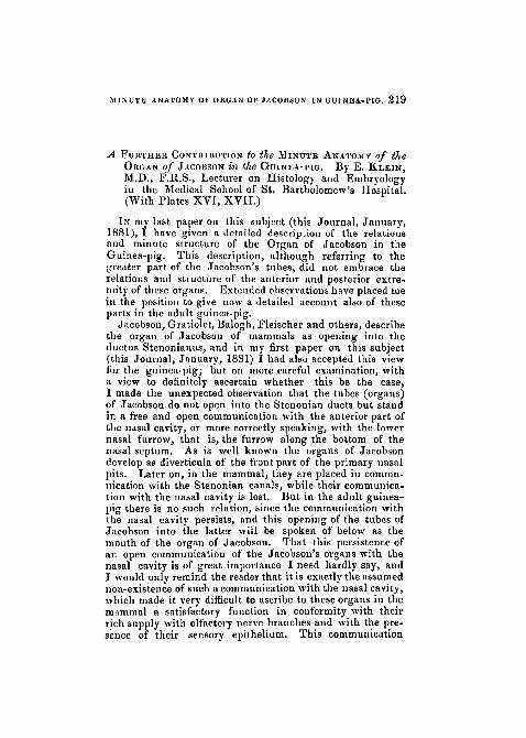
A FURTHER CONTRIBUTION to the MINUTE ANATOMY of theORGAN of JACOBSON in the GUINEA-PIG. By E. KLEIN,M.D., F.R.S., Lecturer on Histology and Embryologyin the Medical School of St. Bartholomew's Hospital.(With Plates XVI, XVII.)
TN my last paper on this subject (this Journal, January,1881), I have given a detailed description of the relationsand minute structure of the Organ of Jacobson in theGuinea-pig. This description, although referring to thegreater part of the Jacobson's tubes, did not embrace therelations and structure of the anterior and posterior extre-mity of these organs. Extended observations have placed mein the position to give now a detailed account also of theseparts in the adult guinea-pig.
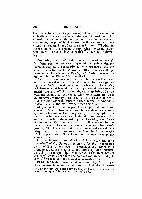
Jacobson, Gratiolet, Balogh, Fleischer and others, describethe organ, of Jacobson of mammals as opening into theductus Stenonianus, and in my first paper on this subject(this Journal, January, 1881) I had also accepted this viewfor the guinea-pig; but on more careful examination, witha view to definitely ascertain whether this be the case,I made the unexpected observation that the tubes (organs)of Jacobson do not open into the Stenonian ducts but standin a free and open communication with the anterior part ofthe nasal cavity, or more correctly speaking, with the lowernasal furrow, that is, the furrow along the bottom of thenasal septum. As is well known the organs of Jacobsondevelop as diverticula of the front part of the primary nasalpits. Later on, in the mammal, they are placed in commu-nication with the Stenonian canals, while their communica-tion with the nasal cavity is lost. But in the adult guinea-pig there is no such relation, since the communication withthe nasal cavity persists, and this opening of the tubes ofJacobson into the latter will be spoken of below as themouth of the organ of Jacobson. That this persistence ofan open communication of the Jacobson's organs with thenasal cavity is of great importance I need hardly say, andI would only remind the reader that it is exactly the assumednon-existence of such a communication with the nasal cavity,which made it very difficult to ascribe to these organs in themammal a satisfactory function in conformity with theirrich supply with olfactory nerve branches and with the pre-sence of their sensory epithelium. This communication
220 DR. E. KLEIN.
being now found in the guinea-pig1 there is of course nodifficulty whatever in ascribing to the organ of Jacobson in thisanimal a function similar to that of the olfactory mucousmembrane, but probably of a more specific nature, as I havealready hinted at in my last communication. Whether inother mammals this communication with the nasal cavitypersists, will be a subject to which I soon hope to devotemyself.
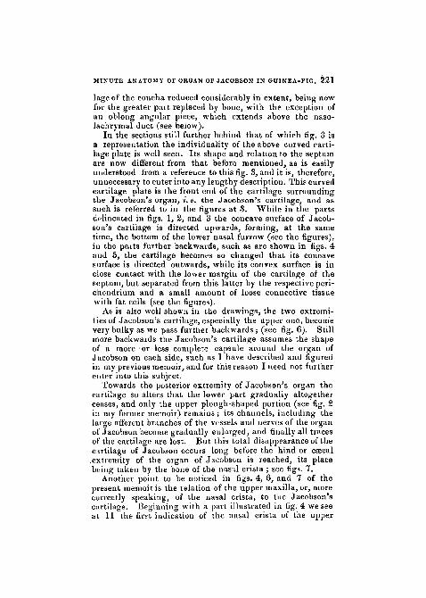
Examining a series of vertical transverse sections throughthe front part of the nasal organ of the guinea-pig, theorgan having been previously suitahly prepared (see mypaper in this Journal for January, 1881), we meet with ap-pearances of the several parts, very accurately shown in thefigures 1 to 6 of Plates XVI and XVII.
Fig. 1 is a transverse section through the most anteriorpart of the nasal organ. The relation of the cartilaginousseptum to the lower turbinated bone, or lower concha nasalis,and further, of this to the alveolar process of the superiormaxilla, are very well illustrated, the drawings being all madewith the camera lucida, the relative proportions and sizesare all very accurately preserved. It will be seen in fig. 1that the cartilaginous septum nasale forms an unbrokencontinuity with the cartilage representing here, i.e. in thefront part of the nasal organ, the support of the lowerconcha. This continuity is brought about, on each side,by a curved, more or less trough-shaped cartilage plate ex-tending on the inner surface of the alveolar process of thesuperior maxilla to the angular piece of cartilage that formsthe support of the lower concha. But this continuation ismore or less broken as we pass a little way backwards(see fig. 2). Here we find the above-named curved car-tilage plate more or less separated from the lower marginof the septum as well as from the cartilage piece of theconcha.
In my former communication I have used the term" concha" of the German anatomists for the " turbinatedbone" of English text books. I consider the former termpreferable, because it gives to the organ a name indepen-dent of its structure. In our case, i. e. in the front part ofthe nasal organ where there is no bone contained in it yet,it would be incorrect to speak of a turbinated ' bone.'
In fig. 3, which is taken a little behind fig. 2, this sepa-ration is complete, and, in addition, we find also the carti-
1 I am in a position to assert also for the rabbit such a free oommuni-eation of the organ of Jacobson with the nasal cavity. .
MINUTE ANATOMY OF ORGAN OF JACOBSON IN GUINEA-PIG. 2 2 1
lage of the concha reduced considerably in extent, being nowfor the greater part replaced by bone, with the exception ofan oblong angular piece, which extends above the naso-lachrymal duct (see below).
In the sections still further behind that of which fig. 3 isa representation the individuality of the above curved carti-lage plate is well seen. Its shape and relation to the septumare now different from that before mentioned, as is easilyunderstood from a reference to this fig. 3, and it is, therefore,unnecessary to enter into any lengthy description. This curvedcartilage plate is the front end of the cartilage surroundingthe Jacobson's organ, i. e. the Jacobson's cartilage, and assuch is referred to in the figures at 3. While in the partsdelineated in figs. 1, 2, and 3 the concave surface of Jacob-son's cartilage is directed upwards, forming, at the sametime, the bottom of the lower nasal furrow (see the figures),iu the parts further backwards, such as are shown in figs. 4and 5, the cartilage becomes so changed that its concavesurface is directed outwards, while its convex surface is inclose contact with the lower margin of the cartilage of theseptum, but separated from this latter by the respective peri-chondrium and a small amount of loose connective tissuewith fat cells (see the figures).
As is also well shown in the drawings, the two extremi-ties of Jacobson's cartilage, especially the upper one, becomevery bulky as we pass further backwards ; (see fig. 6). Stillmore backwards the Jacobsou's cartilage assumes the shapeof a more'or less complete capsule around the organ ofJacobson on each side, such as 1 have described and figuredin my previous memoir, and for this reason I need not furtherenter into this subject.
Towards the posterior extremity of Jacobson's organ thecartilage so alters that the lower part gradually altogetherceases, and only the upper plough-shaped portion (see fig. 2in my former memoir) remains; its channels, including thelarge afferent branches of the vessels and nerves of the organof Jacobson become gradually enlarged, and finally all tracesof the cartilage are lost. But this total disappearance of thecartilage of Jacobson occurs long before the hind or caecal.extremity of the organ of Jacobson is reached, its placebuing taken by the bone of the nasal crista ; see figs. 7.
Another point to be noticed in figs. 4, 6, and 7 of thepresent memoir is the relation of the upper maxilla, or, morecorrectly speaking, of the nasal crista, to the Jacobson'scurtilage. Beginning with a part illustrated in fig. 4 we seeat 11 the first indication of the nasal crista of the upper
222 DR. E. KLEIN.
maxilla, in the shape of a thin lamella of bone, extendingalong the convex surface of Jacobson's cartilage. It soonbecomes very conspicuous, covering not only the convexsurface of the cartilage, but gradually embracing the greaterpart of the circumference of the organ of Jacobson, as shownin figs. 1 and 2 of my paper in the January number of thisJournal. In the posterior portion of the organ of Jacobson,viz. where the cartilage of Jacobson is wanting, as has beenjust mentioned, the former, i. e. the organ of Jacobson, isentirely enclosed in the osseous substance of the nasal crista,as is shown in fig. 7 of the present memoir.
In figs. 1,2, and 3, i. e. in the anterior portion of the nasalorgan, the lower nasal furrow (4) is lined with columnarciliated epithelium, like that of the septum ; it has beenminutely described in my former memoir, and need not,therefore, be referred to here any further. The very bottomof the furrow, however, is lined with stratified pavementepithelium, of which the superficial layers consist of sqamousepithelial cells, each possessing a flattened nucleus. In thepreparations from which the above figures are taken thesesuperficial layers are more or less loosely attached, to therest of the epithelium, hence appear as if desquamating.The stratified epithelium, as a whole, is stained very muchdeeper in these preparations than the columnar epithelium,and is therefore very conspicuous in the drawings.
Such is the state of the epithelium at the bottom of thefurrow in the front part (figs. 1, 2, and 3). But, goingfurther back, we find that this part of the furrow, viz. the onelined with stratified pavement epithelium changes its posi-tion, inasmuch as it does not now occupy the very bottom ofthe furrow, but gradually extends, inwards and upwardstowards the concave surface of the Jacobson's cartilage. Aninspection of figs. 4 and 5 will make this easily understood.At 12 in these figures the changed position of the partcovered with stratified pavement epithelium is well seen.
Now, this pit, viz. marked 12, is the commencement ofthe organ or tube of Jacobson, or, more correctly speaking,the anterior opening of this latter into the nasal furrow ; weshall call it the mouth of Jacobson's organ. Its diameter isabout 0'05 mm., not in'luding the lining epithelium, whosethickness is about 0'057 mm.
A little way further back (see fig. 5) we meet alreadywith the well-formed tube of Jacobson, and we still recog-nise here (he stratified pavement epithelium, occupying, asmust be expected from what has been said before, the upperpart of the tube; the lower part is lined with columnar
MINUTE ANATOMY OF ORGAN OF JACOBSON IN GUINEA-PIG. 2 2 3
ciliated epithelium, like that of the nasal furrow. A littlefurther back the epithelium lining the tube is uniform onall sides, being ciliated columnar epithelium, such as hasbeen minutely described in my former paper as lining thelateral wall of the organ of Jacobson.
Still further back (see fig. 6) we meet with this impor-tant alteration in the epithelium, that the median wall islined with the thick sensory epithelium minutely describedin my first paper. The tube, as a whole, has not, however,got yet its characteristic shape, i. e. so compressed from side toside,that its lumen is kidney shaped in transverse section (seefigs. 1 and 2, in my first paper) ; this shape is, however,soon obtained, and the organ retains.it to near its posteriorextremity. Before this, however^ is reached the directionof the lumen, and consequently of the walls, is more or lef soblique and slanting downwards and inwards. In thisoblique position we find chiefly the epithelium and thecavernous layer.
As regards the structure of the wall of the tube ofJacobson in its commencement, it is similar to that of thenasal furrow, viz. the subepithelial membrane is connectivetissue infiltrated with numerous, lymph-corpuscles, andresembling more or less diffuse adenoid tissue.
Already at. the mouth of Jacobson's tube (12 in figs. 4and 5) we find large veins forming a plexus and repre-senting the rudiment, of. the cavernous tissue described inmy first paper. This plexus becomes more developed as wepass backwards, and from its. first appearance, i. e. at themouth of Jacobson's organ, to the posterior extremity ofthis (see below), does not encroach on the median wall, i.e.the wall nearest the septum. In the parts from which fig. 6 istaken-, but not further in front, we find between the vesselsof the cavernous tissue already bundles of non-striped mus-cular tissue.
Longitudinal sections through the organ of Jacobson provethat a considerable posterior portion is without any carti-lage, and is enclosed altogether in the bone of the cristanasalis, as mentioned above.
The posterior csecal extremity of the organ is slightlycurved inwards and upwards; it is smaller in diameter thanthe rest of the organ ; its lumen is circular in cross section.The median wall of this extremity is not lined with anysensory epithelium, this having previously suddenly cometo an end ; we find here everywhere only columnar epithe-lium, similar to that of the lateral wall of the preceding parts.
224 DR. E. KLEIN.
In my first paper I have shown that the layer of (serous)glands is situated outside the cavernous layer, and extendsin the lateral wall from the lower sulcus to the upper sulcus,but its bulk lies about the lower sulcus, into which the ductschiefly open (see figs. 1 and 2 of my former paper). As wepass backwards, but long before the extremity of the organis reached, we find the area in which the glands extendconsiderably increased, for many glands are now found alsoabout the upper sulcus, and their ducts open into this latter.In the part which we just before described as the poste-rior extremity, viz. the narrow hind part with the cylindricallumen, and without any sensory epithelium on the medianwall, we find the layer of glands forming a complete invest-ment and occupying almost the whole periphery of the organ(see fig. 7).
Another difference between the posterior section of theorgan and the rest lies in the change the cavernous tissueundergoes in that extremity. As is shown in fig. 7, itsposition is not the same that it was before (compare figs. Iand 2 of my first memoir), viz. occupying the lateral wallonly, for, owing to the altered position of the lumen—thislatter being not vertically but obliquely placed—the ca-vernous tissue now occupies the lower wall. At the sametime the number of vessels and the amount of non-strippdmuscle tissue are greatly increased, although the individualvessels are not of so large a size as in preceding portions ofthe organ.
The plexus of non-striped muscular tissue forming thematrix of the cavernous layer is best developed in the extre-mity itself, and, together with the plexus of veins of thecavernous layer and the gland layer outside, extends forsome little distance behind the caecal end. In longitudinalsections comprising the posterior portion of the organ ofJncobson, these plexuses of bundles of non-striped muscularcells, and of veins between them, is very finely seen.
The tube which I described and figured in fig. I1 in myfirst memoir was there named the " accessory organ ofJacobson." But this is the naso-lachrymal duct; this wassuggested to me through the kindness of Dr. Allen Thom-son, by Prof. Kolliker, of Wiirzburg, and on careful dissec-tion I have convinced myself that this is really the case.
It extends anteriorly and posteriorly considerably beyondthe region of the organ of Jacobson. Anteriorly the naso-
1 In the text, p. 106, it is erroneously referred to as "at 8 in fig. 1,"it should read "at 9 iu fig. 1."
MINUTE ANATOMY OF ORGAN OF JACOBSON IN GUINEA-PIG. 2 2 5
lachrymal ducts are met with, in vertical transverse sectionsthrough the nasal organ, some distance in front of Jacob-son's organs, as minute tubes, in the same position and ofthe same nature as described in my first memoir, aswill be easily understood on comparing the figures 1, 2, 3,and 4 accompanying the present paper. Also backwardsfor a very long distance beyond the region of the organ ofJacobson about 10—12 mm.; the naso-lachrymal ductsretain the same position, viz. between the nasal furrow andthe alveolar process of the upper maxilla. As I mentionedin my first paper, this duct is slightly flattened from side toside, and the diameter enlarges considerably posteriorly ; inthe region of the hind extremity of the organ of Jacobsonthe diameters of the naso-lachrymal duct (5 in fig. 7) arethese:
Horizontal transverse diameter of the whole duct . l'Ol mm.Vertical transverse diameter „ „ . . 2-81 mm.Horizontal transverse diameter of the lumen . . 060 mm.Vertical „ „ „ l'Ol mm.Vertical (thickness) diameter of the lower wall . . O"7875 mm.Vertical (thickness) diameter of the upper wall . . 1 01 mm.
Behind the region of the organ of Jacobson the naso-lachrymal ducts increase still more in diameter, the trans-verse diameter of the lumen alone amounting in thebroadest part of the tube to 1'1£5 mm.; they become atthe same time cylindrical, i.e. less flattened from side toside. As regards the structure of the naso-lachrymal ductI have little to add to my previous description as far as thatpart is concerned, that belongs to the region of the organ ofJacobson. In the whole extent of the duct I find in closeproximity to the epithelium lining the interior of the duct,and described minutely in my first memoir, but separatedfrom it (epithelium) by a thin fibrous layer, i. e. the subepi-thelial fibrous layer, a plexus of large veins, most of theserunning a longitudinal course. The wall of these veins isexceedingly thin and appears to consist almost entirely ofthe endothelial membrane lining the lumen. Had I notseen the blood capillaries in connection with them, and had Inot found some of them containing blood, I might easily havemistaken them for lymphatics. This plexus, which I willcall the " subepithelial venous plexus," is well seen n theregion of the posterior extremity of the organ of Jacobson (seefigs. 9, 10); it reaches its greatest development in the regionbehind the organ of Jacobson. In fig. 7 it is also noticedthat the rest of the wall of the naso-lachrymal duct is madeup of a very loose fibrous connective tissue; the thickness of
226 DR. E. KLEIN.
the wall is greatest in an upward and downward verticaldirection. Scattered lymph-corpuscles and more or lesswell-defined accumulations of them, i.e. true lymph-follicles,are met with in the upper wall of the duct close to theepithelium (see figs. 7 and 8). .. Henle1 mentions in the lower portion of the humannaso-lachrymal ducts the richness of its wall in lymphatictissue and in a cavernous tissue, this latter being, a con-tinuation of the cavernous tissue of the lower nasal concha.
In fig. 8 I have given an accurate representation of the wallofithe naso-lachrymal duct, and it will be here ;seen thatthe stratified columnar epithelium, with the vascular in-traepithelial vesicles or cavities,. the . subepithelial fibrouslayerj and the subepithelial venous plex.us are well.marked.
I cannot ascertain whether the superficial cells bear ciliaor not.- In my last paper I have given several reasons whyI think, that they are ciliated but 1 have not been able to getfurther in this matter. Henle3 maintains that in man the epi-thelium lining the duct is simple columnar ciliated epitheliumas against It. Maier,3 who describes it as stratified columnarnon-ciliated epithelium.
The thickness of the epithelium is here about 0"068, the thickness ofthe sub-epithelial fibrous lajer is 0012—0016 mm., and the diameter ofthe veins of the sub-epithelial plexus is between 0028 and 0'04 mm.
The rest of the wall of the duct, in fig. 8, shows anartery ascending towards the internal surface and connectedat its capillary end with a lymph-follicle.
The lymph-follicle, which I described and figured in fig. 1of my first memoir, is not in reality a solitary follicle butis one of a patch of lymph-follicles extending in a longitu-dinal direction, and therefore seen in the figure in trans-verse section. Besides these follicles I find one large patchof lymph-follicles on each side of, and close to the epithe-lium lining the lateral wall of the lower nasal furrow alittle behind the region of the organ of Jacobson. The longdiameter of this patch, i.e. anterio-posteriorly, is about3'4 mm., the thickness diameter, i.e. from side to side isabout 0'45 mm. The individual follicles of the patch arespherical, closely placed side by side, and in some placestheir outlines are indistinct; the diameter of the largestfollicles is about 0*3—0-45 mm.
Both the upper and lower wall of the duct contains a1 ' Eingeweidelehre,' ii, p. 713.s L. c., p. 712.s ' Ueber d. Bau d. Thranenorgane,' Freiburg, 1859, p. 31.
MINUTE ANATOMY OF ORGAN OF JACOBSON IN GUINEA-PIG. 2 2 7
great many wavy and more or less convoluted arterial vesselsconnected with oae another; in fig. 7 they are seen cut invarious directions. The capillaries proceeding from thesearteries are contained in the subepithelial fibrous layerabove mentioned, and some of the capillaries enter the in-traepithelial spaces described in my first memoir. Thecapillaries lead into the subepithelial venous plexus, andsmail veins come out from this plexus and pass into theouter portion of the wall of the naso-lachrymal duct.
In the embryo guinea-pig that I have examined hitherto,at a time when the organ of Jacobson is well developed, alsothe naso-lachrymal ducts are distinctly visible, bearing thesame position and relation to the surrounding parts as in theadult animal.
An embryo guinea-pig, whose total length from vertex ofthe head to the tail was 4 centimetres, while the longdiameter of the head, i. e. from the tip of the nose to theocciput was 2 centimetres, has been the material from whichI have made vertical transverse sections through the nasalorgan. In a section through the anterior parts, containingthe Jacobson's organ as closed tubes, it is noticed that thecartilage of Jacobson forms almost a complete capsule roundthe organ of Jacobson, the lateral and median wall of thelatter being easily distinguishable. Sections made of partsa little further back show, the cartilage capsule of Jacobsonopen on the upper part, so as to allow the entrance of thenervous and vascular branches; the inner or median la-mella of the capsule is at the same time much higher thanthe outer one, and in this state the shape of the cartilageresembles very much that of the adult rabbit, as I shallhave soon an opportunity of showing.
The relations of Jacobson's cartilage to the nasal septumin the front part of the nasal organ and the opening of theanterior extremity of the organ of Jacobson into the nasalfurrow, are exactly the same in this embryo as describedabove of the adult animal. There is, however, this in-teresting condition of the lower nasal furrow throughoutthe front part of the nasal organ, viz. that the furrow itselfis not formed yet, but the stratified epithelium liningthe median and lateral wall of the septum is fused intoone solid membrane. But there are smaller or largercavities visible in this membrane; by confluence of thesealong a definite middle plane the furrow becomes graduallyestablished.. The shape and relation of the naso-lachrymal ducts to the
228 DR. E. KLEIN.
cartilage of the concha and to the upper maxilla is the sameas in the adult animal. The epithelium lining itis stratifiedepithelium composed of a deep layer of columnar cells, andinside these are a good many layers of polyhedral cells.
In the front part the lumen of the duct is not at all dis-tinct; in the posterior portion of the duct the lumen becomesvery conspicuous, and the epithelium resembles, in allrespects, stratified pavement epithelium, the superficial cellsbeing, however, not much flattened.
I measured the thickness of the epithelium lining thenaso-lachrymal duct in this embryo, and I find it to be about0068 mm., the lumen of the duct being0 22—003 mm. Ina part behind the region of the organ of Jacobson the thick-ness of the epithelium is smaller, being 0-056, while thelumen is 012 mm.
For the sake of completeness I append here a few pointsconcerning the structure of the Stenonian ducts, which, asis well known, pass closely side by side, and in the medianline, from the nasal to the oral cavity.
In the adult guinea-pig the nasal extremity of each ofthese ducts is an oblong cleft lined with a continuation ofthe mucous membrane of the lower nasal furrow ; the epithe-lium is ciliated columnar epithelium, and underneath thisis a membrane very vascular and infiltrated with lymph-corpuscles. There are no secreting glands, mucous or other,visible in this part, and the mucous membrane is in imme-diate contact with the bone of the palate.
As the ducts approach the oral cavity, but while still inthe bone of the palate, they become much smaller, but in atransverse section are still cleft-like, and lined with strati-fied pavement epithelium, whose superficial cells are poly-hedral. Having passed the bone and approaching the oralcavity, the ducts become still smaller, they remain cleft-like, and are lined with stratified pavement epithelium, whosesuperficial cells are as much flattened as those of the palatinemucous membrane. The mucosa underneath the epitheliumof the ducts is dense fibrous tissue, and there are indicationsof minute papillae. There is now already to be seen a traceof the Stenonian cartilage in the periphery of the wall ofthe ducts.
Still nearer towards the oral cavity, while the ducts enlargein diameter, and while their shape becomes more cylindrical,the above rudiment of the cartilage forms now for each duct acurved plate, semicircular in transverse section, whose con-cave surface embraces the outer part of the wall of the duct.
MINUTE ANATOM\ OF ORGAN OF JACOBSON IN GU1NEA-PIU. 2 2 9
The two ducts being close side by side, it follows that thetwo semilunar cartilages meet at their extremities, and thusform nearly a complete capsule for the two ducts (see figures9 and 10). There is a smaller or larger apparently isolatednodule of cartilage found between the two ducts.
Just before the ducts open into the oral cavity the lumenbecomes a little smaller, cylindrical, and there are here well-developed papillae, such as those of the palatine mucousmembrane. The Stenonian cartilages have become confluentwith their extremities, and present themselves now in trans-verse section as a beautiful heart-shaped capsule, in each ofwhose cavities lies one of the ducts, the apex of the heartbeing directed forwards, the notch backwards (see fig. 10).In connection with the apex one or more small pieces of,cartilage are seen extending into the tissue separating thetwo ducts.
As regards its structure, the Stenonian cartilage differswidely from that of the cartilage of the nasal septutn and ofJacobson's cartilage, the Stenonian cartilage being elasticcartilage, i. e. dense networks of elastic fibrils, forming asort of capsule around the individual cartilage cells. Theselatter are remarkable for being identical in appearance withwell-formed fat-cells. Of the cartilage of the septum Ihave mentioned, in my first paper, that in many places thecartilage-cells are filled with numbers of minute fat globules,an appearance well known of some other hyaline cartilages;but here, i. e. in the Stenonian cartilages, we find each car-tilage cell filled with one large oil globule. Being elastic orreticular cartilage there is', then, a'great similarity betweenthe Stenonian cartilage of the guinea-pig and the cartilageof the epiglottis of some animals (rabbit, cat, dog), wherethe cells are likewise transformed into fat-cells.
According to Balogh1, the Stenonian cartilage in thesheep is hyaline, and the epithelium lining the duct, speak-ing of the Stenonian duct only as situated in the mucousmembrane of the palatum durum, is stratified and composedof flattened and more or less hexagonal cells.
Kolliker3 found, in the adult human subject, the ductlined with ciliated columnar epithelium. Both Balogh, forthe Stenonian duct of the sheep, and Kolliker, for that ofman, mention mucous glands embedded in the wall of theduct and opening into its lumen.
In the guinea-pig I have searched in vain for these glands ;1 L. c, p. 456.2 L. c, p. 7.
230 DR. E. KLEIN.
except an occasional fold-like unevenness, I cannot find any-thing of glands.
The measurements which I made of transverse sections through theStenonian ducts iu the guinea-pig are these:a. Near their nasal opening:
Anterio-posterior transverse diameter. . 38 mm.Transverse diameter from sid-e to side . 051 mm.Transverse diameter of lumen . . . 0'38 mm.Thickness of epithelium (column ciliated) . 00675 mm.
b. While in the bone of the palate:Anterio-posterior transverse diameter. . 202—2'7 mm.Transverse diameter from side to side . 0335—0 4 mm.Transverse diameter of lumen . . . 0'38 mm.Thickness of epithelium (column ciliated) . 0 0675 mm,
c. Having passed the bone of the palate:Anterio-posterior transverse diameter. . I1]25 mm.Transverse diameter from side to side . . 02475—029 mm.Transverse diameter of lurneo . . . (VI125 nun.Thickness of epithelium (stratiGed pavement) 0'0675—0'09 mm.
d. Further towards the oral cavity, but surrounded laterally by the semi-lunar Stenonian cartilage:
Anterio-posterior transverse diameter. . 1-01 mm.Transverse diameter from side to side . . 061—0'63 mm.Transverse diameter of lumen . . . 0 495 mm.Thickness of epithelium (stratified pavement) 006—0'007 mm.Thickness of the cartilage . . . . O'll—013 mm.Shown connecting the two extremities of
the semi-lunar cartilage . . . 1*35 mm.e. Near the oral extremity, the duct being now cylindrical and the
Stenonian cartilage at the two ducts fused into a heart-shapedcapsule :
Transverse diameter 0'69—0'7 mm.Lumen . 056 mm.Thickness of stratified pavement epithelium 0064 mm.Thickness of cartilage, laterally . . . 015—0"16 mm.
In all these measurements it must be understood that thetransverse diameters, i.e. the anterio-posterior as well as thatfrom side to side, comprise the lumen and the lining epithe-lium only, the fibrous coat forming the proper wall of theducts not being included.