Embed Size (px)
Citation preview

- MANAGEMENT OF NORMAL LABOUR
&- PARTOGRAPHY
By: KATTEY K. A MBBS (Port Harcourt), MPH (Johns Hopkins)

MANAGEMENT OF NORMAL LABOUR
• Introduction• Definitions of labour• Normal labour• Stages of labour• Mechanism of labour• Complications• Management• The Partograph
• WHO Model• Principles of the WHO Model• Components of the Partograph
• ConclusionKATTEY K.A (MBBS, MPH)
© Johns Hopkins University.
Introduction
The simple objective of every pregnancy is the delivery of a healthy baby to a healthy mother. Understanding the birth process and the appropriate management is central to that objective.The use of the partograph aids in early detection of problems with the mother and fetus; and is effective in preventing prolonged labour, in reducing operative intervention and in improving neonatal outcome
KATTEY K.A (MBBS, MPH)
© Johns Hopkins University.
LABOUR
• Labour is a physiologic process during which the fetus and products of conception (membranes, umbilical cord, and placenta) are expelled from the uterus.
• Defined as the onset of regular, intermittent painful contractions with progressive cervical effacement and dilatation of the cervix accompanied by descent of the presenting part.
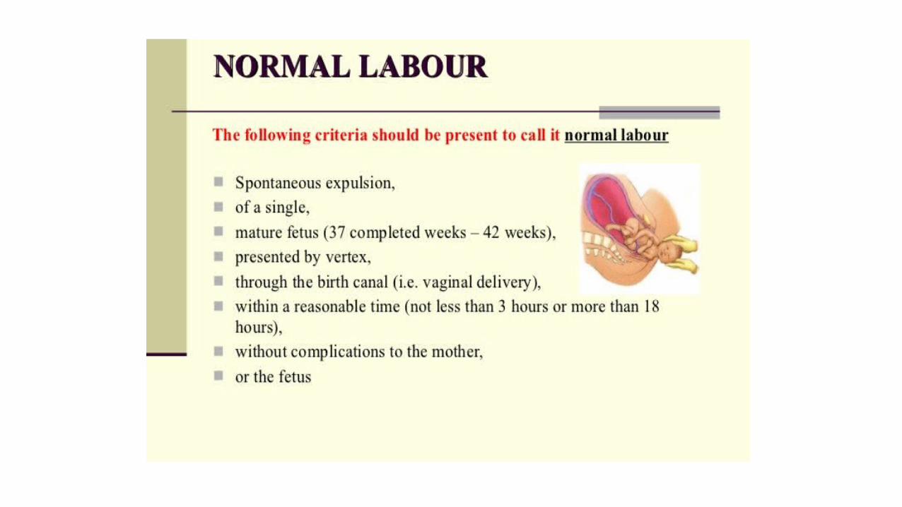
Normal Labour
• WHO defines normal labour as • “spontaneous in onset, low-risk at the start of labour and
remaining so throughout labour and delivery. The infant is born spontaneously in the vertex position between 37 and 42 completed weeks of pregnancy. After birth, mother and infant are in good condition”1
1World Health Organization, Maternal and Newborn Health/Safe Motherhood Unit. Care in normal birth: a practical guide.
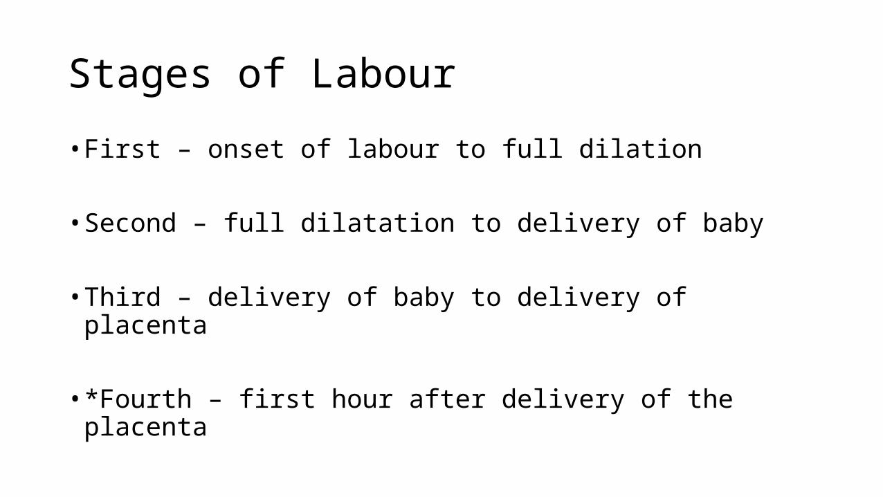
Stages of Labour
• First – onset of labour to full dilation
• Second – full dilatation to delivery of baby
• Third – delivery of baby to delivery of placenta
• *Fourth – first hour after delivery of the placenta
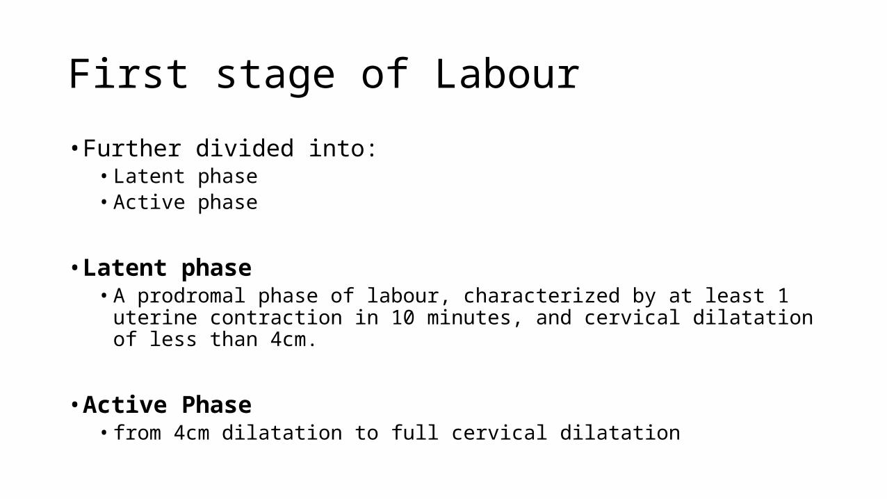
First stage of Labour
• Further divided into:• Latent phase• Active phase
• Latent phase• A prodromal phase of labour, characterized by at least 1 uterine contraction in
10 minutes, and cervical dilatation of less than 4cm.
• Active Phase• from 4cm dilatation to full cervical dilatation
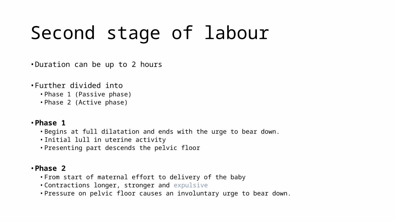
Second stage of labour
• Duration can be up to 2 hours
• Further divided into• Phase 1 (Passive phase)• Phase 2 (Active phase)
• Phase 1• Begins at full dilatation and ends with the urge to bear down.• Initial lull in uterine activity• Presenting part descends the pelvic floor
• Phase 2• From start of maternal effort to delivery of the baby• Contractions longer, stronger and expulsive• Pressure on pelvic floor causes an involuntary urge to bear down.
Third stage of Labour
2 Phases• Separation phase
• Separation of the placenta from the wall of the uterus into the lower uterine segment and/or the vagina
• Expulsion phase• Actual expulsion of the placenta
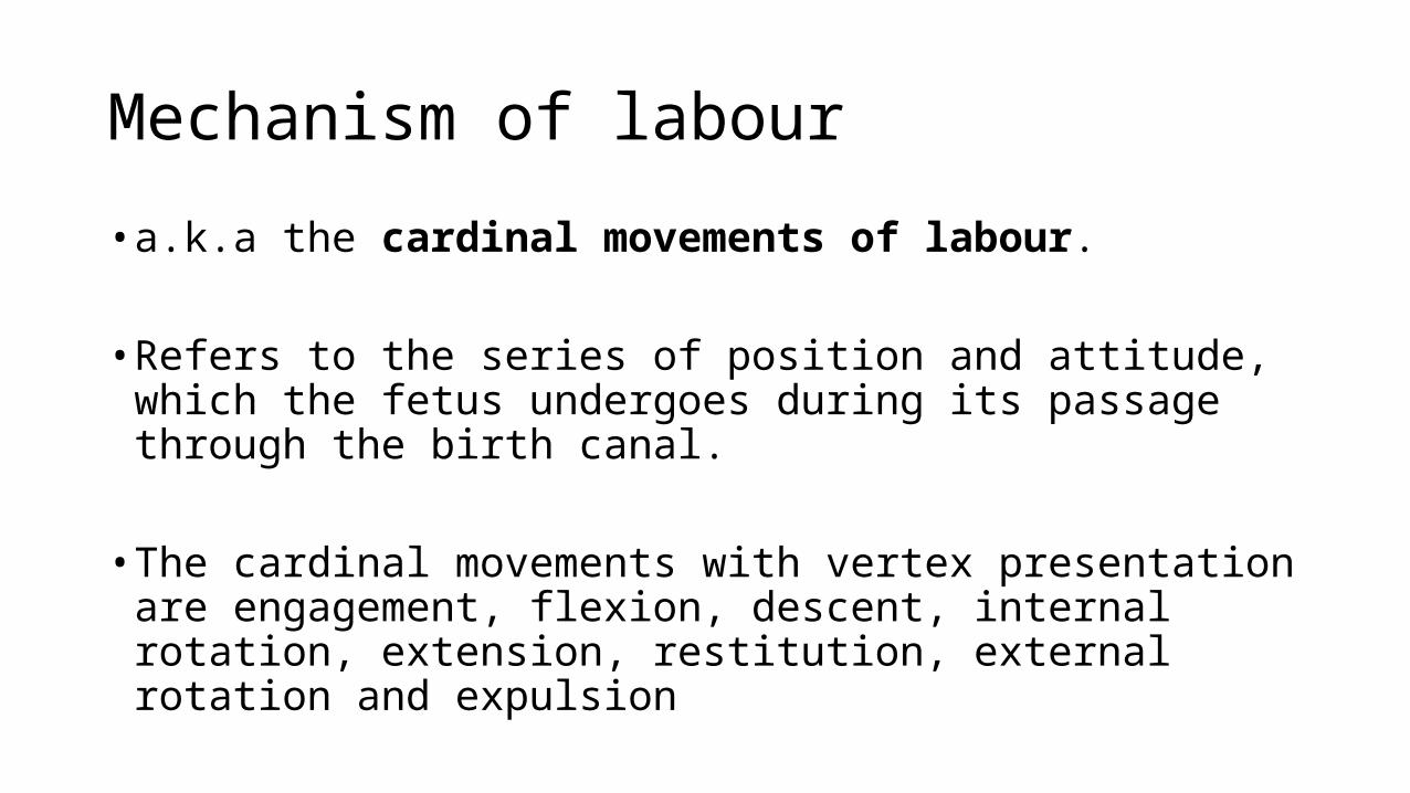
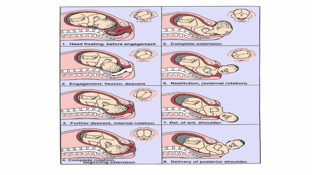
Mechanism of labour
• a.k.a the cardinal movements of labour.
• Refers to the series of position and attitude, which the fetus undergoes during its passage through the birth canal.
• The cardinal movements with vertex presentation are engagement, flexion, descent, internal rotation, extension, restitution, external rotation and expulsion
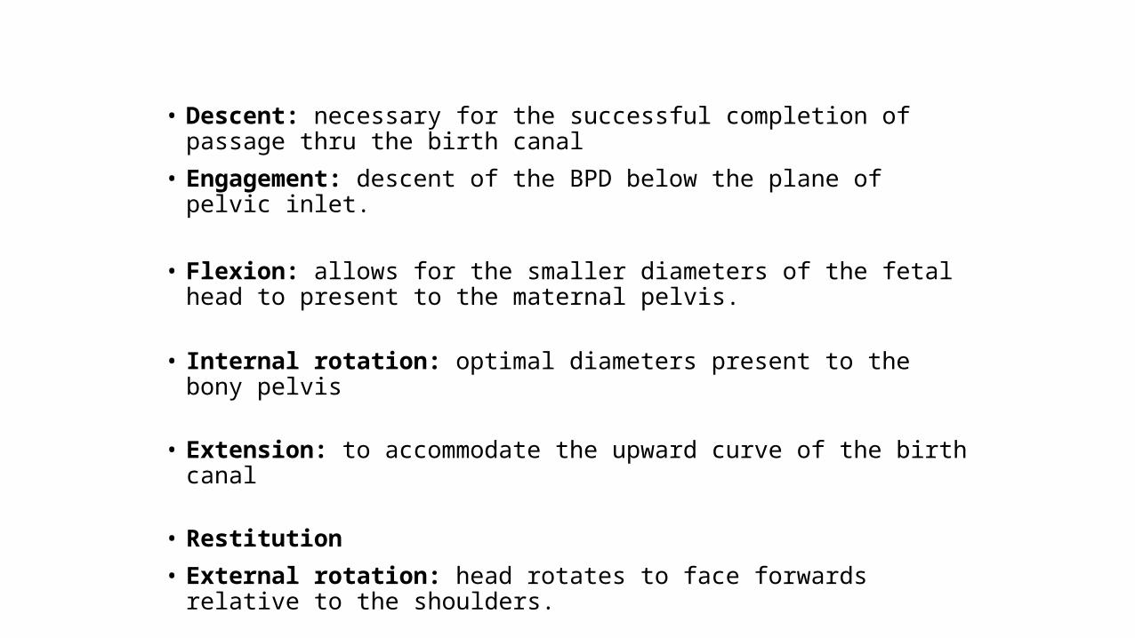
• Descent: necessary for the successful completion of passage thru the birth canal
• Engagement: descent of the BPD below the plane of pelvic inlet.
• Flexion: allows for the smaller diameters of the fetal head to present to the maternal pelvis.
• Internal rotation: optimal diameters present to the bony pelvis
• Extension: to accommodate the upward curve of the birth canal
• Restitution
• External rotation: head rotates to face forwards relative to the shoulders.
• Expulsion

MANAGEMENT OF LABOUR
Principles are
• Diagnosis of labour
• Monitoring progress of labour
• Ensuring maternal well-being
• Ensuring fetal well-being
Management• History
• Booked: ANC history, including investigations• Unbooked: obtain a history, do investigations• History of labour pains, passage of show and drainage of liqour
• General Examination
• Assess mother and fetus (BP, Temp, Lie, Presentation, FHR, cervical dilatation etc)
• Admit if labor is confirmed
• Investigations• PCV, RVS, Urinalysis
Management Cont’d
• Bowel preparation• Enema can be given if no bowel action for 24 hours or rectum feels loaded on VE
• Bladder care• Encouraged to void every 2-4 hours during labour• Measure volume and do analysis
• Nutrition in early labor
• Positioning of laboring mother.• Left lateral in bed
• Pain relief
• Analgesic drugs
• Inhalational analgesia (e.g Entonox)
• Epidural anaesthesia
• Monitor labour progress (partograph)
MANAGEMENT IN THE SECOND STAGE• The mother should not be left unattended
• Observe the following• Maternal condition (BP, pulse)• Fetal conditions• Uterine contractions• Progress of descent
• Ideally delivery is an antiseptic procedure.
Third stage of Labour
• Expectant (physiologic) management.• Spontaneous expulsion after separation.
• Signs of placental separation:• Uterus becomes globular and hard• Uterus rises in the abdomen• Gush of blood• Lengthening of the umbilical cord
• Active management includes the use of oxytocics, CCT and fundal massage
3rd Stage Management
• Active Management is advocated to prevent PPH
• Use of oxytocics (oxytocin, ergometrine)• Early cord clamping• Controlled cord traction• Uterine massage
• Examination of the placenta, umbilical cord for completeness and anomalies.
• Examination of the perineum for lacerations
• Repair episiotomy if given
Complications of labour
• FIRST STAGE• Cord prolapse• Fetal distress• Maternal dehydration/exhaustion• Poor progress in labour• Obstructed labour• Ruptured uterus
• SECOND STAGE• Perineal lacerations• Fetal distress• Shoulder dystocia
• THIRD STAGE• PPH• Retained placenta
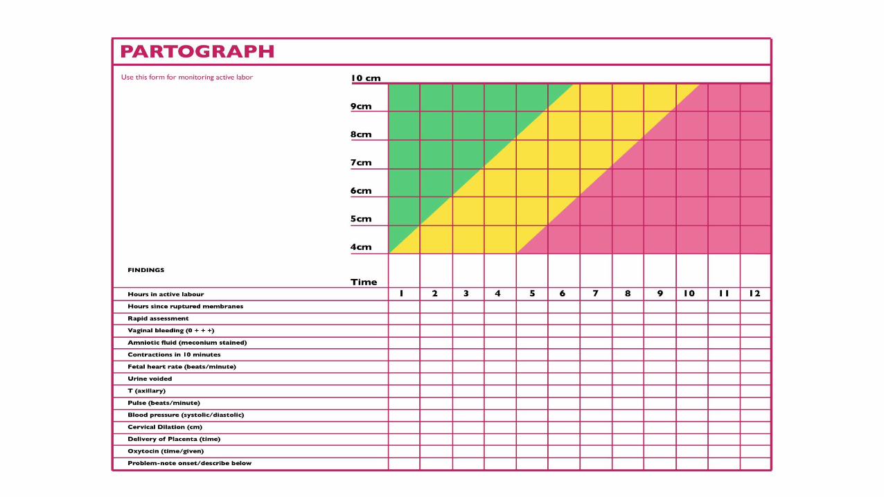
PARTOGRAPH
• Greek word meaning ‘labor curve’
• A graphic recording of progress of labour and salient features in the mother and fetus.
• It serves as an early warning system and assists in early decision of transfer, augmentation or termination of labour.
• Increases the quality and regularity of observations of the fetus and mother in labour.
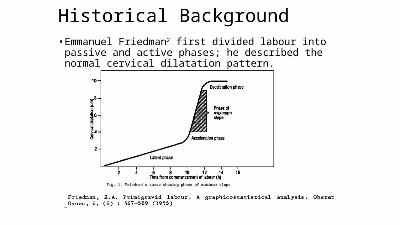
Historical Background• Emmanuel Friedman2 first divided labour into passive and active
phases; he described the normal cervical dilatation pattern.
2
Fig. 1. Friedman’s curve showing phase of maximum slope
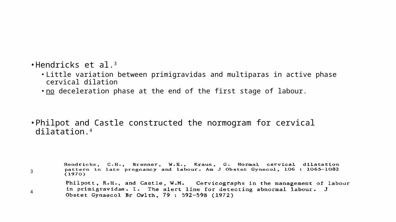
• Hendricks et al.3 • Little variation between primigravidas and multiparas in active phase cervical dilation• no deceleration phase at the end of the first stage of labour.
• Philpot and Castle constructed the normogram for cervical dilatation.4
3
4
• The WHO model of the partograph was developed as an international standard in 1988 following the launch of the worldwide Safe Motherhood Initiative.
• Following a multicenter trial in 1990-1991, WHO recommended that the partograph be used in monitoring all labours.
The Principles of WHO Partograph
1. The active phase of labour commences at 3cm cervical dilatation.2. The latent phase of labour should not last longer than 8 hours.3. During active labour, the rate of cervical dilatation should not be
slower than 1cm/hr.4. A lag time of 4 hours between a slowing of labour and the need for
intervention is unlikely to compromise the fetus or mother and avoids unnecessary intervention.
5. Vaginal examinations should be performed as infrequently as is compatible with safe practice (once in 4 hrs)
6. Partograph with preset lines.
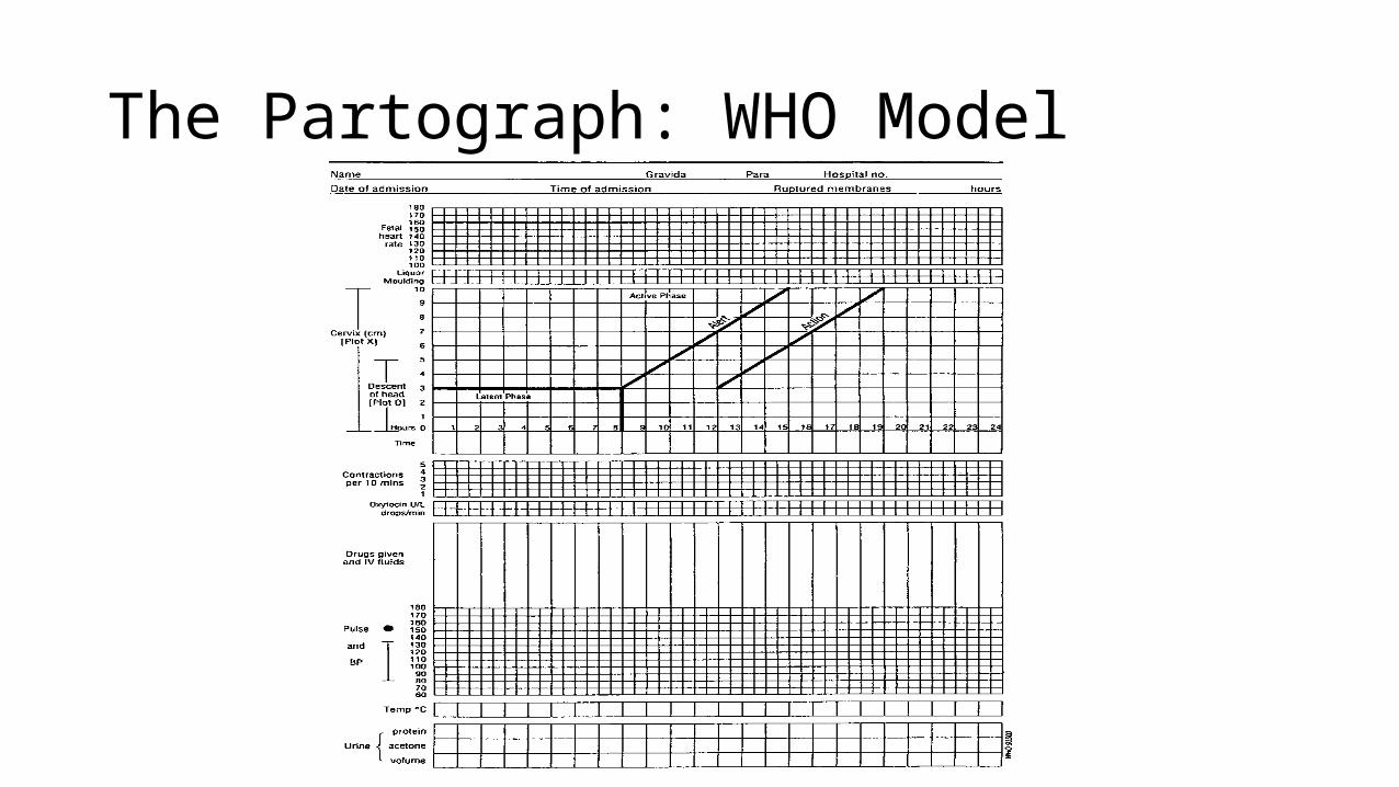
The Partograph: WHO Model
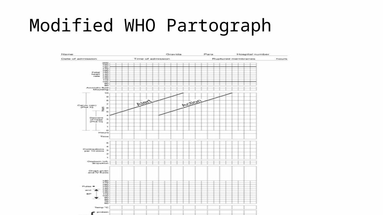
Modified WHO Partograph
Components of the Partograph
1. The progress of labour2. The fetal condition3. The maternal condition
1. PROGRESS OF LABOUR
Rate of cervical dilatation of the cervix (cervicograph)
Rate of descent of the presenting part.
CERVICOGRAPH is divided into the latent and active phase.Modified WHO partograph has only the active phase
Assess cervical dilatation every 4 hours2 hourly if oxytocin augmentation is ongoing.
Cervical dilatation marked with an X
Cervicogram
• Used between cervical dilatation of 4cm – 10cm.
• Cervix dilates at the rate of 1cm/hour
• Has the alert and action lines
Alert line
• An alert line is a visual representation of a cervical os dilatation rate of 1 cm/hour labor progress sustained throughout the active phase
• It is the slowest rate of active phase labor progress for normal labor outcome
Action Line
• Drawn 4 hours to the right of the alert line (WHO Partograph)
• The critical point at which specific management decisions must be made.
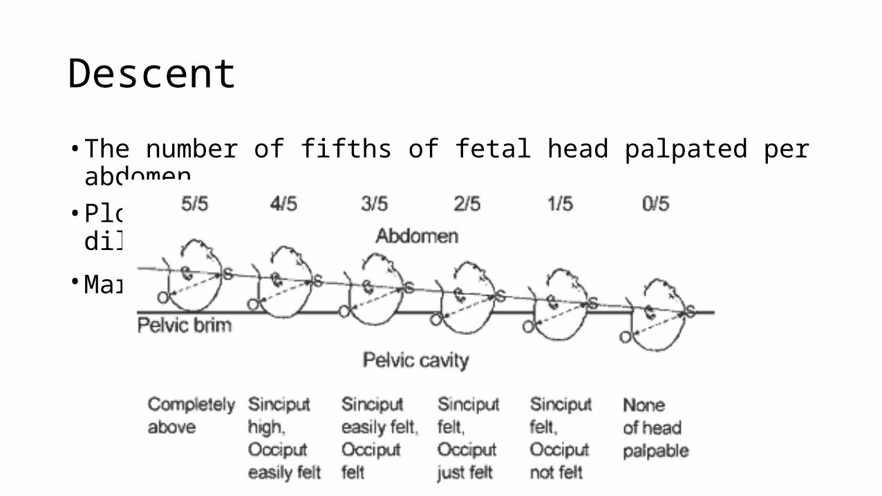
Descent
• The number of fifths of fetal head palpated per abdomen.• Plotted on the same graph as the cervical dilatation.
• Marked with an O or a w
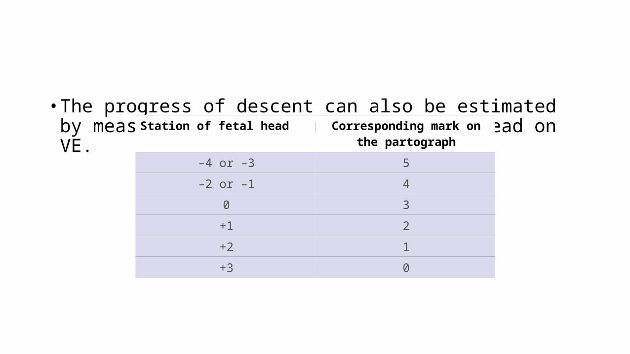
• The progress of descent can also be estimated by measuring the station of the fetal head on VE.Station of fetal head Corresponding mark on
the partograph
–4 or –3 5
–2 or –1 4
0 3
+1 2
+2 1
+3 0
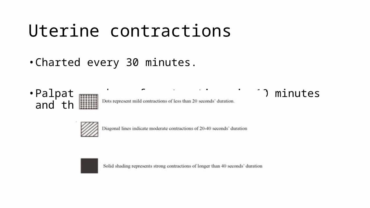
Uterine contractions
• Charted every 30 minutes.
• Palpate number of contractions in 10 minutes and the duration in seconds.
2. THE FETAL CONDITION
Fetal Heart rate:• Record every 30 minutes• Every 15 minutes in cases of fetal bradycardia or tachycardia• Baseline fetal heart rate is 120-160 beats per minute.
The amniotic fluid:• record the colour at every vaginal examination• I: membranes intact• C: membranes ruptured, clear fluid• M: meconium-stained fluid• B: blood-stained fluid
• Moulding:• Is the overriding of the skull bones when the fetal head passes through the
birth canal.
• An important indication of how adequately the pelvis can accommodate the fetal head.
• Degrees:• Zero: bones apart• 1+: bones touching• 2+: bones overlap, reducible• 3+: overlap, irreducible
3. MATERNAL CONDITION
• Pulse• Every 30 minutes• Mark with a dot. (.)
• Blood pressure• Every 4 hours• Mark with arrows
• Temperature• Every 2-4 hours
• Urinalysis• Every 2-4 hours; for protein, ketones, glucose or blood• Measure volume
• Oxytocin: amount and number of drops/ minute every 30 minutes
• Other drugs and IV fluids are also charted on the partograph
ConclusionLabour is an important human experience, which could give intense joy or sorrow depending on the outcome.
It is important for providers of antenatal and intrapartum care to have an understanding of what constitutes normal labour, and to watch out for problems that may arise.
The partograph is an inexpensive tool that aids in early recognition of problems with the mother and fetus during labor. All health workers should be conversant with its use.

….. On a lighter note
I like trying to get pregnant, I’m not so sure about childbirth. ~~~Lauren Holly
The pain of childbirth is not remembered, it is the child that is remembered. ~~~ Freeman Dyson

KATTEY K.A (MBBS, MPH)