Embed Size (px)
Citation preview

PRESENTED BY:
DR.N.BENTHUNGO TUNGOEP.G, MS(ORTHOPEDICS)
CENTRAL INSTITUTE OF ORTHOPEDICSVMMC & SAFDARJUNG HOSPITAL
NEW DELHI
SCAPHOID NON UNION
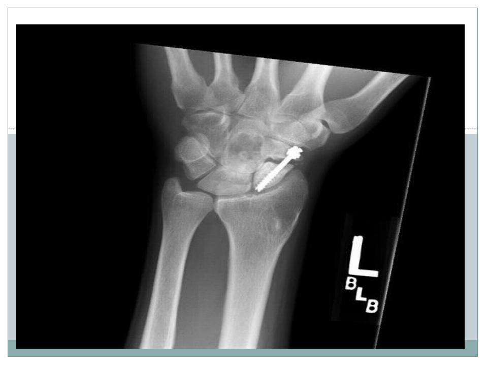
Management of scaphoid non union
Introduction
The incidence of scaphoid nonunion for undisplaced fx is 5-10%
The incidence increases up to 90% in displaced proximal pole fractures
Failure to heal after 6 months establishes the Dx of nonunion
Recent studies indicated that virtually that all unstable nonunions lead to carpal collapse and posttraumatic arthritis,, for this reason treatment is recommended for all scaphoid nonunions even if asymptomatic


INTRODUCTION
According to FISK, in established non unions of the scaphoid, proximal scaphoid rotates dorsally into extension and the distal part faces downward in flexion leading totriplane angulation and subsequent humpback deformity of the scaphoid.
Impingement between the palmar-flexed scaphoid distal pole and the radial styloid process leads to the development of radiocarpal osteoarthritis.
At the same time, the unsupported carpus collapses into a DISI deformity with increasing subluxation and secondary arthritis of the midcarpal joint.
CAUSES/FACTORS
Delayed diagnosis: upto 40% undiagnosed at time of injury
Gross displacement: upto nonunion rate of 92% in displaced fractures
Associated injuries of the carpus and ligaments.Impaired blood supply(30% to 40% of
osteonecrosis occur most frequently in fractures of the proximal third)
Inadequate immobilization/ poor fixation techniques
Immunocompromised states/ smoking etc
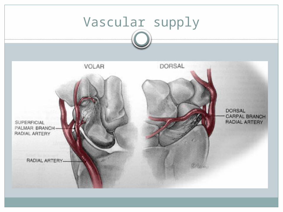
Vascular supply

SYMPTOMS
radial-sided wrist pain,
reduced wrist motion with pain at the limits of motion,
reduced grip strength
Radiological assesment
Xray
CT Scan
Gadolinium enhanced MRI
Bone scan

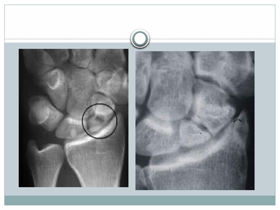
Radiographic findings
Classic findings of nonunion,
including widening of the fracture gap,
cystic changes,
fracture line sclerosis even when the fracture is healing

Hump back deformity
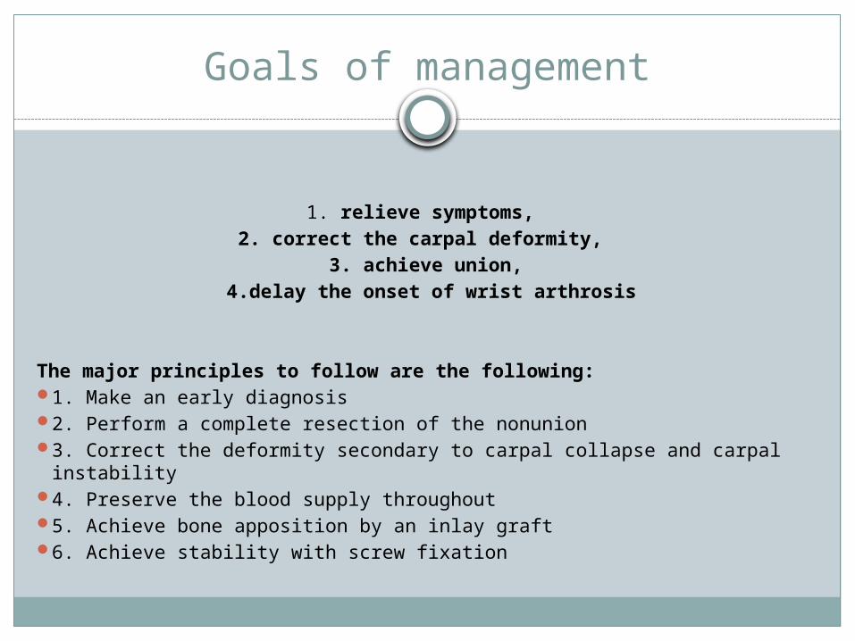
Goals of management
1. relieve symptoms, 2. correct the carpal deformity,
3. achieve union, 4.delay the onset of wrist arthrosis
The major principles to follow are the following: 1. Make an early diagnosis 2. Perform a complete resection of the nonunion 3. Correct the deformity secondary to carpal collapse and carpal instability 4. Preserve the blood supply throughout 5. Achieve bone apposition by an inlay graft 6. Achieve stability with screw fixation
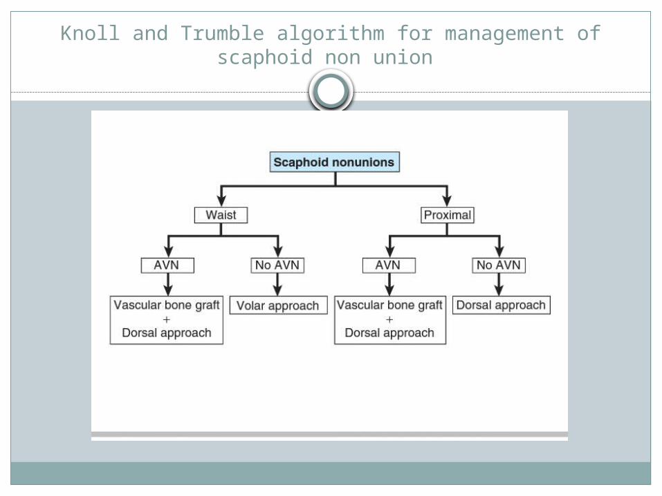
Knoll and Trumble algorithm for management of scaphoid non union
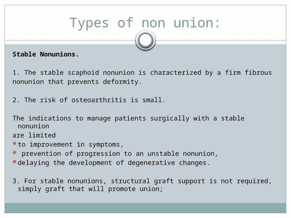
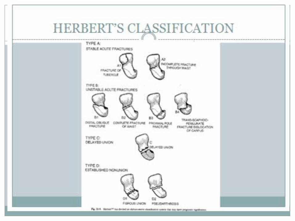
Types of non union:
Stable Nonunions.
1. The stable scaphoid nonunion is characterized by a firm fibrousnonunion that prevents deformity.
2. The risk of osteoarthritis is small.
The indications to manage patients surgically with a stable nonunionare limited to improvement in symptoms, prevention of progression to an unstable nonunion, delaying the development of degenerative changes.
3. For stable nonunions, structural graft support is not required, simply graft that will promote union;

Unstable Nonunions. The quoted success rates of achieving union
with internal fixation and bone grafting for unstable nonunions range from 60% to 95%.
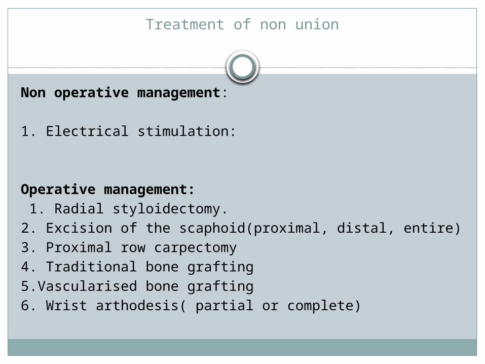
Treatment of non union
Non operative management:
1. Electrical stimulation:
Operative management: 1. Radial styloidectomy.2. Excision of the scaphoid(proximal, distal, entire)3. Proximal row carpectomy4. Traditional bone grafting5.Vascularised bone grafting6. Wrist arthodesis( partial or complete)

STYLOIDECTOMY
Styloidectomy alone probably is of little value in treating nonunions of the scaphoid.
If arthritic changes involve only the scaphoid fossa of the radiocarpal joint, however, styloidectomy is indicated in conjunction with any grafting of the scaphoid or excision of its ulnar fragment.
Technique: Stewart
EXCISION OF THE PROXIMALFRAGMENT
Excising both fragments of the scaphoid as the only procedure is unwise; although the immediate result may be satisfactory, eventual derangement of the wrist is likely.
Soto-Hall and Haldeman reported gradual migration of the capitate into the space previously occupied by the scaphoid.
If excision of both fragments is considered, it is preferable to add some other procedure to stabilize the capitolunate joint (e.g., capitolunate or capital-lunate-triquetral-hamate fusions).
Excising the proximal scaphoid fragment usually is satisfactory; the loss of one fourth or less of the scaphoid usually causes minimal impairment of wrist motion. Because postoperative immobilization is brief, function usually returns rapidly.
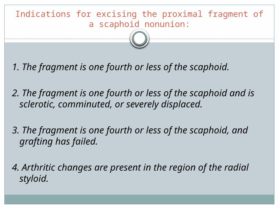
Indications for excising the proximal fragment of a scaphoid nonunion:
1. The fragment is one fourth or less of the scaphoid.
2. The fragment is one fourth or less of the scaphoid and is sclerotic, comminuted, or severely displaced.
3. The fragment is one fourth or less of the scaphoid, and grafting has failed.
4. Arthritic changes are present in the region of the radial styloid.

Excision of the Distal Scaphoid
Satisfactory results have been reported with distal scaphoid resection for the treatment of scaphoid nonunions with radioscaphoid arthritis treated with distal scaphoid resection.
If capitolunate arthritis is present, an additional procedure (e.g., limited intercarpal arthrodesis) should be added to distal scaphoid excision.
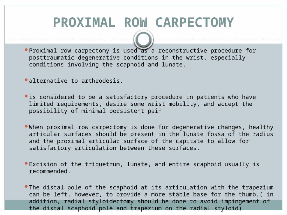
PROXIMAL ROW CARPECTOMY Proximal row carpectomy is used as a reconstructive procedure for
posttraumatic degenerative conditions in the wrist, especially conditions involving the scaphoid and lunate.
alternative to arthrodesis.
is considered to be a satisfactory procedure in patients who have limited requirements, desire some wrist mobility, and accept the possibility of minimal persistent pain
When proximal row carpectomy is done for degenerative changes, healthy articular surfaces should be present in the lunate fossa of the radius and the proximal articular surface of the capitate to allow for satisfactory articulation between these surfaces.
Excision of the triquetrum, lunate, and entire scaphoid usually is recommended.
The distal pole of the scaphoid at its articulation with the trapezium can be left, however, to provide a more stable base for the thumb.( in addition, radial styloidectomy should be done to avoid impingement of the distal scaphoid pole and trapezium on the radial styloid)
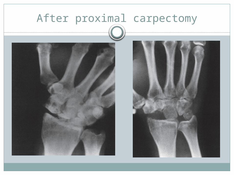
After proximal carpectomy
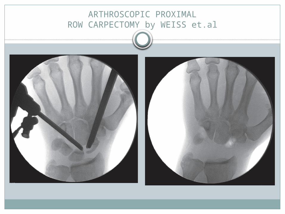
ARTHROSCOPIC PROXIMALROW CARPECTOMY by WEISS et.al
Grafting operations
Cancellous bone grafting for scaphoid nonunion, as first described by Matti and modified by Russe, has proved to be a reliable procedure, producing bony union in 80% to 97% of patients. This technique is most useful for ununited fractures that do not have associated shortening or angulation.

TYPES OF BONE GRAFTING
1. Russe bone graft (Inlay):Used for stable nonunions . The initial procedure used a single corticocancellousstrut across the fracture line;a later modificationinvolved two corticocancellous struts inserted intothe scaphoid excavation with their cancellous sidesfacing each other,the remainder of the cavity is filledwith cancellous chips. Usually k-wires are added tosecure the construct.

The time to union with this procedure is relatively long ,generally requiring cast immobilization for 6-4 months
Healing rates of 85-90 % have been reportedSatisfactory relief of symptoms has been
reported ; 78 % of painful wrist became free of symptoms and 88 % of patients were satisfied with the results.

Inlay graft

2. Fernandez bone graft (interpositional graft):
angulated nonunions with a dorsal humpback deformity require interpositional grafting.
Fernandez has described the use of a trapezoidal iliac graft to correct the angulation and carpal collapse pattern.Fixation is achieved with screws or k-wires
In both types of bone grafting ,a volar approach is used, and care must be taken to preserve the vascularity of the fragments
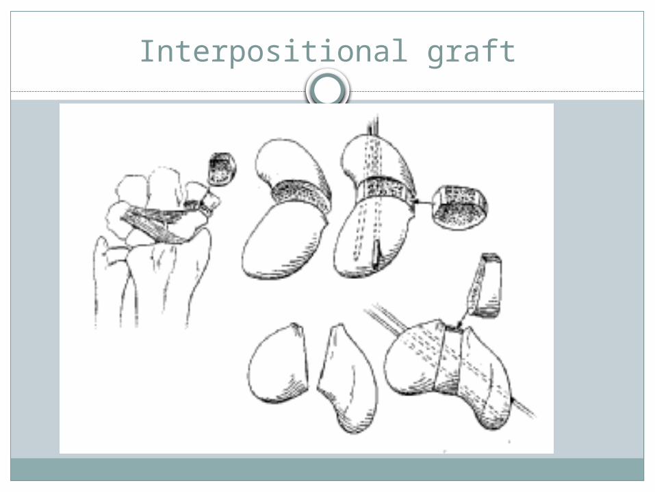
Interpositional graft

Malpositioned Nonunion of Scaphoid Fractures (“Humpback” Deformity).
Due to resorption or comminution, shortening and angulation, with its convexity dorsal and radial occurs in non union fractures of scaphoid leading to “humpback” deformity
The deformity includes extension of the proximal pole of the scaphoid, resulting extension of the lunate, and a form of dorsal intercalated instability pattern seen on lateral plain radiographs

Electrical stimulation: Pulsed Electromagnetic Field ( PEMF ) stimulation
has been investigated as a noninvasive treatment for scaphoid nonunion .Although controversial, there appears to be some benefit (shorter healing time)when electric stimulation is combined with bone grafting procedures
C) Proximal pole excision: when a small proximal fragment is not amenable to bone grafting ,proximal pole excision and fascial hemiarthroplasty are recommended
) Salvage procedures :Are indicated when nonunion has lead to carpal collapse and
secondary degenerative changesProximal row carpectomy,intercarpal arthrodesis, or radiocarpal
arthrodesis is recommended in patients with chronic wrist pain and stiffness
Radial styloidectomy and scaphoid interposition arthroplasty may be combined with other procedures or performed independently in the younger patient with less severe symptoms
Silicone implants have been used in the past but are now avoided because of silicone synovitis
GRAFTING OPERATIONS TECHNIQUES
FERNANDEZTOMAINO ET AL.STARK ET AL.
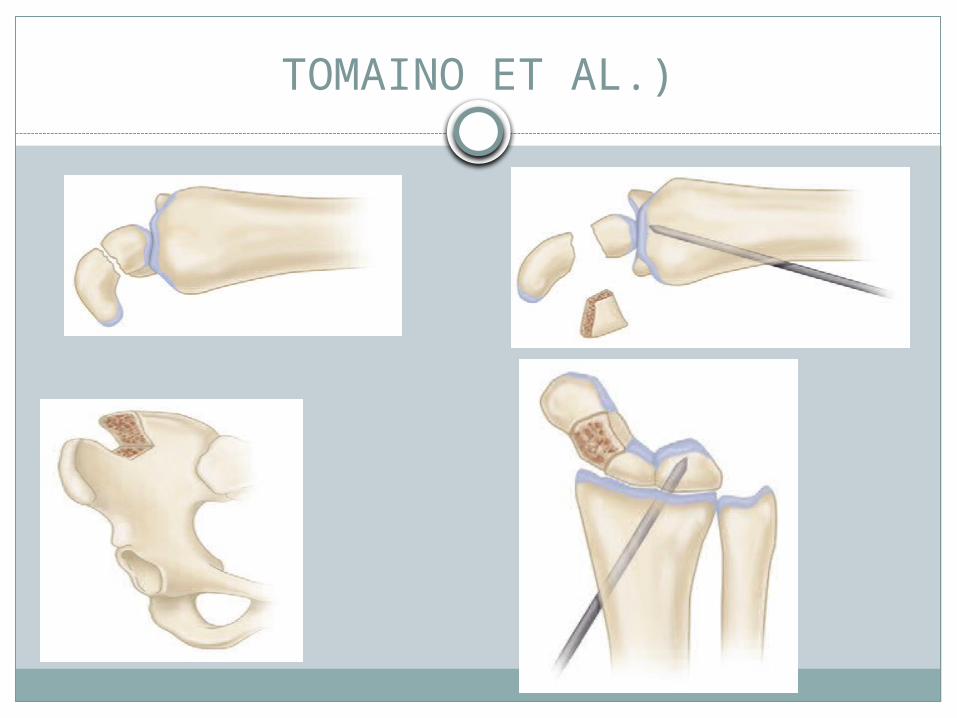
TOMAINO ET AL.)
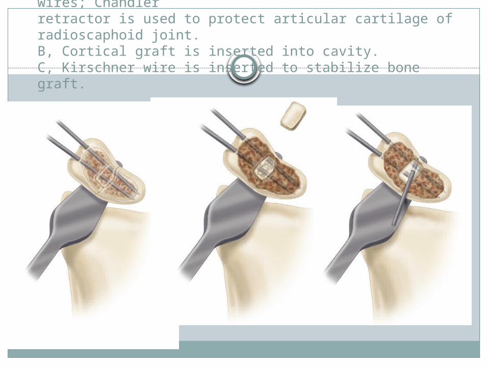
STARK et.al TECHNIQUE: A, Excavation of scaphoid and placement ofKirschner wires; Chandlerretractor is used to protect articular cartilage of radioscaphoid joint.B, Cortical graft is inserted into cavity. C, Kirschner wire is inserted to stabilize bone graft.
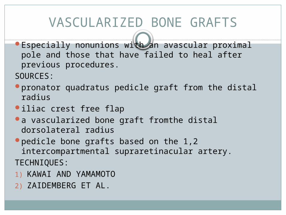
VASCULARIZED BONE GRAFTSEspecially nonunions with an avascular proximal pole
and those that have failed to heal after previous procedures.
SOURCES:pronator quadratus pedicle graft from the distal radiusiliac crest free flapa vascularized bone graft fromthe distal dorsolateral
radiuspedicle bone grafts based on the 1,2
intercompartmental supraretinacular artery.TECHNIQUES:1) KAWAI AND YAMAMOTO2) ZAIDEMBERG ET AL.
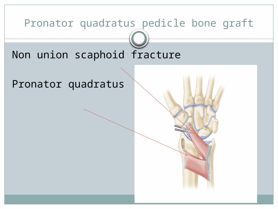
Pronator quadratus pedicle bone graft
Non union scaphoid fracture
Pronator quadratus

Arthrodesis of the Wrist
a salvage procedure for old ununited or malunited fractures of the scaphoid with associated radiocarpal traumatic arthritis.

THANK YOU