Embed Size (px)
Citation preview

Genito-Urinary Tumors
Episode 3

Multilocular Cystic Nephroma
• Rare benign tumor.
• Age & Sex:
- 3 m.-2 Yrs.→ ♀:♂ = 1:2
- 40-50 Yrs. → ♀:♂ = 8:1

Multilocular Cystic Nephroma
Pathology:
• Solitary, well-circumscribed,
multiseptated mass of
noncommunicating fluid-filled
loculi that is surrounded by a
thick fibrous capsule and
compressed renal parenchyma.

Multilocular Cystic Nephroma
Pathology:
Multilocular cystic renal tumor:
1. Cystic nephroma: a multicystic tumor lacking
blastemal or other embryonal elements.
2. Cystic partially differentiated nephroblastoma
(CPDN): a multicystic tumor in which the septa
contain blastemal or other embryonal elements.

Multilocular Cystic Nephroma
Plain X-Ray:
• Soft tissue mass lesion.
• Displacement of the bowel
and adjacent structures if the
lesion is of sufficient size.
Multilocular Cystic Nephroma
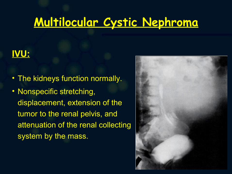
IVU:
• The kidneys function normally.
• Nonspecific stretching,
displacement, extension of the
tumor to the renal pelvis, and
attenuation of the renal collecting
system by the mass.
Multilocular Cystic Nephroma
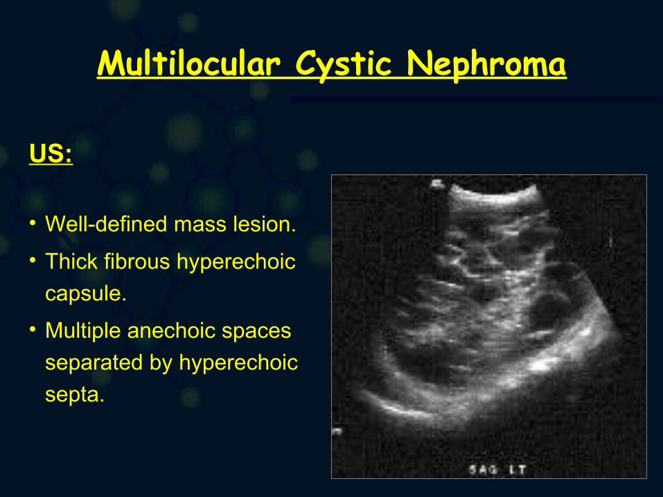
US:
• Well-defined mass lesion.
• Thick fibrous hyperechoic
capsule.
• Multiple anechoic spaces
separated by hyperechoic
septa.

Multilocular Cystic Nephroma
US:
• Well-defined mass lesion.
• Thick fibrous hyperechoic
capsule.
• Multiple anechoic spaces
separated by hyperechoic
septa.

Multilocular Cystic Nephroma
CT:
• Well-defined margins
• Multicystic architecture
• Enhancing septa
• Herniation into the renal
collecting system.
• Cystic spaces are not enhancing and
demonstrate CT numbers slightly
higher than those of water.

Multilocular Cystic Nephroma
CT:

Multilocular Cystic Nephroma
CT:

Multilocular Cystic Nephroma
CT:

Multilocular Cystic Nephroma
CT:

Multilocular Cystic Nephroma
MRI:
• MRI demonstrates the low signal intensity of the tumor
capsule.
• Observations of nonenhanced MRIs have shown
encapsulated masses with dividing septa between
cystic spaces.
Multilocular Cystic Nephroma
MRI:
• On T1-weighted sequences, signal intensity varies from
low to very high and from low to intermediate.
• T2-weighted sequencing resulted in low signal intensity
in the tumor capsule and the intermediate septations.
• The signal intensity of the cysts is high in all cases.

Multilocular Cystic Nephroma
MRI:

Multilocular Cystic Nephroma
MRI:

Multilocular Cystic Nephroma
Angiography:
•Hypovascular mass.