Embed Size (px)
Citation preview
Multi-modality imaging and
Cancer
Parminder S. Basran, PhD, FCCPMSenior Medical PhysicistBCCA- Vancouver Island CentreAdjunct Associate ProfessorUniversity of Victoria- Dept. Physics & Astronomy
Outline
Introduction
Clinical Example: Lung cancer
Prostate
Conclusions / Future Directions
Introduction
Introduction
Multi-modality imaging is changing the way we treat cancer patients.
The information content of these images is vast and almost overwhelming.
Even at a qualitative level, multi-modality imaging is a powerful tool.
The challenge for the next decade will be on how we most efficiently deploy multi-modal imaging, and integrate it –quantitatively- with patient care.

Introduction
Cancer: what is it?
http://en.wikipedia.org/wiki/Cancer
“a class of diseases in which a group of cells display uncontrolled growth, invasion that intrudes upon and destroys adjacent tissues, and sometimes metastasis, or spreading to other locations in the body via lymph or blood.”
Introduction
Cancer: how do you detect it?
Most cancers are initially recognized either because signs or symptoms appear or through screening (blood tests, X-rays, CT scans and endoscopy). Confirmation requires pathologic testing. People with suspected cancer are investigated with medical tests.
Introduction
Cancer: how do you detect it?
Multi-modality imaging is beginning to play a central role in detecting cancer.
Not only is imaging used to measure the extent of the disease, it is commonly used to screen patients (generally higher risk patients) to mitigate morbidity from cancer.
There is always a risk in an imaging procedure. A screening program requires a careful assessment of the risks.
Introduction
As our understanding of cancer increases, so does the recognition that each tumor is unique.
In the last decade, the focus in cancer research on the genetic make-up of cancer
Now it is clear that the tumor microenvironment, cellular and protein interactions affect disease progression, aggressiveness and response to treatment.
Introduction
Subsequently, imaging the •tumor micro-environment•host/stem cell interactions,•various proteins
can be used to assess •disease progression•aggressiveness •response to treatment.
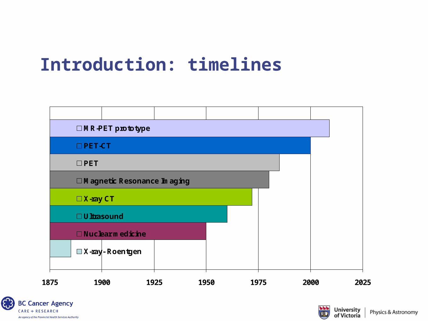
Introduction: timelines
1875 1900 1925 1950 1975 2000 2025
MR-PET prototype
PET-CT
PET
Magnetic Resonance Imaging
X-ray CT
Ultrasound
Nuclear medicine
X-ray- Roentgen
Introduction
Multi-modality imaging (in cancer): what does that really mean?
Incorporation of two or more imaging modalities sometimes within the setting of a single examination in
• assessing disease progression / aggressiveness • delivering efficacious treatment• assessing response to treatment
Maybe with a single or several imaging devices obtained at (nearly) the same time points.
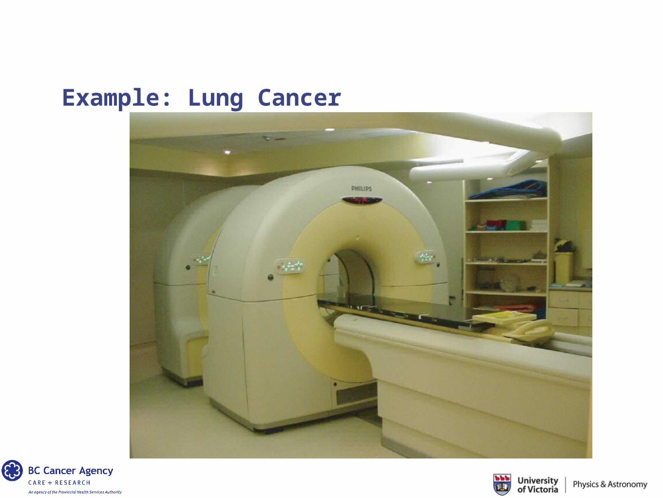
Example: Lung Cancer
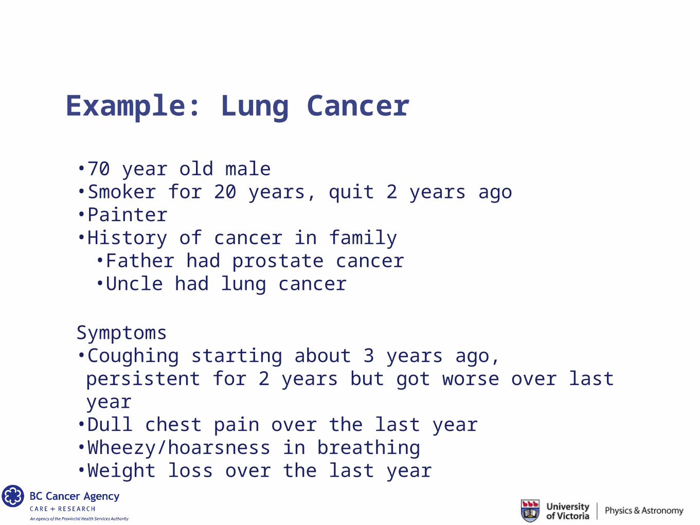
Example: Lung Cancer
•70 year old male•Smoker for 20 years, quit 2 years ago•Painter•History of cancer in family
•Father had prostate cancer•Uncle had lung cancer
Symptoms•Coughing starting about 3 years ago, persistent for 2 years but got worse over last year
•Dull chest pain over the last year•Wheezy/hoarsness in breathing •Weight loss over the last year
Example: Lung Cancer
Tests that may be performed include:
* Chest x-ray * Sputum cytology test to look for cancer cells * Blood work
Chest x-ray
Example: Lung Cancer
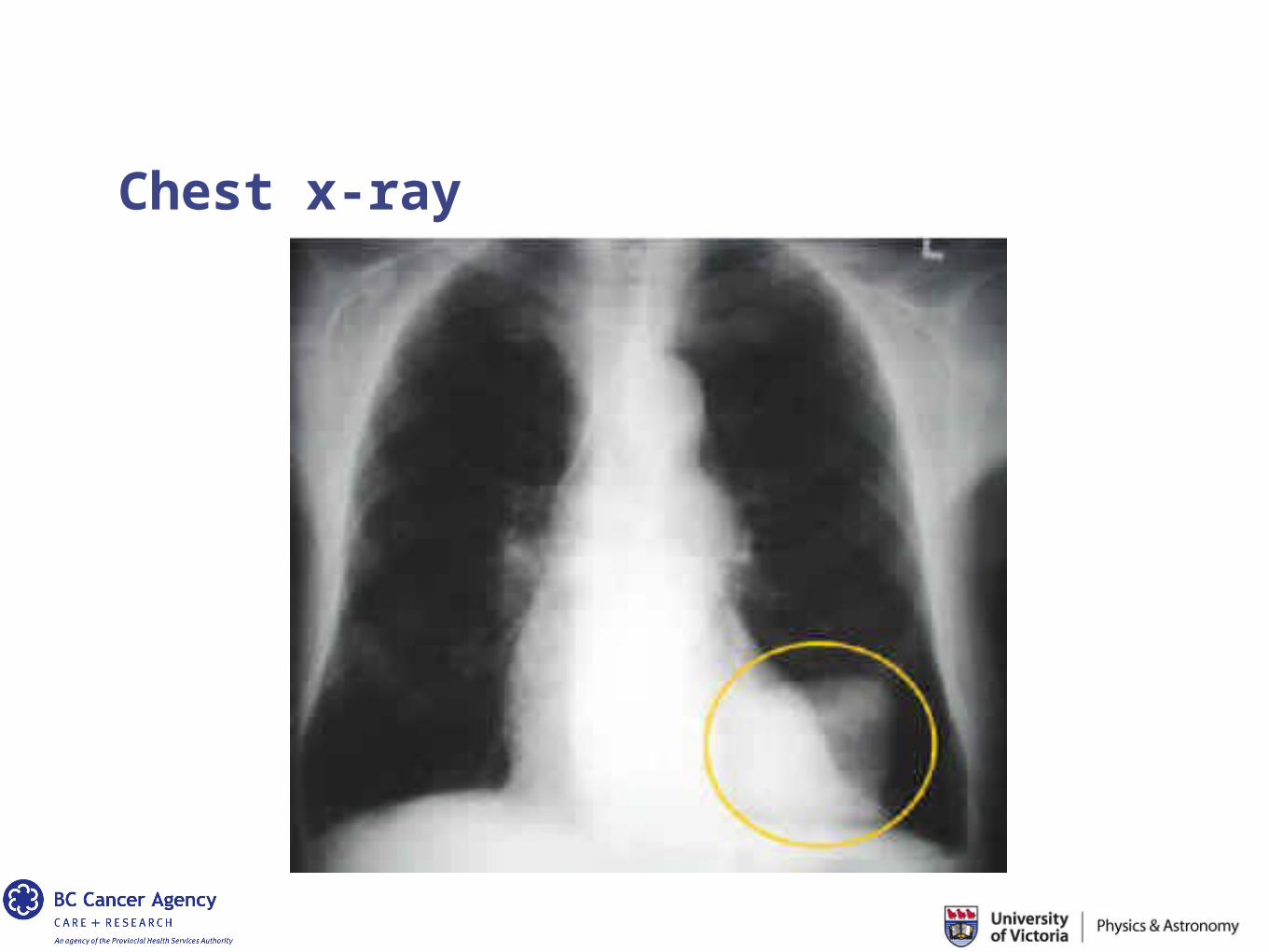
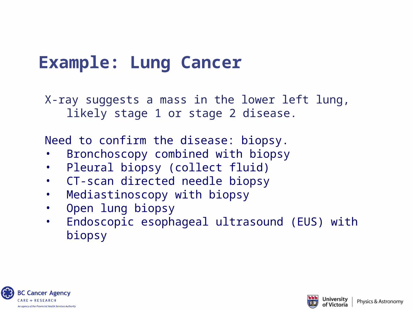
X-ray suggests a mass in the lower left lung, likely stage 1 or stage 2 disease.
Need to confirm the disease: biopsy. • Bronchoscopy combined with biopsy• Pleural biopsy (collect fluid)• CT-scan directed needle biopsy• Mediastinoscopy with biopsy• Open lung biopsy• Endoscopic esophageal ultrasound (EUS) with
biopsy
Example: Lung Cancer
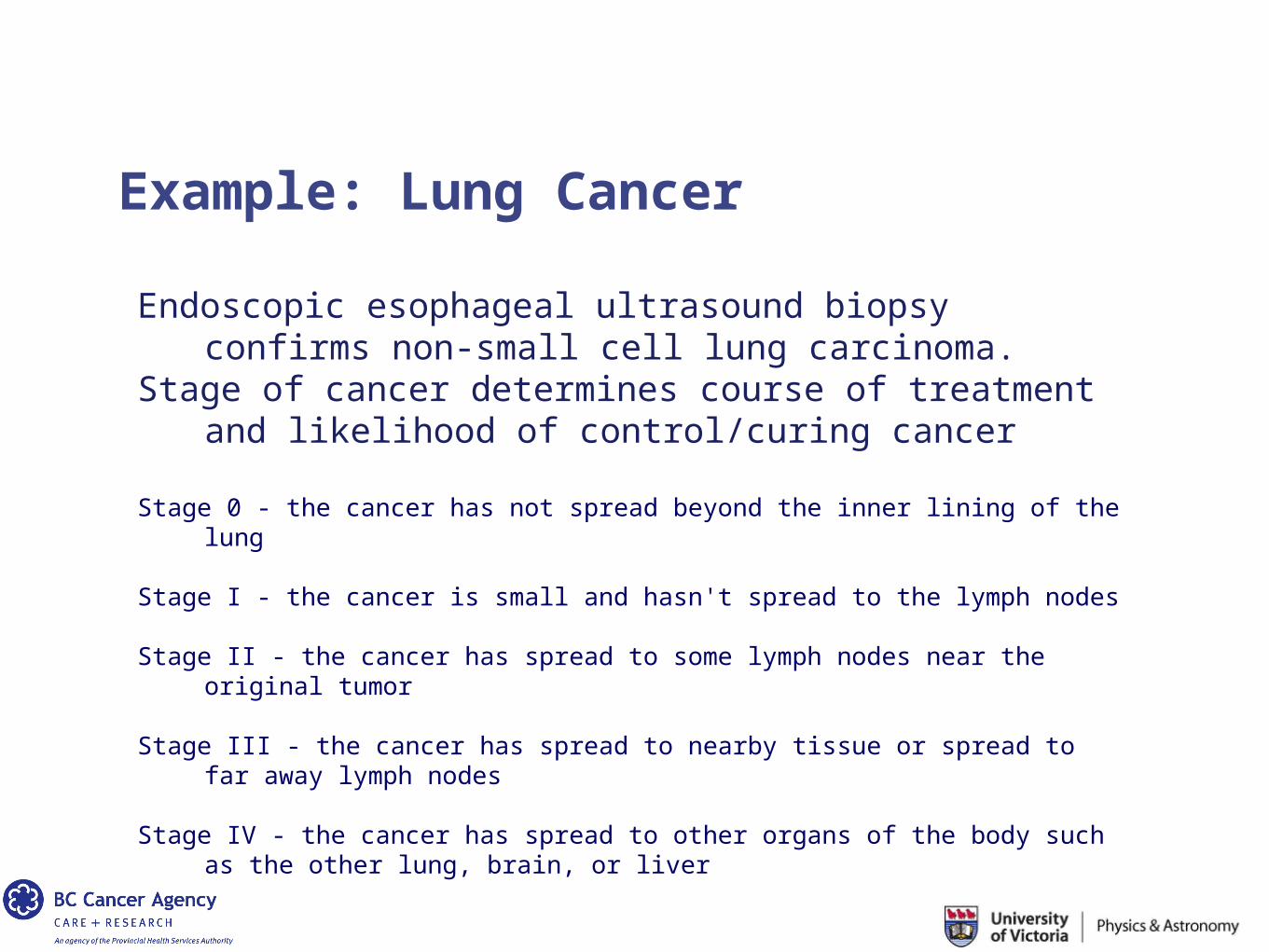
Endoscopic esophageal ultrasound biopsy confirms non-small cell lung carcinoma.
Stage of cancer determines course of treatment and likelihood of control/curing cancer
Stage 0 - the cancer has not spread beyond the inner lining of the lung Stage I - the cancer is small and hasn't spread to the lymph nodes
Stage II - the cancer has spread to some lymph nodes near the original tumor
Stage III - the cancer has spread to nearby tissue or spread to far away lymph nodes
Stage IV - the cancer has spread to other organs of the body such as the other
lung, brain, or liver
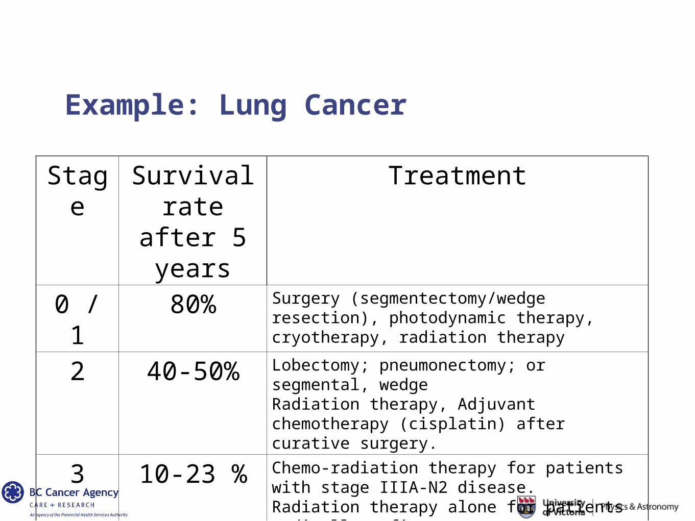
Example: Lung Cancer
Stage
Survival rate after 5 years
Treatment
0 / 1 80% Surgery (segmentectomy/wedge resection), photodynamic therapy, cryotherapy, radiation therapy
2 40-50% Lobectomy; pneumonectomy; or segmental, wedge Radiation therapy, Adjuvant chemotherapy (cisplatin) after curative surgery.
3 10-23 % Chemo-radiation therapy for patients with stage IIIA-N2 disease.Radiation therapy alone for patients medically unfit
4 <10% Chemotherapy, Radiation for palliative/symptom relief
Example: Lung Cancer
Prognosis:
- Likely stage 2 NSCLC- Pretty good chance of treating / controling the disease- Treatment options are optimistic
- Surgery: Lobectomy; pneumonectomy; or segmental, wedge resection
- Radiation therapy- Adjuvant chemotherapy (cisplatin) after curative surgery.
Example: Lung Cancer
Treatment Strategy:
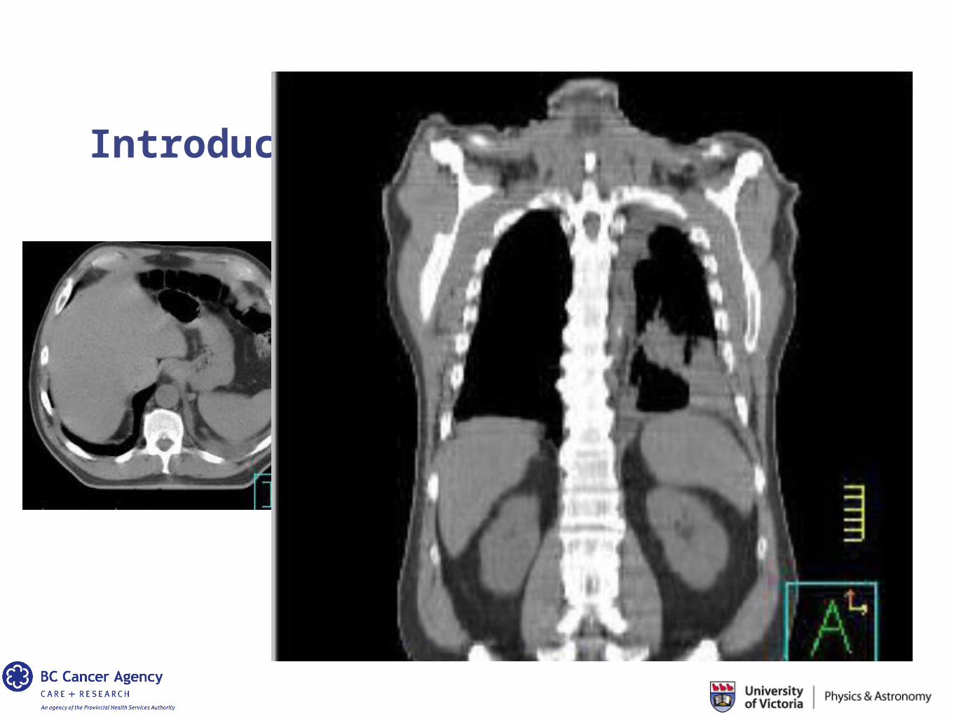
Radiation therapy• Deliver 60 Gray over 30 days (2 Gy/day)• Use high energy photons (6 MeV range)• Need to simulate the treatment virtually• Obtain a CT scan to define the tumor and normal tissues• Simulate the radiation beams in the CT dataset
Example: Lung Cancer
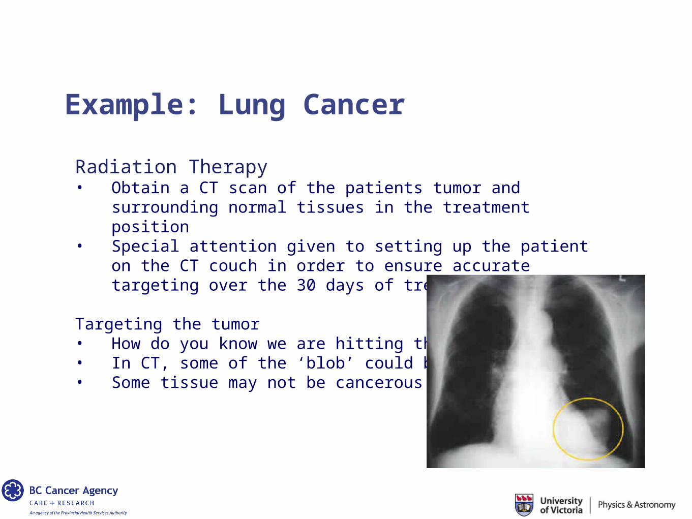
Radiation Therapy• Obtain a CT scan of the patients tumor and surrounding
normal tissues in the treatment position• Special attention given to setting up the patient on the CT
couch in order to ensure accurate targeting over the 30 days of treatment delivery
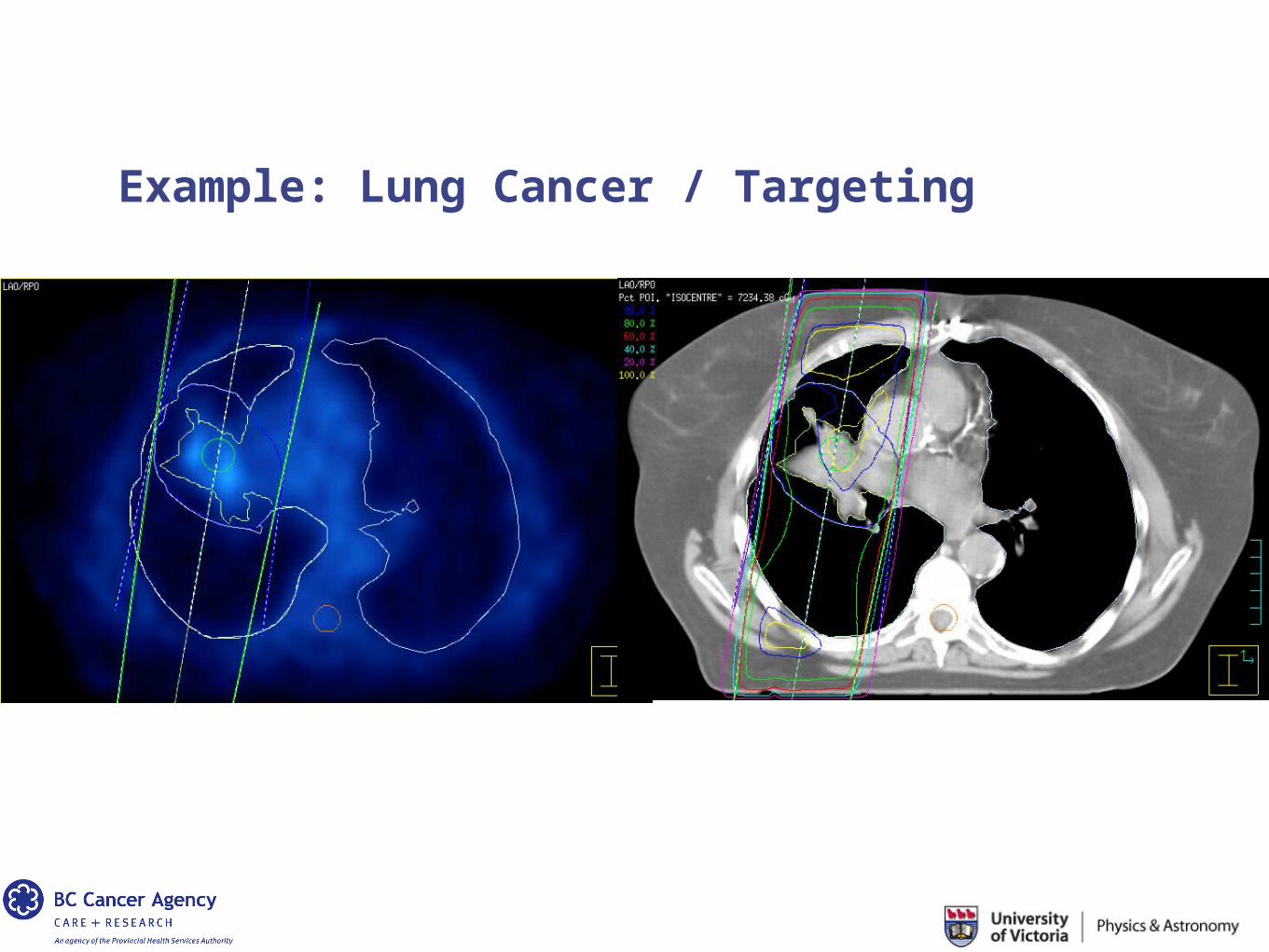
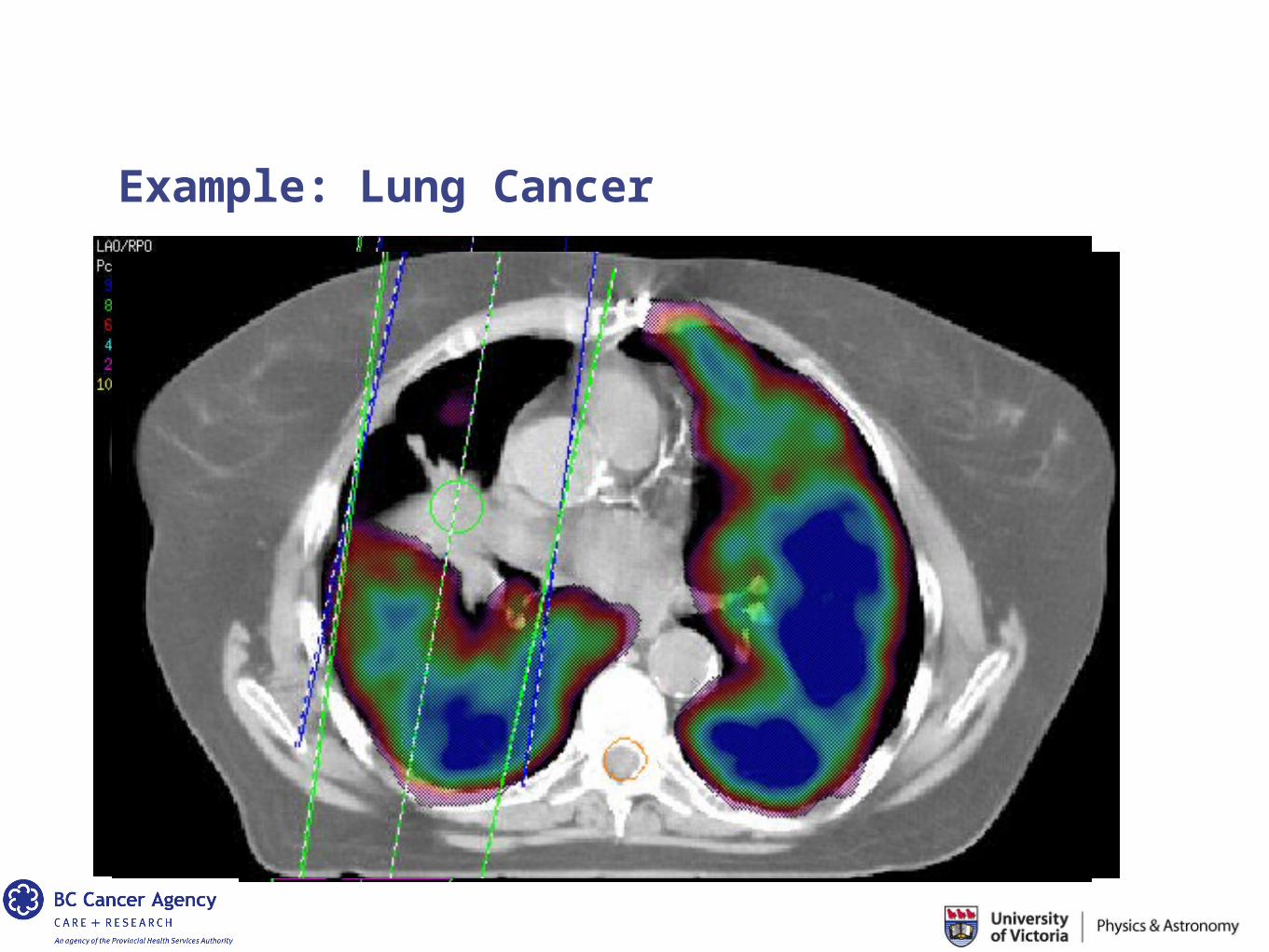
Targeting the tumor• How do you know we are hitting the tumor?• In CT, some of the ‘blob’ could be water• Some tissue may not be cancerous
Example: Lung Cancer
As our understanding of cancer increases, so does the recognition that each tumor is unique.
In the last decade, the focus in cancer research on the genetic make-up of cancer
Now it is clear that the tumor microenvironment, cellular and protein interactions affect disease progression, aggressiveness and response to treatment.
Introduction: 1 + 1 ≠ 2
Example: Lung Cancer
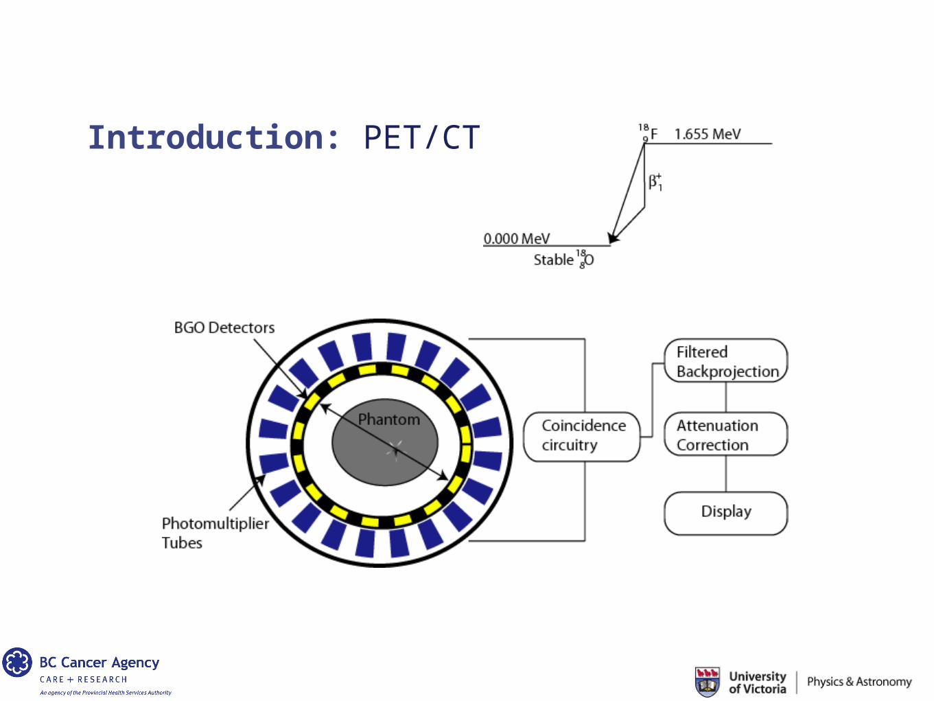
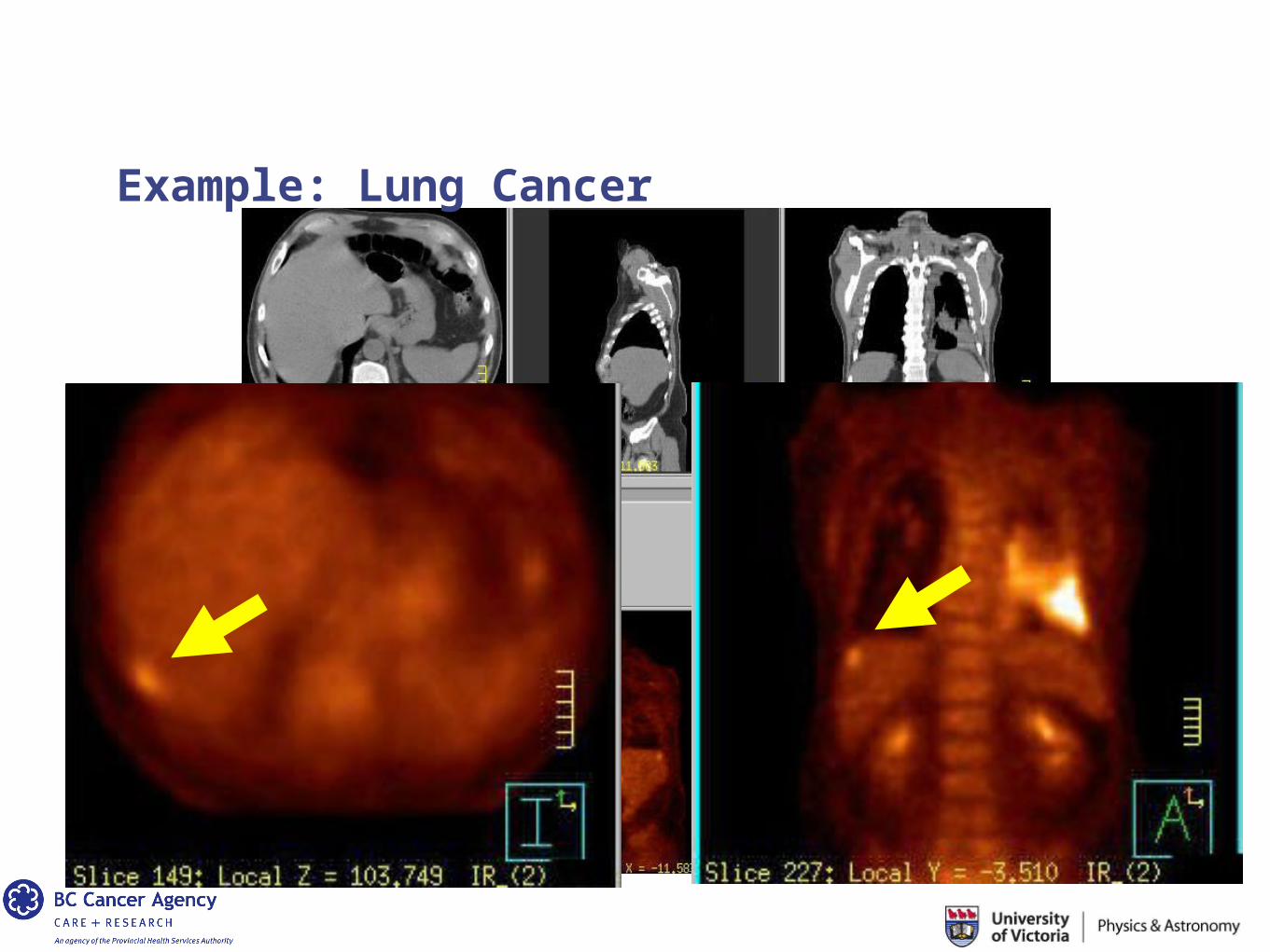
Positron Emission Tomography (PET), is a non-invasive molecular imaging technique that uses various radio-labeled compounds and visualizes metabolic differences between tissues, thus depicting the functional status of a suspicious lesion.
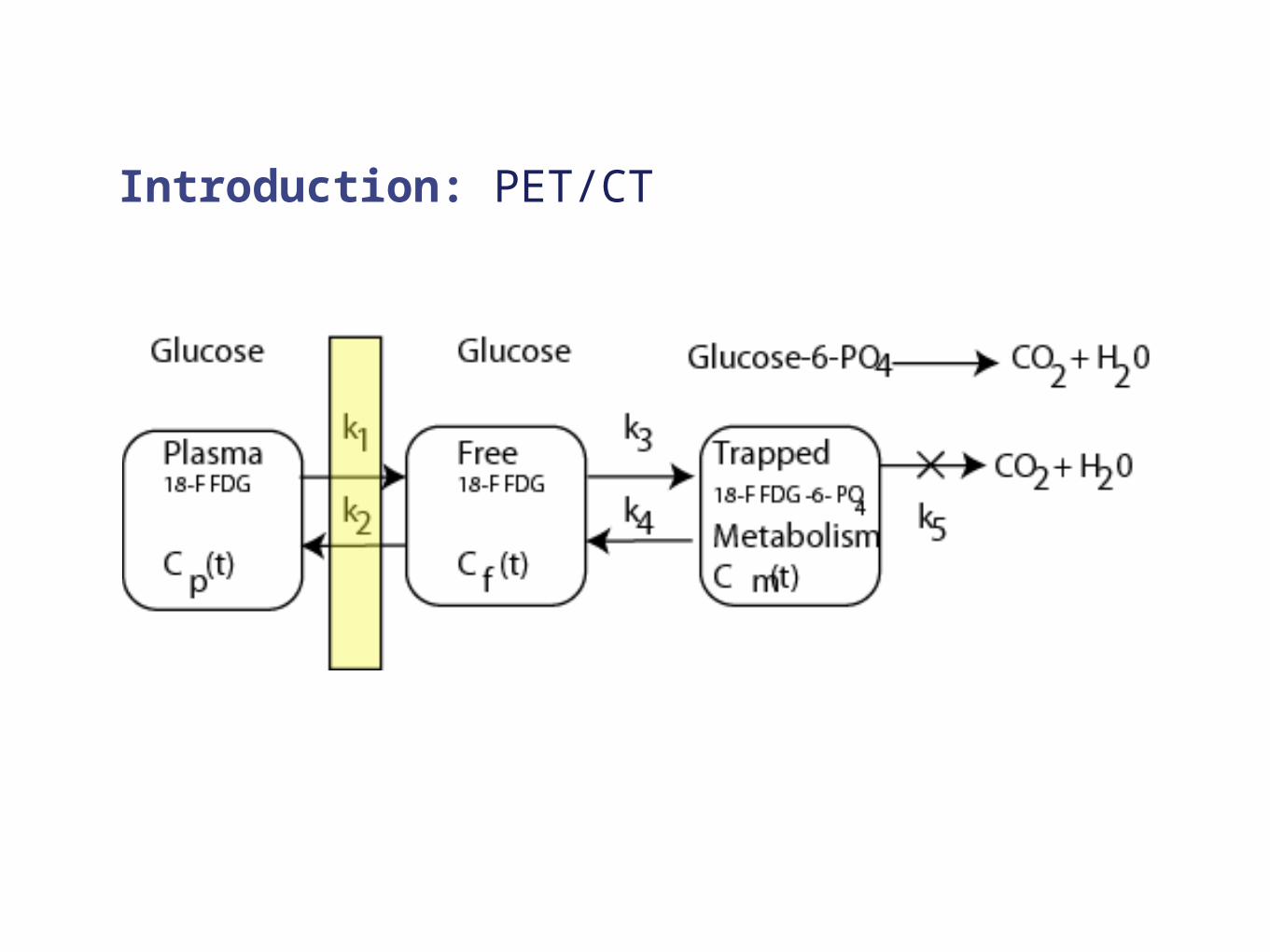
Malignant cells have an increased glycolytic rate.
Radio labeled 18F-fluorodeoxyglucose (18F-FDG) is a glucose analogue that has the same cellular uptake as glucose but is metabolically trapped within the cell after enzymatic phosphorylation to FDG-6-phosphate.
Therefore, FDG can be used to quantify glucose metabolic rates.
Example: Lung Cancer
What happens if you combine PET with the gold standard CT?
Does 1 + 1 = 2 ?
CT + PET = better targeting? Better planning?
Example: Lung Cancer
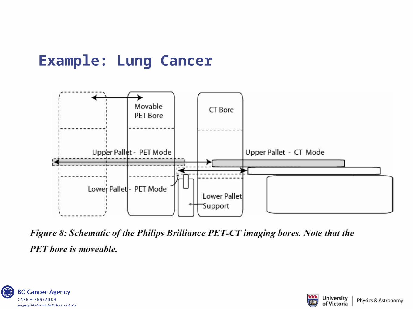
Example: Lung Cancer
Introduction: PET/CT
Introduction: PET/CT
Example: Lung Cancer
Introduction: 1 + 1 ≠ 2
Non-small cell carcinoma of the lung
- Metastatic spread to the liver
- Upstaged the disease and therefore the management of the disease
- No longer a radical treatment: palliative treatment with emphasis on quality of life
- Huge implications for the patient, family, patient care, health care costs.
Example: Lung Cancer / Targeting
Example: Lung Cancer
Clinically, the best example of multimodality imaging is seen in the rapid evolution of PET-CT and SPECT-CT scanner hybrids.
SPECT = Single Photon Emission Computed Tomography
Simpler version of PET scanning
Again, the main utility in SPECT-CT is the merging of functional with anatomical data (one of which is typically the gold standard).
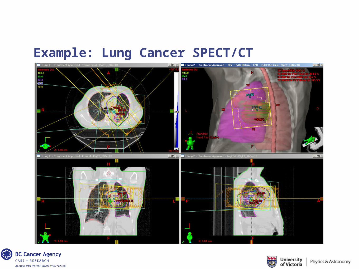
Example: Lung Cancer SPECT/CT
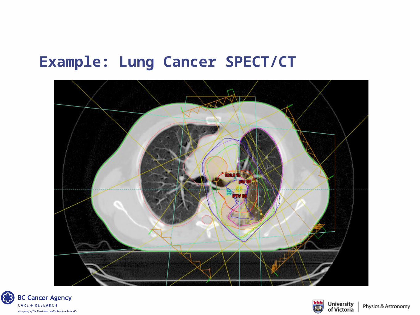
Example: Lung Cancer SPECT/CT
Example: Lung Cancer
What value might functional imaging have in such a case?
In radiation therapy, you can always eradicate the tumor… but you might suffer significant side effects.
In lung cancer RT, the most significant side-effect is the loss of lung function…
Obtain a SPECT image to assess healthy lung function.
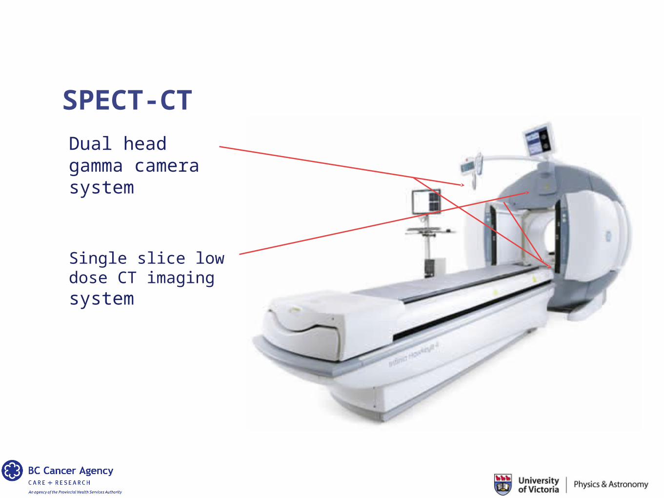
SPECT-CT
Dual head gamma camera system
Single slice low dose CT imaging system
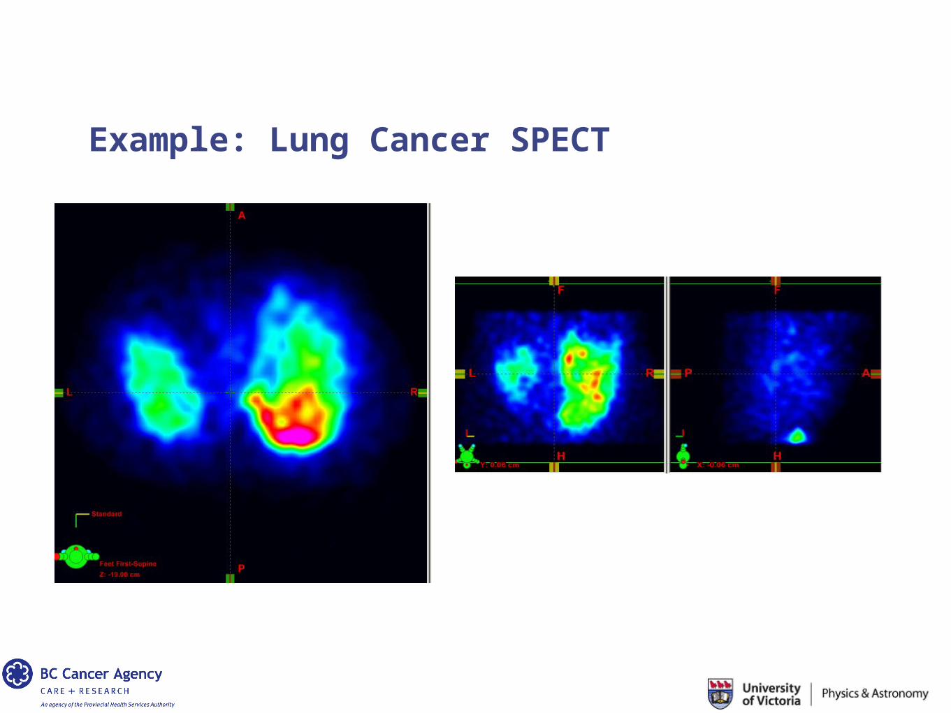
Example: Lung Cancer SPECT
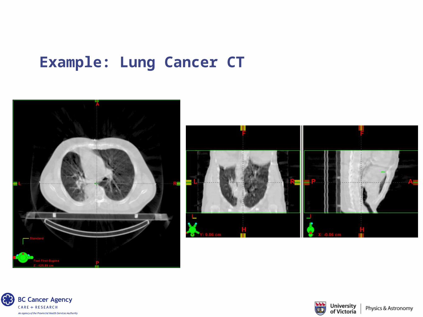
Example: Lung Cancer CT
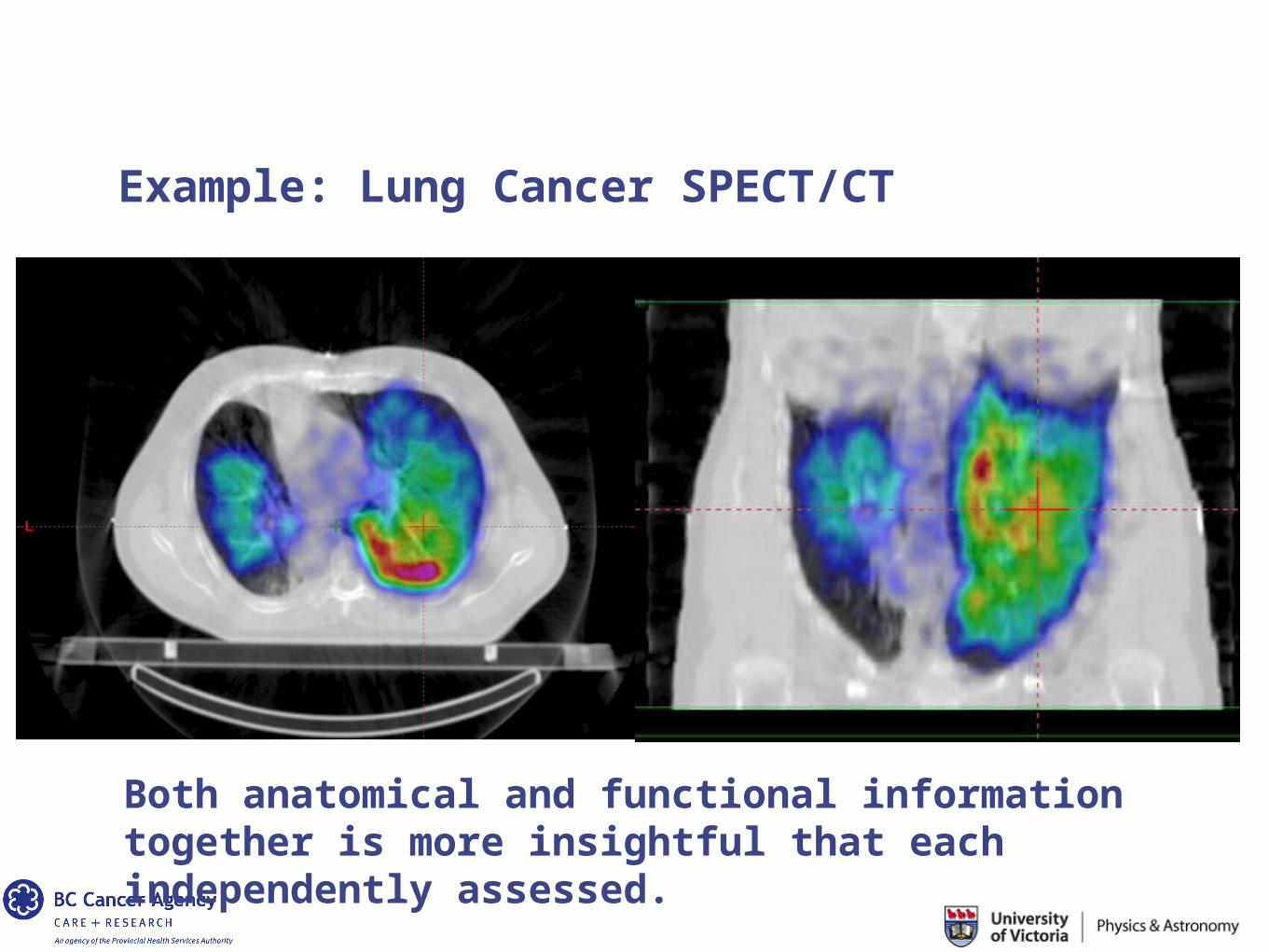
Example: Lung Cancer SPECT/CT
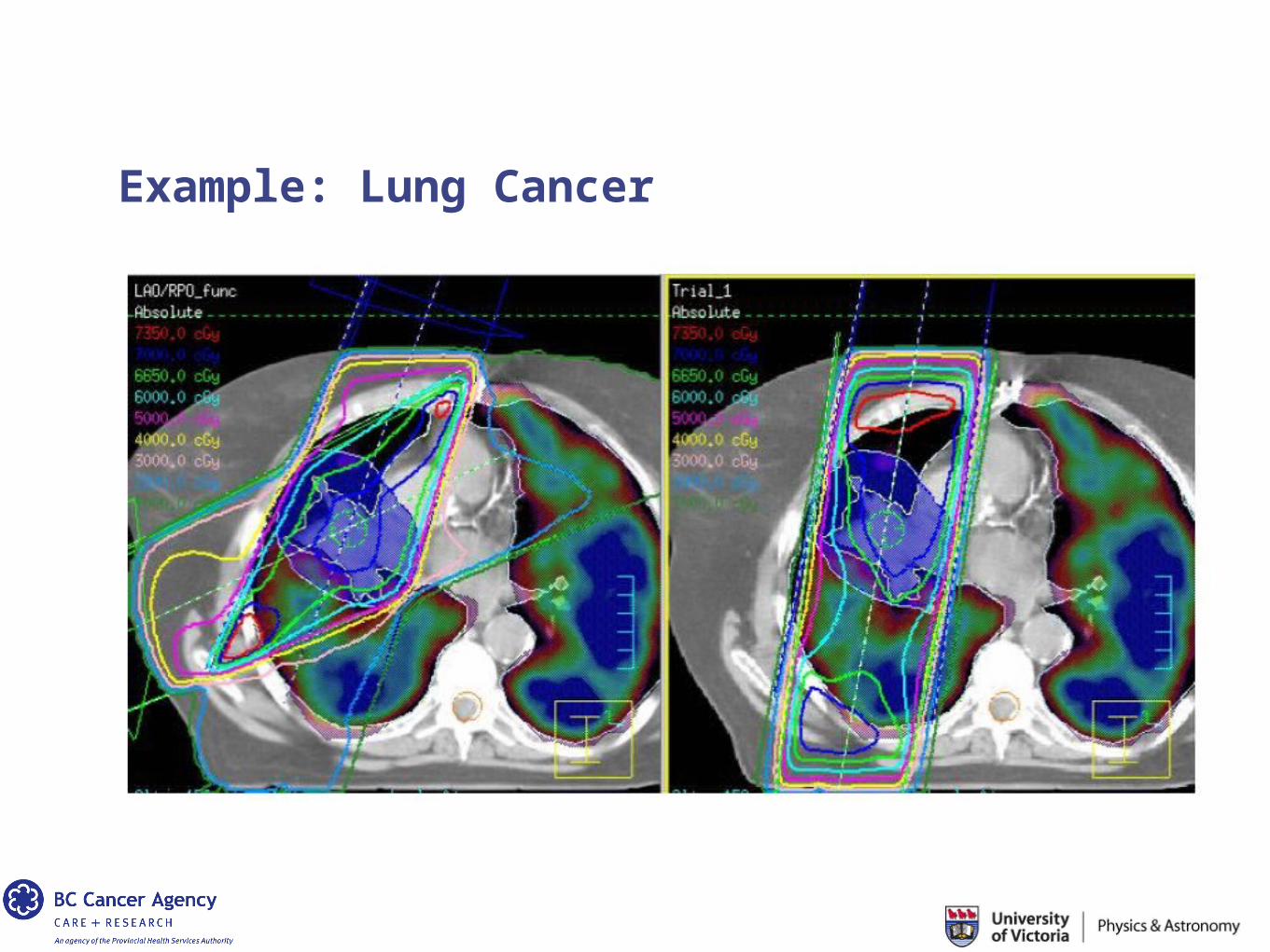
Both anatomical and functional information together is more insightful that each independently assessed.
Example: Lung Cancer
In the absence of SPECT information, completely functioning parts of the lung would be needlessly irradiated.
High doses of radiation can destroy healthy lung tissue.
It might make sense to direct the beams through parts of the lung that are not functioning.
Example: Lung Cancer
Example: Lung Cancer
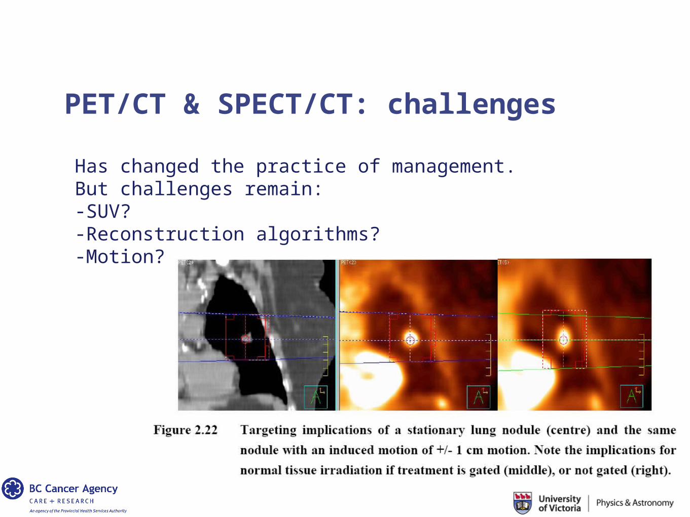
Has changed the practice of management.But challenges remain:-SUV?-Reconstruction algorithms?-Motion?
PET/CT & SPECT/CT: challenges
Example: Prostate Cancer
Example: Prostate Cancer
A variety of multi-modality applications in the management and treatment of prostate cancer.
MR imaging has become a powerful tool in imaging the prostate.
Even adding different types of imaging sequences within the MR poses some exciting possibilities.
Example: Prostate Cancer
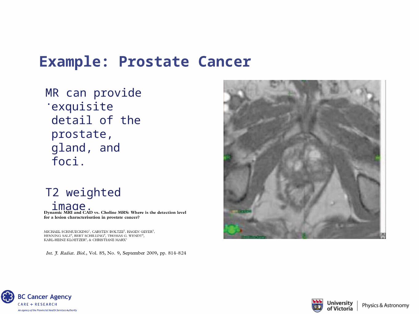
.MR can provide exquisite detail of the prostate, gland, and foci.
T2 weighted image.
Example: Prostate Cancer
.
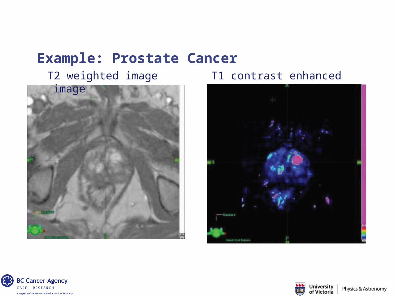
T2 weighted image T1 contrast enhanced image
Example: Prostate Cancer
.
Example: Prostate Cancer
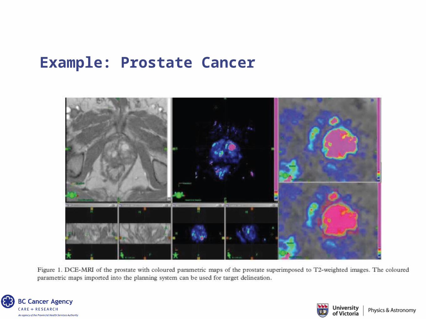
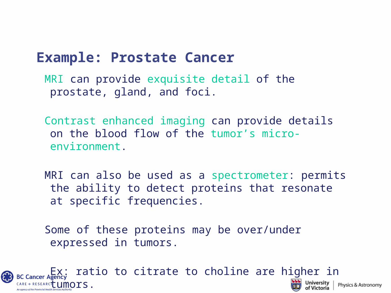
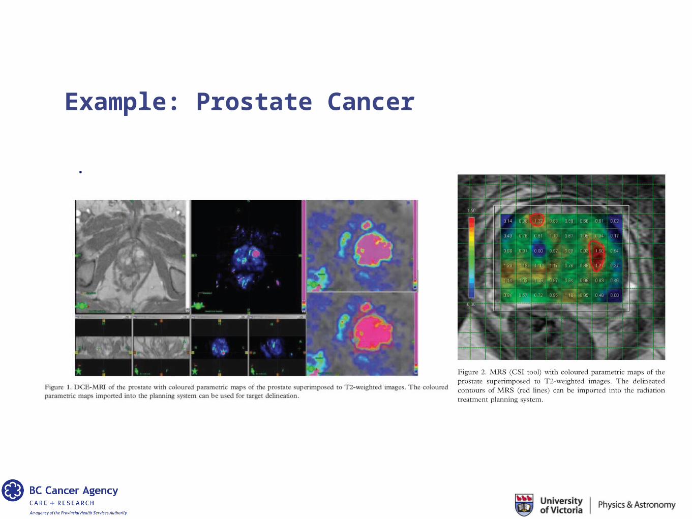
MRI can provide exquisite detail of the prostate, gland, and foci.
Contrast enhanced imaging can provide details on the blood flow of the tumor’s micro-environment.
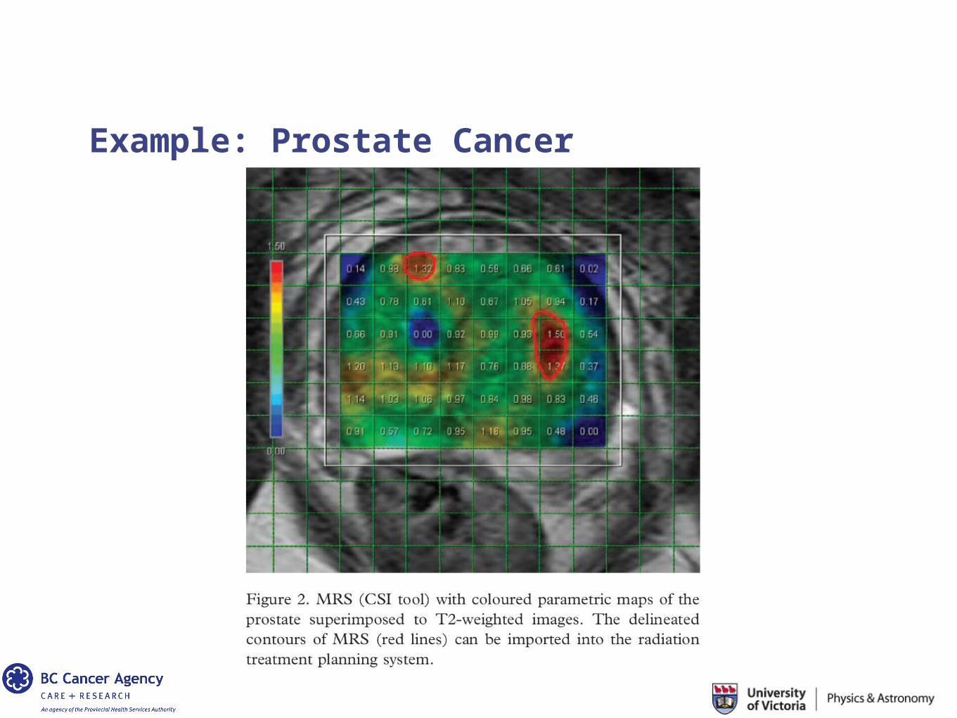
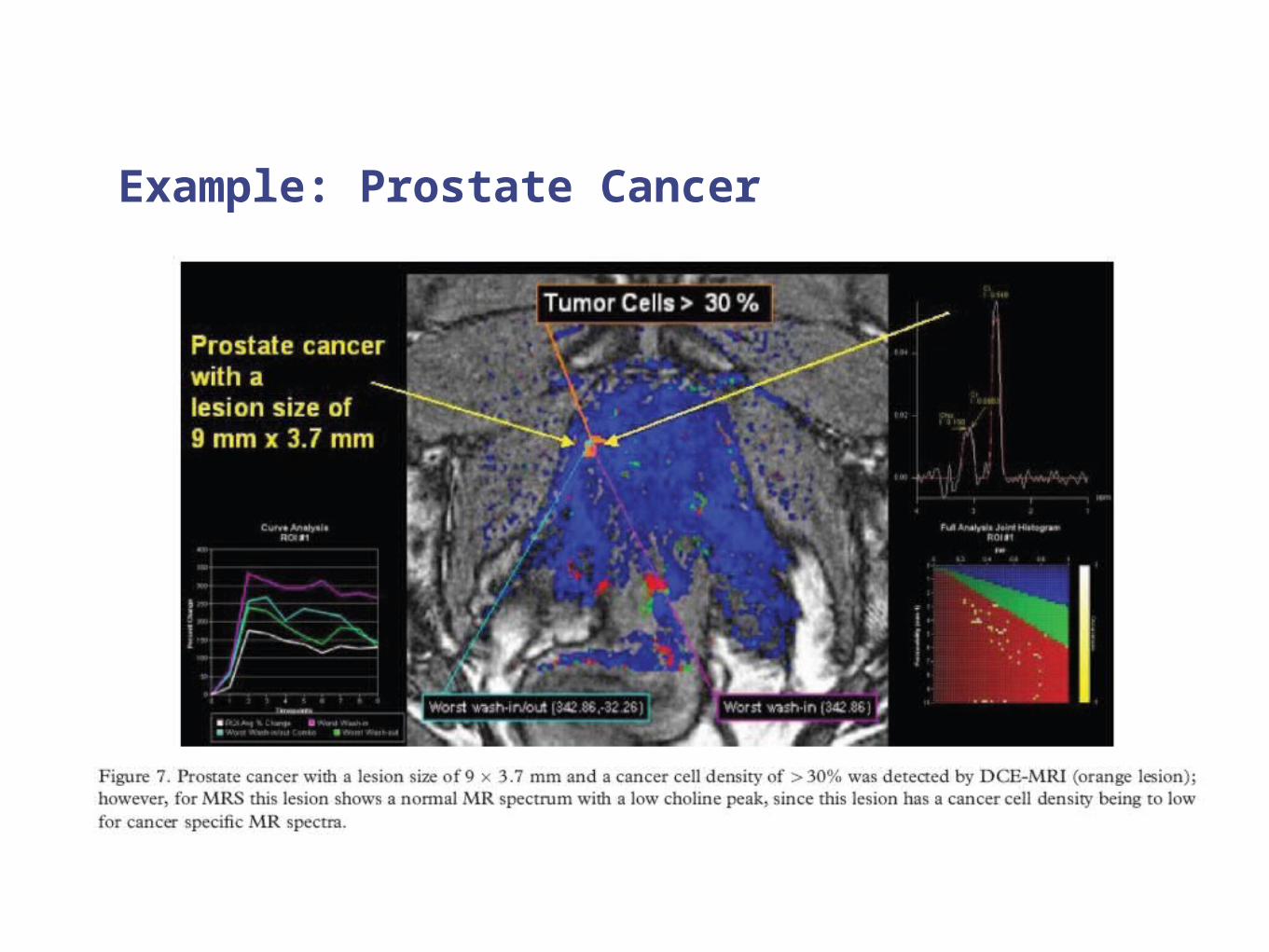
MRI can also be used as a spectrometer: permits the ability to detect proteins that resonate at specific frequencies.
Some of these proteins may be over/under expressed in tumors.
Ex: ratio to citrate to choline are higher in tumors.
Example: Prostate Cancer
Example: Prostate Cancer
.
Example: Prostate Cancer
Example: Prostate Cancer
… that the tumor microenvironment, cellular and protein interactions affect disease progression, aggressiveness and response to treatment.
Conclusions / Future Considerations
Conclusions / Future Directions
Certainly, these are not the only innovations on the horizon…
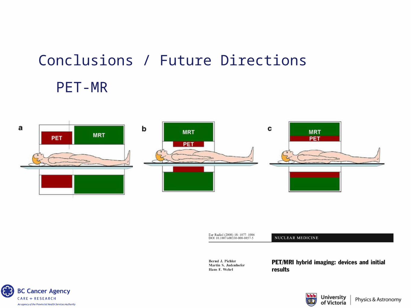
Conclusions / Future Directions
PET-MR
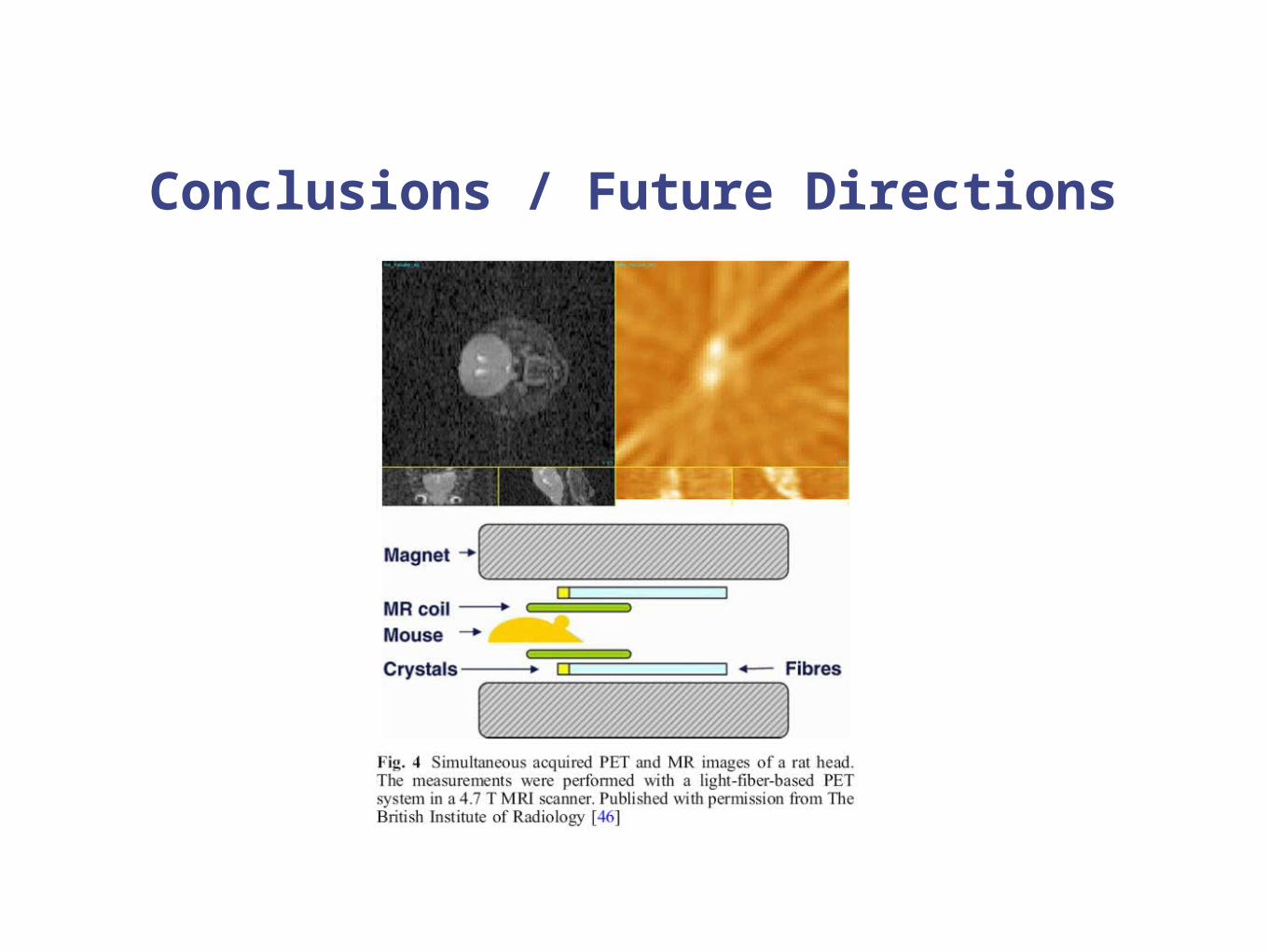
Conclusions / Future Directions
Conclusions / Future Directions
PET / MR
http://medicalphysicsweb.org/cws/article/research/43526
http://medicalphysicsweb.org/cws/article/research/41868
SPECT / MR
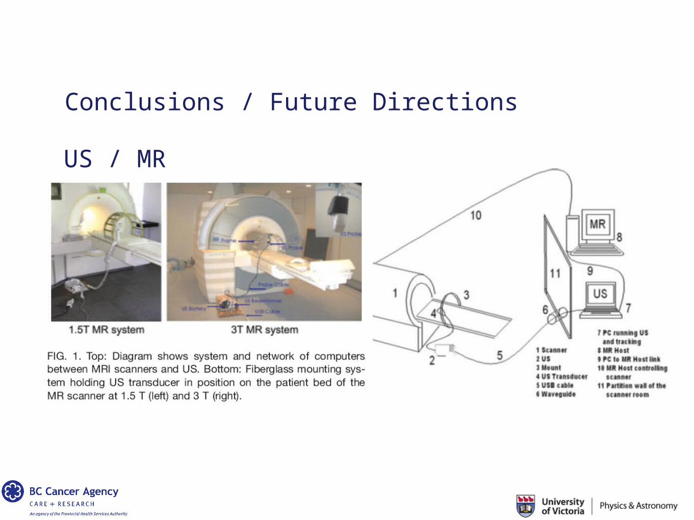
Conclusions / Future Directions
US / MR
Conclusions / Future Directions
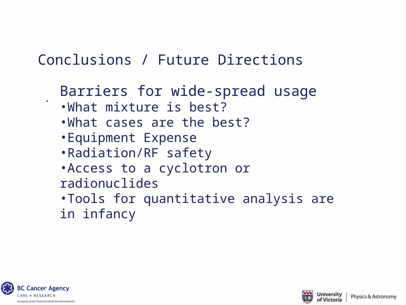
. Barriers for wide-spread usage•What mixture is best?•What cases are the best?•Equipment Expense•Radiation/RF safety•Access to a cyclotron or radionuclides•Tools for quantitative analysis are in infancy
Conclusions / Future Directions
Barriers for wide-spread usage•What mixture is best?
It remains unclear what combination of modalities should be used.
PET/CT clearly shows benefits in lung, head and neck cancers.
MRI/PET could equally show such benefits in, example, prostate, but clinical cost-benefit has to be demonstrated.
Conclusions / Future Directions
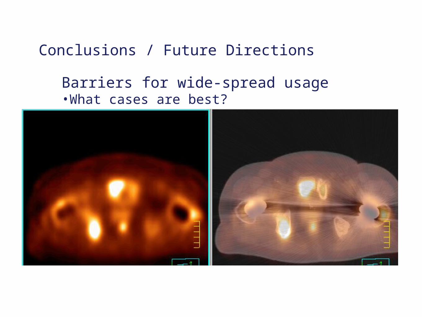
Barriers for wide-spread usage•What cases are best?
Conclusions / Future Directions
Barriers for wide-spread usage•Expense
PET / SPECT detectors are the key source of costs in these systems.
Advances in material sciences and production efficiency will drive these costs lower.
For MRI, roughly costs ~ 1M / Tesla, and proportional to scan length
Most clinical MR scanners have settled in the 1-3 T range.
Operating costs remain high.
CT/US systems are very mature; slice-wars are over…costs are relatively stable.

Conclusions / Future Directions
Barriers for wide-spread usage•Radiation and RF Safety
Often one already has a ‘room’ and one would like to renovate the room to accommodate the multi-modality imager. Addressing safety issues will require re-assessment of RF/Radiation safety operations, equipment usage, licensing, and resource expertise.
Typically an x-ray or CT room.
MR: need to provide radiofrequency barrier, or cage, around the scanner.
PET/SPECT: room shielding requirements are minimal, but operational costs are high (radiation chemist, detectors, personnel dosimetry monitoring, etc. )
Conclusions / Future Directions
Barriers for wide-spread usage•Access to a cyclotron or radionuclides (PET/SPECT)
PET requires ready access to a cyclotron, F-18 ~ 110 minutesC-11 ~ 20 minutes0-15 ~ 2 minutes
SPECT requires access to radio-isotopes
Conclusions / Future Directions
Barriers for wide-spread usage•Tools for quantitative analysis are in infancySome additional challenges:
Radiology lingo ≠ Radiation Oncology lingoRadiology technology ≠ Radiation Oncology technology
Bridging these fields is not a bad idea
Conclusions / Future Directions
.EX: NCI:PAR-08-225: Quantitative Imaging for Evaluation of Responses to Cancer Therapies (U01)
NCI: Reference Image Database to Evaluate Response
- initiative seeking input from RSNA / AAPM / ACR /- started with 30 longitudinal CT studies, primarily from
MDACC- currently CT, PET CT, DCE MRI, DW MRI, for lung, breast,
neuro
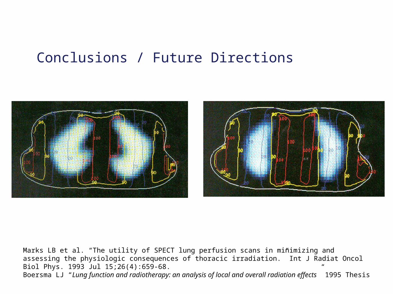
Marks LB et al. “The utility of SPECT lung perfusion scans in minimizing and assessing the physiologic consequences of thoracic irradiation.” Int J Radiat Oncol Biol Phys. 1993 Jul 15;26(4):659-68.Boersma LJ “Lung function and radiotherapy: an analysis of local and overall radiation effects” 1995 Thesis
Conclusions / Future Directions
Conclusions / Future Directions
Bold Prediction
Multi-modality imaging will replace ‘gold standard’
• assessing disease progression / aggressiveness
• delivering efficacious treatment ?• assessing response to treatment …

Thank you…questions?