Embed Size (px)
Citation preview
DIAGNOSIS
OF MARFAN
SYNDROME
Dr. Satyam Rajvanshi
SR Cardiology, Dr. RML Hospital, New Delhi
Introduction
Marfan syndrome - autosomal dominant inherited disorder of
connective tissue, characterised by loss of elastic tissue,
affects numerous body systems, including the
musculoskeletal, cardiovascular, neurological, and respiratory
systems, and the skin and eyes.
History
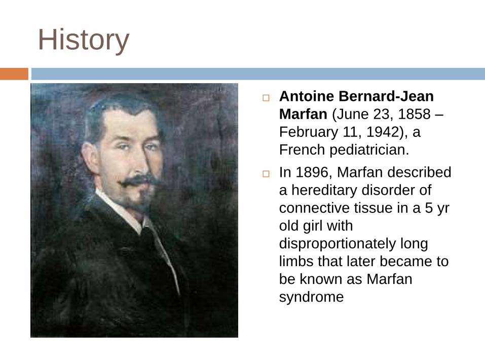
Antoine Bernard-Jean
Marfan (June 23, 1858 –
February 11, 1942), a
French pediatrician.
In 1896, Marfan described
a hereditary disorder of
connective tissue in a 5 yr
old girl with
disproportionately long
limbs that later became to
be known as Marfan
syndrome
Epidemiology
One of the most common inherited disorders of connective
tissue
Incidence: 1 in 3000-5000 individuals
Prevalence is thought to be similar
Regardless of sex
Regardless of ethnicity
Marfan syndrome-diagnosis and management.
Curr Probl Cardiol. Jan 2008;33(1):7-39.
Aetiology
Caused by a variety of mutations in the FBN1 gene. FBN1
mutations have been identified in over 90 percent patients
In 75% of patients - autosomal dominant, although the
appearance of family members and degree of pathological
features may vary.
In 25% of patients - mutation occurs spontaneously and
may be associated with older paternal age.
The first fibrillin-1 gene mutation was identified in 1990.
Subsequently, over 1000 different mutations have been
identified.
About 10 percent of individuals with suspected MFS have no
defined FBN1 mutation. Some of these individuals may have
TGFBR1 or TGFBR2 mutations. TGFBR1/TGFBR2 mutations
more typically cause LoeysDietz syndrome (LDS), with rare
reports in association with familial thoracic aortic aneurysm
(FTAA) syndrome.
Some patients with FBN1 gene mutations do not have MFS
and instead have a related disorder such as ectopia lentis
syndrome or other diseases such as ShprintzenGoldberg
syndrome, WeillMarchesani syndrome, or stiff skin syndrome.
Pathophysiology
Mutations in the fibrillin-1 gene result in the production of an abnormal fibrillin protein, leading to abnormalities in the mechanical stability and elastic properties of connective tissue.
More recently, research suggests that transforming growth factor-beta is implicated in the failure of normal elastic tissue formation.
TGFBR 1 and 2 mutations – may have similar manifestations
Cystic medial necrosis - cysts being fluid collections of mucinand ground substance - lead to a weakening of the aortic wall with subsequent aortic dilation and potentially aortic dissection, aneurysms, and rupture. They also lead to a reduction of the structural integrity of the skin, ligaments, eye lenses, lung airways, and the spinal dura.
Clinical Manifestations
Diagnostic Criteria
Step by step Approach
Diagnosis of Marfan
Syndrome
First description
5 year girl, Gabrielle P
Prominent Skeletal features - disproportionately long limbs.
She Probably had Congenital Contractual arachnodactyly!
- Not Marfan!
Revised diagnostic criteria for Marfan syndrome.
American Journal of Medical Genetics. 62 (1996)
Additional features described during 20th century
Ectopia Lentis (Borger; 1914)
Autosomal Dominant inheritance (Weve; 1931)
Aortic Dialatation (Etter and grover; 1943)
Aortic Dissection (Baer; 1943)
Mitral Valve Prolapse (Brown; 1975)
Dural Ectasia (Pyeritz; 1988)
Revised diagnostic criteria for Marfan syndrome.
American Journal of Medical Genetics. 62 (1996)
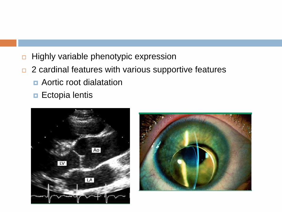
Highly variable phenotypic expression
2 cardinal features with various supportive features
Aortic root dialatation
Ectopia lentis
The Berlin Nosology
‘Clinical’ Classification of Heritable connective tissue
disorders (HCTD)
Described Marfan Syndrome
Major and Minor manifestations (in decreasing order of
specificity)
Skeletal
Ocular
Cardiovascular
Pulmonary
Skin
CNS
Autosomal Dominant InheritanceInternational Nosology of HCTD,1986
American Journal of Medical Genetics. (1988)
Relied completely on clinical criteria
Led to overdiagnosis
Specially in family members of index cases
Overlapping HCTDs
International Nosology of HCTD,1986
American Journal of Medical Genetics. (1988)
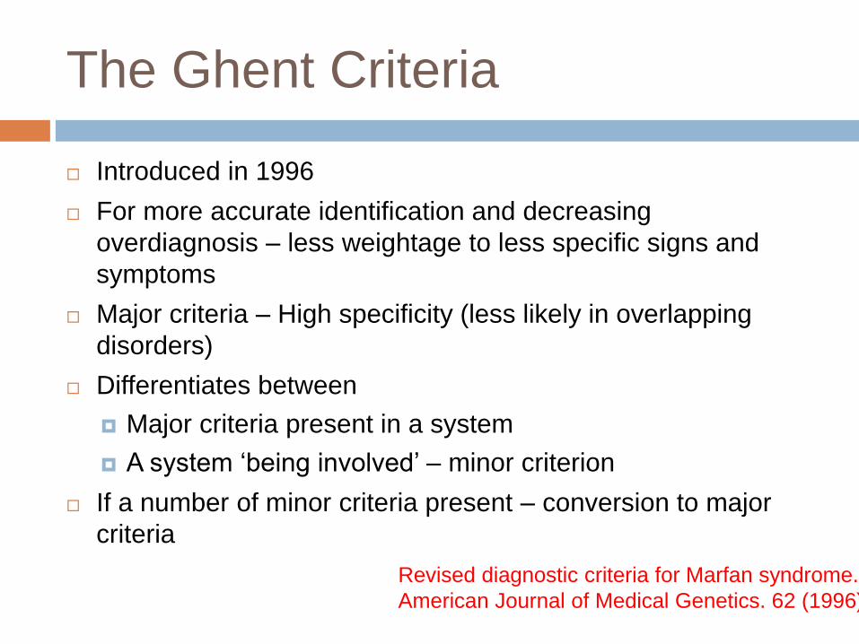
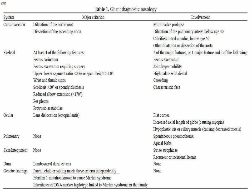
The Ghent Criteria
Introduced in 1996
For more accurate identification and decreasing
overdiagnosis – less weightage to less specific signs and
symptoms
Major criteria – High specificity (less likely in overlapping
disorders)
Differentiates between
Major criteria present in a system
A system ‘being involved’ – minor criterion
If a number of minor criteria present – conversion to major
criteria
Revised diagnostic criteria for Marfan syndrome.
American Journal of Medical Genetics. 62 (1996)
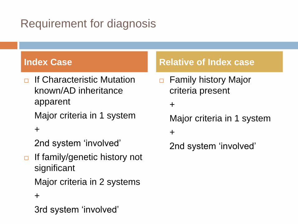
Requirement for diagnosis
If Characteristic Mutation
known/AD inheritance
apparent
Major criteria in 1 system
+
2nd system ‘involved’
If family/genetic history not
significant
Major criteria in 2 systems
+
3rd system ‘involved’
Family history Major
criteria present
+
Major criteria in 1 system
+
2nd system ‘involved’
Index Case Relative of Index case
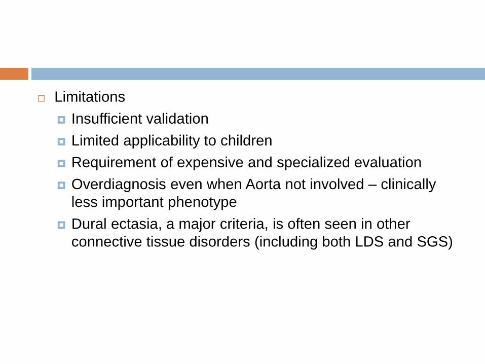
Limitations
Insufficient validation
Limited applicability to children
Requirement of expensive and specialized evaluation
Overdiagnosis even when Aorta not involved – clinically
less important phenotype
Dural ectasia, a major criteria, is often seen in other
connective tissue disorders (including both LDS and SGS)
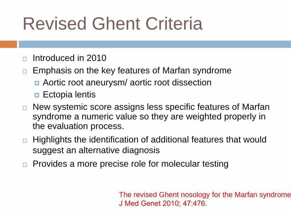
Revised Ghent Criteria
Introduced in 2010
Emphasis on the key features of Marfan syndrome
Aortic root aneurysm/ aortic root dissection
Ectopia lentis
New systemic score assigns less specific features of Marfansyndrome a numeric value so they are weighted properly in the evaluation process.
Highlights the identification of additional features that would
suggest an alternative diagnosis
Provides a more precise role for molecular testing
The revised Ghent nosology for the Marfan syndrome.
J Med Genet 2010; 47:476.
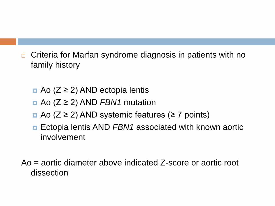
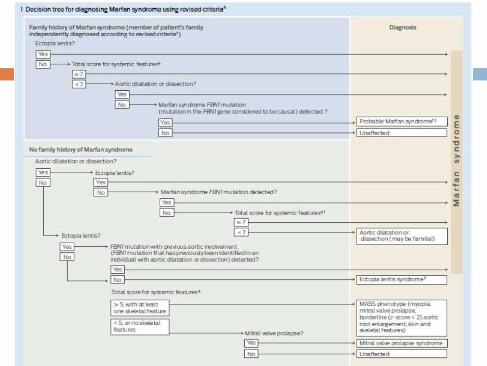
Criteria for Marfan syndrome diagnosis in patients with no
family history
Ao (Z ≥ 2) AND ectopia lentis
Ao (Z ≥ 2) AND FBN1 mutation
Ao (Z ≥ 2) AND systemic features (≥ 7 points)
Ectopia lentis AND FBN1 associated with known aortic
involvement
Ao = aortic diameter above indicated Z-score or aortic root
dissection
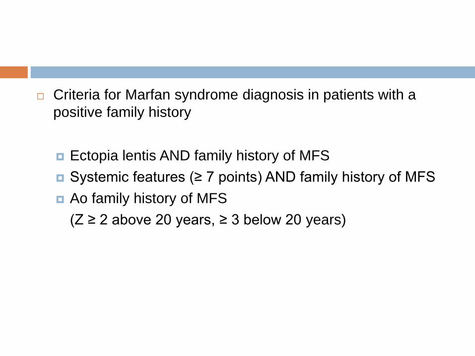
Criteria for Marfan syndrome diagnosis in patients with a
positive family history
Ectopia lentis AND family history of MFS
Systemic features (≥ 7 points) AND family history of MFS
Ao family history of MFS
(Z ≥ 2 above 20 years, ≥ 3 below 20 years)
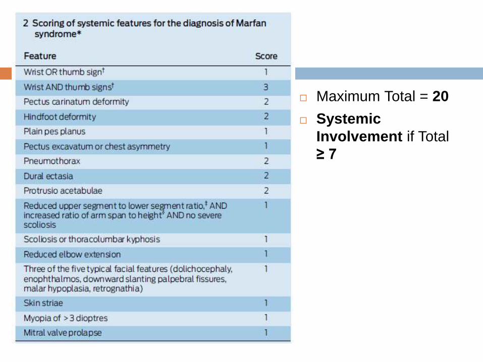
Maximum Total = 20
Systemic
Involvement if Total
≥ 7
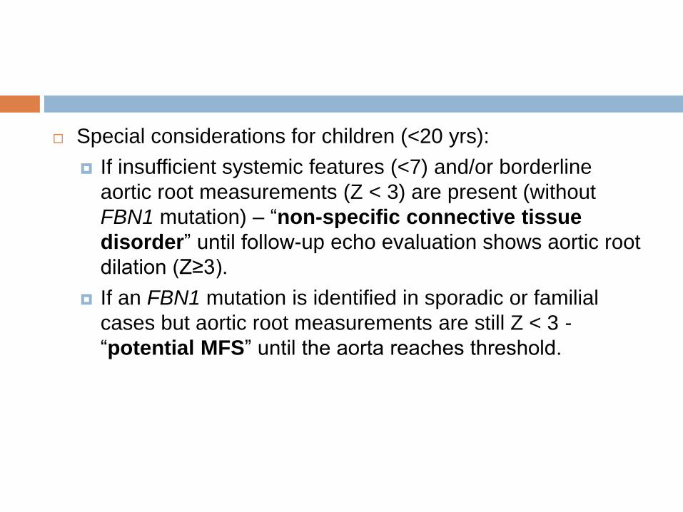
Special considerations for children (<20 yrs):
If insufficient systemic features (<7) and/or borderline
aortic root measurements (Z < 3) are present (without
FBN1 mutation) – “non-specific connective tissue
disorder” until follow-up echo evaluation shows aortic root
dilation (Z≥3).
If an FBN1 mutation is identified in sporadic or familial
cases but aortic root measurements are still Z < 3 -
“potential MFS” until the aorta reaches threshold.
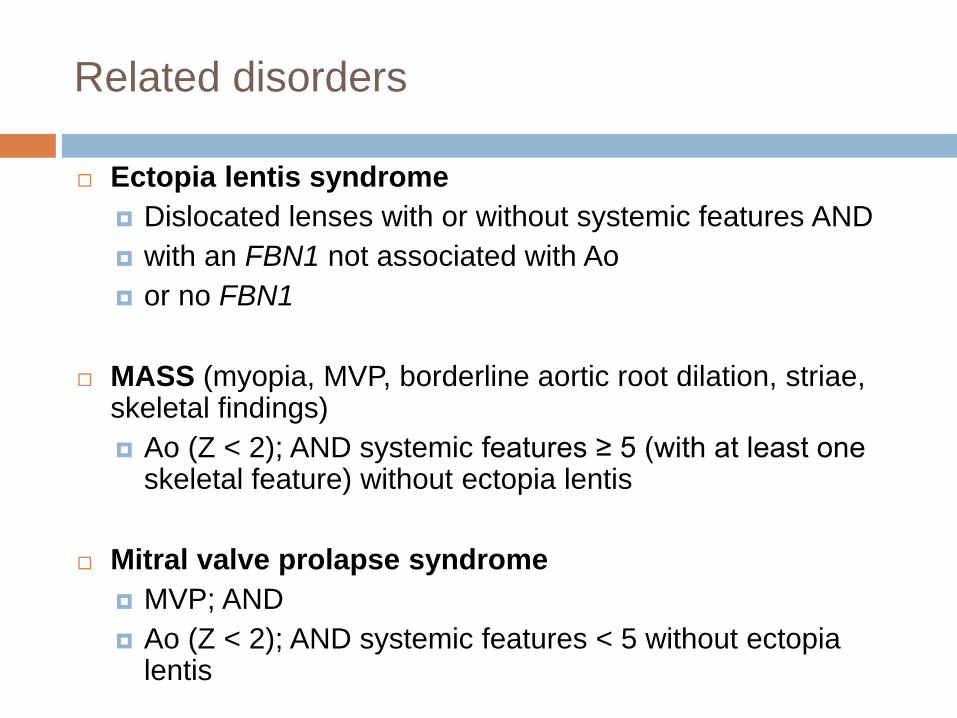
Related disorders
Ectopia lentis syndrome
Dislocated lenses with or without systemic features AND
with an FBN1 not associated with Ao
or no FBN1
MASS (myopia, MVP, borderline aortic root dilation, striae, skeletal findings)
Ao (Z < 2); AND systemic features ≥ 5 (with at least one skeletal feature) without ectopia lentis
Mitral valve prolapse syndrome
MVP; AND
Ao (Z < 2); AND systemic features < 5 without ectopialentis
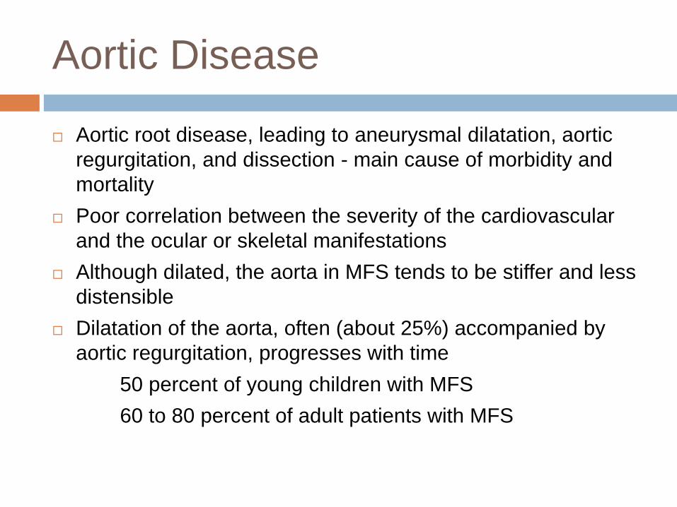
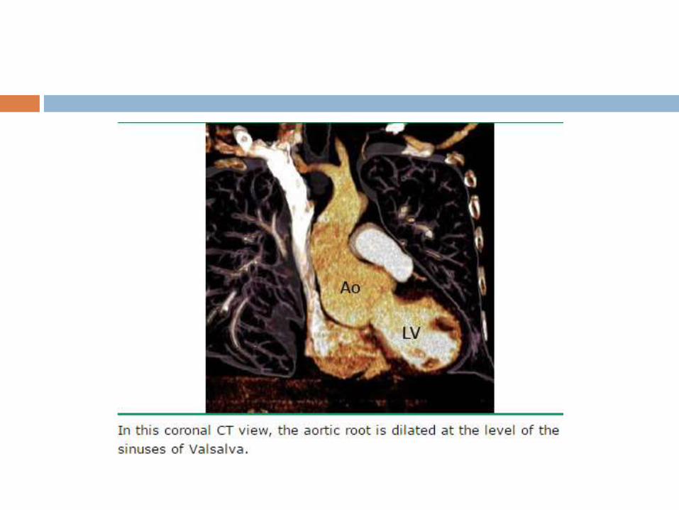
Aortic Disease
Aortic root disease, leading to aneurysmal dilatation, aortic
regurgitation, and dissection - main cause of morbidity and
mortality
Poor correlation between the severity of the cardiovascular
and the ocular or skeletal manifestations
Although dilated, the aorta in MFS tends to be stiffer and less
distensible
Dilatation of the aorta, often (about 25%) accompanied by
aortic regurgitation, progresses with time
50 percent of young children with MFS
60 to 80 percent of adult patients with MFS
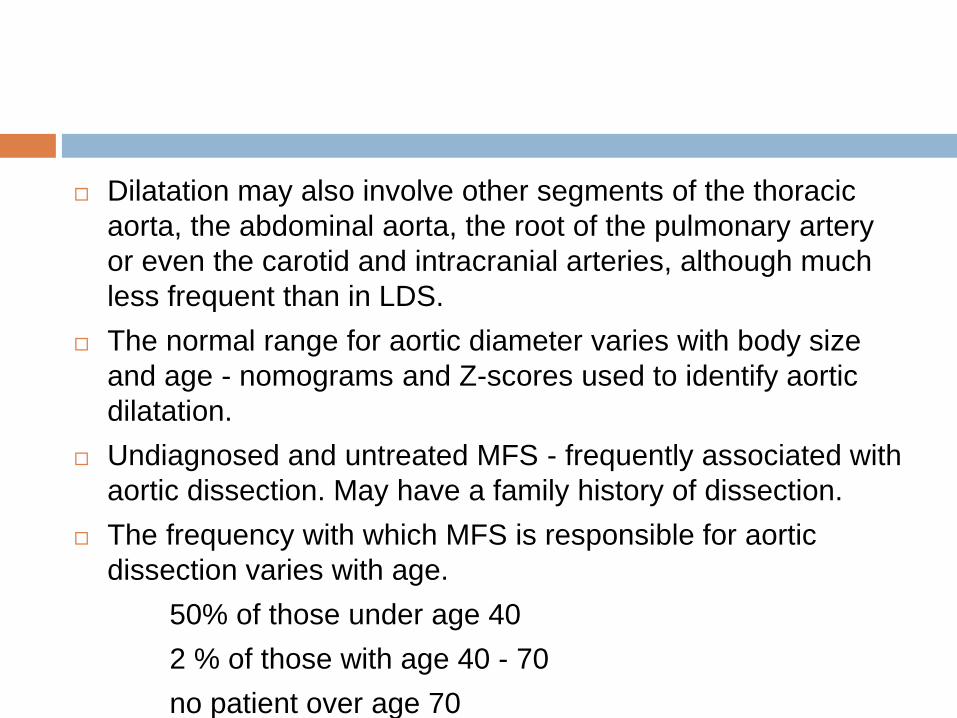
Dilatation may also involve other segments of the thoracic
aorta, the abdominal aorta, the root of the pulmonary artery
or even the carotid and intracranial arteries, although much
less frequent than in LDS.
The normal range for aortic diameter varies with body size
and age - nomograms and Z-scores used to identify aortic
dilatation.
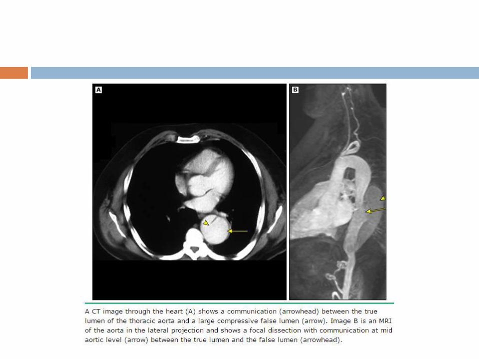
Undiagnosed and untreated MFS - frequently associated with
aortic dissection. May have a family history of dissection.
The frequency with which MFS is responsible for aortic
dissection varies with age.
50% of those under age 40
2 % of those with age 40 - 70
no patient over age 70
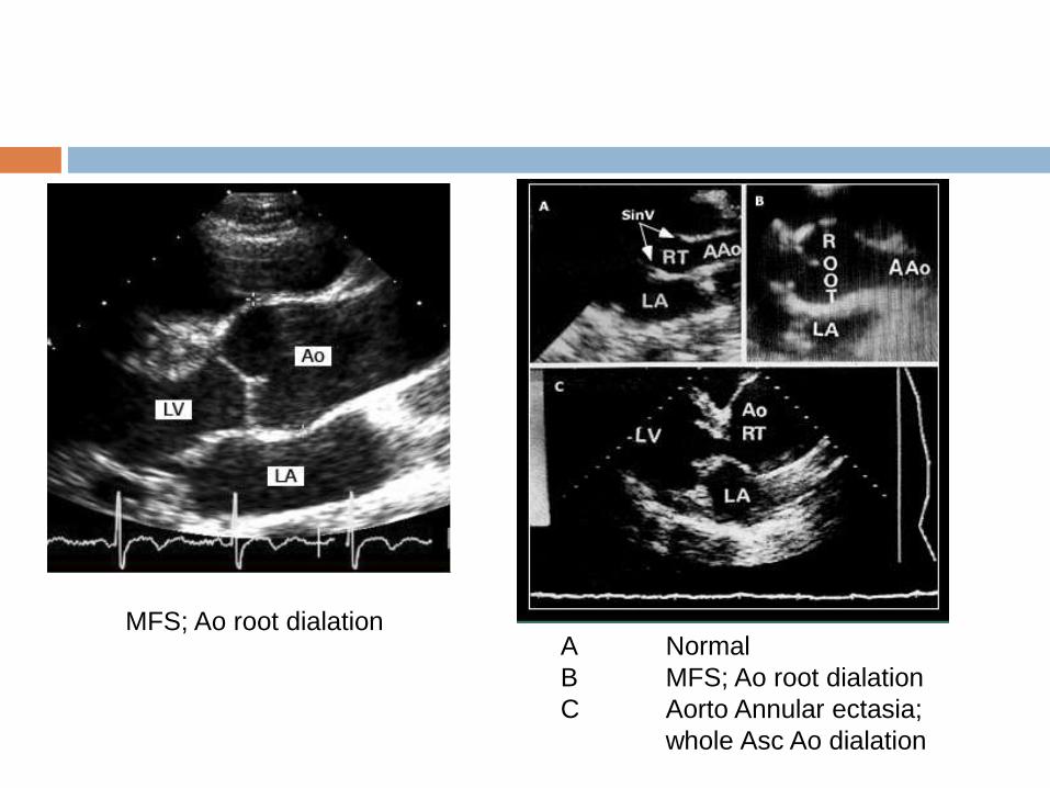
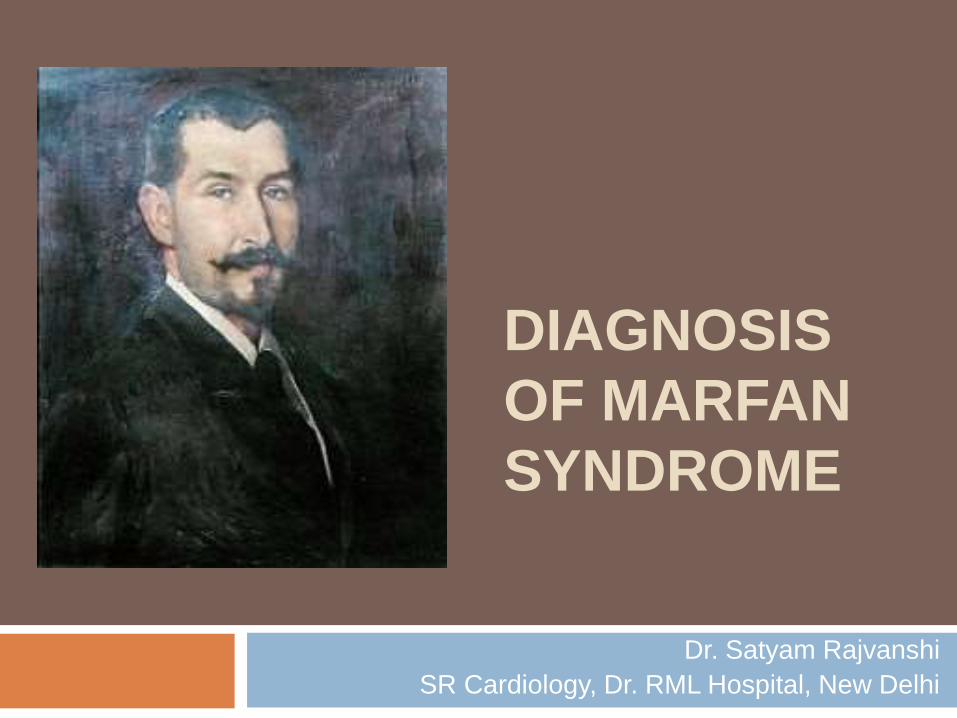
A Normal
B MFS; Ao root dialation
C Aorto Annular ectasia;
whole Asc Ao dialation
MFS; Ao root dialation
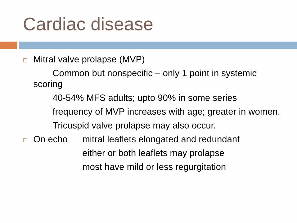
Cardiac disease
Mitral valve prolapse (MVP)
Common but nonspecific – only 1 point in systemic
scoring
40-54% MFS adults; upto 90% in some series
frequency of MVP increases with age; greater in women.
Tricuspid valve prolapse may also occur.
On echo mitral leaflets elongated and redundant
either or both leaflets may prolapse
most have mild or less regurgitation
Approximately 25 percent of patients with MVP have
progressive disease - defined by the appearance or
worsening of clinical symptoms of mitral regurgitation or
worsening on echocardiography.
Heart failure attributable to mitral valve prolapse and
regurgitation represents a major source of morbidity and
mortality in young children with the most extreme and rapidly
progressive presentation of MFS.
Some report suggest - some patients may have a
cardiomyopathy with biventricular enlargement and generally
asymptomatic mild systolic dysfunction unrelated to valvular
disease
Skeletal disease
Excess linear growth of the long bones – Individuals taller
than predicted by their genetic background
Joint laxity
Paradoxically, some individuals with MFS have reduced joint
mobility, particularly of the elbow and digits - reduced elbow
extension (≤170 degrees with full extension) – 1 point to the
systemic score
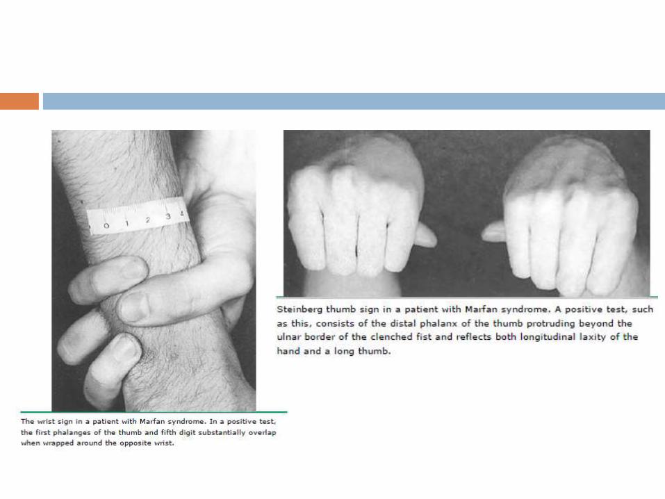
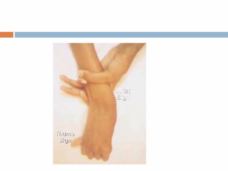
Arachnodactyly — abnomally long and slender fingers
Thumb sign - entire distal phalanx protrudes beyond the
ulnar border of a clenched fist with or without the assistance
of the patient or examiner to achieve maximum adduction
Wrist sign - the top of the thumb covers the entire fingernail
of the fifth finger when wrapped around the contralateral wrist
Pectus deformity — Pectus carinatum - more specific for
MFS than pectus excavatum or chest asymmetry,
Hindfoot valgus — occurs with forefoot abduction and
lowering of the midfoot and should be evaluated from anterior
and posterior views.
Pes planus (flat foot) without hindfoot valgus is less specific
Generalized joint hypermobility also may occur, producing
findings that overlap with the much more common benign
joint hypermobility syndrome.
Abnormal US/LS and arm span/height —
disproportionately long extremities in comparison to the
length of the trunk (dolichostenomelia) - upper segment to
lower segment (US/LS) ratio is decreased and the arm span
to height ratio is increased.
The lower segment is defined as the distance from the top of
the symphysis pubis to the floor in the standing position; The
upper segment is the height minus the lower segment.
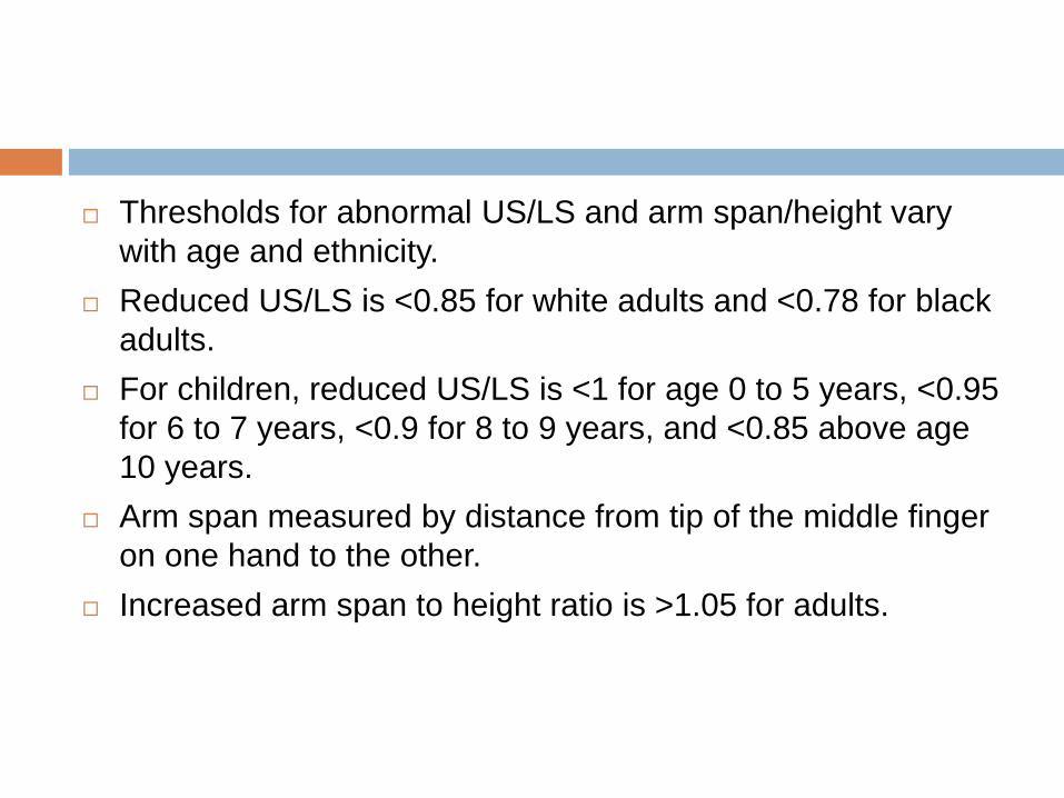
Thresholds for abnormal US/LS and arm span/height vary
with age and ethnicity.
Reduced US/LS is <0.85 for white adults and <0.78 for black
adults.
For children, reduced US/LS is <1 for age 0 to 5 years, <0.95
for 6 to 7 years, <0.9 for 8 to 9 years, and <0.85 above age
10 years.
Arm span measured by distance from tip of the middle finger
on one hand to the other.
Increased arm span to height ratio is >1.05 for adults.
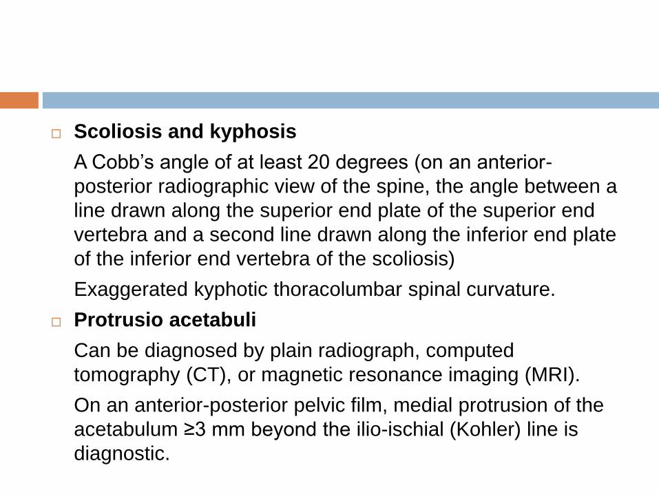
Scoliosis and kyphosis
A Cobb’s angle of at least 20 degrees (on an anterior-
posterior radiographic view of the spine, the angle between a
line drawn along the superior end plate of the superior end
vertebra and a second line drawn along the inferior end plate
of the inferior end vertebra of the scoliosis)
Exaggerated kyphotic thoracolumbar spinal curvature.
Protrusio acetabuli
Can be diagnosed by plain radiograph, computed
tomography (CT), or magnetic resonance imaging (MRI).
On an anterior-posterior pelvic film, medial protrusion of the
acetabulum ≥3 mm beyond the ilio-ischial (Kohler) line is
diagnostic.
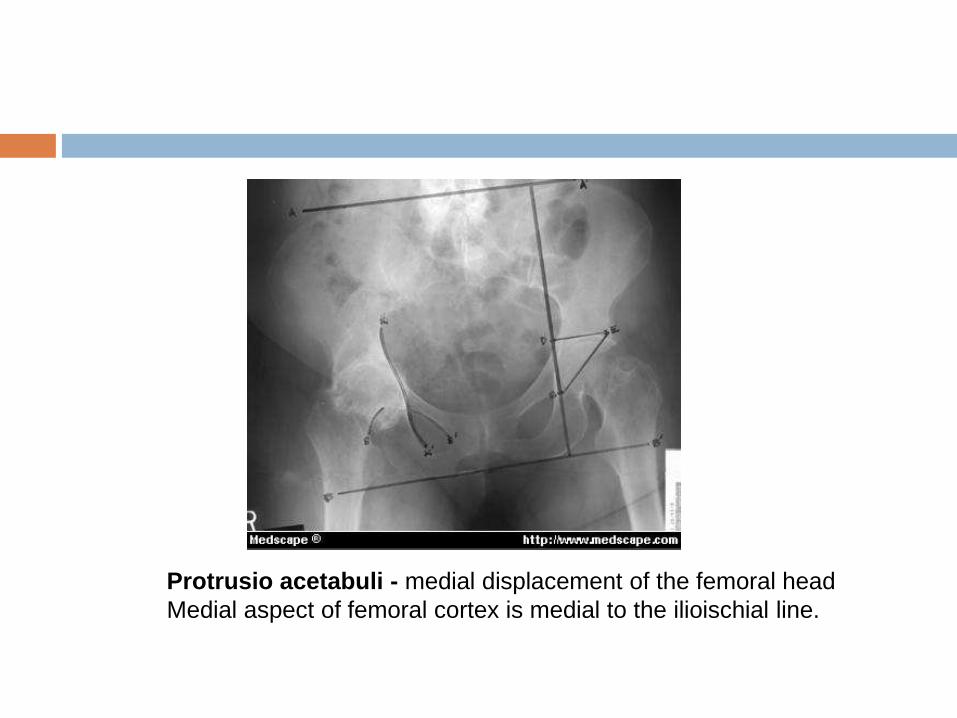
Protrusio acetabuli - medial displacement of the femoral head
Medial aspect of femoral cortex is medial to the ilioischial line.
Facial features — dolichocephaly (reduced cephalic index or
head width/length ratio), enophthalmos, downslanting
palpebral fissures, malar hypoplasia, and retrognathia.
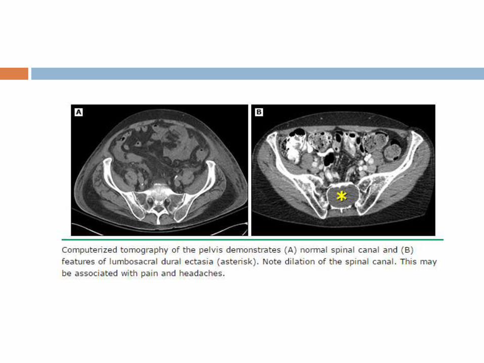
Dural ectasia
Enlargement of the spinal canal owing to progressive ectasia
of dura and neural foramina and to erosion of vertebral bone.
Usually involves the lumbosacral spine
60-90% pts on MRI/CT - sensitive but not specific sign of
MFS, is commonly seen in Loeys-Dietz syndrome and
Shprintzen-Goldberg syndrome, has been reported in the
vascular form of Ehlers-Danlos syndrome.
MRI most sensitive technique.
No correlation appears to exist between the severity of dural
ectasia and the degree of aortic dilatation.
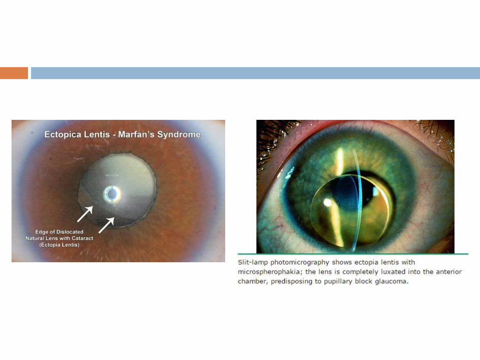
Ocular abnormalities
Ectopia lentis - 50 to 80 percent.
Detected on slit-lamp examination after maximal dilatation of
the pupil and the lens is usually displaced upward and
temporally. It is caused by failure of the supporting ciliary
zonules.
Myopia >3 diopters - secondary myopia due to increased axis
globe length.
Flat cornea, hypoplastic iris, hypoplastic ciliary muscle
causing decreased miosis, retinal detachment, glaucoma,
and early cataract formation. Retinal tears and detachment
are commonly bilateral.
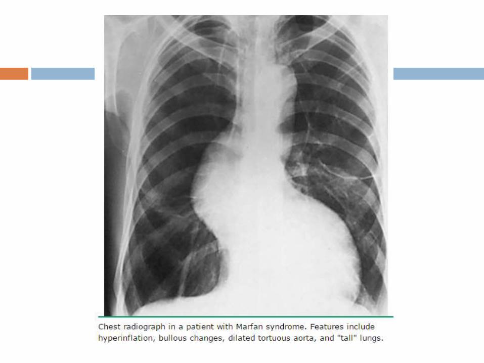
Pulmonary disease — Some patients develop
emphysematous changes with lung bullae predominantly in
the upper lobes, can predispose to spontaneous
pneumothorax
Skin striae — The presence of striae atrophicae contributes
one point to the systemic score if not associated with
pronounced weight changes or pregnancy and if in
uncommon location such as the mid back, lumbar region,
upper arm, axillary region, or thigh
Other — Recurrent or incisional herniae, joint hypermobility,
and high arched palate may occur but are not included in the
systemic score - nonspecific.
Step-by-step diagnostic
approach History and physical examination (including slit-lamp
ophthalmic examination with pupil dilation) in conjunction with
imaging of the aortic root and the ascending, descending,
and abdominal aorta (echo, CT, MRI) are usually sufficient for
diagnosis.
Identification of risk factors
Family history of Marfan's syndrome, or of aortic dissection
or aneurysm.
There is also a weak association with high parental age.
Other historical considerations
Family history of myopia, astigmatism, strabismus,
amblyopia, premature cataract or other lens abnormalities,
glaucoma, retinal detachment, dental extraction or braces
for dental crowding, hernias, or spontaneous
pneumothorax. Patients may have a history of joint pain or
Physical examination
Tall stature, wide arm span, high level of pubic bone, high
arched palate, arachnodactyly, positive wrist and thumb
sign, pectus excavatum, pectus carinatum, scoliosis,
striae, flat feet, thick spectacles for myopia, hernias, aortic
or mitral valve murmur may be present.
Spontaneous pneumothorax or emphysematous bullae
may present as dyspnoea.
There may be signs of heart failure due to valve disease or
cardiomyopathy.
Complete ophthalmic examination, including fundus
examination with pupil dilation - signs of lens subluxation
or dislocation, cataract, glaucoma, or retinal detachment.
May present with acute aortic dissection or rupture.
Initial investigations
Echocardiography, thorax CT, and thorax MRI are used
initially for aortic root imaging.
Abdominal ultrasound, CT, and MRI are used for
visualisation of the descending aorta.
CXR is performed to exclude the presence of a
pneumothorax, and may reveal emphysematous bullae.
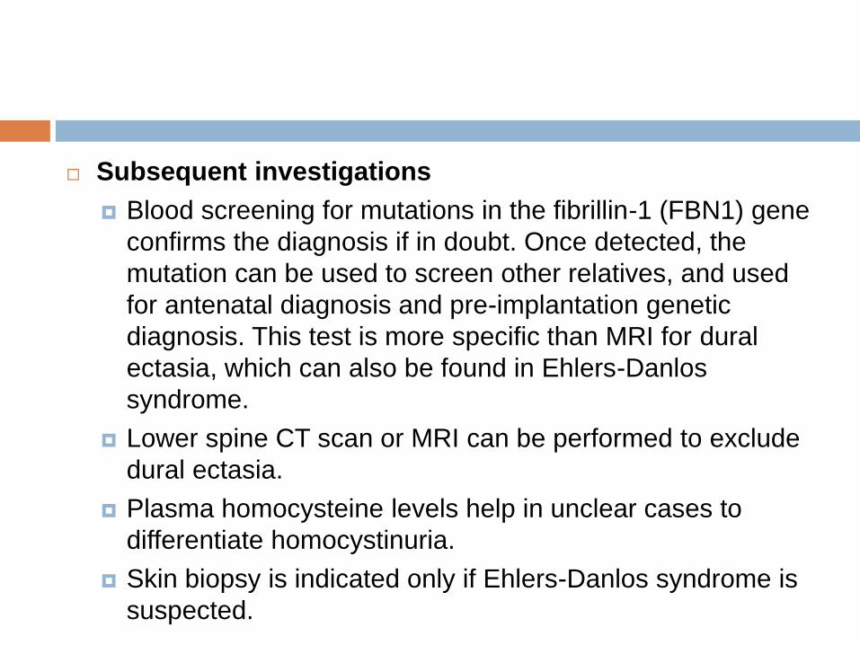
Subsequent investigations
Blood screening for mutations in the fibrillin-1 (FBN1) gene
confirms the diagnosis if in doubt. Once detected, the
mutation can be used to screen other relatives, and used
for antenatal diagnosis and pre-implantation genetic
diagnosis. This test is more specific than MRI for dural
ectasia, which can also be found in Ehlers-Danlos
syndrome.
Lower spine CT scan or MRI can be performed to exclude
dural ectasia.
Plasma homocysteine levels help in unclear cases to
differentiate homocystinuria.
Skin biopsy is indicated only if Ehlers-Danlos syndrome is
suspected.

Summary and
Recommendations
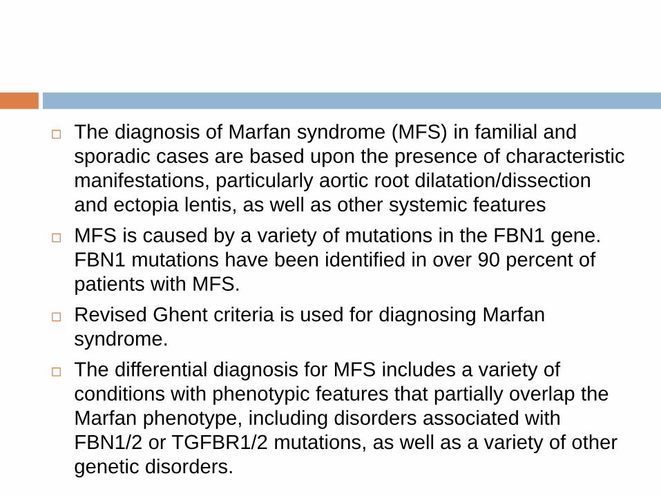
The diagnosis of Marfan syndrome (MFS) in familial and
sporadic cases are based upon the presence of characteristic
manifestations, particularly aortic root dilatation/dissection
and ectopia lentis, as well as other systemic features
MFS is caused by a variety of mutations in the FBN1 gene.
FBN1 mutations have been identified in over 90 percent of
patients with MFS.
Revised Ghent criteria is used for diagnosing Marfan
syndrome.
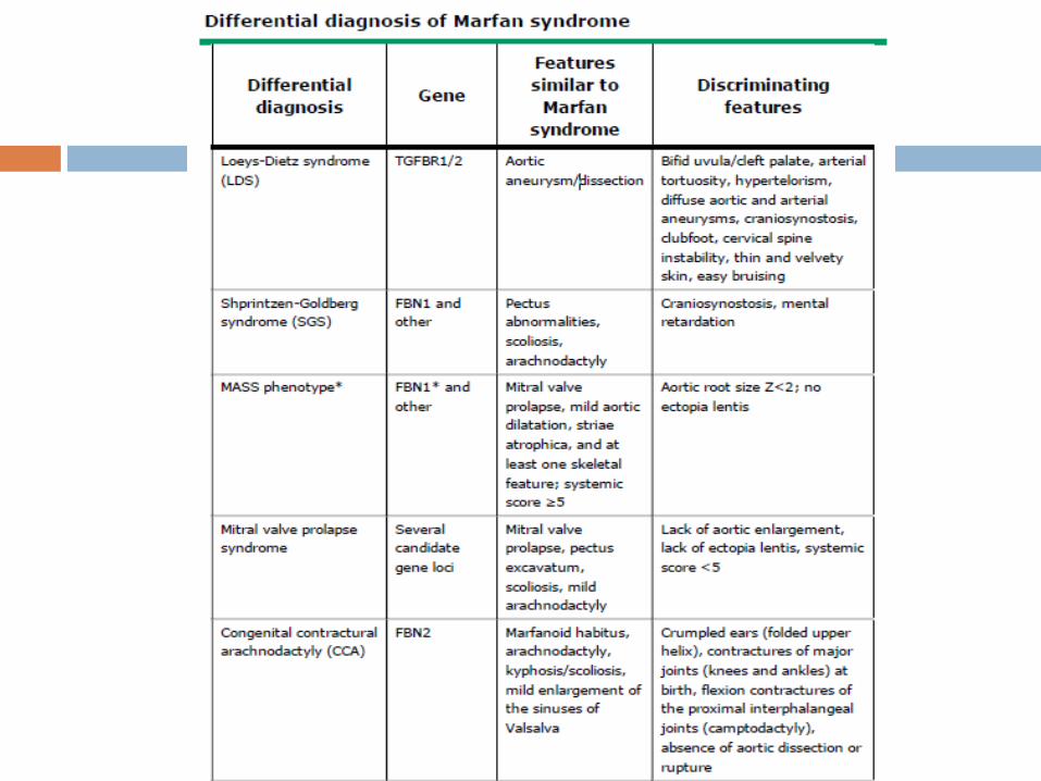
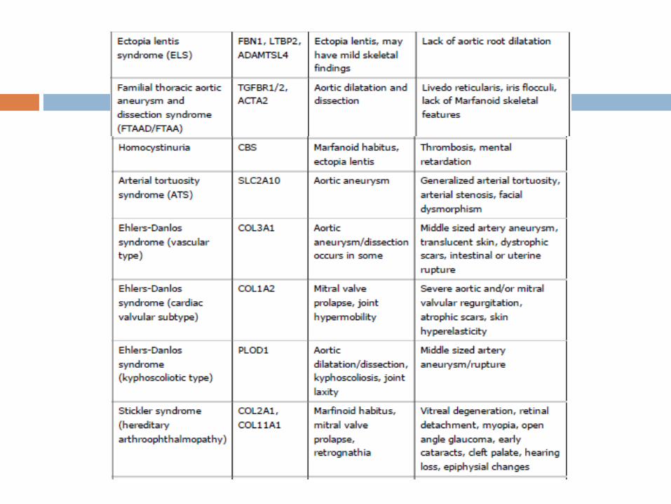
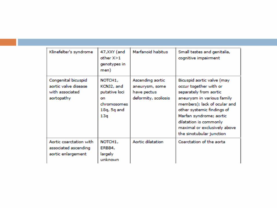
The differential diagnosis for MFS includes a variety of
conditions with phenotypic features that partially overlap the
Marfan phenotype, including disorders associated with
FBN1/2 or TGFBR1/2 mutations, as well as a variety of other
genetic disorders.
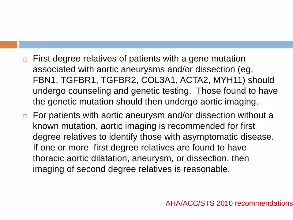
First degree relatives of patients with a gene mutation
associated with aortic aneurysms and/or dissection (eg,
FBN1, TGFBR1, TGFBR2, COL3A1, ACTA2, MYH11) should
undergo counseling and genetic testing. Those found to have
the genetic mutation should then undergo aortic imaging.
For patients with aortic aneurysm and/or dissection without a
known mutation, aortic imaging is recommended for first
degree relatives to identify those with asymptomatic disease.
If one or more first degree relatives are found to have
thoracic aortic dilatation, aneurysm, or dissection, then
imaging of second degree relatives is reasonable.
AHA/ACC/STS 2010 recommendations
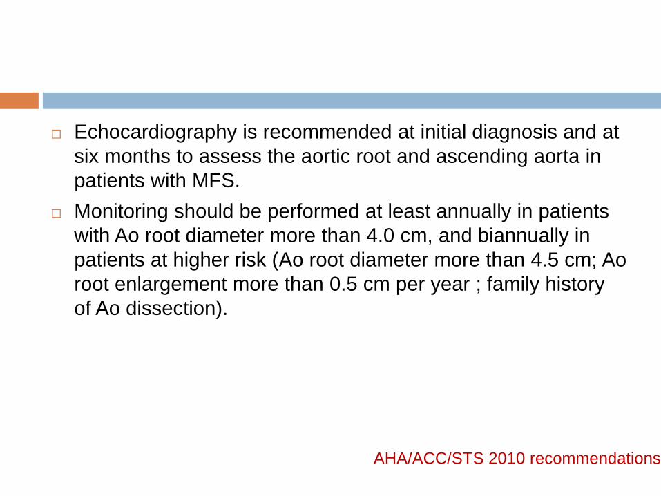
Echocardiography is recommended at initial diagnosis and at
six months to assess the aortic root and ascending aorta in
patients with MFS.
Monitoring should be performed at least annually in patients
with Ao root diameter more than 4.0 cm, and biannually in
patients at higher risk (Ao root diameter more than 4.5 cm; Ao
root enlargement more than 0.5 cm per year ; family history
of Ao dissection).
AHA/ACC/STS 2010 recommendations