Embed Size (px)
Citation preview
Male Genital System(Part-II)
Congenital anomalies of Testis
• Cryptorchidism :Failure of descent of testis from the abdominal cavity through theinguinal canal.
• Causes- Most common idiopathic
• Epidemiology– about1% of males– right > left, 25% bilateral
• Pathogenesis– Hormonal abnormalities– Testicular abnormalities– Mechanical problems
• Clinical course
– Testicular descent occurs in two morphologically and hormonally distinct phases.
– During the first, the transabdominal, phase, the testis comes to lie within the lower abdomen or brim of the pelvis. This phase is believed to be controlled by a hormone called müllerian-inhibiting substance.
– In the second, or the inguinoscrotal, phase, the testes descend through the inguinal canal into the scrotal sac. This phase is androgen dependent
– When unilateral, may see atrophy in contralateral testis.– sterility– concomitant inguinal hernia– increased risk of testicular malignancy
• Morphology
– Atrophic changes by 2 yrs of age;
– Arrest in the development of germ cells
– Hyalinization and thickening of seminiferoustubules & interstitial fibrosis
– Sparing Leydig cells which become prominent
– With progressive tubular atrophy the testis becomes small and firm in consistency.
– Similar changes - contralateral descended testis
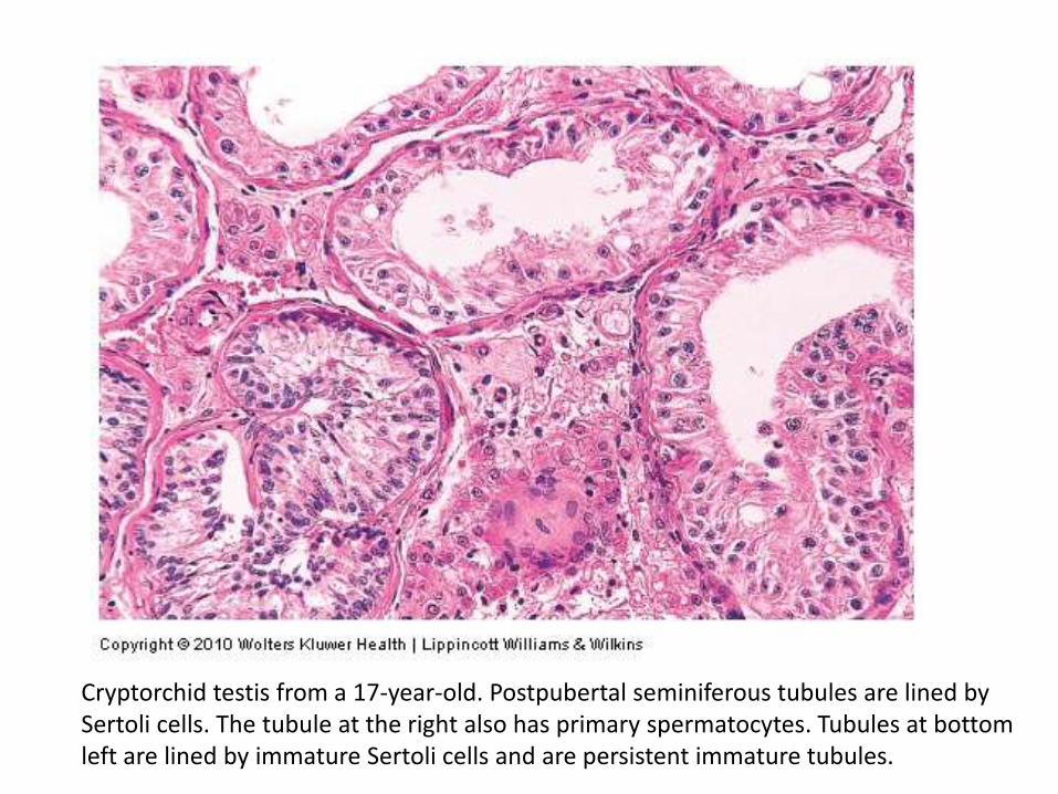
Cryptorchid testis from a 17-year-old. Postpubertal seminiferous tubules are lined by Sertoli cells. The tubule at the right also has primary spermatocytes. Tubules at bottom left are lined by immature Sertoli cells and are persistent immature tubules.
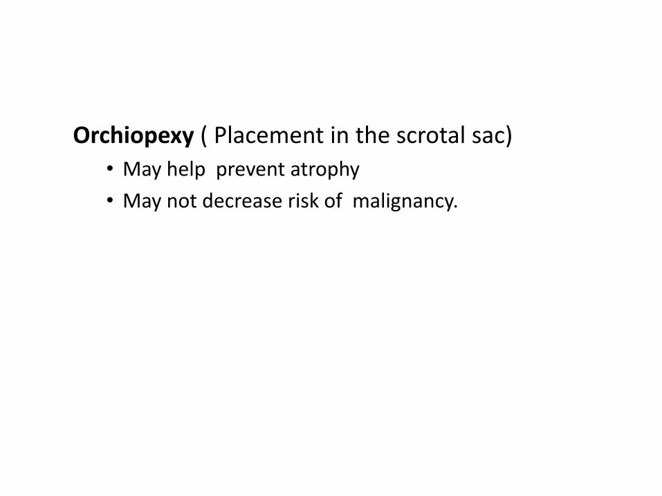
Orchiopexy ( Placement in the scrotal sac)
• May help prevent atrophy
• May not decrease risk of malignancy.
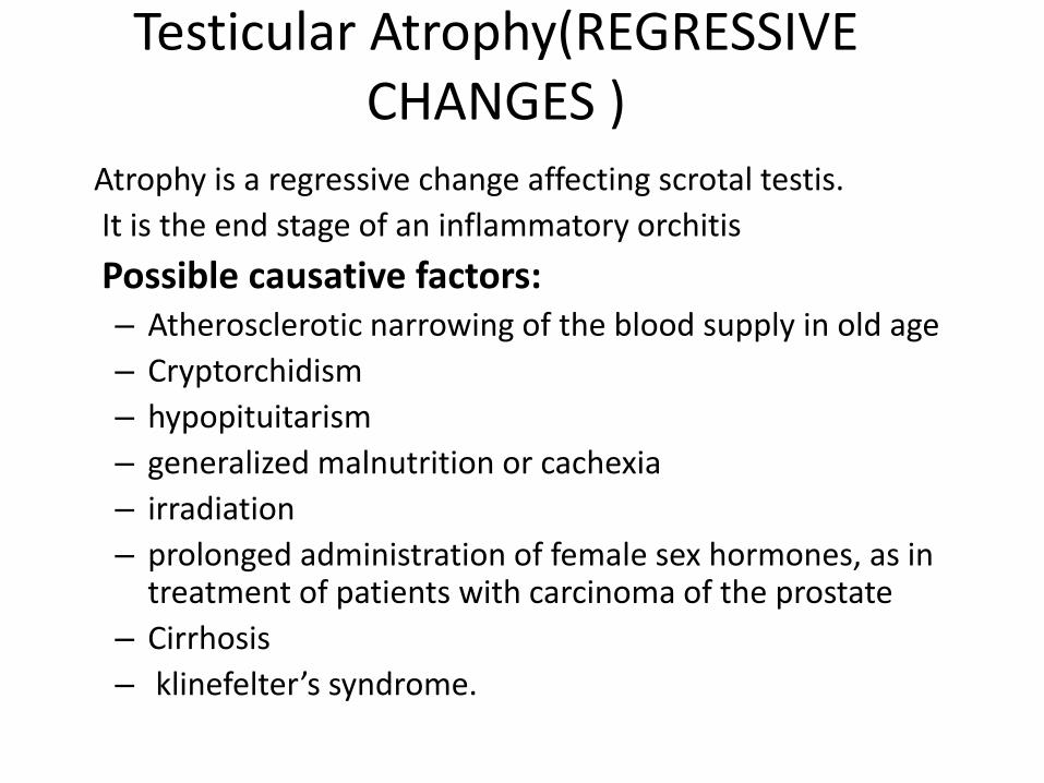
Testicular Atrophy(REGRESSIVE CHANGES )
Atrophy is a regressive change affecting scrotal testis.
It is the end stage of an inflammatory orchitis
Possible causative factors:– Atherosclerotic narrowing of the blood supply in old age
– Cryptorchidism
– hypopituitarism
– generalized malnutrition or cachexia
– irradiation
– prolonged administration of female sex hormones, as in treatment of patients with carcinoma of the prostate
– Cirrhosis
– klinefelter’s syndrome.
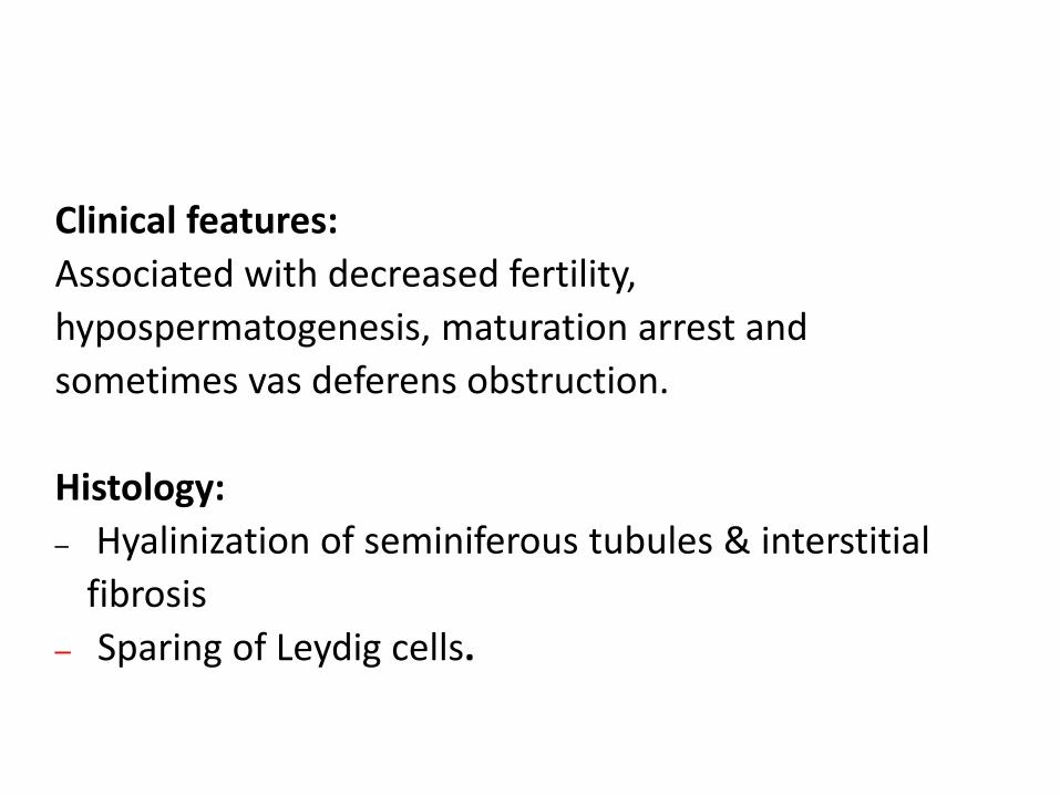
Clinical features:
Associated with decreased fertility,
hypospermatogenesis, maturation arrest and
sometimes vas deferens obstruction.
Histology:
– Hyalinization of seminiferous tubules & interstitial
fibrosis
– Sparing of Leydig cells.
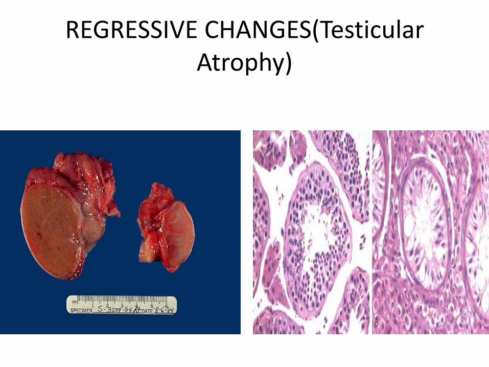
REGRESSIVE CHANGES(Testicular Atrophy)
INFLAMMATION
• Inflammations are distinctly more common in the epididymis than in the testis.
• Of the three major specific inflammatory states that affect the testis and epididymis, gonorrhea and tuberculosis almost invariably arise in the epididymis, whereas syphilis affects first the testis.
Nonspecific Epididymitis and Orchitis
• Epididymitis and possible subsequent orchitis are commonly related to infections in the urinary tract (cystitis, urethritis, prostatitis), which reach the epididymis and the testis through either the vas deferens or the lymphatics of the spermatic cord.
• The cause of epididymitis varies with the age of the patient. Though uncommon in children, epididymitis in childhood is usually associated with a congenital genitourinary abnormality and infection with gram-negative rods .
• In sexually active men younger than age 35 years, the sexually transmitted pathogens C. trachomatis and Neisseria gonorrhoeaeare the most frequent culprits. In men older than age 35 the common urinary tract pathogens, such as E. coli and Pseudomonas, are responsible for most infections.
• Morphology. The bacterial invasion induces nonspecific acute inflammation characterized by congestion, edema, and infiltration by neutrophils, macrophages, and lymphocytes.
• Although the infection, in the early stage, is more or less limited to the interstitial connective tissue, it rapidly extends to involve the tubules and may progress to frank abscess formation or complete suppurative necrosis of the entire epididymis
• Such inflammatory involvement of the epididymis and testis is often followed by fibrous scarring, which in many cases leads to sterility.
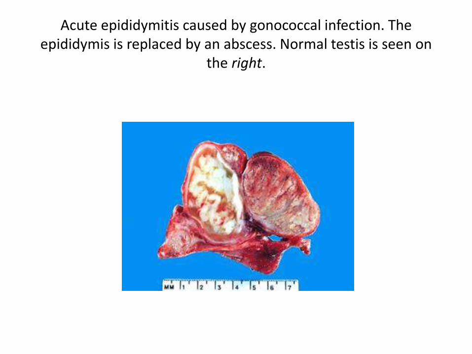
Acute epididymitis caused by gonococcal infection. The epididymis is replaced by an abscess. Normal testis is seen on
the right.
Granulomatous (Autoimmune) Orchitis
• Idiopathic granulomatous orchitis presents in middle age as a moderately tender testicular mass of sudden onset sometimes associated with fever.
• It may appear insidiously, however, as a painless testicular mass mimicking a testicular tumor, hence its importance.
• Histologically the orchitis is distinguished by granulomas restricted to spermatic tubules.
Specific Inflammations
• Gonorrhea Extension of infection from the posterior urethra to the prostate, seminal vesicles, and then to the epididymis is the usual course of a neglected gonococcal infection.Inflammatory changes similar to those described for nonspecific
infections occur, with the development of frank abscesses in the epididymis, which may lead to extensive destruction of this organ.
• Mumps Mumps is a systemic viral disease that most commonly affects school-aged children. Testicular involvement is extremely uncommon in this age group.In postpubertal males, however, orchitis may develop and has been
reported in 20% to 30% of male patients. Most often, acute interstitial orchitis develops about 1 week after the onset of swelling of the parotid glands.
• Tuberculosis Tuberculosis almost invariably begins in the epididymis and may spread to the testis.The infection invokes the classic morphologic reactions of caseating
granulomatous inflammation characteristic of tuberculosis elsewhere.
• Syphilis The testis and epididymis are affected in both acquired and congenital syphilis, but almost invariably the testis is involved first by the infection. In many cases, the orchitis is not accompanied by epididymitis. The morphologic pattern of the reaction takes two forms: the production of gummas or a diffuse interstitial inflammation characterized by edema and lymphocytic and plasma cell infiltration with the characteristic hallmark of all syphilitic infections
VASCULAR DISEASES(Torsion of testis)
• Twisting of the spermatic cord which typically cuts off the venous drainage of the testis
• Bilateral anatomic defect where the testis has increased mobility giving rise to “bell-clapper” abnormality
• Infracted testicle and epididymis due to torsion• Neonatal torsion occurs either in utero or shortly after birth. It lacks
any associated anatomic defect to account for its occurrence.• Adult torsion is typically seen in adolescence presenting as sudden
onset of testicular pain. It often occurs without any inciting injury; sudden pain heralding the torsion may even occur during sleep.
• Morphology. Depending on the duration of the process, the morphologic changes range from intense congestion to widespread extravasation of blood into the interstitial tissue to hemorrhagic testicular infarction
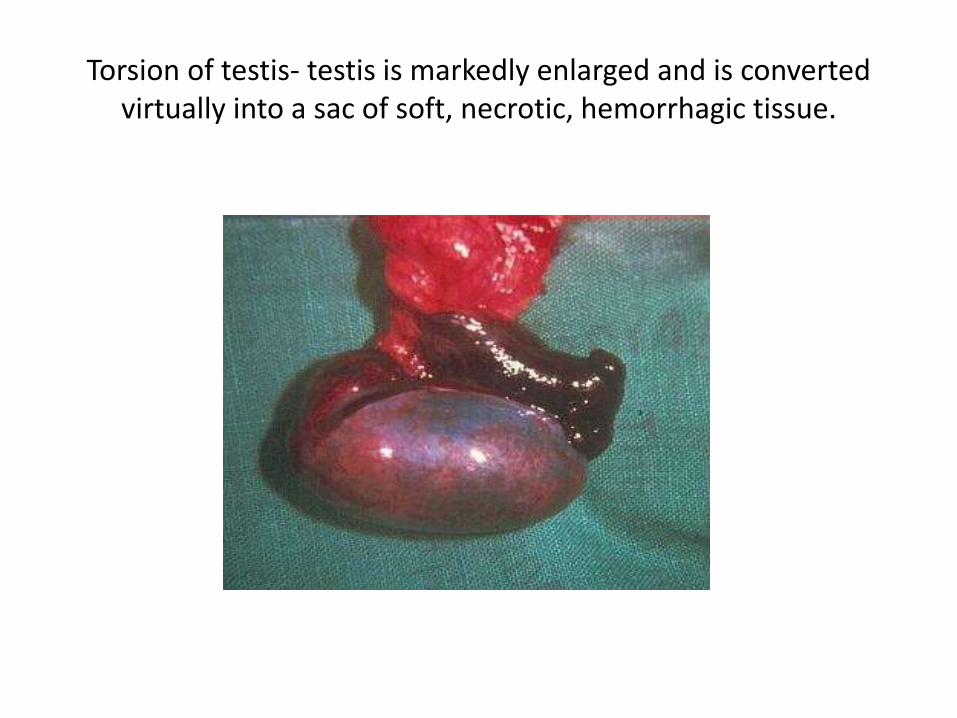
Torsion of testis- testis is markedly enlarged and is converted virtually into a sac of soft, necrotic, hemorrhagic tissue.
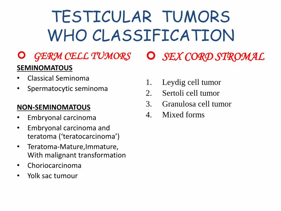
TESTICULAR TUMORSWHO CLASSIFICATION
GERM CELL TUMORSSEMINOMATOUS
• Classical Seminoma
• Spermatocytic seminoma
NON-SEMINOMATOUS
• Embryonal carcinoma
• Embryonal carcinoma and teratoma (‘teratocarcinoma’)
• Teratoma-Mature,Immature,With malignant transformation
• Choriocarcinoma
• Yolk sac tumour
SEX CORD STROMAL TUMORS
1. Leydig cell tumor
2. Sertoli cell tumor
3. Granulosa cell tumor
4. Mixed forms
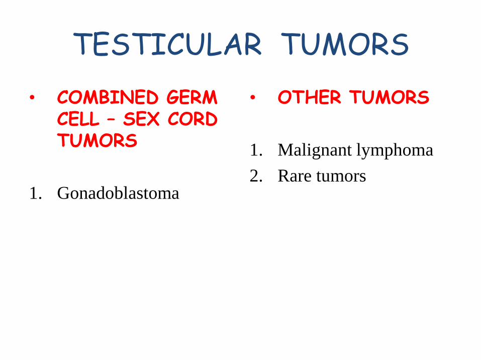
TESTICULAR TUMORS
• COMBINED GERM CELL – SEX CORD TUMORS
1. Gonadoblastoma
• OTHER TUMORS
1. Malignant lymphoma
2. Rare tumors
SEMINOMA
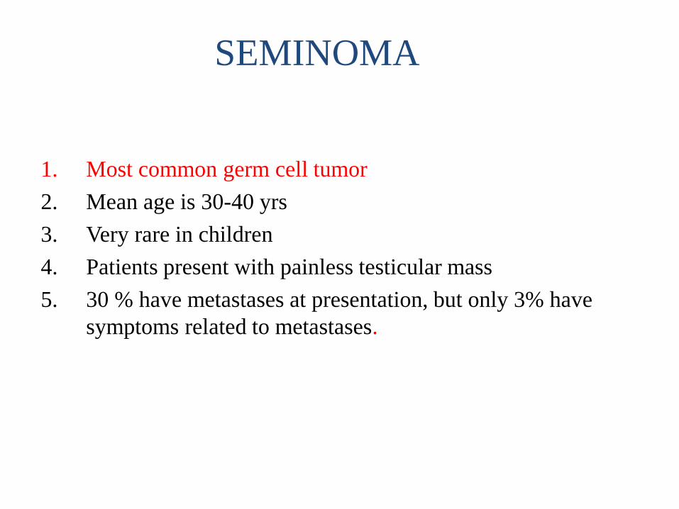
1. Most common germ cell tumor
2. Mean age is 30-40 yrs
3. Very rare in children
4. Patients present with painless testicular mass
5. 30 % have metastases at presentation, but only 3% have
symptoms related to metastases.
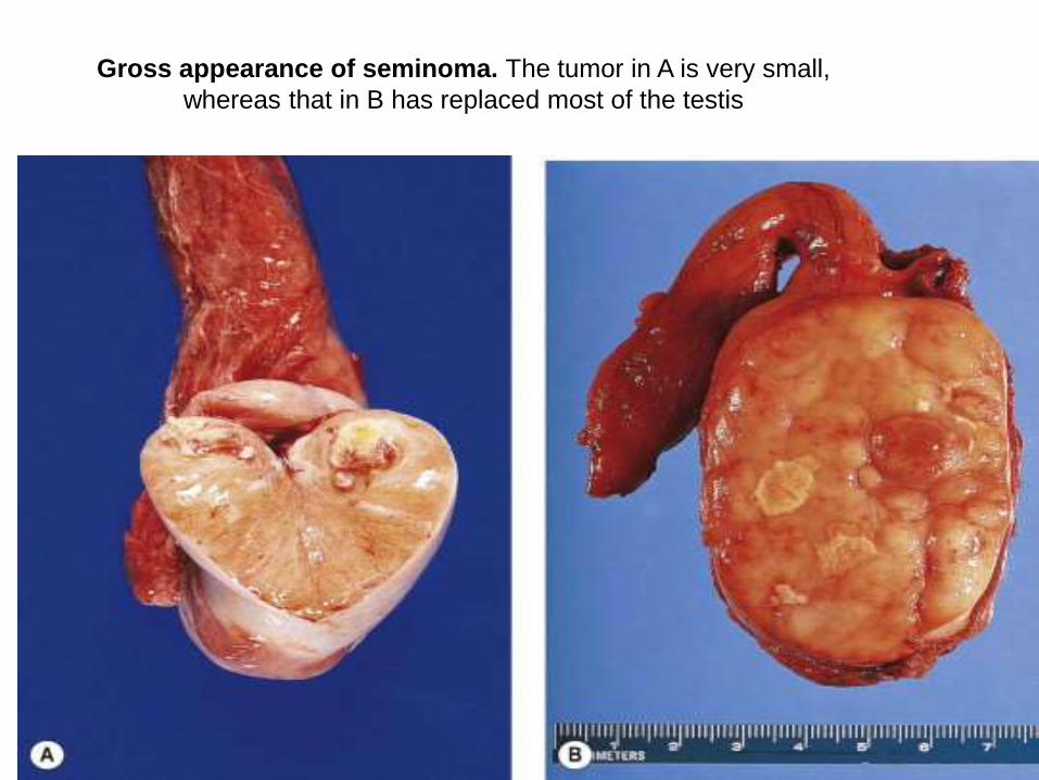
Gross appearance of seminoma. The tumor in A is very small,
whereas that in B has replaced most of the testis
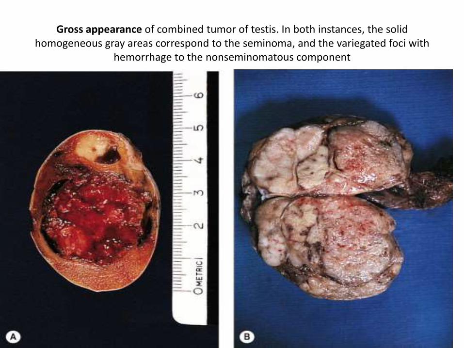
Gross appearance of combined tumor of testis. In both instances, the solid homogeneous gray areas correspond to the seminoma, and the variegated foci with
hemorrhage to the nonseminomatous component
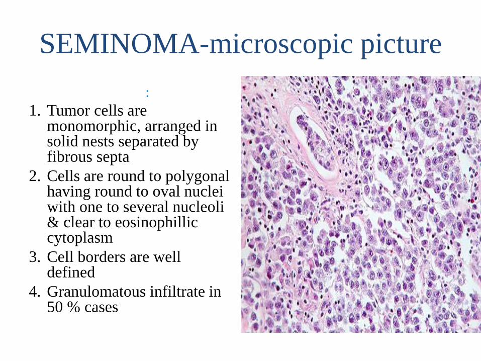
SEMINOMA-microscopic picture
• MICROSCOPIC :
1. Tumor cells are monomorphic, arranged in solid nests separated by fibrous septa
2. Cells are round to polygonal having round to oval nuclei with one to several nucleoli & clear to eosinophilliccytoplasm
3. Cell borders are well defined
4. Granulomatous infiltrate in 50 % cases
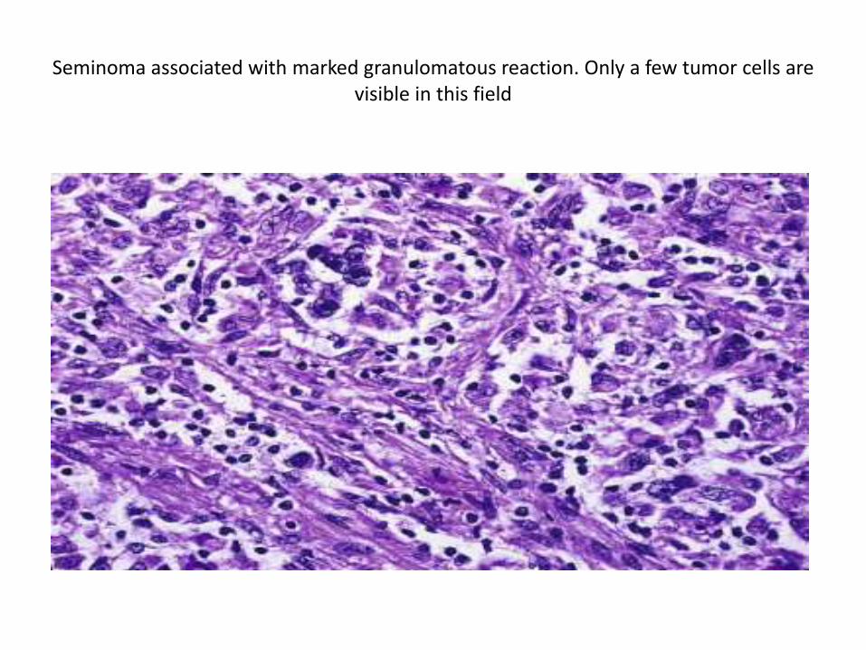
Seminoma associated with marked granulomatous reaction. Only a few tumor cells are visible in this field
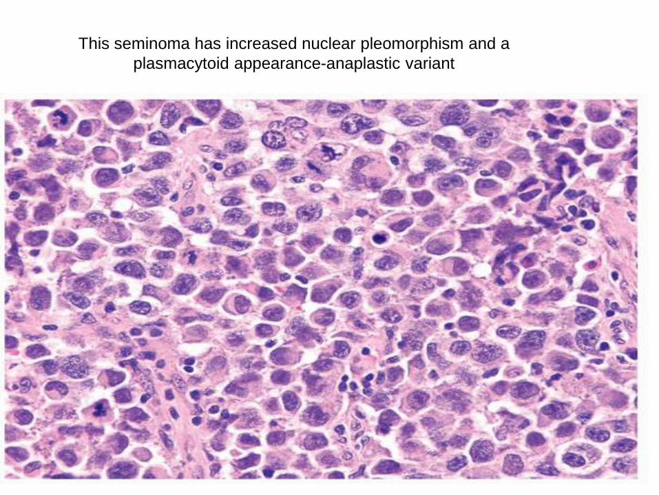
This seminoma has increased nuclear pleomorphism and a
plasmacytoid appearance-anaplastic variant
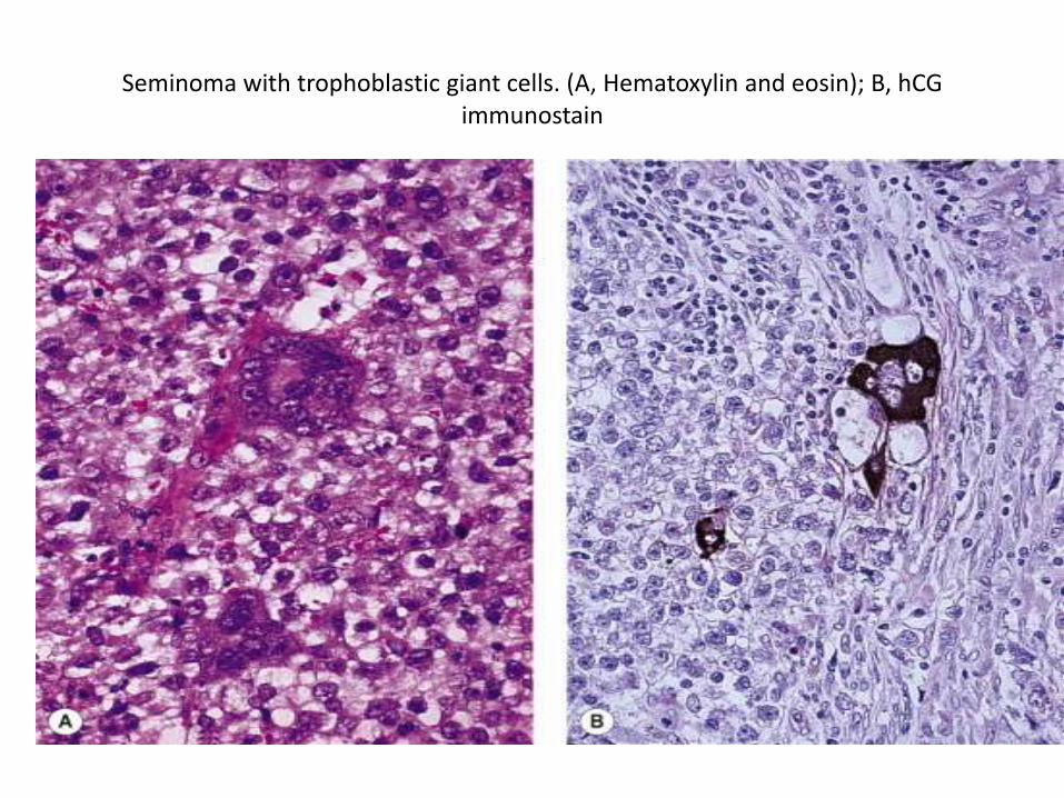
Seminoma with trophoblastic giant cells. (A, Hematoxylin and eosin); B, hCGimmunostain
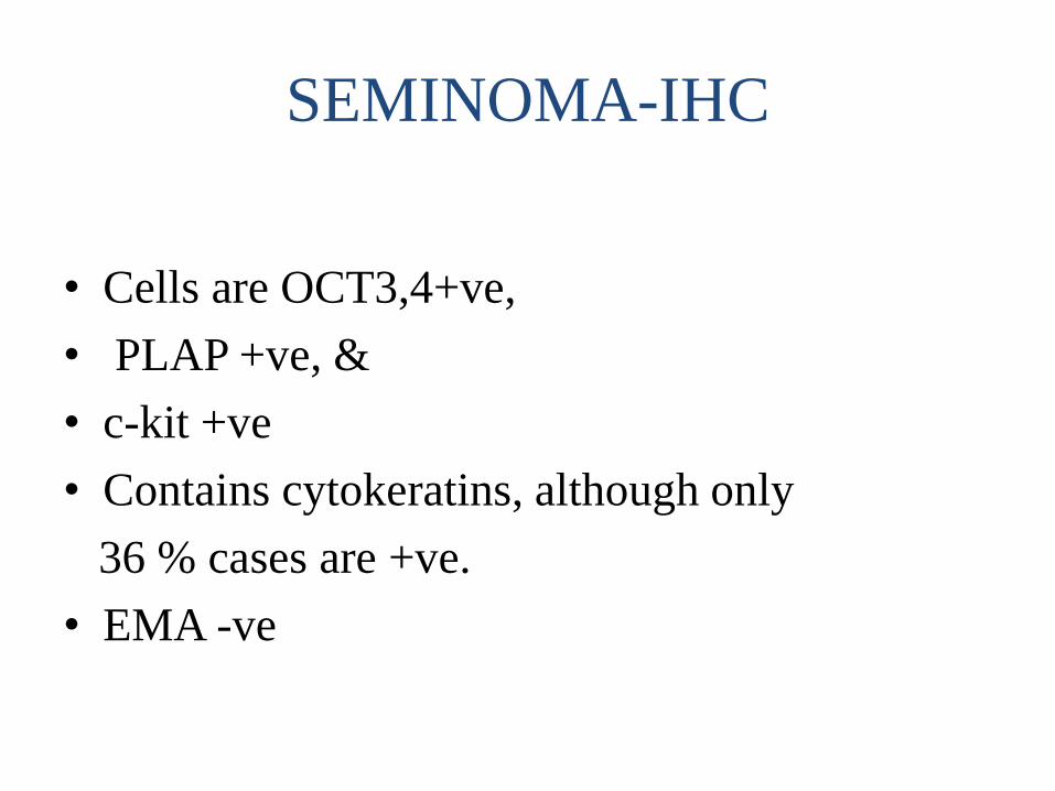
SEMINOMA-IHC
• IMMUNOHISTO CHEMISTRY
• Cells are OCT3,4+ve,
• PLAP +ve, &
• c-kit +ve
• Contains cytokeratins, although only
36 % cases are +ve.
• EMA -ve
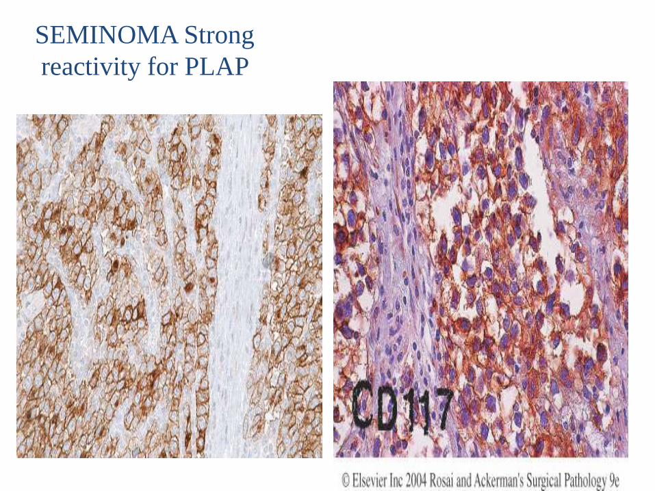
SEMINOMA Strong
reactivity for PLAP
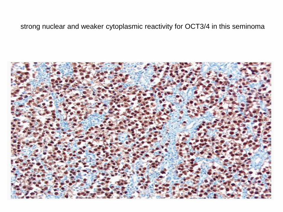
strong nuclear and weaker cytoplasmic reactivity for OCT3/4 in this seminoma
SPERMATOCYTIC SEMINOMA
1. Occurs only in testis & represents 2 % of germ cell
tumors
2. Patients are in 60s & present with testicular mass
3. Very rarely metastasize.
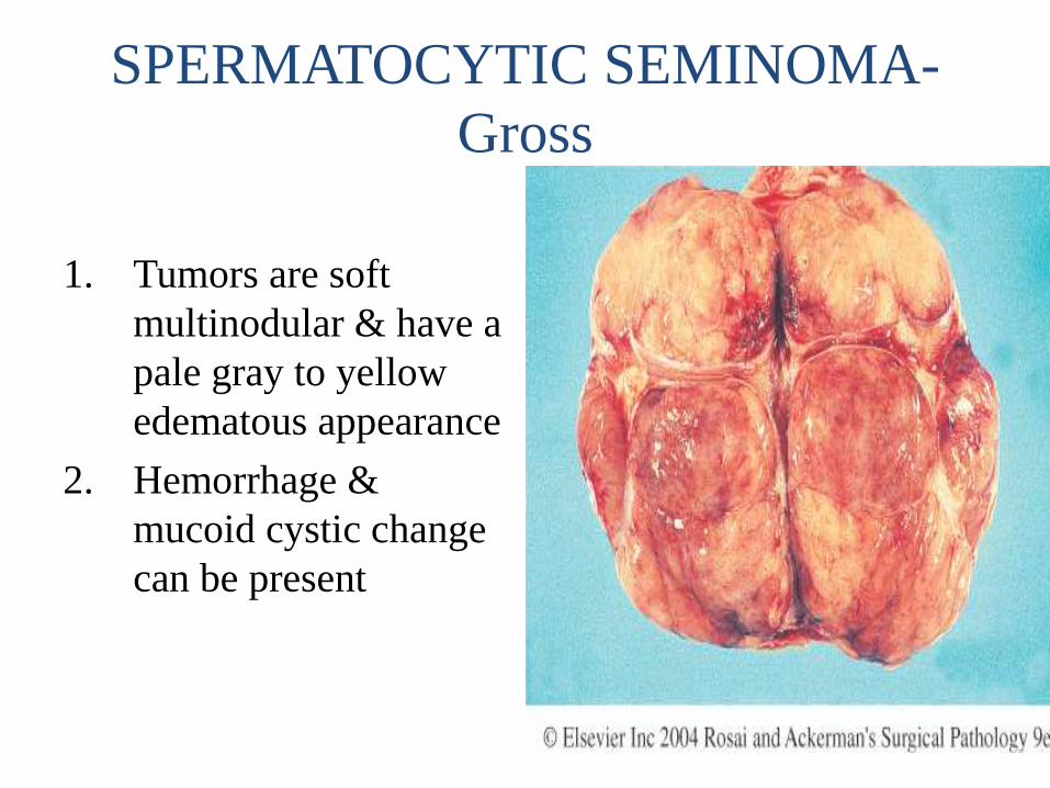
SPERMATOCYTIC SEMINOMA-
Gross
• MACROSCOPIC
1. Tumors are soft
multinodular & have a
pale gray to yellow
edematous appearance
2. Hemorrhage &
mucoid cystic change
can be present
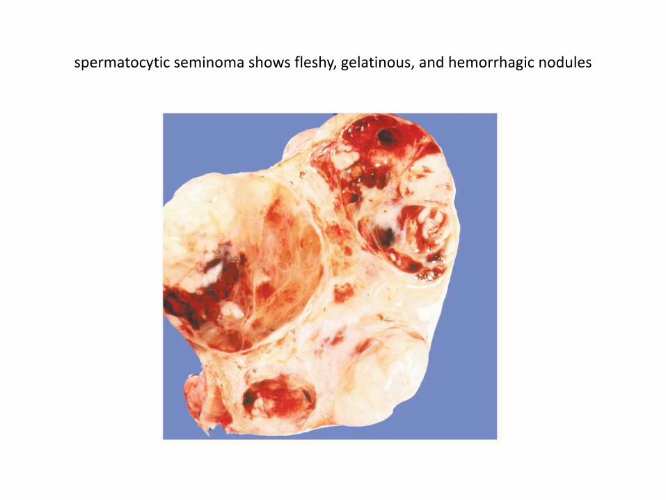
spermatocytic seminoma shows fleshy, gelatinous, and hemorrhagic nodules
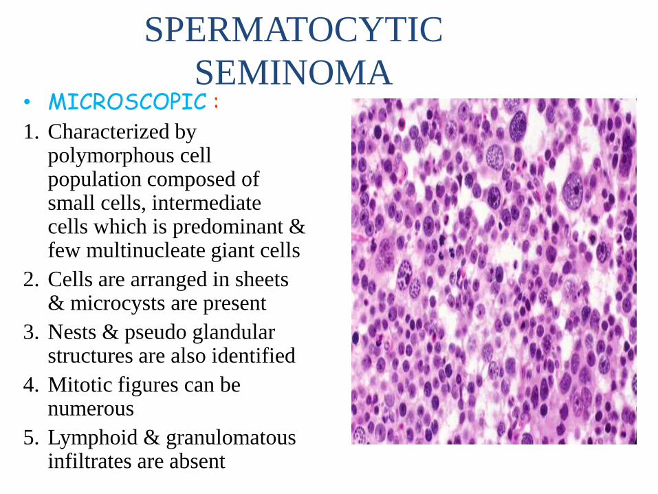
SPERMATOCYTIC
SEMINOMA• MICROSCOPIC :1. Characterized by
polymorphous cell population composed of small cells, intermediate cells which is predominant & few multinucleate giant cells
2. Cells are arranged in sheets & microcysts are present
3. Nests & pseudo glandular structures are also identified
4. Mitotic figures can be numerous
5. Lymphoid & granulomatousinfiltrates are absent
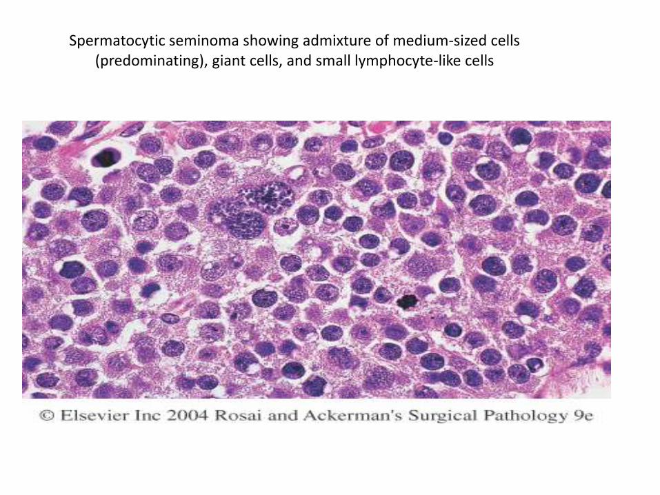
Spermatocytic seminoma showing admixture of medium-sized cells (predominating), giant cells, and small lymphocyte-like cells
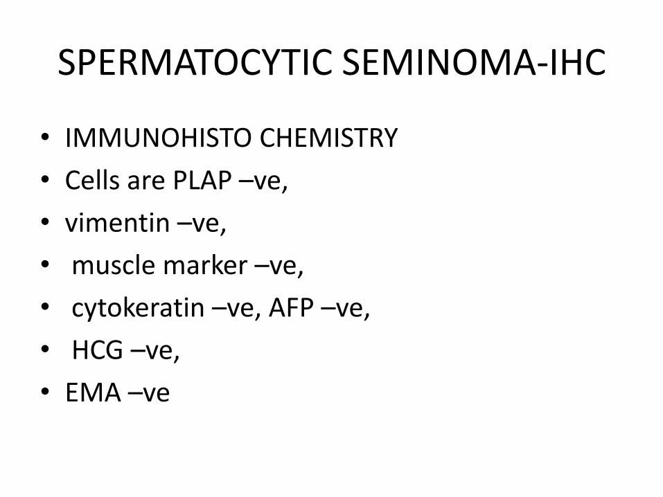
SPERMATOCYTIC SEMINOMA-IHC
• IMMUNOHISTO CHEMISTRY
• Cells are PLAP –ve,
• vimentin –ve,
• muscle marker –ve,
• cytokeratin –ve, AFP –ve,
• HCG –ve,
• EMA –ve
EMBRYONAL CARCINOMA
1. 2nd most common germ cell tumor,
comprising approx. 20 % cases
2. Present in majority of mixed germ cell
tumors
3. Most men present in their 20s to 30s with a
testicular mass
4. More than 2/3rds of patients have
metastases, but only 10 % have symptom
related to metastases.
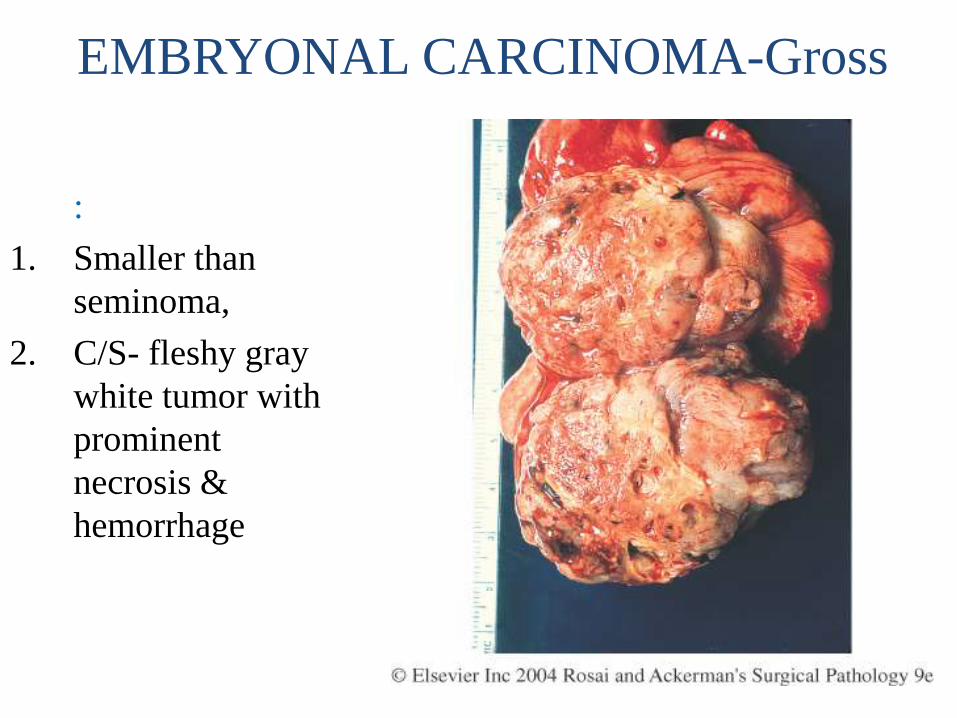
EMBRYONAL CARCINOMA-Gross
• MACROSCOPIC
:
1. Smaller than
seminoma,
2. C/S- fleshy gray
white tumor with
prominent
necrosis &
hemorrhage
EMBRYONAL CARCINOMA-
microscopy• MICROSCOPIC :1. Cells are large with vesicular nuclei, prominent nucleoli, &
indistinct cell borders
2. Tumor cells are arranged in sheets, cords & glandular structure
3. Necrosis & hemorrhage may be prominent
4. May be intimately admixed with a yolk sac tumor
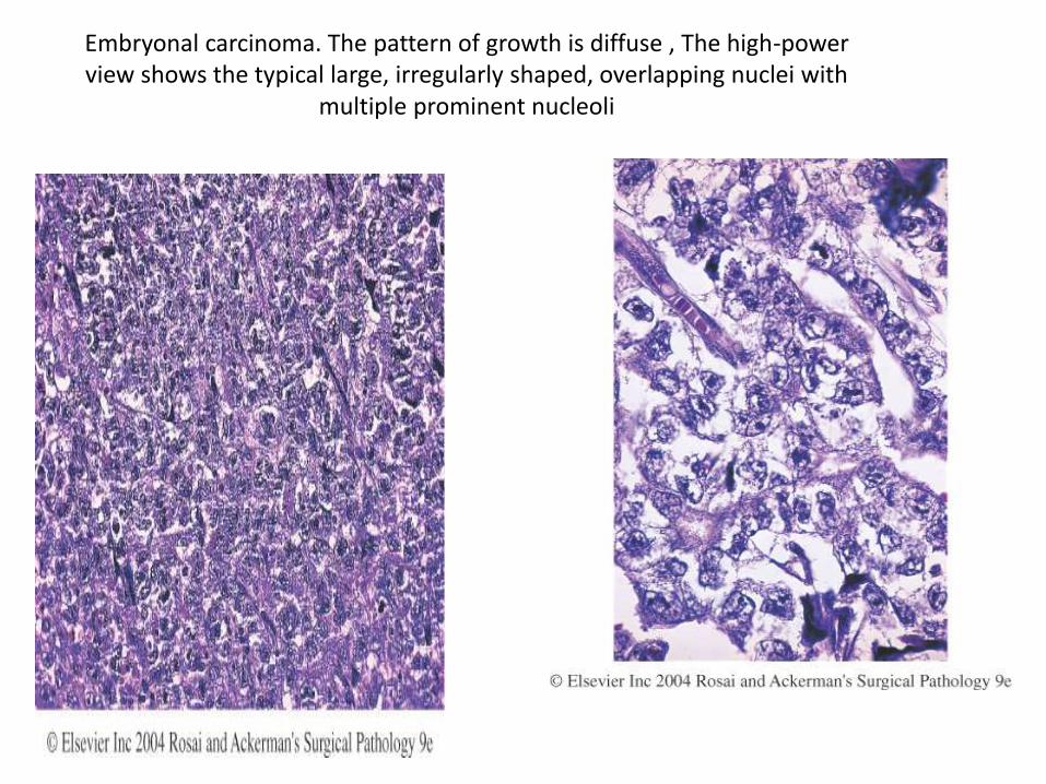
Embryonal carcinoma. The pattern of growth is diffuse , The high-power view shows the typical large, irregularly shaped, overlapping nuclei with
multiple prominent nucleoli
EMBRYONAL CARCINOMA-
IHC
• IMMUNOHISTO CHEMISTRY
• Tumor cells are CD 30 +ve, a finding unique to Embryonal carcinoma, and useful in ruling out solid pattern of Embryonal carcinoma, which can simulate Seminoma .
• OCT 4 +ve,
• PLAP +ve,
• cytokeratin +ve,
c-kit –ve, and EMA -ve
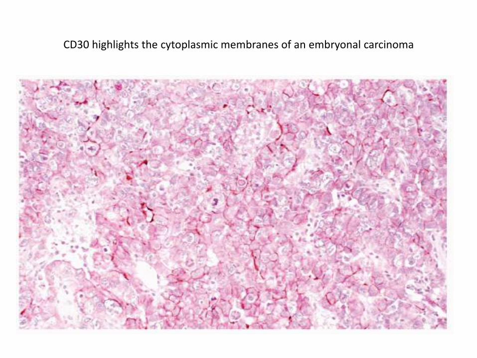
CD30 highlights the cytoplasmic membranes of an embryonal carcinoma
Choriocarcinoma
• Choriocarcinoma is a highly malignant form of testicular tumor. In its “pure” form choriocarcinoma is rare, constituting less than 1% of all germ cell tumors.
• Morphology. Often they cause no testicular enlargement and are detected only as a small palpable nodule. Typically, these tumors are small, rarely larger than 5 cm in diameter. Hemorrhage and necrosis are extremely common.
• Histologically the tumors contain two cell types- cytotrophoblastsand syncytiotrophoblasts
• Syncytiotrophoblast appears as a large cell having many irregular or lobular hyperchromatic nuclei and an abundant eosinophilicvacuolated cytoplasm
• Cytotrophoblasts are more regular and polygonal with distinct borders and clear cytoplasm.
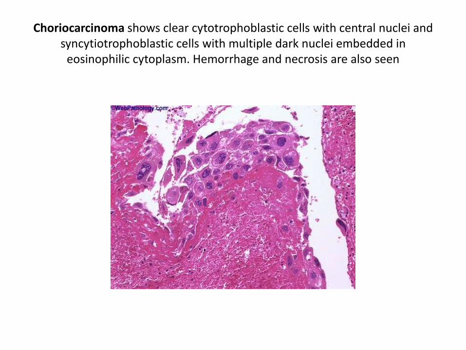
Choriocarcinoma shows clear cytotrophoblastic cells with central nuclei and syncytiotrophoblastic cells with multiple dark nuclei embedded in
eosinophilic cytoplasm. Hemorrhage and necrosis are also seen
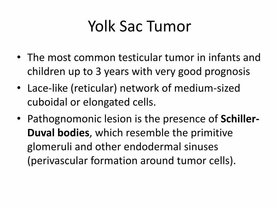
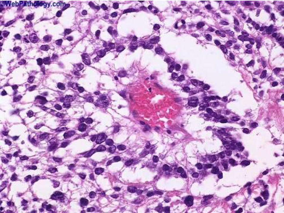
Yolk Sac Tumor
• The most common testicular tumor in infants and children up to 3 years with very good prognosis
• Lace-like (reticular) network of medium-sized cuboidal or elongated cells.
• Pathognomonic lesion is the presence of Schiller-Duval bodies, which resemble the primitive glomeruli and other endodermal sinuses (perivascular formation around tumor cells).
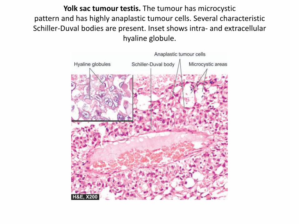
Yolk sac tumour testis. The tumour has microcysticpattern and has highly anaplastic tumour cells. Several characteristicSchiller-Duval bodies are present. Inset shows intra- and extracellular
hyaline globule.
Teratoma
• The designation teratoma refers to a group of complex testicular tumors having various cellular or organoid components reminiscent of normal derivatives from more than one germ layer.
• They may occur at any age from infancy to adult life. Pure forms of teratoma are fairly common in infants and children, second in frequency only to yolk sac tumors.
• In adults, pure teratomas are rare, constituting 2% to 3% of germ cell tumors.
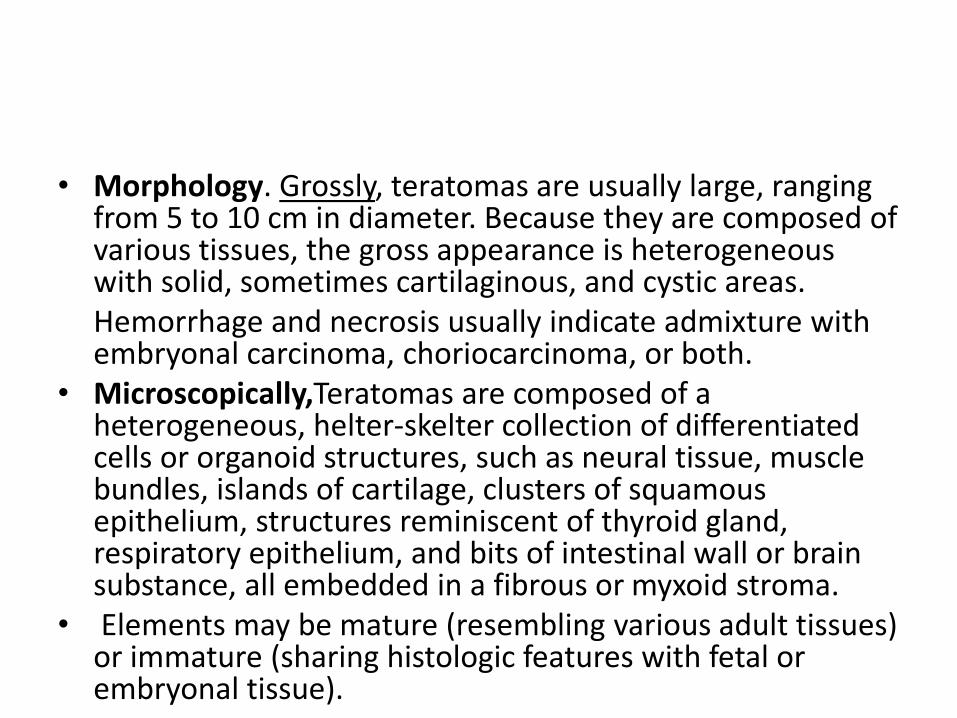
• Morphology. Grossly, teratomas are usually large, ranging from 5 to 10 cm in diameter. Because they are composed of various tissues, the gross appearance is heterogeneous with solid, sometimes cartilaginous, and cystic areas. Hemorrhage and necrosis usually indicate admixture with embryonal carcinoma, choriocarcinoma, or both.
• Microscopically,Teratomas are composed of a heterogeneous, helter-skelter collection of differentiated cells or organoid structures, such as neural tissue, muscle bundles, islands of cartilage, clusters of squamousepithelium, structures reminiscent of thyroid gland, respiratory epithelium, and bits of intestinal wall or brain substance, all embedded in a fibrous or myxoid stroma.
• Elements may be mature (resembling various adult tissues) or immature (sharing histologic features with fetal or embryonal tissue).
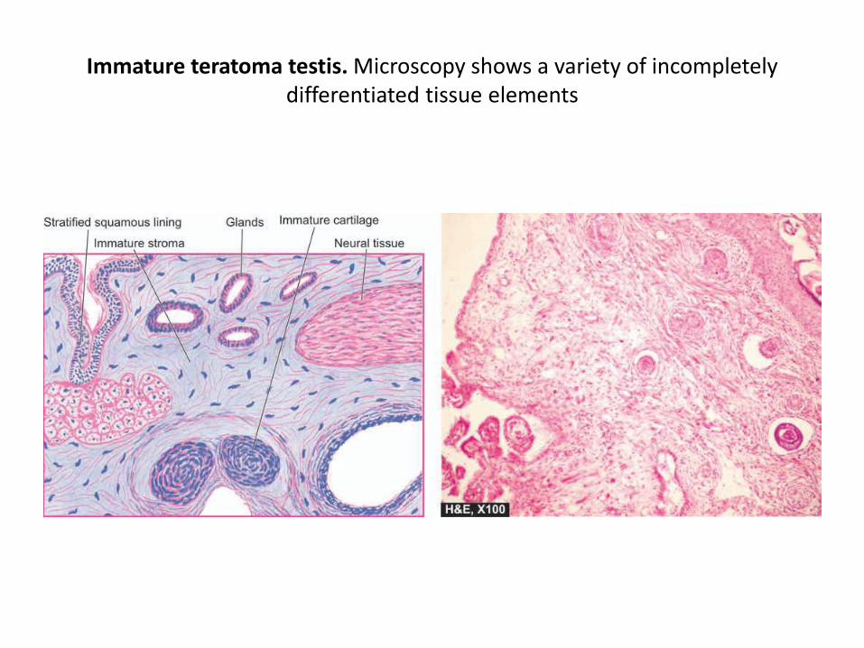
Immature teratoma testis. Microscopy shows a variety of incompletely differentiated tissue elements
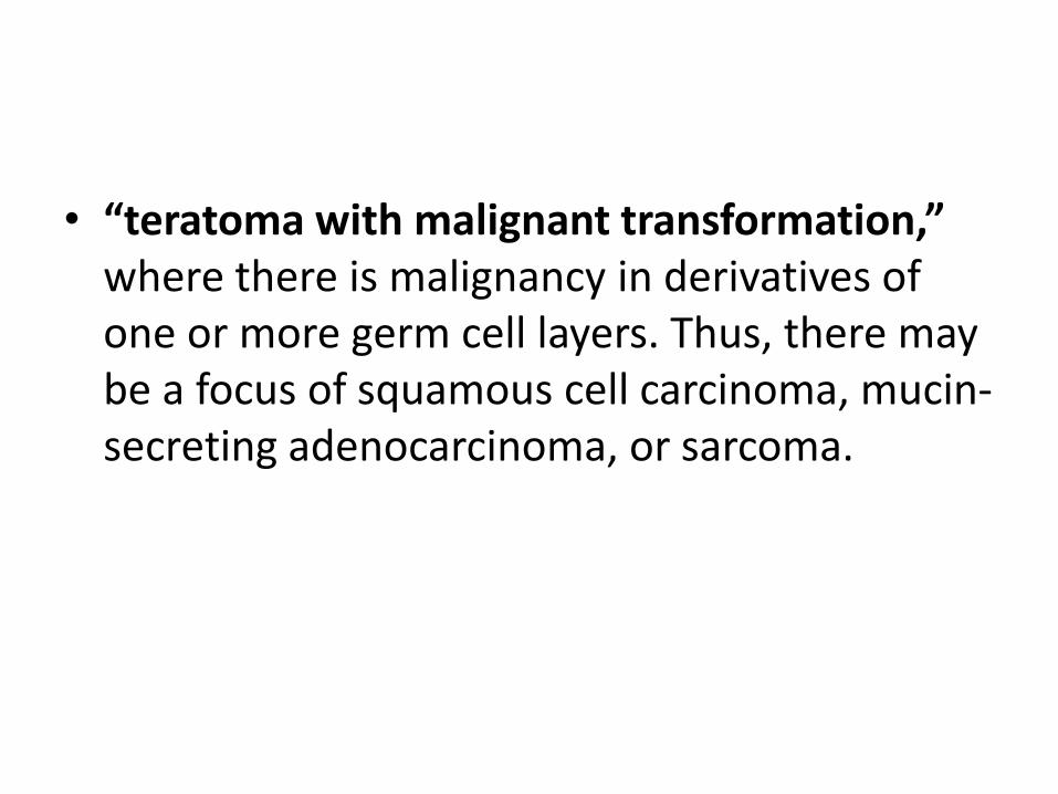
• “teratoma with malignant transformation,” where there is malignancy in derivatives of one or more germ cell layers. Thus, there may be a focus of squamous cell carcinoma, mucin-secreting adenocarcinoma, or sarcoma.
Tumors of Sex Cord–Gonadal Stroma
• Sex cord–gonadal stroma tumors are subclassifiedbased on their presumed histogenesis and differentiation. The two most important members of this group—Leydig cell tumors and Sertoli cell tumors.
Leydig Cell Tumors • Tumors of Leydig cells are particularly interesting,
because they may elaborate androgens and in some cases both androgens and estrogens, and even corticosteroids.
• They may arise at any age, although most cases occur between 20 and 60 years of age.
• As with other testicular tumors, the most common presenting feature is testicular swelling, but in some patients gynecomastia may be the first symptom.
• Morphology. These neoplasms form circumscribed nodules, usually less than 5 cm in diameter. They have a distinctive golden brown, homogeneous cut surface. Histologically, neoplastic Leydig cells usually are remarkably similar to their normal counterparts in that they are large and round or polygonal, and they have an abundant granular eosinophilic cytoplasm with a round central nucleus. The cytoplasm frequently contains lipid granules, vacuoles, or lipofuscin pigment, and, most characteristically, rod-shaped crystalloids of Reinke occur in about 25% of the tumors.Approximately 10% of the tumors in adults are invasive and produce metastases; most are benign.
Sertoli Cell Tumors
• Most Sertoli cell tumors are hormonally silent and present as a testicular mass.
• Morphology. These neoplasms appear as firm, small nodules with a homogeneous gray-white to yellow cut surface. Histologically the tumor cells are arranged in distinctive trabeculae that tend to form cordlike structures and tubules. Most Sertoli cell tumors are benign, but occasional tumors (∼10%) pursue a malignant course.
Gonadoblastoma• Gonadoblastomas are rare neoplasms containing a mixture of germ cells and
gonadal stromal elements, that almost always arise in gonads with some form of testicular dysgenesis .
• In some cases the germ cell component becomes malignant, giving rise to seminoma.
Testicular Lymphoma • Although an uncommon tumor of the testis, testicular lymphoma is included here
because affected patients present with only a testicular mass, mimicking other, more common, testicular tumors.
• Aggressive non-Hodgkin lymphomas account for 5% of testicular neoplasms, and are the most common form of testicular neoplasms in men over the age of 60. In most cases, the disease is already disseminated at the time of detection.
• The most common testicular lyphomas, in decreasing order of frequency, are diffuse large B cell lymphoma, Burkitt Lymphoma, and EBV-positive extranodalNK/T cell lymphoma.
• Patients with testicular lymphomas have a higher incidence of central nervous system involvement than those similar tumors located elsewhere.
MISCELLANEOUS LESIONS OF TUNICA VAGINALIS
• Tunica vaginalis is a mesothelial-lined surface exterior to the testis that may accumulate serous fluid (hydrocele) causing considerable enlargement of the scrotal sac. By transillumination it is usually possible to define the clear, translucent character of the contained fluid. Hydrocelesacs are frequently lined by mesothelial cells.
• Rarely, malignant mesotheliomas also can be seen arising from the tunica vaginalis.
• Hematocele indicates the presence of blood in the tunica vaginalis. It is an uncommon condition usually encountered only when there has been either direct trauma to the testis or torsion of the testis.
• Chylocele refers to the accumulation of lymph in the tunica and is almost always found in patients with elephantiasis who have widespread, severe lymphatic obstruction caused by filariasis.
• Spermatocele refers to a small cystic accumulation of semen in dilated efferent ducts or ducts of the rete testis.
• Varicocele is a dilated vein in the spermatic cord.

THANK YOU….