Embed Size (px)
Citation preview

Applied Anatomy
Physiology &
Pathology of
Lymphoreticular
system
Dr. Khaing zay aung
16. 2. 2015

The lymphatic system comprises:
Lymphatic chanals
Lymphoid organs &
(lymph nodes, Peyer’s patches, spleen, thymus &
tonsils)
Circulating elements
(lymphocytes and other mononuclear immune cells)

Developmental Anatomy
6 to 7 week of fetal life
The origin of the lymphatic vessels
is unclear
May arise from sac like outgrowth
of the endothelium of the veins

6 primary lymph sacs
2 JUGULAR
2 ILIAC
1 RETROPERITONEAL
1 CISTERNA CHILI

These lymph-sacs are developed by the
confluence of numerous venous capillaries
Which at first lose their connections with
the venous system
But subsequently, on the formation of the
sacs, regain them

Two main chanals
The rt and lt thoracic ducts
They join the jugular sacs with the cisterna chili
Drain into the venous system at the junction of the internal jugular vein and the subclavian vein
Numerous anastomoses produce many variations in the final form of the thoracic duct.

Development of thymus
Develops from the third and
fourth pharyngeal pouches
The stroma arises out of the
endodermal and also
ectodermal in origin

5. Bud of thymus


Development of tonsils
The tonsil buds appear with the formation
of the pharyngeal pouches
Located in the throat region
Palatine, lingual and unpaired pharyngeal
tonsils

8.Tonsilar buds

The lymphatic tissues of the intestine,
Payer’s patches, appear later than tonsils.
The second half of the pregnancy

Development of the lymphnodes

The origin of the lymphatic vessels is unclear
May arise from sac like outgrowth of the endothelium of the veins
The primary lymph nodes develop in regions that are occupied by lymphatic sacs.

Development of spleen
From the thickening of the visceral
mesothelium
Within it there is an accumulation of the
mesenchymal cells
Along the leftward shift of the stomach, it
resided on the left side of the abdomen
During the first trimester, macrophages and
precursor cells of erythropoiesis enter into
the spleen
After 15 wks of gestation, the white pulp and
red pulp appears

Development of lymphocytes
Largest part of the lymphocyte
development occurs in bone marrow,
thymus and the primary lymphatic organs
Large number of immunocompetent
lymphocytes are produced that colonize
the secondary lymphatic organs,
lymphnodes, tonsils, MALT and spleen
Distinguish into two types; T & B
lymphocytes


Anatomy of lymphatic system
the lymphatic system parallels the
cardiovascular system
One way system
Convey lymph from end organs to the
cardiovascular system

Lymph contains nutrients, oxygen,
hormones, fatty acids, toxins and waste
products.

Functions
Worked together with the immune system
As immune surveillance that Produce,
maintain, and distribute lymphocyte
Alternative route of Collection and
transportation of fluid , nutrient ,
proteins and hormones
Part in maintenance of normal blood
volume
(There is a small net movement of fluid
from the plasma into the interstitial fluid
along every systemic capillary. The total
volume is 3.6 l/day.)

Collection of lymph

Formation of lymph
ISF forms at the arterial end of the capillaries
Most of it returns to its venous ends and venules; the
rest (10—20%) enters the lymph capillaries as lymph.
As it flows through the lymph nodes, however, it comes
in contact with blood and tends to accumulate more
cells (particularly lymphocytes) and proteins.

Lymphatic vessels
Blind ended tubes
Endothelial lined (single layer)
Lymphatic capillaries coalesce to form
larger meshlike network of tubes k\a
lymphatic vessels


The lymphatic system

Lymphatic capillaries
Absent in bone, bone marrow, teeth,
CNS
Enter lymphatic collecting vessels
Lacteals – specialized lymph capillaries
present in intestinal mucosa
Absorb digested fat and deliver chyle
to the blood

Lymphnodes
Beanshaped structures
Throughout the lymphatic system
App: 600 to 700
Concentrated in the neck, axilla, groin, mediastinum & mesenteries of the GI tract.
Main line of defense by 2 types of cell lines
T & B lymphocytes

A lymph node has an outer capsule of
connective tissue from which trabeculae
pass into the deeper tissue.
Beneath the capsule is a space, the
subcapsular sinus into which the afferent
lymphatics drain after penetrating the
capsule.
Lymph from the subcapsular sinus passes
via the medullary cords to the hilum of
the lymph node from which the efferent
lymphatics drain.

Both afferent and efferent vessels have
valves which allow only forward flow.
The node consists of an outer cortex and an
inner medulla and contains lymphatic
sinuses.

Three distinct microanatomical
regions within a lymph node.
Cortex
Paracortex
Medulla

1. Cortex: which contains either primary
or secondary lymphoid follicles;
2. Paracortex: which is the T-cell
dependent region of the lymph node; and
3. Medulla: which contains the medullary
cords and sinuses and also contains
lymphocytes which are much less densely
packed than in the cortex, together with
macrophages, plasma cells and a small
number of granulocytes.

Cortex
consists of primary lymphoid follicles which
are unstimulated follicles, spherical in
shape, containing densely packed
lymphocytes.
Secondary follicles are present after
lymphocytes have been stimulated
antigenically.

These follicles have an outer ring of small
B lymphocytes surrounding the germinal
centre, which contains largely dividing
lymphoblasts, macrophages and dendritic
cells.

Antigen is trapped upon the surface of the dendritic cells and presented to ‘virgin’ B lymphocytes in the presence of T helper cells.
These B cells subsequently undergo a series of morphological and functional changes.
The function of germinal centre is to generate immunoglobulin-secreting plasma cells in response to antigenic challenge.


Paracortex
T-cell-dependent region of the lymph node.
When a T cell response occurs there is marked
proliferation of cells in this area.
contains large number of T lymphocytes with
a predominance of helper/inducer cells.
The cluster of differentiation (CD4) is
expressed by helper/inducer T cells.

Medulla
Lymph enters the marginal sinus of the node and drains
to the hilum through the sinuses which converge into
the medullary region.
The sinuses are lined by macrophages which
phagocytose foreign or abnormal particles from the
lymph passing through the node, i.e. the filtering
function.

Between the sinuses in the medulla lie
the medullary cords which contain
numerous plasma cells.
The medullary cords are one of the main
sites of antibody secretion within the
lymph node.


Waxing and waning of lymph
nodes
Enlargement on infections occurs in the
corresponding areas
Inflammation – swollen glands
Lymphadenopathy– chronic or excessive
enlargement of lymph nodes
They received the metastasizing cancer cells
Spread along the lymphatics
Nodal status is important
Lymphatic vessels
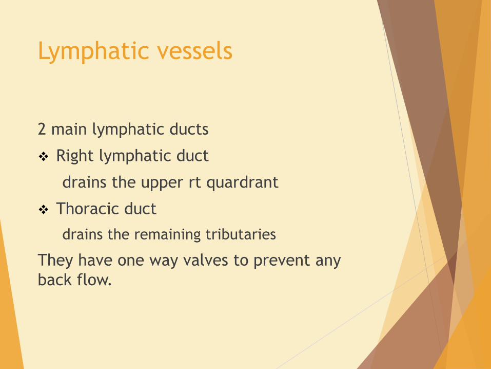
2 main lymphatic ducts
Right lymphatic duct
drains the upper rt quardrant
Thoracic duct
drains the remaining tributaries
They have one way valves to prevent any
back flow.

Cisterna chyli
It is a lymphatic sac at the base of the
thoracic duct
Anterior to the body of L1 or L2
Formed by the convergence of the lumbar
lymphatic trunks and intestinal lymphatic
trunks

Cisterna chyli

Thoracic Duct
Main lymphatic duct of the body
Originates from the cisterna chili
Enters into the thorax via the aortic foramen of the diaphragm
Situated in the posterior mediastinum
Receives lymph from the left side of the head & neck, lt upper limb & lt chest wall
Empty into the junction of the ltsubclavian vein and internal jugular vein
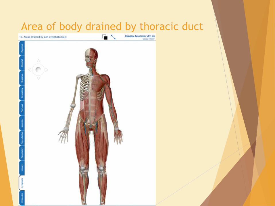
Area of body drained by thoracic duct

area of body drained by the right lymphatic duct

Thymus
Bilobed lymphoid organ
Located in the superior
mediastinum
maximum absolute size during
puberty between 30 and 40 g
It regresses after the puberty

Two lobes covered by capsules
Fibrous septa – divided 2 mm area of lobules
on each lobe
Each lobule
dense cortex
Pale medulla
Lymphoid stem cells in cortex
Divided rapidly and daughter T cells become
matured
Migrated into Medulla -
T cells sensitive to normal tissue are destroyed


Spleen
Largest lymphoid organ
75-250 g
Lies in lt hypochondrium with its long axis
along the 10th rib
Mainly over the 9th , 10th and 11th ribs
There is a notch in its inferolateral
surface

Blood supply is from the tortious splenic
artery from the coeliac axis
Which gives off branches to stomach and
pancreas within the gastrosplenic
ligament
Divides into superior and inferior
branches

Splenic vein is formed by several
tributaries within the splenic substance
Joins with the superior mesenteric vein to
form portal vein behind the neck of the
pancreas

Efferent lymphatics in white pulp joins
with the arterioles
Emerge as nodes at the hilum
Drains via the retropancreatic nodes to
the coeliac nodes


Tonsils
Aggregates of lymphnodes under the
epithelial lining of the oral and
pharyngeal areas
Pharyngeal tonsils
On the roof of the nasopharynx
Also called the adenoids

Palatine tonsils &
Lingual tonsils- at the base of posterior
surface of tongue
These are collectively known as
Waldeyer’s ring

Bld supply is principally from the tonsilar
artery which is the branch of the facial
artery
Entering at the lower pole of the tonsil
Also from lingual, ascending palatine and
ascending pharyngeal arteries
Lymphatic drainage is nodes around the
internal jugular vein to the
jugulodigestric or tonsillar nodes


Physiology

Functions of LYMPHATIC SYSTEM
The principal function of the lymphatic
system is the return of protein rich fluid to
the circulation through the lymphatic venous
junctions in the jugular area.

Water
Electrolytes
low molecular weight molecules (polypeptides, cytokines, growth factors)
Macromolecules - fibrinogen, albumin, globulins, coagulation and fibrinolyticfactors
From the interstitial fluid (ISF) return to the circulation via the lymphatics

Intestinal lymph (chyle) transports
cholesterol, long-chain fatty acids,
triglycerides and the fat-soluble vitamins (A,
D, E and K) directly to the circulation,
bypassing the liver.
Lymphocytes and other immune cells also
circulate within the lymphatic system.


Innate immunity
Also called natural or native immunity
Defense mechanisms that are present
before the infection
First line of defence
Always ready

Innate immunity consists of:
• physical barriers
• secretions with antibacterial activity, including
lactoferrin;
• phagocytic cells: monocytes, macrophages and
neutrophils;
• NK cells (lymphocytes capable of non
MHC restricted killing);

soluble mediators which can enhance the
activity of innate and specific responses:
C-reactive protein (CRP)
mannose-binding lectin (MBL)
cytokines
soluble enzymatic cascades such as the
complement system

The innate immune system is non-adaptive,
i.e. it cannot adapt its receptors to
recognize an organism which has evolved
and mutated its antigens to evade binding.
It does not develop memory
It does not possess antigen specificity
through the specialized and mutable antigen
receptors of immunoglobulins.

Adaptive immunity
Also called acquired or specific immunity
Mechanisms that are stimulated by
microbes
Capable of recognizing nonmicrobial
substances called ‘antigens’.

These are more effective than innate
ones
Mediated by lymphocytes and antibodies
which amplify and focus non-specific
responses and provide additional effector
functions
These cells are organised into lymphoid
tissues
Cellular (cell mediated) immunity refers
to lymphocyte-mediated effector
responses (T helper (Th) and cytotoxic
cells) of the specific immune response

Cellular immunity refers to lymphocyte-
mediated effector responses (T helper
and cytotoxic T cells) of the specific
immune response
Humoral immunity often refers to the
antibody arm of the specific immune
response.

Antibodies are usually not produced without
some cell-mediated response to the same
antigen
T and B lymphocytes possess infinitely
variable antigen receptors which can
clonally expand.
Antigen receptors which can be secreted
into interstitial fluid and onto mucosal
surfaces are called antibodies.
Antibodies can activate complement and
also enhance opsonization of antigen to
facilitate phagocytosis.

Both innate and adaptive mechanisms
exponentially amplify the immune response
since clonal expansion of lymphocytes
increases the number of cells reactive with
an antigen.
Cytokines and complement components
recruit other immune effector mechanisms
and antibodies activate complement and
phagocytes.
The specific adaptive immune response is thus
flexible and adaptable.
Capable of responding to antigens which
have not been previously encountered
Including those generated in organisms by
the selecting pressures of an effective
adaptive immune response.
Many pathogens have specific
adaptations/mutations to evade previous
immunological memory responses (e.g.
influenza antigen variability) or to suppress
the normal mechanisms of immune
destruction.

ANTIGENS
An antigen is any substance which can
elicit a specific immune response.
Consists of many epitopes.
An epitope is a specific sequence of a
protein or carbohydrate recognized by the
receptor molecules of the immune system
(antibody or T cell receptor)

Antigens can be divided into
foreign - non-self, allogeneic,
xenogeneic, etc.
Autoantigens – self antigens

ANTIBODIES
An antibody is a soluble protein immune
receptor produced by B lymphocytes,
consisting of two identical antigen-
binding sites .
The antigen specificity of the antibody
resides in the antigen-binding variable
regions (the fragment antigen-binding,
Fab, portion).

Antibodies are divided into different
isotypes (classes) which have different
functional attributes according to Fc
fragments
Antibodies which bind to antigen or cells
and activate complement via the Fc region
thus recruit, activate, amplify and target
non-specific defense mechanisms.


Functions of the spleen
1. Haematological function
2. Immunological function

Haematological functions
Site of quality control of erythrocyte
population
Removes fragmented or damaged red
cells from circulation k/a culling
Remodeling of surface of maturing
erythroctytes where by maintaining the
membrane surface area and volume ratio

Removal of intraerythrocyte inclusions s/a
Heinz’s bodies, Howel-Jolly bodies k/a
pitting
Clearance of particulate matter from the
circulation – imp function for the timely
immune response to blood borne antigens
Sequestration of plalets

Haemopoiesis
Only in fetal life
No bld formation in the after birth
Revision of fetal pattern of haemopoiesis
in certain diseases

Immunological function
Each population of lymphocyte is a
constant flux
¼ of body’s population of T lymphocytes
is in the spleen at a point time
Humoral response following antigenic
stimulation involves co-operation
between T & B lymphocytes on the
surface of large dentritic cells

Germinal centres ( secondary follicles)
later appear within the primary follicle
Reach their maximum development in
about 8 wks following antigenic
stimulation
Antibody response is relatively decreased
after splenectomy

Also influence the opsonization of
pneumococci in non immune individuals
Susceptible to them after splenectomy


Pathology Reactive lymphadenitis
Tuberculous lymphadenitis
Lymphoedema
Lymphomas

Reactive Lymphadenitis
Infections and nonmicrobial inflammatory
stimuli not only cause leukocytosis but
also involve the lymph nodes
often associated with lymph node
enlargement (lymphadenopathy)
may be acute or chronic
histologic appearance of the nodes is
entirely nonspecific

Tuberculous lymphadenitis
Especially neck glands
Present as cervical lymphadenopathy
Cold abscess
Lymphnodes are rubbery and matted
together
Eventually it can progress to collar stud
abscess formation & sinus

Tissue diagnosis by excisional biopsy
Granuloma formation with grossly
caseation necrosis
Definitive Rx is antituberculous
chemotherapy

Lymphoedema
Abnormal lymph swelling
Caused by accumulation of increased
amount of high protein ISF
Secondary to defective lymphatic
drainage in the presence of near normal
net capillary function

Pathophysiology
Normal capillary function
Oedema fluid is high protein content
Results from
Lymphatic aplasia
Hypoplasia
Dysmotility
Obliteration by inflammation
Infective or neoplastic process
Surgical extirpation

Two main types
Primary
Unknown cause
Thought to be congenital lymphatic
dysplasia
Secondary
Clear underlying cause

Aetiological classification of
lymphoedema
Primary
Congenital (onset <2 years old):
sporadic
familial (Nonne–Milroy’s disease)
Praecox (onset 2–35 years old):
sporadic
familial (Letessier–Meige’s disease)
Tarda (onset after 35 years old)
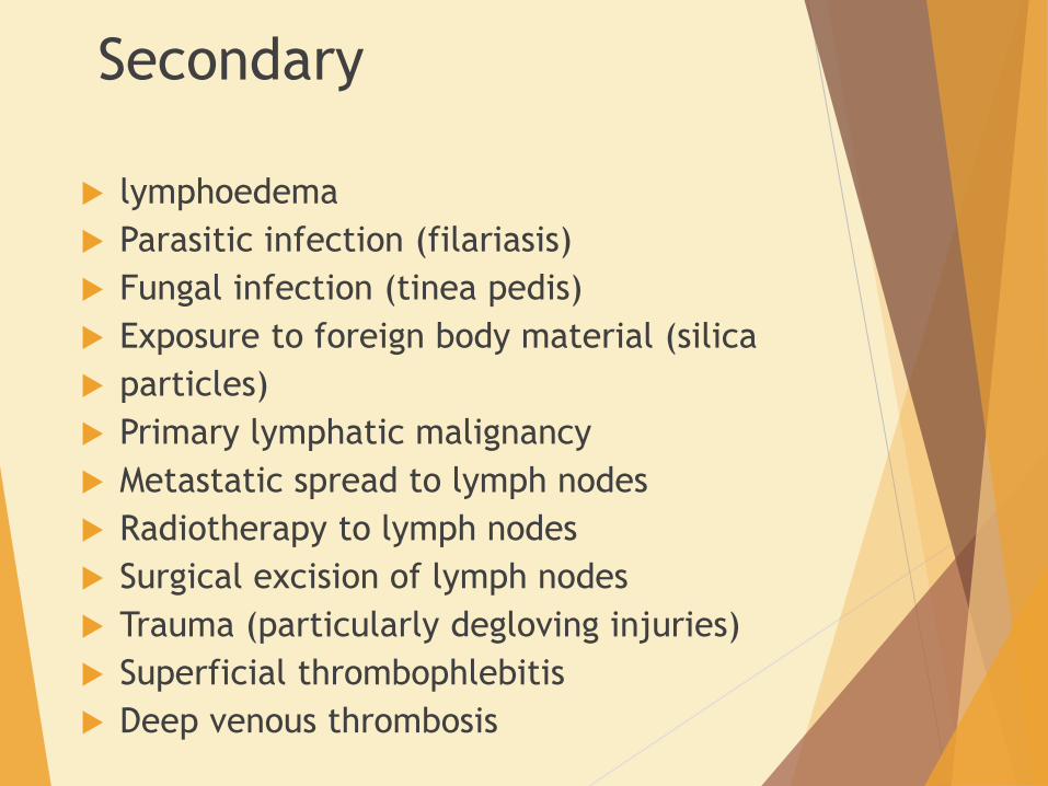
Secondary
lymphoedema
Parasitic infection (filariasis)
Fungal infection (tinea pedis)
Exposure to foreign body material (silica
particles)
Primary lymphatic malignancy
Metastatic spread to lymph nodes
Radiotherapy to lymph nodes
Surgical excision of lymph nodes
Trauma (particularly degloving injuries)
Superficial thrombophlebitis
Deep venous thrombosis

Grading of lymphoedema
(Brunner)
Subclinical
excess interstitial fluid and histological
abnormalities in lymphatics and lymph nodes
but no clinically apparent lymphoedema
Grade I
Oedema pits on pressure and swelling largely
or completely disappears on elevation and bed
rest

Grade II
Oedema doesn’t pit and doesn’t reduce
upon elevation
Grade III
Oedema is associated with irreversible skin
changes e.g; fibrosis or papillae

Clinical features
Characteristically involves foots
Contour of ankle is lost
Toes appears square
Skin on the dorsum of the toes cannot be
pinched “Stemmer’s Sigh”
Ulceration is unusual
Ulceration and non healing bruises should
raise the suspicion of malignancy


Lymphangiosarcoma was originally
described in post mastectomy oedema
“Stewart-Treves” syndrome
0.5% of patients
Mean onset is 10 yrs
Can develop in longstanding lymphedema
“20 yrs”

Malignancies associated with
lymphoedema
Lymphangiosarcoma
Kaposi’s sarcoma
SCC
Liposarcoma
Malignant melanoma
Malignant fibrous histocytoma
BCC
lymphoma

Filariasis
Common cause of lymphedema worldwide
Infectious disease caused by viviparous
nematodes “Wucheria bancrofti”
Vector “mosquitos”
Parasites enters the lymphatics via blood
Lodges in lymphnodes
Causing fibrosis and obstruction

Either by direct damage or by immune
response of host
Degree of lymphedema is often massive
referring to as elephantiasis
Diethylcarbamazine destroys the
parasites but unable to reverse the
lymphedema


Investigations
Routine tests
CP, U&E, creatinine, LFT, T & DP, albumin, CRP
Lymphangiography
Lymphoscintigraphy
CT
MRI
USG
Limb volume measurement

Management
Relief of pain
Control of swelling
Skin care
Manual lymphatic drainage
Multilayer lymphedema bandaging and
compression garments
Exercise
Drugs
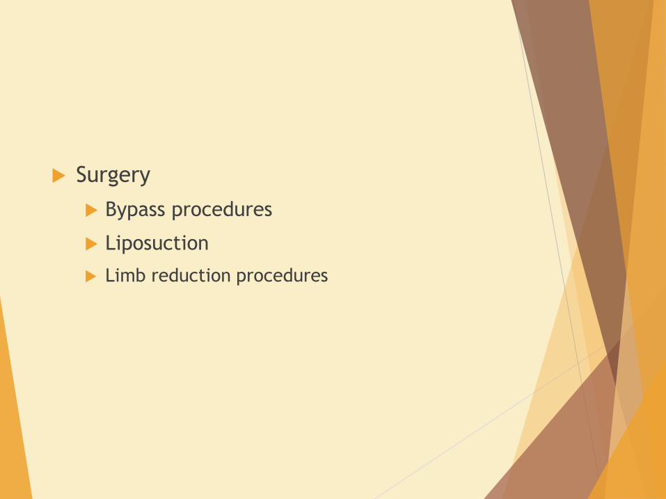
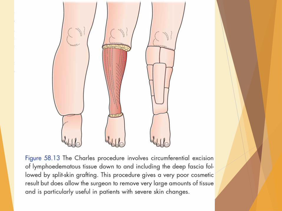
Surgery
Bypass procedures
Liposuction
Limb reduction procedures


lymphomas
Hodgkin’s lyphomas
Non Hodgkin’s lymphomas

Hodgkin’s lymphomas
Reed Stemberg giant cell
Mainly arise from B lymphocytes
Lymph node enlargement is often cervical
Rubbery consistency
Hepatosplenomegaly
General symptoms of malignancies
Diagnosed by lymph node histology and
bonemarrow aspirate and trephine biopsy

12 yrs old boy

Non Hodgkin’s lymphomas
70% are B cell origin
May present without typical lymphnode
enlargement
Hepatosplenomegaly and other features
of malignancy
Invx as in HL
Treatment is mainly by chemotherapy
regimens

Thymic tumours
May arise from either epithelium or
lymphoid tissue or both
May present as
Mediastinum mass
Associated myasthenia gravis
Associated immune deficiency states
Treatment is by thymectomy

Gastric lymphoma
Primary gastric lymphoma ~ 5% of all
gastric neoplasms
most prevalent in the sixth decade of
life.
most commonly occur in the gastric
antrum
Primary gastric lymphomas are B cell-
derived
arising from the mucosa-associated
lymphoid tissue (MALT)

At an early stage, the disease takes the
form of a diffuse mucosal thickening,
which may ulcerate
Presented as – pain, weight loss , bleeding
as s/s of Ca stomach

Diffuse large B-cell lymphoma (55%)
Associated with immunodeficiencies and H. pylori
infection
Extranodal marginal cell lymphoma (MALT) (40%)
Burkitt’s lymphoma (3%)
associated with Epstein-Barr virus infections, highly
aggressive , younger age,
Site – cardia or body of stomach
Mantle cell and follicular lymphomas (each < 1%).

Management OGDS
endoscopic biopsy ,not specify with endoscopic features
Type of lymphoma
Imaging
EUS
CT chest and abdomen
H. pylori testing
by histology and, if negative, confirmed by serology

Treatment
multimodality treatment program
Resection - controversial
Chemotherapy plus radiation therapy: CHOP
(cyclophosphamide, hydroxy-daunomycin, Oncovin,
prednisolone)

Early-stage MALT lymphomas
Diffuse large B-cell lymphoma
H. pylori eradication alone – Successful
eradication resulted in remission in more
than 75% of cases
Follow up
repeat endoscopy in 2 months to
document clearance of the infection as
well as biannual endoscopy for 3 years to
document regression