Embed Size (px)
Citation preview
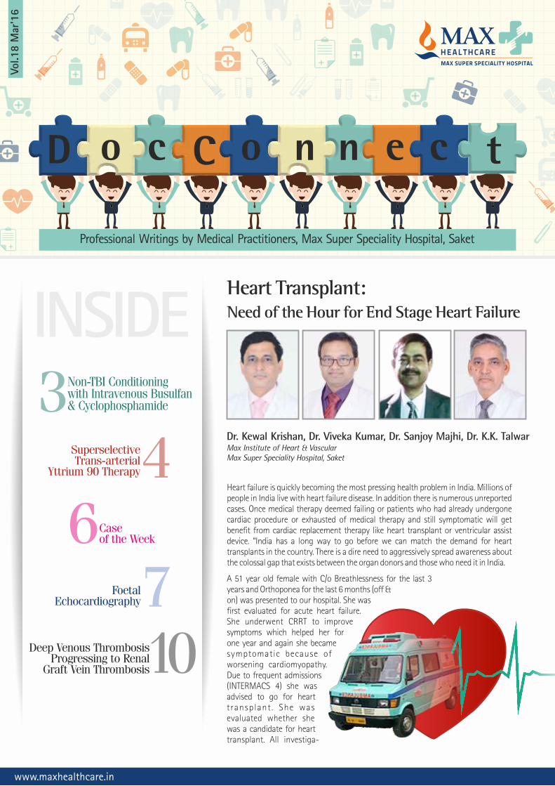
INSIDEHeart Transplant: Need of the Hour for End Stage Heart Failure
Dr. Kewal Krishan, Dr. Viveka Kumar, Dr. Sanjoy Majhi, Dr. K.K. TalwarMax Institute of Heart & VascularMax Super Speciality Hospital, Saket
www.maxhealthcare.in
Professional Writings by Medical Practitioners, Max Super Speciality Hospital, Saket
Vol.1
8 M
ar'1
6
o c C o n n e c tD
6Caseof the Week
3Non-TBI Conditioning with Intravenous Busulfan& Cyclophosphamide
4Superselective Trans-arterial
Yttrium 90 Therapy
7FoetalEchocardiography
10Deep Venous ThrombosisProgressing to Renal
Graft Vein Thrombosis
Heart failure is quickly becoming the most pressing health problem in India. Millions of people in India live with heart failure disease. In addition there is numerous unreported cases. Once medical therapy deemed failing or patients who had already undergone cardiac procedure or exhausted of medical therapy and still symptomatic will get benefit from cardiac replacement therapy like heart transplant or ventricular assist device. “India has a long way to go before we can match the demand for heart transplants in the country. There is a dire need to aggressively spread awareness about the colossal gap that exists between the organ donors and those who need it in India.
A 51 year old female with C/o Breathlessness for the last 3 years and Orthoponea for the last 6 months (off & on) was presented to our hospital. She was first evaluated for acute heart failure. She underwent CRRT to improve symptoms which helped her for one year and again she became symptomatic because of worsening cardiomyopathy. Due to frequent admissions (INTERMACS 4) she was advised to go for heart transplant . She was evaluated whether she was a candidate for heart transplant. All investiga-

2
A 32km green corridor was set up from Max-Shalimar Bagh
to Max-Saket on Thursday, 7th Jan’16 at 10.20 am to transport the heart
of a 17-year-old road accident victim in 45 min 27 sec. The donor's two kidneys
successfully saved lives of two patients at Max-Shalimar Bagh, the two corneas were sent to
AIIMS and the liver was transported to the Institute of Liver and Biliary Sciences (ILBS) for transplantation.
5 year female with osteopetrosis presented to us with optic atrophy A(VEP showed perception of light). Hearing was preserved till now. Her X-Rays and CT show diffuse bony thickening suggestive of
(2)osteopetrosis. She was diagnosed with osteopetrosis and admitted for BMT. In view of her age we wished to avoid TBI and used IV Busulfan
(3)and cyclophosphamide conditioning . GVHD prophylaxis consisted of cyclosporine and short course methotrexate. She had a HLA
identical 2 year old sister. A bone marrow harvest was done under GA , total volume infused was 220 ml, TNC= 11400, MNC-48%, CD 34-3.42 %; 390/ul; CD 34 dose: 5.36 x 10⁶/kg. Neutrophil engraftment was attained on day +19 and platelet engraftment was attained on day+29. Her transplant course was smooth without any fever or infection. There was no VOD and no GVHD. She did develop grade 1 mucositis during conditioning.
Non-TBI Conditioning with Intravenous Busulfan& Cyclophosphamide is Feasible in Bone MarrowTransplantation for Osteopetrosis
Dr. Rahul NaithaniSenior Consultant, Hematology & Bone Marrow TransplantationMax Super Speciality Hospital, Saket
tions including right heart cath and immunological tests were performed. Her pulmonary vascular resistance and other parameters were in acceptable limits of transplant. She was put on waiting list for transplant. During this period she was admitted twice for acute heart failure. On Jan 7, 2016 we received call for the donor (A+ve) from Max Hospital, Shalimar Bagh. CTVS team went over there to evaluate the donor. Meanwhile recipient was called immediately to get admitted to Max hospital, Saket to make her ready for the surgery. After the complete evaluation of donor it was decided to take the heart. The local police acted swiftly and created a green corridor, which allowed us to travel a distance of 32 kms in a mere 45 mins. As soon as we reached to operation theatre recipient was put on cardiopulmonary bypass and recipient cardiectomy was done. New heart was sutured with bicaval technique and cross clamp was removed. Heart started beating in no time. Slowly CPB was weaned off and chest closed in layers. Patient was extubated next morning. Over the course of next few days ionotropes were weaned off and all lines were removed. Patient has been doing fine since then.
A miraculous chain of events, led to this path-breaking surgery, helping save a life of a middle aged very sick lady.
"Our team at Max Healthcare worked round-the-clock to ensure a seamless and successful heart transplant. There are numerous precautions that need to be kept in mind while performing a heart transplant. Before the heart is retrieved from the donor, the donor heart needs to undergo echocardiography to ascertain that the heart is healthy and has no previous damage. The heart also needs to be checked on table and cleared for suitability along with any diseases that can preclude the retrieval. Given the abysmal number of heart donors in India, it was crucial to ensure that every step of the transplant was carried out without any road blocks.
Heart transplant in India is still very rare, not because of the lack of medical skills, but unfortu-nately, because of the lack of organ donation. Once harvested, the heart has to be transplanted with-in a very tight window of four hours. It is still considered a taboo and family members of brain dead patients do not volunteer to donate the organs of their loved ones. People need to be educated on how the organs of the brain dead patient can provide life to another needy patient and provide a second lease of life.
3
On day 53 she presented with anemia and thrombocytopenia (Hb 45g/L,platelet 35 x 109/l, TLC 3.4 x 10⁹/l). Peripheral smear showed macrocytosis and reticulocyte count of 10% and 5 nRBCs/100 WBCs. Her LDH was high 353 U/l (<192U/l). Bone marrow aspirate showed cellular bone marrow with eryhroid hyperplasia with adequate megakaryocytes. It also showed an osteoclast. A diagnosis of post transplant autoimmune cytopenia was made which responded to addition of steroids within a week. Overall she continued to improve with normalisation of serum calcium and improvement in vision in form of perception of light despite having optic atrophy at diagnosis. There was no more watery discharge
from mouth (due to malocclusion of teeth) which was present pre BMT, no more snoring and some regression of proptosis.
Article by Behfar ET AL and this case demonstrate that non- (1)radiation based conditioning regime is feasible in osteopetrosis .
This helps lot of centres in developing countries who may not have infrastructural facility or expertise for total body irradiation. Secondly, immune cytopenias seems not uncommon in children with osteopetrosis undergoing BMT as was the case in previous
(1)series also where one child developed hemolytic anaemia . She did show some visual improvement in form of perception of light in initial phase post BMT. We did not have chimerism facilities at that time, however clinical improvement suggest that the BMT was successful in ameliorating the osteopetrosis phenotype.
REFERENCES1. Behfar M, Dehghani SS, Hosseini AS, Jalali A, Hamidieh AA, Ghavamzadeh A. Non
total body irradiation myeloablative conditioning with intravenous busulfan and cyclophosphamide in hematopoietic stem cell transplantation for malignant infantile osteopetrosis. Pediatr Transplant. 2015 Jun;19(4):422-7.
2. Srinivasan M, Abinun M, Cant AJ, Tan K, Oakhill A, Steward CG. Malignant infantile osteopetrosis presenting with neonatal hypocalcaemia. Arch Dis Child Fetal Neonatal Ed 2000:83: F21–F23
3. Driessen GJ, Gerritsen EJ, Fischer A, Fasth A, Hop WC, Veys P, Porta F, Cant A, Steward CG, Vossen JM,Uckan D, Friedrich W. Long-term outcome of haematopoietic stem cell transplantation in autosomal recessive osteopetrosis: An EBMT report. Bone Marrow Transplant 2003: 32: 657–663.
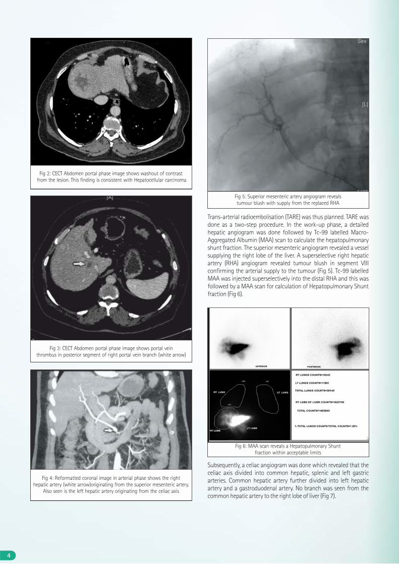
CASE REPORT64 year old male patient with post-necrotic liver cirrhosis came with complaints of weakness for a few weeks. Triple Phase CECT revealed a large arterial hyper-enhancing lesion in segment VIII which showed washout in porto-venous phase (Fig 1 and 2) and tumour thrombus in anterior branch of right portal vein (Fig 3). Liver showed features of cirrhosis and there was mild ascites. Reformatted CT angiographic images revealed a replaced right hepatic artery from the superior mesenteric artery (Fig 4). AFP levels were 394 and USG guided percutaneous biopsy also revealed Hepatocellular carci-noma. Child-Pugh scoring classified the patient into category A.
Surgical resection was not done owing to the portal vein thrombosis and co-morbidities. Patient was unwilling for Liver transplantation. Trans-arterial chemo-embolisation (TACE) was not considered due to portal vein thrombosis.
Superselective Trans-arterial Yttrium 90 Therapy:A New Hope in Hepatic Malignancies
Dr. Vivek Saxena, Dr. Lakshay Mehta, Dr. Bharat AggarwalDepartment of RadiologyMax Super Speciality Hospital, Saket
Fig 1: CECT Abdomen arterial phase image shows a solitary enhancing lesion in segment VIII of liver
4
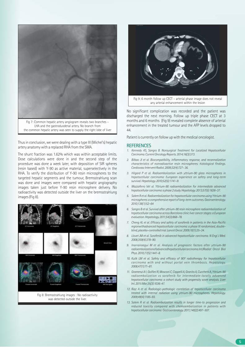
Trans-arterial radioembolisation (TARE) was thus planned. TARE was done as a two-step procedure. In the work-up phase, a detailed hepatic angiogram was done followed by Tc-99 labelled Macro-Aggregated Albumin (MAA) scan to calculate the hepatopulmonary shunt fraction. The superior mesenteric angiogram revealed a vessel supplying the right lobe of the liver. A superselective right hepatic artery (RHA) angiogram revealed tumour blush in segment VIII confirming the arterial supply to the tumour (Fig 5). Tc-99 labelled MAA was injected superselectively into the distal RHA and this was followed by a MAA scan for calculation of Hepatopulmonary Shunt fraction (Fig 6).
Subsequently, a celiac angiogram was done which revealed that the celiac axis divided into common hepatic, splenic and left gastric arteries. Common hepatic artery further divided into left hepatic artery and a gastroduodenal artery. No branch was seen from the common hepatic artery to the right lobe of liver (Fig 7).
Fig 3: CECT Abdomen portal phase image shows portal vein thrombus in posterior segment of right portal vein branch (white arrow)
Fig 4: Reformatted coronal image in arterial phase shows the right hepatic artery (white arrow)originating from the superior mesenteric artery.
Also seen is the left hepatic artery originating from the celiac axis
Fig 6: MAA scan reveals a Hepatopulmonary Shunt fraction within acceptable limits
Fig 2: CECT Abdomen portal phase image shows washout of contrast from the lesion. This finding is consistent with Hepatocellular carcinoma
Fig 5: Superior mesenteric artery angiogram reveals tumour blush with supply from the replaced RHA
5
Fig 7: Common hepatic artery angiogram reveals two branches – LHA and the gastroduodenal artery. No branch from
the common hepatic artery was seen to supply the right lobe of liver
Thus in conclusion, we were dealing with a type III (Michel's) hepatic artery anatomy with a replaced RHA from the SMA.
The shunt fraction was 1.62% which was within acceptable limits. Dose calculations were done in and the second step of the procedure was done a week later, with deposition of SIR spheres (resin based) with Y-90 as active material, superselectively in the RHA. To verify the distribution of Y-90 resin microspheres to the targeted hepatic segments and the tumour, Bremsstrahlung scan was done and images were compared with hepatic angiography images taken just before Y-90 resin microsphere delivery. No radioactivity was detected outside the liver on the bremsstrahlung images (Fig 8).
No significant complication was recorded and the patient was discharged the next morning. Follow up triple phase CECT at 3 months and 6 months (Fig 9) revealed complete absence of arterial enhancement in the treated tumour and the AFP levels dropped to 44.
Patient is currently on follow up with the medical oncologist.
REFERENCES1. Kennedy AS, Sangro B. Nonsurgical Treatment for Localized Hepatocellular
Carcinoma. Current Oncology Reports. 2014;16(3):373.
2. Bilbao JI et al. Biocompatibility, inflammatory response, and recannalization characteristics of nonradioactive resin microspheres: histological findings. Cardiovasc Intervent Radiol. 2009;32(4):727– 36.
3. Hilgard P et al. Radioembolization with yttrium-90 glass microspheres in hepatocellular carcinoma: European experience on safety and long-term survival. Hepatology. 2010;52(5):1741–9.
4. Mazzaferro Vet al. Yttrium-90 radioembolization for intermediate advanced hepatocellular carcinoma: a phase 2 study. Hepatology. 2013;57(5):1826–37.
5. Salem R et al. Radioembolization for hepatocellular carcinoma using Yttrium-90 microspheres: a comprehensive report of long-term outcomes. Gastroenterology. 2010;138(1):52–64
6. Sangro B et al. Survival after yttrium-90 resin microsphere radioembolization of hepatocellular carcinoma across Barcelona clinic liver cancer stages: a European evaluation. Hepatology. 2011;54(3):868–78.
7. Cheng AL et al. Efficacy and safety of sorafenib in patients in the Asia-Pacific regionwithadvanced hepatocellular carcinoma: a phase III randomised, double-blind, placebo-controlled trial. Lancet Oncol. 2009;10(1):25–34.
8. Llovet JM et al. Sorafenib in advanced hepatocellular carcinoma. N Engl J Med. 2008;359(4):378–90.
9. Inarrairaegui M et al. Analysis of prognostic factors after yttrium-90 radioembolizationofadvancedhepatocellularcarcinoma.IntJRadiat Oncol Biol Phys. 2010;77(5):1441–8.
10. Kulik LM et al. Safety and efficacy of 90Y radiotherapy for hepatocellular carcinoma with and without portal vein thrombosis. Hepatology. 2008;47(1):71–81.
11. Gramenzi A1, Golfieri R, Mosconi C, Cappelli A, Granito A, Cucchetti A, Yttrium-90 radioembolization vs sorafenib for intermediate-locally advanced hepatocellular carcinoma: a cohort study with propensity score analysis. Liver Int. 2015 Mar;35(3):1036-47.
12. Riaz A et al. Radiologic-pathologic correlation of hepatocellular carcinoma treated with internal radiation using yttrium-90 microspheres. Hepatology. 2009;49(4):1185–93.
13. Salem R et al. Radioembolization results in longer time-to progression and reduced toxicity compared with chemoembolization in patients with hepatocellular carcinoma. Gastroenterology. 2011;140(2):497–507.
Fig 9: 6 month follow up CECT – arterial phase image does not reveal any arterial enhancement within the lesion
Fig 8: Bremsstrahlung images : No radioactivity was detected outside the liver.
RADIOLOGY CASE OF THE MONTH
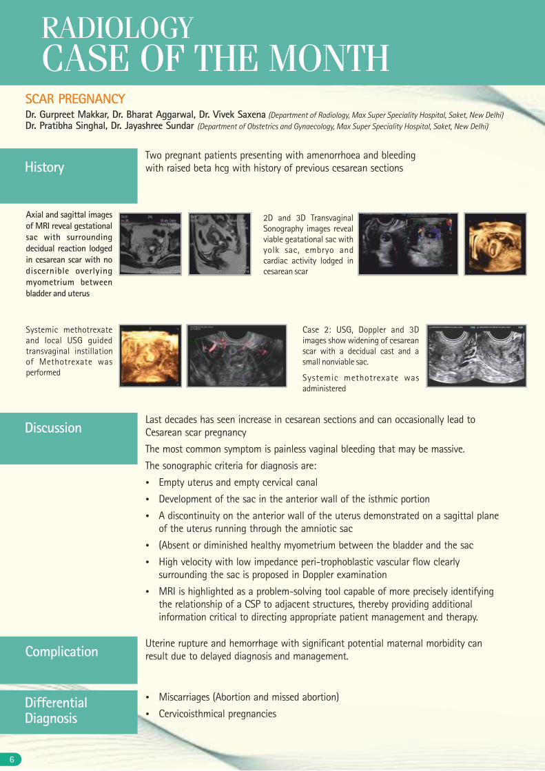
HistoryTwo pregnant patients presenting with amenorrhoea and bleeding with raised beta hcg with history of previous cesarean sections
SCAR PREGNANCYDr. Gurpreet Makkar, Dr. Bharat Aggarwal, Dr. Vivek Saxena (Department of Radiology, Max Super Speciality Hospital, Saket, New Delhi)Dr. Pratibha Singhal, Dr. Jayashree Sundar (Department of Obstetrics and Gynaecology, Max Super Speciality Hospital, Saket, New Delhi)
Discussion
ComplicationUterine rupture and hemorrhage with significant potential maternal morbidity can result due to delayed diagnosis and management.
Last decades has seen increase in cesarean sections and can occasionally lead to Cesarean scar pregnancy
The most common symptom is painless vaginal bleeding that may be massive.
The sonographic criteria for diagnosis are:
Ÿ Empty uterus and empty cervical canal
Ÿ Development of the sac in the anterior wall of the isthmic portion
Ÿ A discontinuity on the anterior wall of the uterus demonstrated on a sagittal plane of the uterus running through the amniotic sac
Ÿ (Absent or diminished healthy myometrium between the bladder and the sac
Ÿ High velocity with low impedance peri-trophoblastic vascular flow clearly surrounding the sac is proposed in Doppler examination
Ÿ MRI is highlighted as a problem-solving tool capable of more precisely identifying the relationship of a CSP to adjacent structures, thereby providing additional information critical to directing appropriate patient management and therapy.
Ÿ Miscarriages (Abortion and missed abortion)
Ÿ Cervicoisthmical pregnanciesDifferential Diagnosis
Axial and sagittal images of MRI reveal gestational sac with surrounding decidual reaction lodged in cesarean scar with no discernible overlying myometrium between bladder and uterus
2D and 3D Transvaginal Sonography images reveal viable geatational sac with yolk sac, embryo and cardiac activity lodged in cesarean scar
Systemic methotrexate and local USG guided transvaginal instillation of Methotrexate was performed
Case 2: USG, Doppler and 3D images show widening of cesarean scar with a decidual cast and a small nonviable sac.
Systemic methotrexate was administered
6
7
Foetal echocardiography has seen the revolution and is now coming up as a very promising tool in the diagnosis and evaluation of various congenital cardiac defects. Almost all the
cardiac abnormalities can now be diagnosed appropriately at 16 weeks of gestation. We report an interesting case of the distressed parents who were referred as suspected congenital cardiac abnormality at 23 weeks of gestation.
CASE REPORTA 30 year old primigravida with gestation of 23 weeks with precious pregnancy presented for fetal echocardiography. Her antenatal ultrasound was suggestive of suggestive of ventrciulo septal defect and parents were very anxious for the same. They were told of the very poor outcome of the pregnancy, need for immediate surgery after birth and poor long term outcome therefore they were understandably very disturbed. Fetal echocardiography (done using Philips IE 33 echo machine) showed a case of Tetrology of Fallot with good pulmonary artery anatomy. Child had isolated large ventriculoseptal defect with aortic override. He had anterior malalignment of the septum. His pulmonary artery annulus was adequate and distal branch pulmonary arteries were: right pulmonary artery: 3mm, left pulmonary artery: 2.6mm. There was a good antegrade flow in the pulmonary arteries and normal ventricular function. Parents were recounseled about the presence of Tetrology of Fallot. They were told as the need of elective total correction (single stage) with normal long term outcomes. Parents were highly thankful and decided to continue. The child was delivered as a healthy female child with a birth weight of 3kg. Child was discharged as per the protocol. She had an oxygen saturation of 97% at discharge. Her postoperative echocardiography was suggestive of Tetrology of Fallot, large ventriculo septal defect with aortic override. There was mild anterior malalignment with right ventricular outflow tract gradient of 14mmHg. The gradient increased to 30mm hg by next day with decrease in neonatal pulmonary artery pressures. She had adequate pulmonary artery annulus of: 6mm (expected: 6mm) with confluent branch pulmo-nary arteries of RPA: 4mm, LPA:3.6 (expected: 4mm). The parents were recounseled regarding the disease and that the elective surgery will be planned at a later date ( as was explained antentally). Child is being planned for a regular saturation monitoring. There was no other structural or systemic abnormality postnatally.
DISCUSSIONFoetal echocardiography is an important diagnostic tool whose application has made it possible for the precise diagnostic evalua-tion of various congenital heart diseases. By its ability to allow, to
make conscious decision about the continuation of pregnancy and hence influence the outcome it has greatly modified the natural history of the disease. Sensitivity and specificity of the foetal echocardiographic examination appears maximal at 16-20 weeks of gestation. This timing also allows for repeat examination when first examination is incomplete before 22 weeks (maximum time limit of termination of pregnancy). Foetal echocardiography can be performed as early as 8-10 weeks transvaginaly. In the present case the precise diagnosis not only allayed the anxiety in the family, it also helped to make a conscious decision about the planning of delivery in appropriate neonatal setting. TOF with adequate annulus and branch pulmonary arteries is a diagnosis with good long term outcome and it was this decision that helped family take a conscious decision about their precious pregnancy.
Tetralogy of Fallot includes non-restrictive perimembranous ventricular septal defect with overriding aorta, and pulmonary stenosis. Best views to detect this anomaly are five-chamber view and long axis view of left ventricular outflow tract. These views show ventricular septal defect with overriding aorta, and dilated ascending aorta. Further sweep from five-chamber view shows narrowing of right ventricular outflow tract. We observed good antegrade flow in the present case. Although it needs to be counseled that the pulmonary stenosis in TOF can be progressive, hence I believe it is justifiable to repeat the echocardiography in each trimester in case family decides for the continuation of pregnancy as in the present case. Short axis view at the level of great vessels shows small pulmonary arteries and dilated aorta. Doppler interrogation of right ventricular outflow tract shows increased pulmonary outflow velocity. All these findings can be progressive with increasing right ventricle outflow tract obstruction, decreasing size of pulmonary artery. Common differential diagnosis is truncus arteriosus in which single great artery with large conotruncal ventricular septal defect is present. The present case highlights the importance of foetal echo. The diagnosis of good anatomy TOF not only allayed parental anxiety after adequate counseling it also enabled delivery proximity to the paediatric cardiac centre.
FIGURE 1 (PRE): Foetal echocardiography images done at 23 weeks showing Tetrology of Fallot in various views. Fig 1a showing modified apical 5c view with flow acceleration in RVOT, Fig 1b showing VSD (marked by arrow) with aortic override, Fig 1c showing 4 chamber view with well formed 2 ventricles, Fig 1d showing parasternal short axis view with RVOT.
RVOT: right ventricular outflow tract, PA: pulmonary artery, RA: right atrium, RV: right ventricle, LA: left atrium, LV: left ventricle, VSD: ventricular septal defect, AO: aorta.
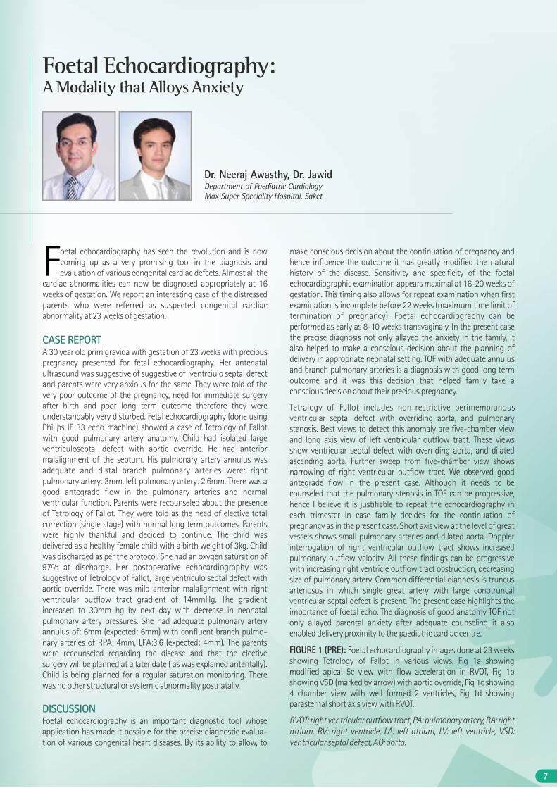
Dr. Neeraj Awasthy, Dr. Jawid Department of Paediatric CardiologyMax Super Speciality Hospital, Saket
Foetal Echocardiography: A Modality that Alloys Anxiety
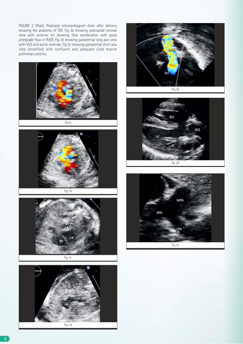
Fig 1d
Fig 2c
Fig 2a
Fig 2b
FIGURE 2 (Post): Postnatal echocardiogram done after delivery showing the anatomy of TOF. Fig 2a showing subcoastal coronal view with anterior tilt showing flow acceleration with good antegrade flow in RVOT, Fig 2b showing parasternal long axis view with VSD and aortic override, Fig 2c showing parasternal short axis view (modified) with confluent and adequate sized branch pulmonary arteries.
Fig1a
Fig 1b
Fig 1c
8
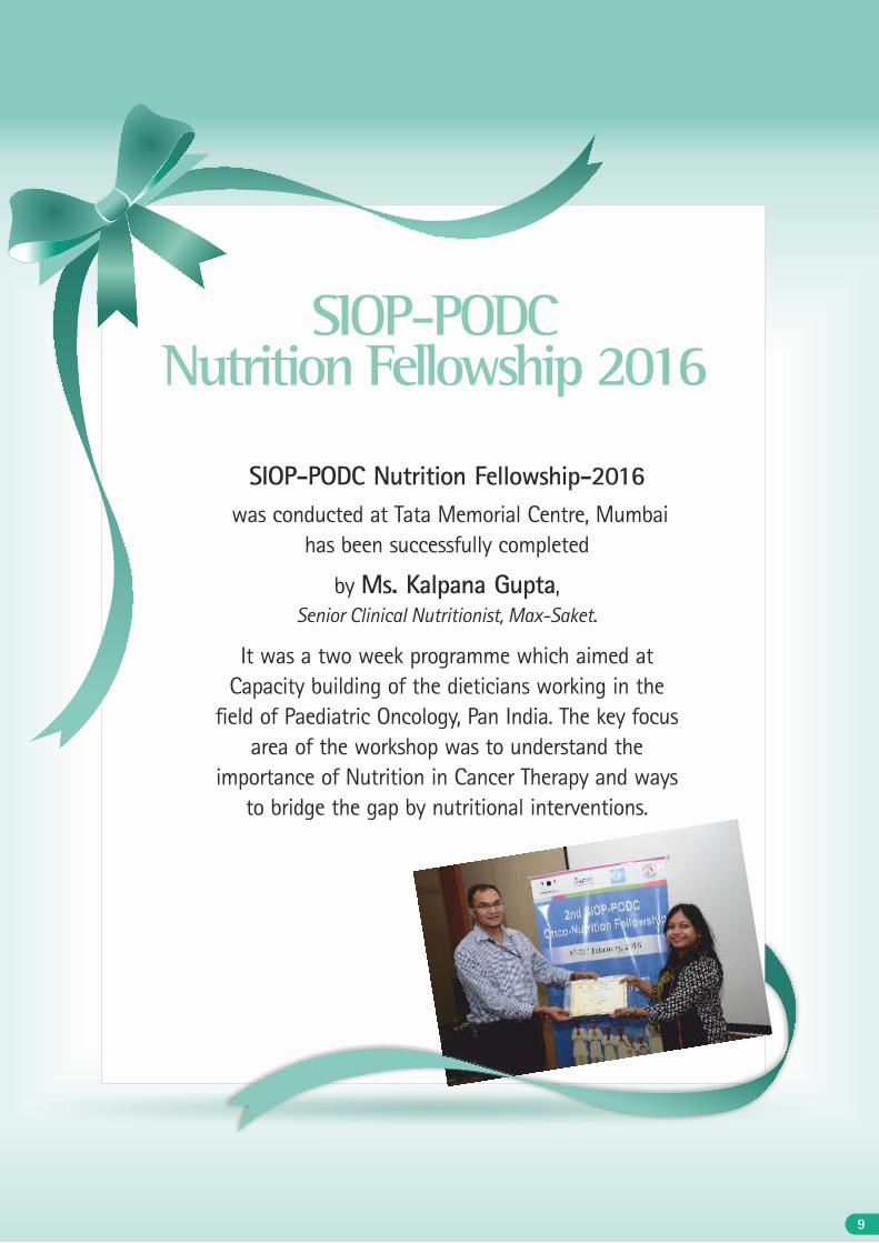
SIOP-PODC Nutrition Fellowship-2016 was conducted at Tata Memorial Centre, Mumbai
has been successfully completed
by Ms. Kalpana Gupta, Senior Clinical Nutritionist, Max-Saket.
It was a two week programme which aimed at Capacity building of the dieticians working in the
field of Paediatric Oncology, Pan India. The key focus area of the workshop was to understand the
importance of Nutrition in Cancer Therapy and ways to bridge the gap by nutritional interventions.
SIOP-PODC Nutrition Fellowship 2016
9
10
ABSTRACTWe present a case of diabetes and hypertension with history of diabetic retinopathy and nephropathy with chronic kidney disease stage V (D) who underwent live related renal transplantation. In post transplant period he had delayed graft function which gradually recovered and was being planned for discharge. At the time he developed right lower limb deep venous thrombosis which rapidly progressed to graft vein stenosis in matter of hours and was successfully managed with thrombolysis, PTA and pulverization of the thrombus. The patient recovered good renal function and is presently having a serum creatinine of 1.7 mg/dl.
CONCLUSIONTimely and aggressive management of acute complications like graft vein stenosis can have favorable graft outcomes.
Mr. A, a known case of diabetes since 1998 and hypertension since 2012, with diabetic nephropathy and retinopathy with chronic kidney disease stage V was on maintenance hemodialysis for past few months. He was being worked up for renal transplant with prospective donor being his wife. After a thorough pre-transplant work up and T and B cell CDC cross match negative his renal transplant surgery was performed. He was given ATG induction and kept on triple drug immunosupression tacrolimus, mycophenolate mofetil and steroids. Total ischemic time was 25 minutes and warm ischemic time was 5 minutes. Post-operative he had urine output of less than 100 ml over next 2 hours. Ultrasound Doppler done showed good flows at hilum with RI of 0.77and 0.72. Local causes like catheter site obstruction were also ruled out. His urine output remained low and hemodialysis was done at night. A clinical diagnosis of ATN was made and tacrolimus was withheld and second dose of ATG was given. Tacrolimus levels sent were 29.7 ng/ml. Over a period of next 2 days his urine output improved. His tacrolimus levels started decreasing and gradually tacrolimus was reintroduced. On postoperative day 7th his serum creatinine came down to 1.7 mg/dl. His blood sugars fluctuated and required insulin infusions intermittently. His urine culture grew E Coli sensitive to colistin and colistin and meropenem were added. He complained of breathlessness and was evaluated. Chest medicine opinion was also
taken. CT thorax and Pulmonary function test done were within normal limits. Breathlessness gradually improved by fluid manage-ment only. He was planned for discharge on a serum creatinine of 1.8 mg/dl when he complained of mild pain in right lower limb. On examination right lower limb had mild swelling. A venous doppler done was suggestive of deep venous thrombosis extending up to right external iliac vein but renal artery flows were normal. He was immediately started on low molecular weight heparin. In a period of few hours he had a fall in urine output. A high clinical suspicion of graft vein thrombosis was kept and after explaining to family his CT venogram was done which showed deep venous thrombosis extending from right external iliac vein up to below knee including anastomoses of allograft renal vein. Cardiologist and vascular surgeons were involved and after discussing also possible options and there outcomes with the family TPA thrombolysis and vascular intervention was planned. On fluoroscopy thrombus filled right allograft renal vein and right common femoral vein without any flows to common iliac vein were detected. PTA and pulverisation of the thrombus was done and TPA was injected. TPA thrombolysis was continued post intervention and heparin infusion with regular monitoring of APTT was done. Post procedure urine output remained low and his hemodialysis was done. Gradually over next 24 hrs his urine output improved. He required one more session of hemodialysis before his serum creatinine started decreasing. He was gradually shifted to acitrome. His INR was monitored and patient discharged on a serum creatinine of 2 mg/dl. He is on regular follow up and maintaining a serum creatinine of 1.7 mg/dl.
REVIEWRenal allograft thrombosis may be responsible for 2–7% of early
[1, 2, 3]allograft losses in adults and up to 35% in children . In a study conducted by Zilinska et al out of 103 renal transplant patients (january 2008 to december 2009) studied they detected renal vein thrombosis in 3 cases (2.9%), artery thrombosis in 4 cases (3.9%), one time intrarenal pseudoaneurysm (1%) and renal artery stenosis
[4]in ten patients (9.7%) . Most cases of renal allograft thrombosis occur early in the postoperative period with a peak incidence of 48
A Rare Delayed Presentation ofDeep Venous Thrombosis Progressing toRenal Graft Vein Thrombosis
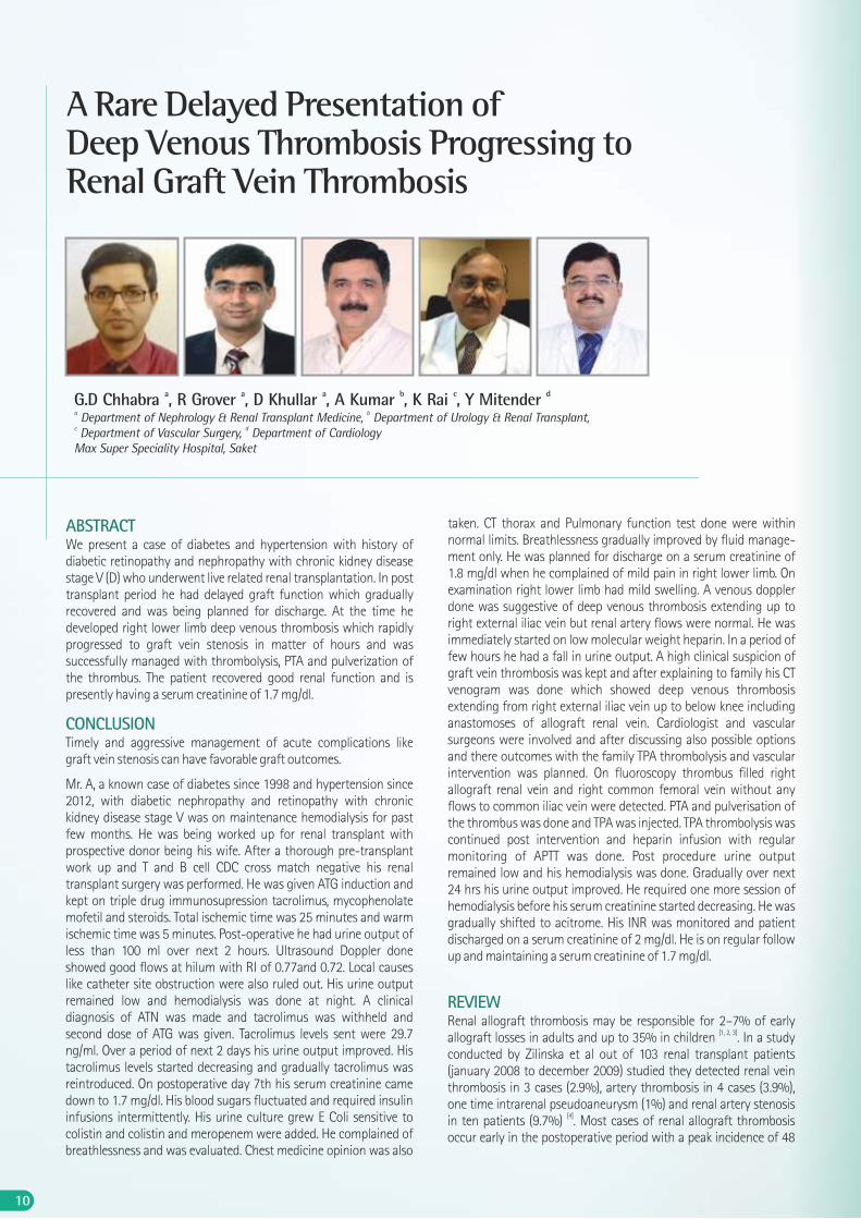
a a a b c dG.D Chhabra , R Grover , D Khullar , A Kumar , K Rai , Y Mitender a b Department of Nephrology & Renal Transplant Medicine, Department of Urology & Renal Transplant, c d Department of Vascular Surgery, Department of CardiologyMax Super Speciality Hospital, Saket
11
hours. However, thrombus formation may be delayed until after the [1]first week . Thrombosis may initially involve the renal artery or
more frequently the renal vein, but in some cases it is difficult to ascertain where the thrombosis originated. Predisposing factors for renal allograft thrombosis include:
Ÿ Hypovolaemia
Ÿ Atherosclerosis
Ÿ Technique error
Ÿ OKT3 (plus high-dose methylprednisolone)
Ÿ Antiphospholipid antibodies
Ÿ High dose steroids
Ÿ Long cold ischaemia time
Ÿ Delayed graft function recovery
Ÿ Elderly donors
Late allograft thrombosis has been defined as occurring later than [1,5]14 days postoperatively , but rarely renal artery thrombosis may
develop a few months post transplantation. Renal allograft vein thrombosis may be induced by renal vein kinking or by renal vein compression caused by lymphocele or other fluid collection, and often results from extension of deep vein thrombosis to the renal
[1,6] [1, 7]allograft vein . A review of the USRDS data found that in renal transplant recipients deep vein thrombosis had an incidence of 2.9 episodes/1000 persons year; the risk was greater for patients with renal insufficiency and with nephrotic syndrome, increased haematocrit, rejection, infection or factor V Leiden mutation. The prognosis is poor because many patients lose their graft function, but some may be rescued depending on the timelines of the diagnosis. Pulmonary embolism is a complication of renal vein thrombosis especially with deep vein thrombosis. Treatment with streptokinase or urokinase may be useful particularly in case of acute or partial vein thrombosis. Percutaneous mechanical thrombectomy and localised catheter-directed thrombolysis may
[1, 8]also allow the return of kidney function in some patients .
DISCUSSIONAs clear from above review our patient had some predisposing factors in form of an infection and delayed graft recovery. The above factors may have pre-disposed our patient to a thrombotic state vis-a vis all patients with these predisposing factors do not have thrombotic events. But early diagnosis and prompt and aggressive management led to resolution of the thrombosis and recovery of renal function.
CONCLUSIONComplications like renal graft vein thrombosis can present atypically and in late period as extensions of deep venous thrombosis but aggressive and timely intervention can have very satisfactory results and salvage renal graft.
REFERENCES(1) Claudio Ponticelli, Marco Moia and Giuseppe Montagnino Renal allograft
thrombosis Nephrol Dial Transplant (2009) 24: 1388–1393.
(2) Irish A. Renal allograft thrombosis: can thrombophilia explain the inexplicable Nephrol Dial Transplant 1999; 14: 2297–2303.
(3) Smith JM, Stablein D, Singh A et al. Decreased risk of renal allograft thrombosis associated with interleukin-2 receptor antagonists: a report of the NAPRTCS. Am J Transplant 2006; 6: 585–588.
(4) Zilinska Z, Chrastina M, Trebaticky B, Breza j et al. Vascular comlications after renal transplantation: clinical study. Bratisl Lek Listy 2010; 111 (11): 586-589.
(5) Friedman GS, Meier-Kriesche HU, Kaplan B et al. Hypercoagulable states in renal transplant candidates: impact of anticoagulation upon incidence of renal allograft thrombosis. Transplantation 2001; 72: 1073–1078.
(6) Ramirez PJ, Gohn RY,KestinAet al. Renal allograft loss due to proximal extension of ileofemoral deep venous thrombosis. Clin Transplant 2002; 16: 310–313
(7) Abbott KC, Cruess DF, Agodoa LY et al. Early renal insufficiency and late venous thromboembolism after renal transplantation in the United States. Am J Kidney Dis 2004; 43: 120–130.
(8) Melamed ML, Kim HS, Jaar BG et al. Combined percutaneous mechanical and chemical thrombectomy for renal vein thrombosis in kidney transplant recipients. Am J. Transplant 2005; 5: 621– 626.
12


![PETITIONER: KEWAL KRISHAN PURI & ANR. · PDF filewas passed by the composite State of Punjab is an Act for the better regulation of ... Liberty. Cinema [1965] 2 ... Micanite Industries](https://img.pdfslide.us/doc/110x75/5ab29b997f8b9a1d168da484/petitioner-kewal-krishan-puri-anr-was-passed-by-the-composite-state-of-punjab.jpg)