Embed Size (px)
Citation preview
Inflammatory ArthritisMichael Aref, MD, PhDHospitalist, Carle Physician Group
Department of Nuclear, Plasma, and Radiological Engineering, UIUCDepartment of Medicine, UICOM-UC
Objectives
• Review the history and physical examination findings of inflammatory arthritis
• Define and categorize inflammatory arthritis
• Understand the initial laboratory and radiological evaluation of suspected rheumatological disease
• Increase clinical sensitivity and specificity to rheumatological disease
Chest pain rule-out• 45 y/o WM
• Persistent, bilateral shoulder, elbow, and hand pain, worse with activity. Associated fatigue.
• Smoker, mother has history of rheumatoid arthritis.
• VSS, decreased ROM of shoulders, with bilateral finger, elbow, and shoulder synovitis.
• Mild normochromic, normocytic anemia and hypokalemia; EKG and chest x-ray benign.
• What test(s) would you like to order next?
Objectives
• Review the history and physical examination findings of inflammatory arthritis
• Define and categorize inflammatory arthritis
• Understand the initial laboratory and radiological evaluation of suspected rheumatological disease
• Increase clinical sensitivity and specificity to rheumatological disease
HistoryAggravating and alleviating factors
Severity
Character
Location
Associated symptoms
Setting
Timing
• Does activity make your symptoms better or worse?
• Probably pretty bad, or they wouldn’t be in the hospital.
• Are the same joints affected persistently or are different joints affected?
• How many joints are affected? 1 (monoarticular), 2-4 (oligoarticular), or more (polyarticular)?
• Any association with your back, particularly your spine?
• Joint swelling? Low grade temperature? Rash? Fatigue?
• When you wake up in the morning, how long do your symptoms last?
• How long have you been noticing these symptoms?
Physical Examination• Main signs of active inflammation includeErythema (Rubor)Warmth (Calor)Swelling (Tumor)Tenderness (Dolor)Loss of Function
• EnthesopathyPathology or lesions of enthesis (the site where ligament or tendon inserts into bone) Examples include: plantar fasciitis, Achilles tendonitis.
• DislocationArticulating surfaces are displaced and no longer in contact
• SubluxationPartial dislocation
• SynovitisInflammation of the synovial membrane
• ValgusLower limb deformity whereby distal part is directed away from the midline e.g. hallux valgus
• VarusLower limb deformity whereby distal part is directed towards the midline e.g. varus knee with medial compartment OA
GALS Screen Basic Rheumatologic Examination
The Clinical Examination of the Rheumatic Disease Patient
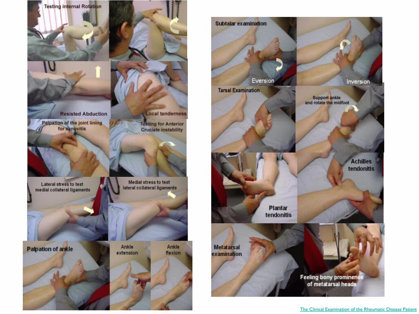
The Clinical Examination of the Rheumatic Disease Patient
Objectives
• Review the history and physical examination findings of inflammatory arthritis
• Define and categorize inflammatory arthritis
• Understand the initial laboratory and radiological evaluation of suspected rheumatological disease
• Increase clinical sensitivity and specificity to rheumatological disease
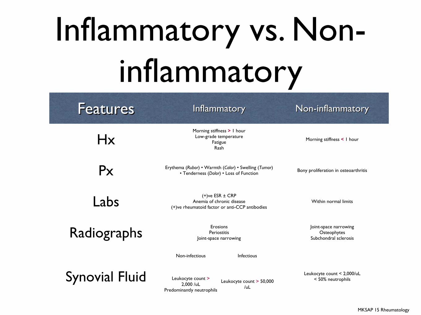
Inflammatory vs. Non-inflammatory
FeaturesFeatures InflammatoryInflammatory Non-inflammatoryNon-inflammatory
HxMorning stiffness > 1 hourLow-grade temperature
FatigueRash
Morning stiffness < 1 hour
Px Erythema (Rubor) • Warmth (Calor) • Swelling (Tumor) • Tenderness (Dolor) • Loss of Function Bony proliferation in osteoarthritis
Labs(+)ve ESR ± CRP
Anemia of chronic disease(+)ve rheumatoid factor or anti-CCP antibodies
Within normal limits
RadiographsErosionsPeriostitis
Joint-space narrowing
Joint-space narrowingOsteophytes
Subchondral sclerosis
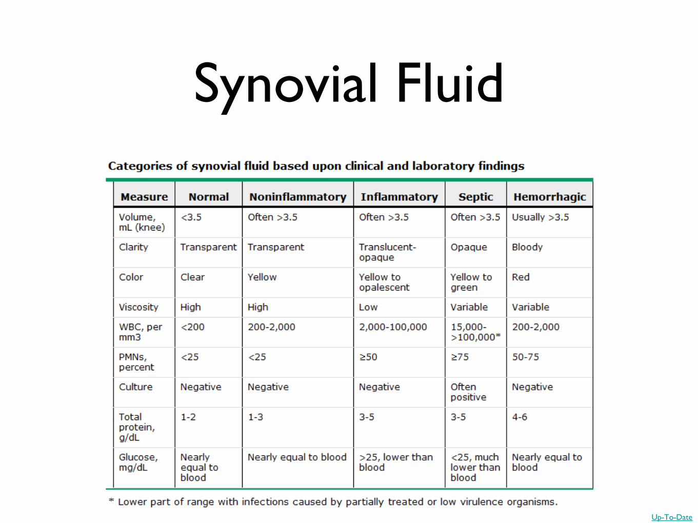
Synovial Fluid
Non-infectious Infectious
Leukocyte count < 2,000/uL< 50% neutrophilsLeukocyte count >
2,000 /uLPredominantly neutrophils
Leukocyte count > 50,000 /uL
MKSAP 15 Rheumatology
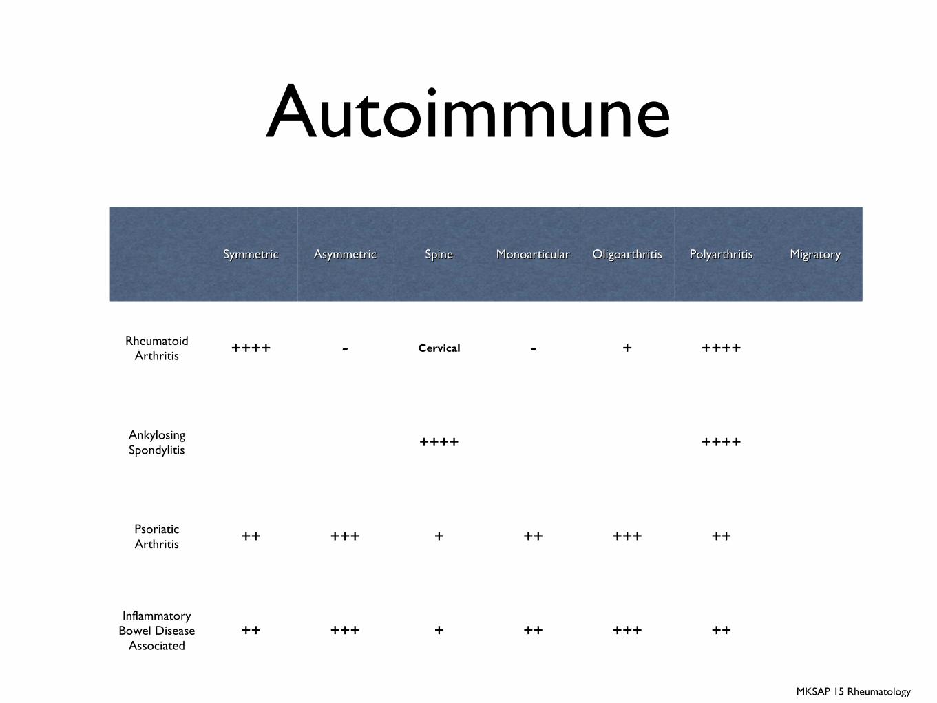
Autoimmune
SymmetricSymmetric AsymmetricAsymmetric SpineSpine MonoarticularMonoarticular OligoarthritisOligoarthritis PolyarthritisPolyarthritis MigratoryMigratory
Rheumatoid Arthritis ++++ - Cervical - + ++++
Ankylosing Spondylitis ++++ ++++
Psoriatic Arthritis ++ +++ + ++ +++ ++
Inflammatory Bowel Disease
Associated++ +++ + ++ +++ ++
MKSAP 15 Rheumatology
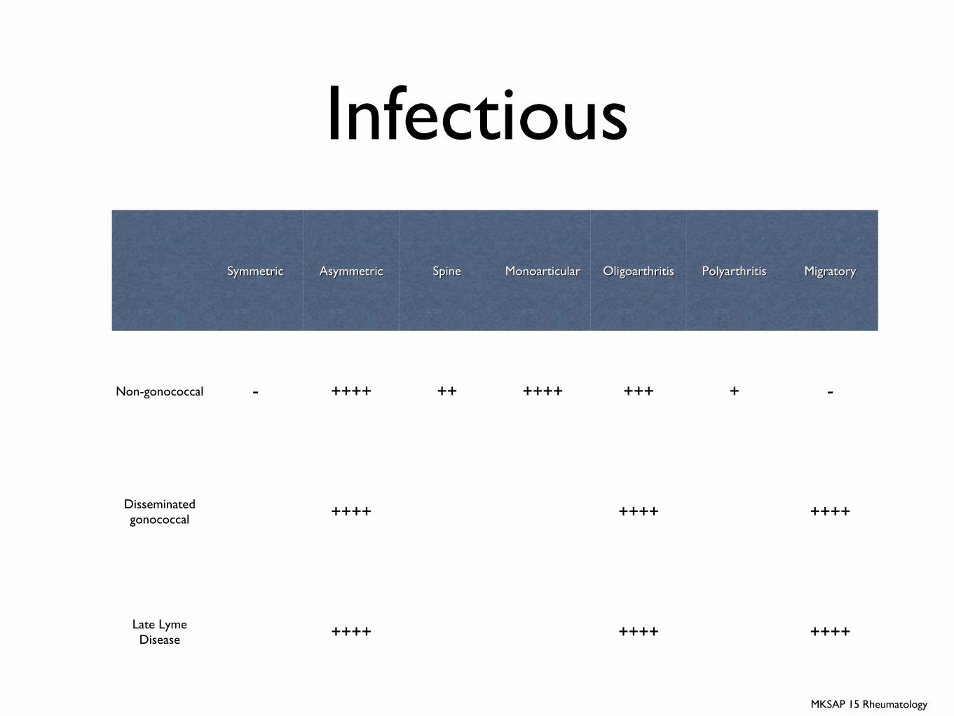
Infectious
SymmetricSymmetric AsymmetricAsymmetric SpineSpine MonoarticularMonoarticular OligoarthritisOligoarthritis PolyarthritisPolyarthritis MigratoryMigratory
Non-gonococcal - ++++ ++ ++++ +++ + -
Disseminated gonococcal ++++ ++++ ++++
Late Lyme Disease ++++ ++++ ++++
MKSAP 15 Rheumatology
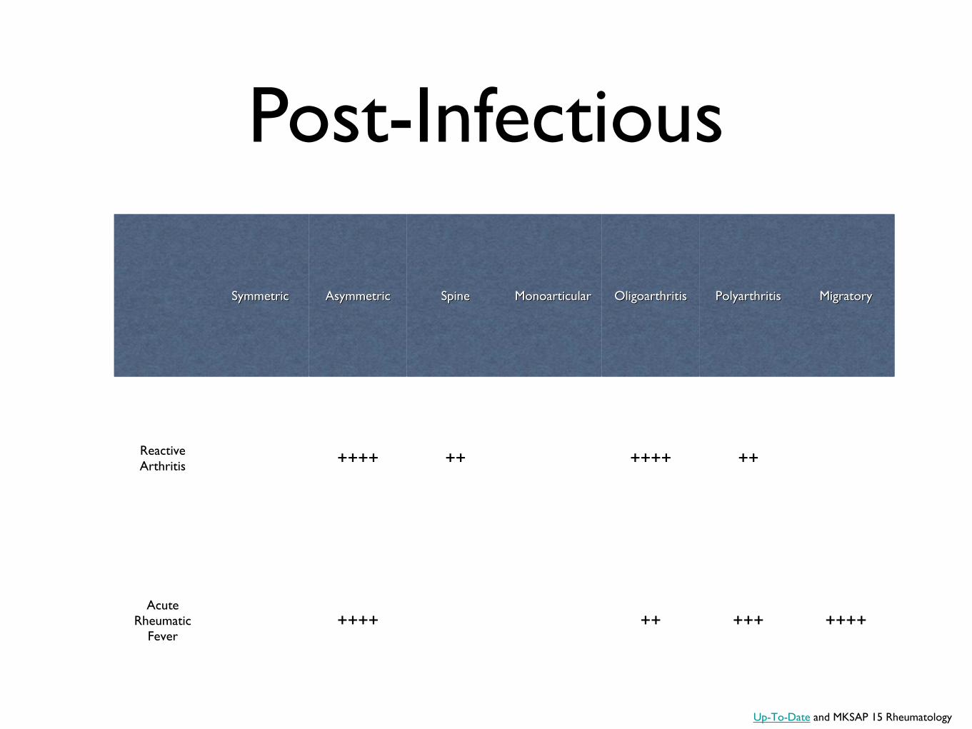
Post-Infectious
SymmetricSymmetric AsymmetricAsymmetric SpineSpine MonoarticularMonoarticular OligoarthritisOligoarthritis PolyarthritisPolyarthritis MigratoryMigratory
Reactive Arthritis ++++ ++ ++++ ++
Acute Rheumatic
Fever++++ ++ +++ ++++
Up-To-Date and MKSAP 15 Rheumatology
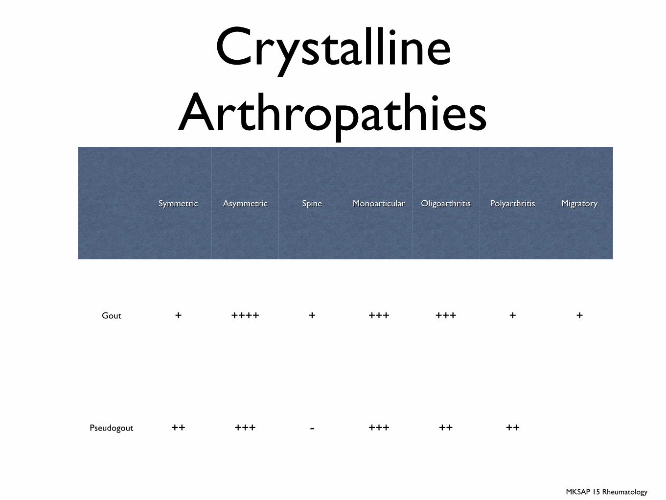
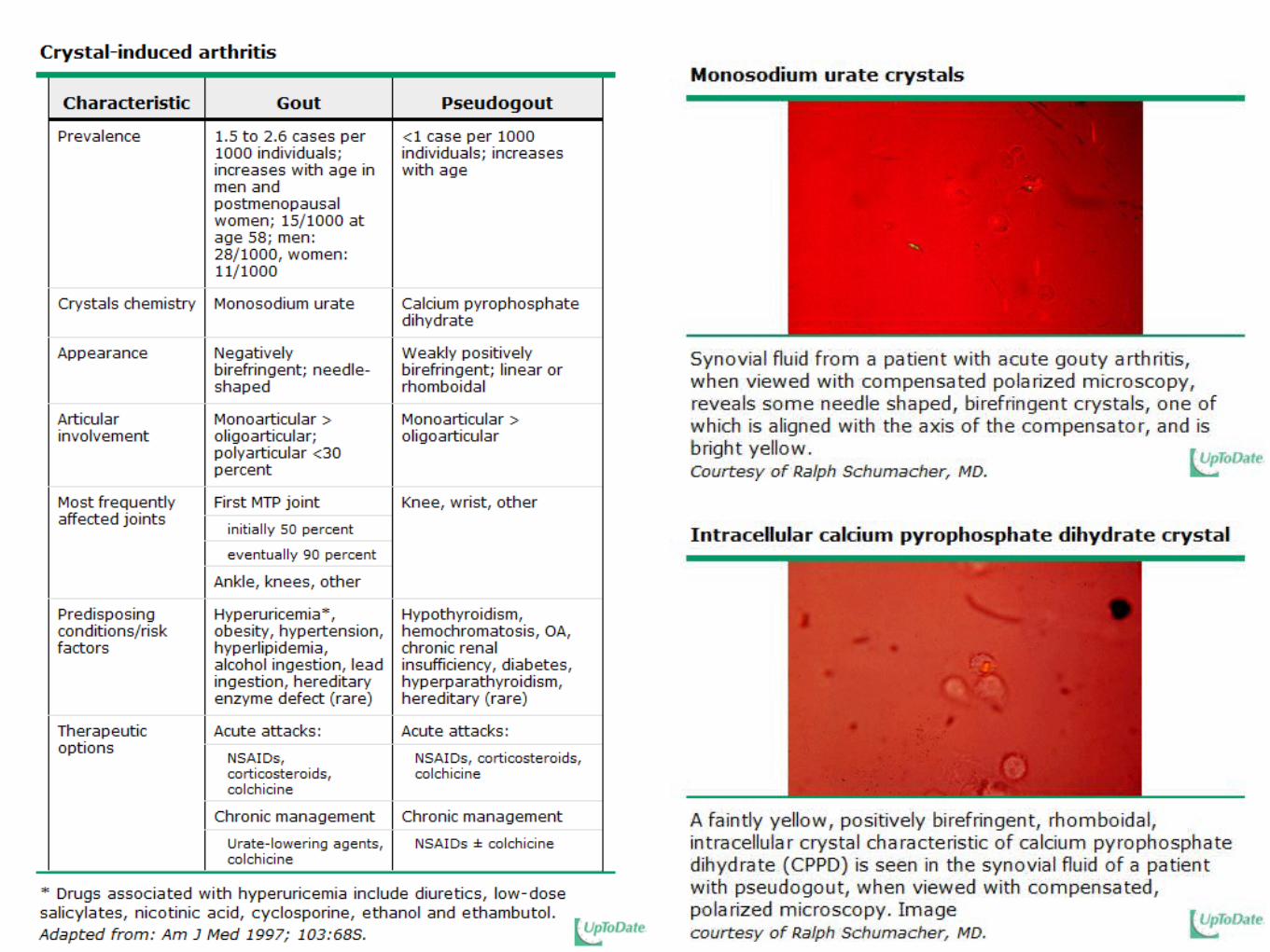
Crystalline Arthropathies
SymmetricSymmetric AsymmetricAsymmetric SpineSpine MonoarticularMonoarticular OligoarthritisOligoarthritis PolyarthritisPolyarthritis MigratoryMigratory
Gout + ++++ + +++ +++ + +
Pseudogout ++ +++ - +++ ++ ++
MKSAP 15 Rheumatology
Objectives
• Review the history and physical examination findings of inflammatory arthritis
• Define and categorize inflammatory arthritis
• Understand the initial laboratory and radiological evaluation of suspected rheumatological disease
• Increase clinical sensitivity and specificity to rheumatological disease
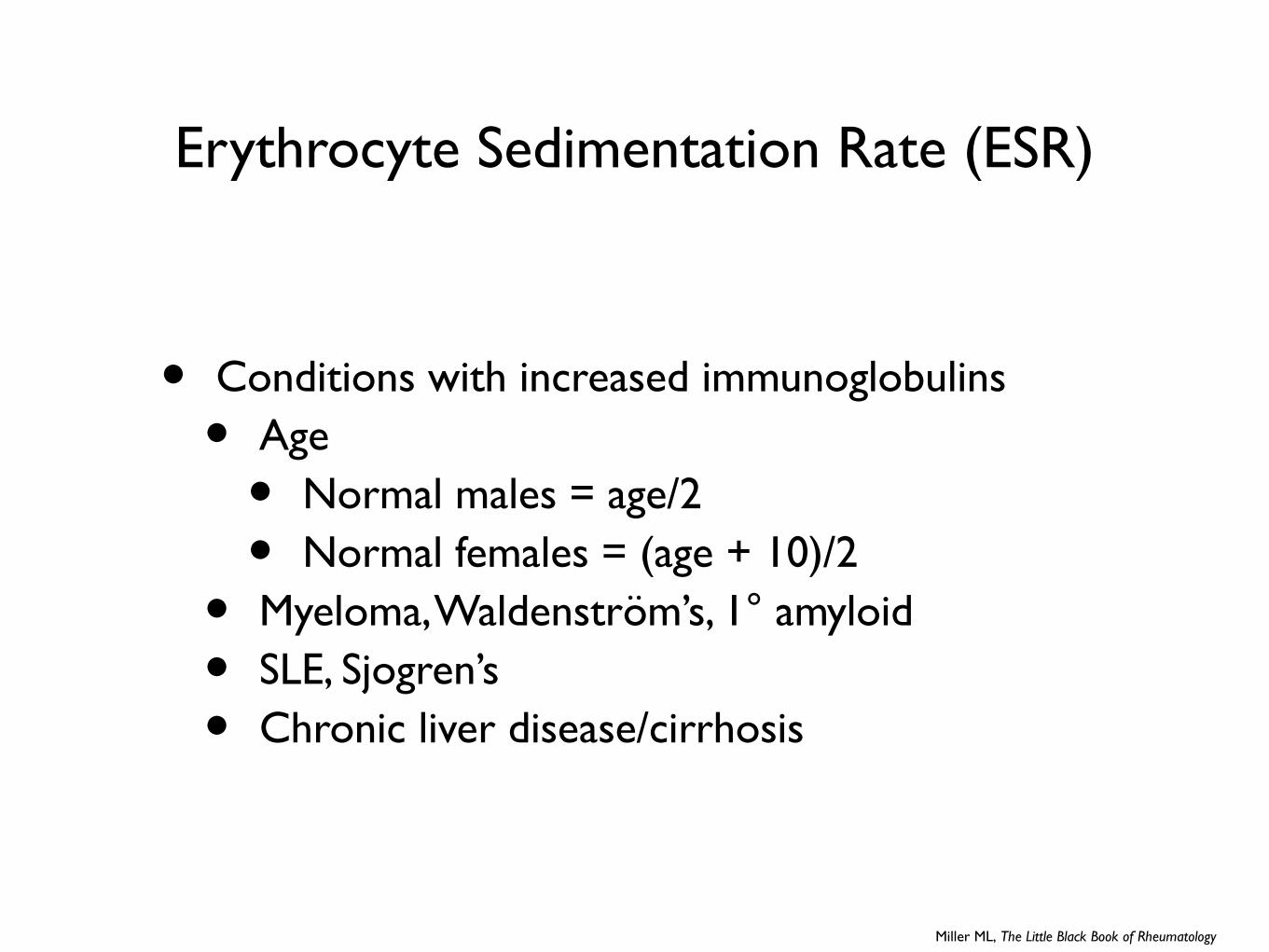
Erythrocyte Sedimentation Rate (ESR)
• Conditions with increased immunoglobulins
• Age
• Normal males = age/2
• Normal females = (age + 10)/2
• Myeloma, Waldenström’s, 1° amyloid
• SLE, Sjogren’s
• Chronic liver disease/cirrhosis
Miller ML, The Little Black Book of Rheumatology
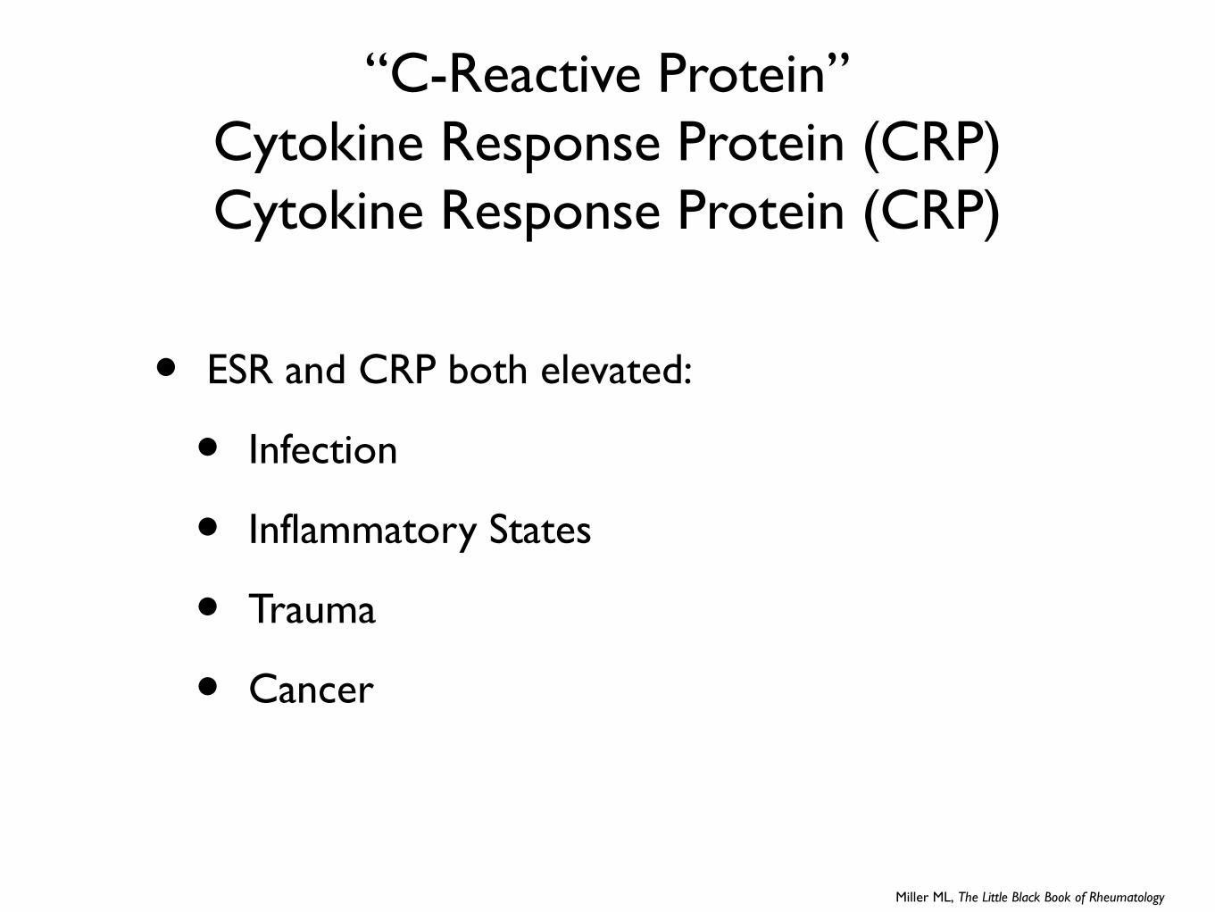
“C-Reactive Protein”Cytokine Response Protein (CRP)Cytokine Response Protein (CRP)
• ESR and CRP both elevated:
• Infection
• Inflammatory States
• Trauma
• Cancer
Miller ML, The Little Black Book of Rheumatology
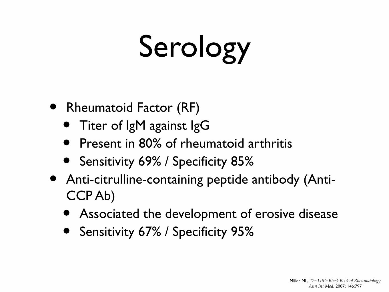
Serology
• Rheumatoid Factor (RF)
• Titer of IgM against IgG
• Present in 80% of rheumatoid arthritis
• Sensitivity 69% / Specificity 85%
• Anti-citrulline-containing peptide antibody (Anti-CCP Ab)
• Associated the development of erosive disease
• Sensitivity 67% / Specificity 95%
Miller ML, The Little Black Book of RheumatologyAnn Int Med, 2007; 146:797
Objectives
• Review the history and physical examination findings of inflammatory arthritis
• Define and categorize inflammatory arthritis
• Understand the initial laboratory and radiological evaluation of suspected rheumatological disease
• Increase clinical sensitivity and specificity to rheumatological disease
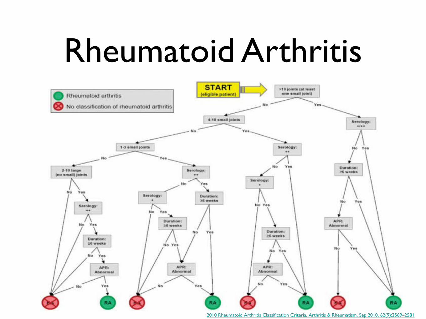
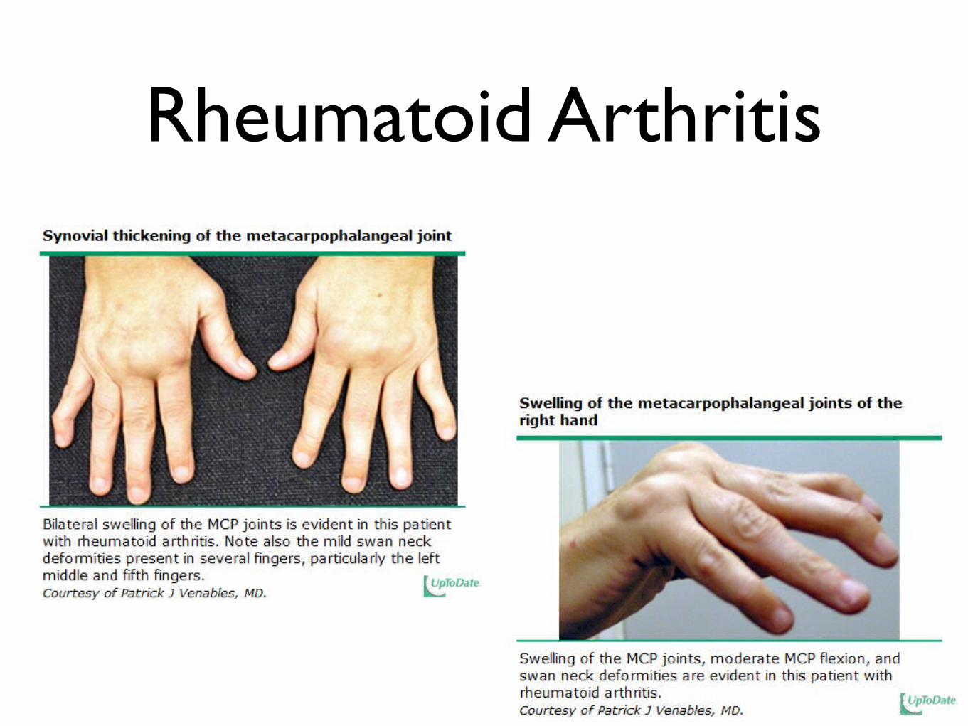
Rheumatoid Arthritis
2010 Rheumatoid Arthritis Classification Criteria, Arthritis & Rheumatism, Sep 2010, 62(9):2569–2581
Rheumatoid Arthritis
Ankylosing Spondylitis
Modified New York Criteria (1984)
• Definite ankylosing spondylitis is present if
• Radiologic criterion is present in addition to at least one clinical criterion.
• Probable ankylosing spondylitis is present if
• Three clinical criterion are present alone or if
• The radiologic criterion is present but no clinical criteria are present.
Clinical criteria
• Low back pain: present for more than 3 months, improved by exercise but not relieved by rest.
• Limitation of lumbar spine motion in sagittal and frontal planes.
• Limitation of chest expansion relative to normal values for age and sex.
Radiologic criterion
• Sacroiliitis on radiographs
http://emedicine.medscape.com/article/386639-overview
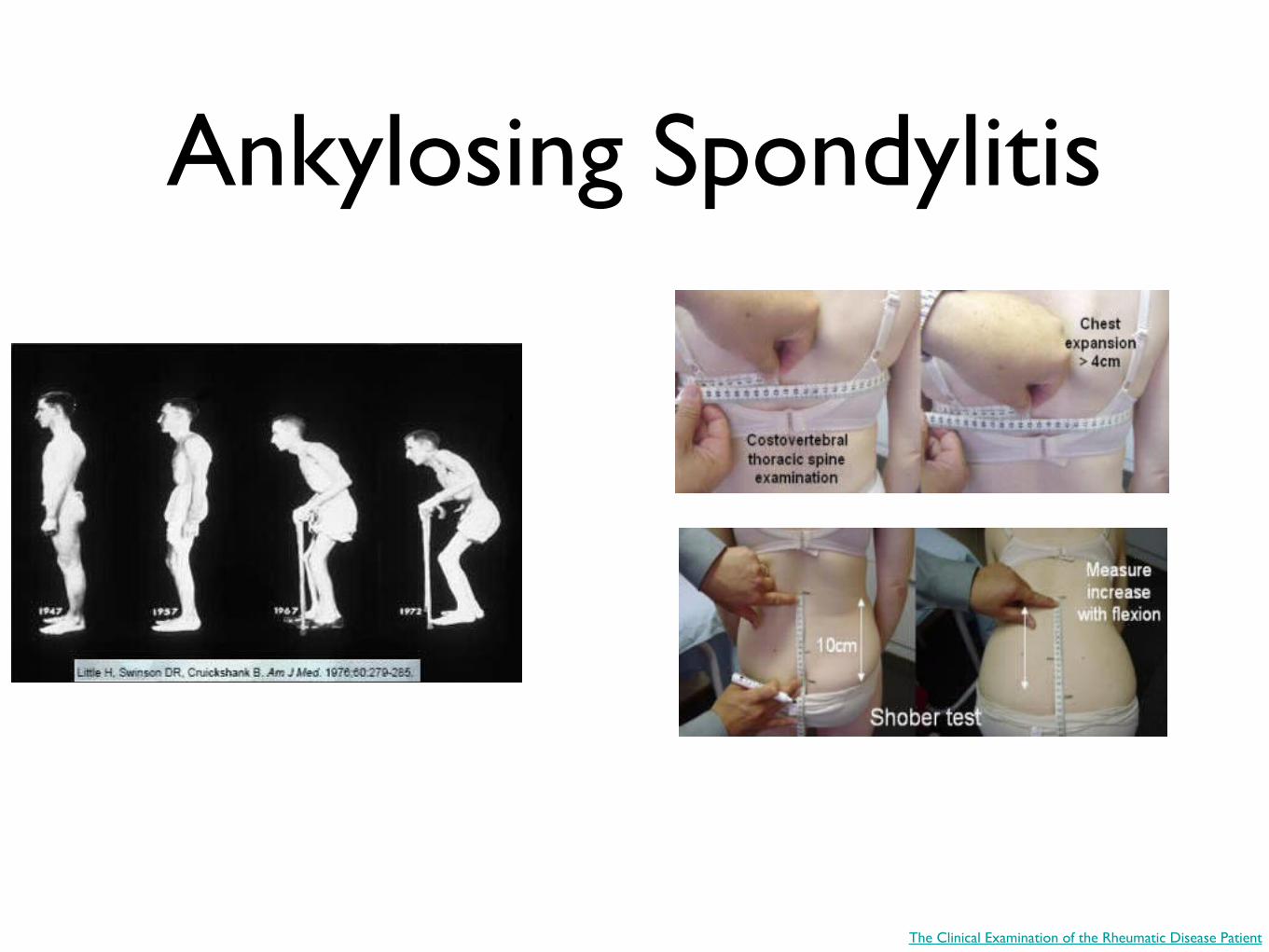
Ankylosing Spondylitis
The Clinical Examination of the Rheumatic Disease Patient
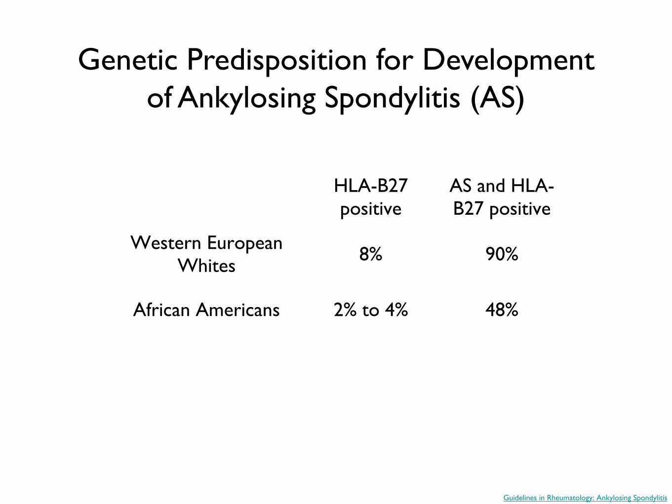
Genetic Predisposition for Development of Ankylosing Spondylitis (AS)
HLA-B27 positive
AS and HLA-B27 positive
Western European Whites 8% 90%
African Americans 2% to 4% 48%
Guidelines in Rheumatology: Ankylosing Spondylitis
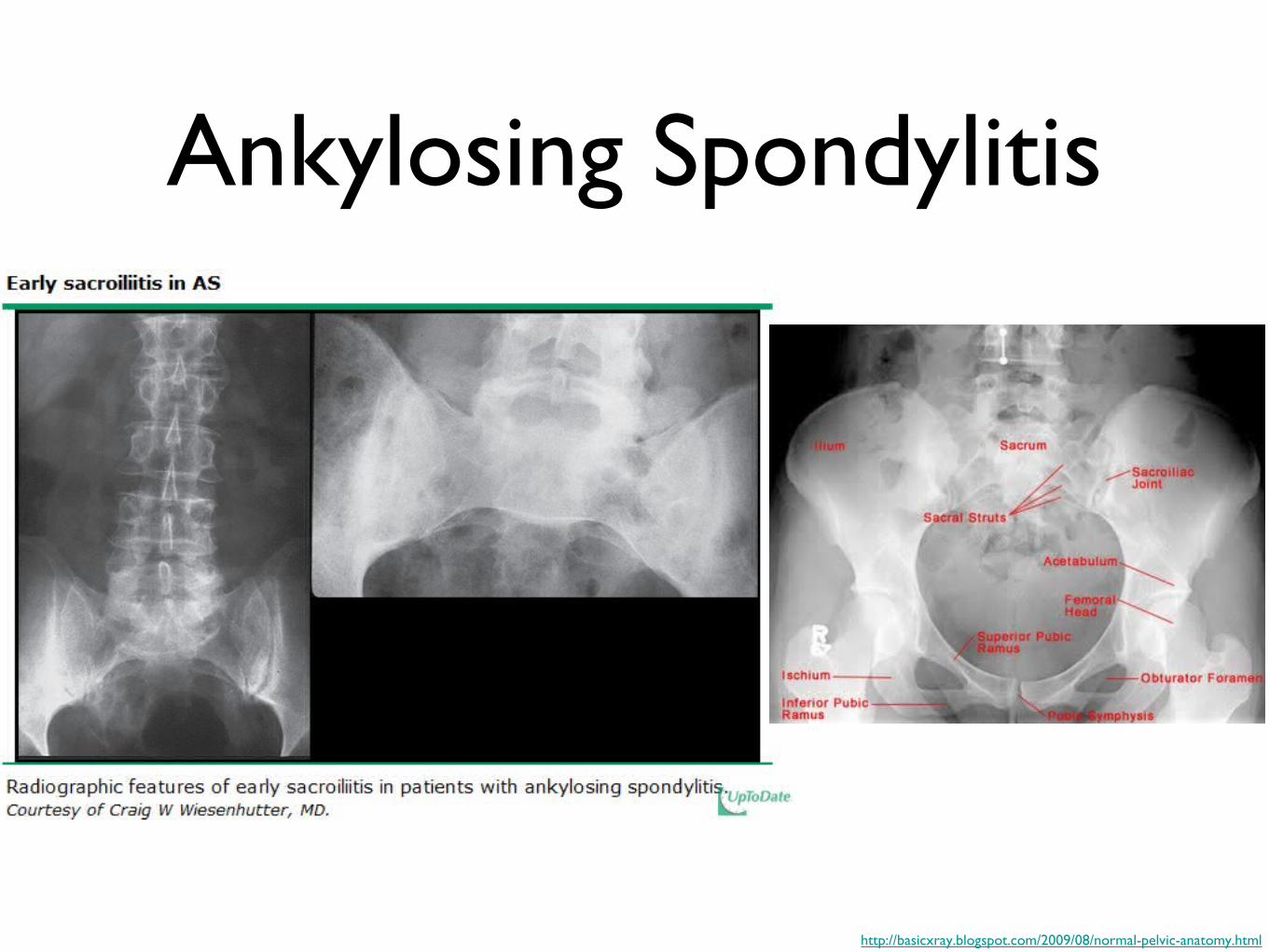
Ankylosing Spondylitis
http://basicxray.blogspot.com/2009/08/normal-pelvic-anatomy.html
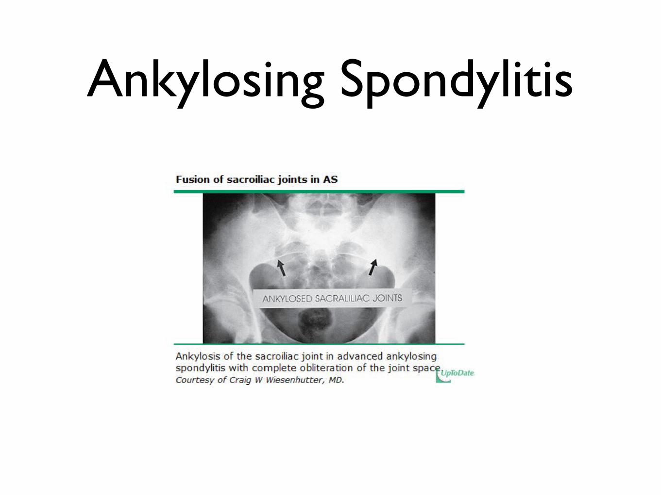
Ankylosing Spondylitis
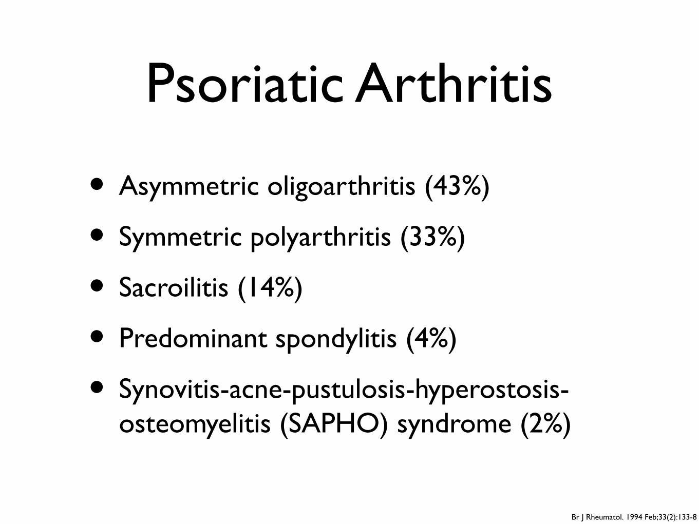
Psoriatic Arthritis
• Asymmetric oligoarthritis (43%)
• Symmetric polyarthritis (33%)
• Sacroilitis (14%)
• Predominant spondylitis (4%)
• Synovitis-acne-pustulosis-hyperostosis-osteomyelitis (SAPHO) syndrome (2%)
Br J Rheumatol. 1994 Feb;33(2):133-8
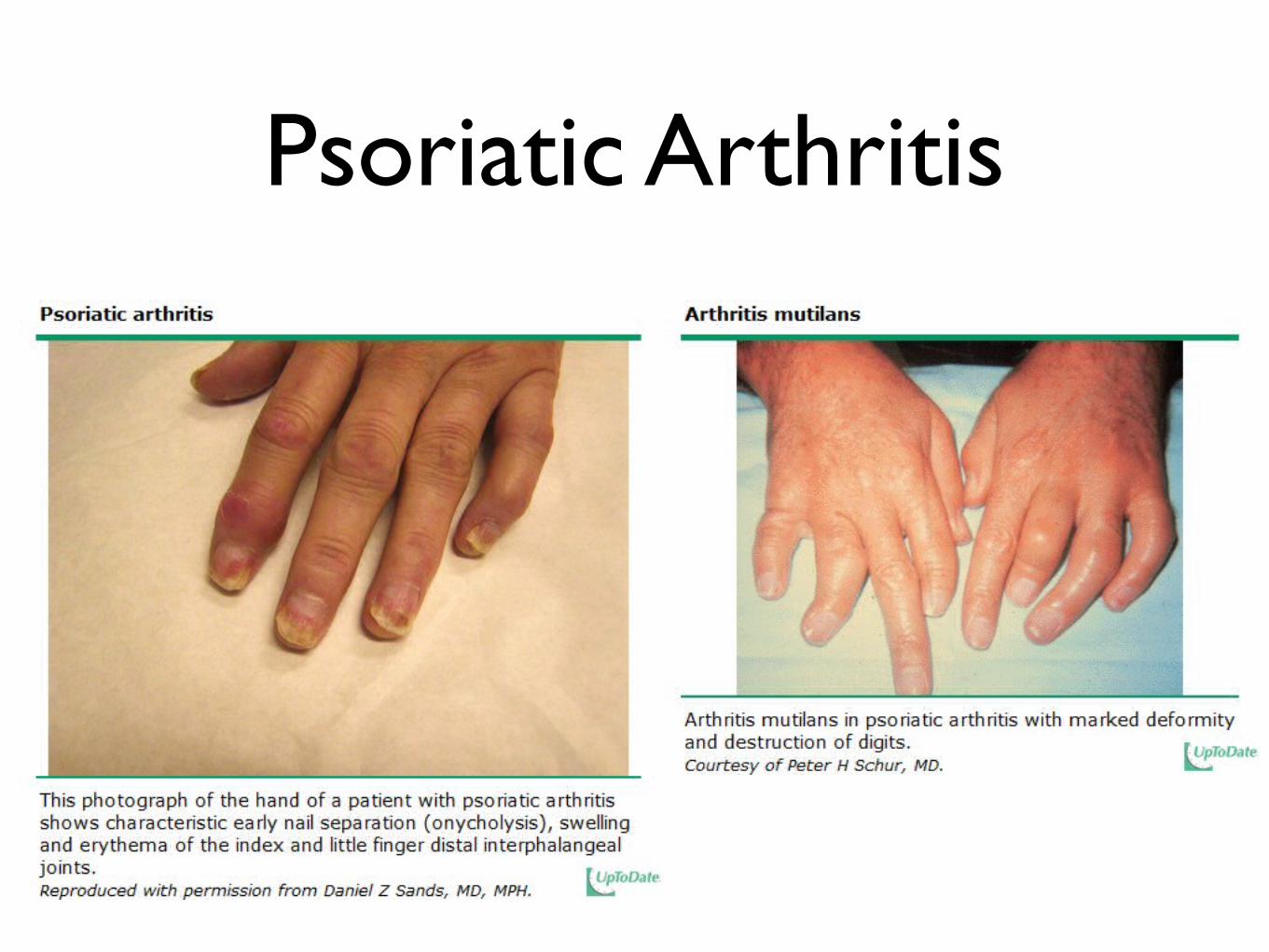
Psoriatic Arthritis
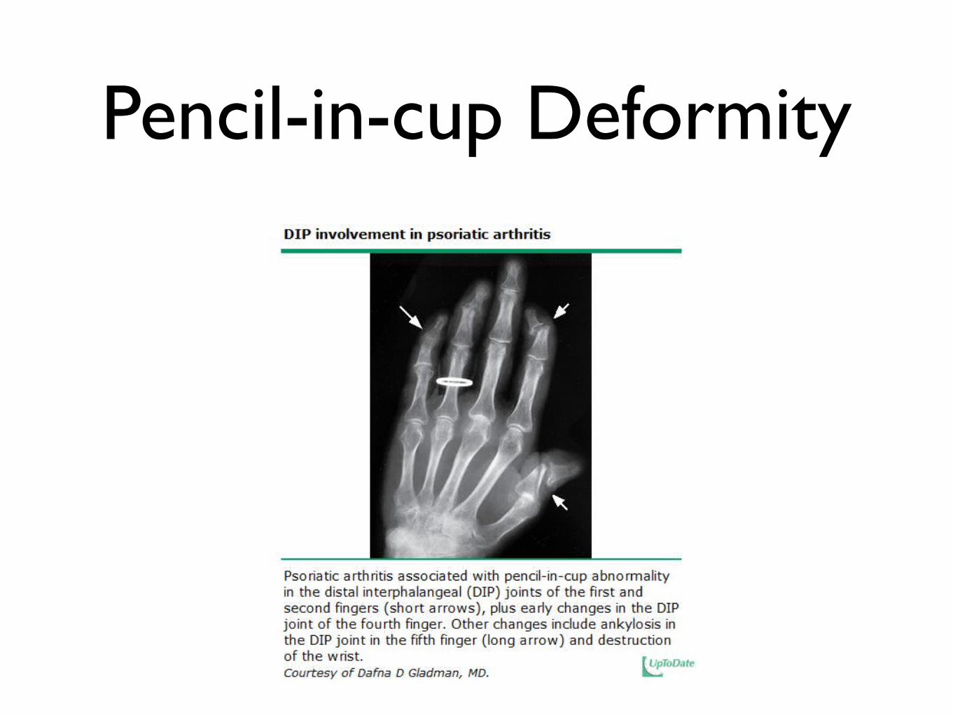
Pencil-in-cup Deformity
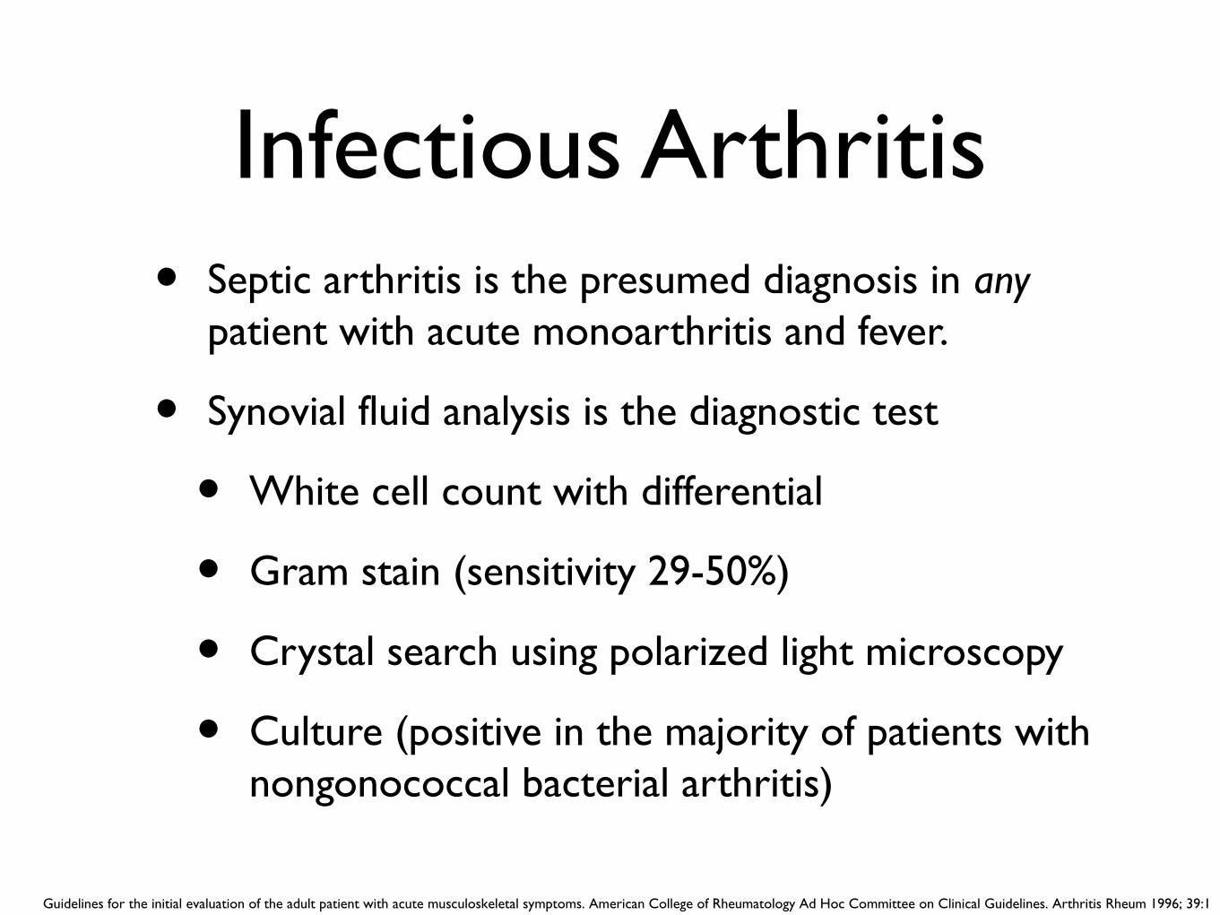
Infectious Arthritis• Septic arthritis is the presumed diagnosis in any
patient with acute monoarthritis and fever.
• Synovial fluid analysis is the diagnostic test
• White cell count with differential
• Gram stain (sensitivity 29-50%)
• Crystal search using polarized light microscopy
• Culture (positive in the majority of patients with nongonococcal bacterial arthritis)
Guidelines for the initial evaluation of the adult patient with acute musculoskeletal symptoms. American College of Rheumatology Ad Hoc Committee on Clinical Guidelines. Arthritis Rheum 1996; 39:1
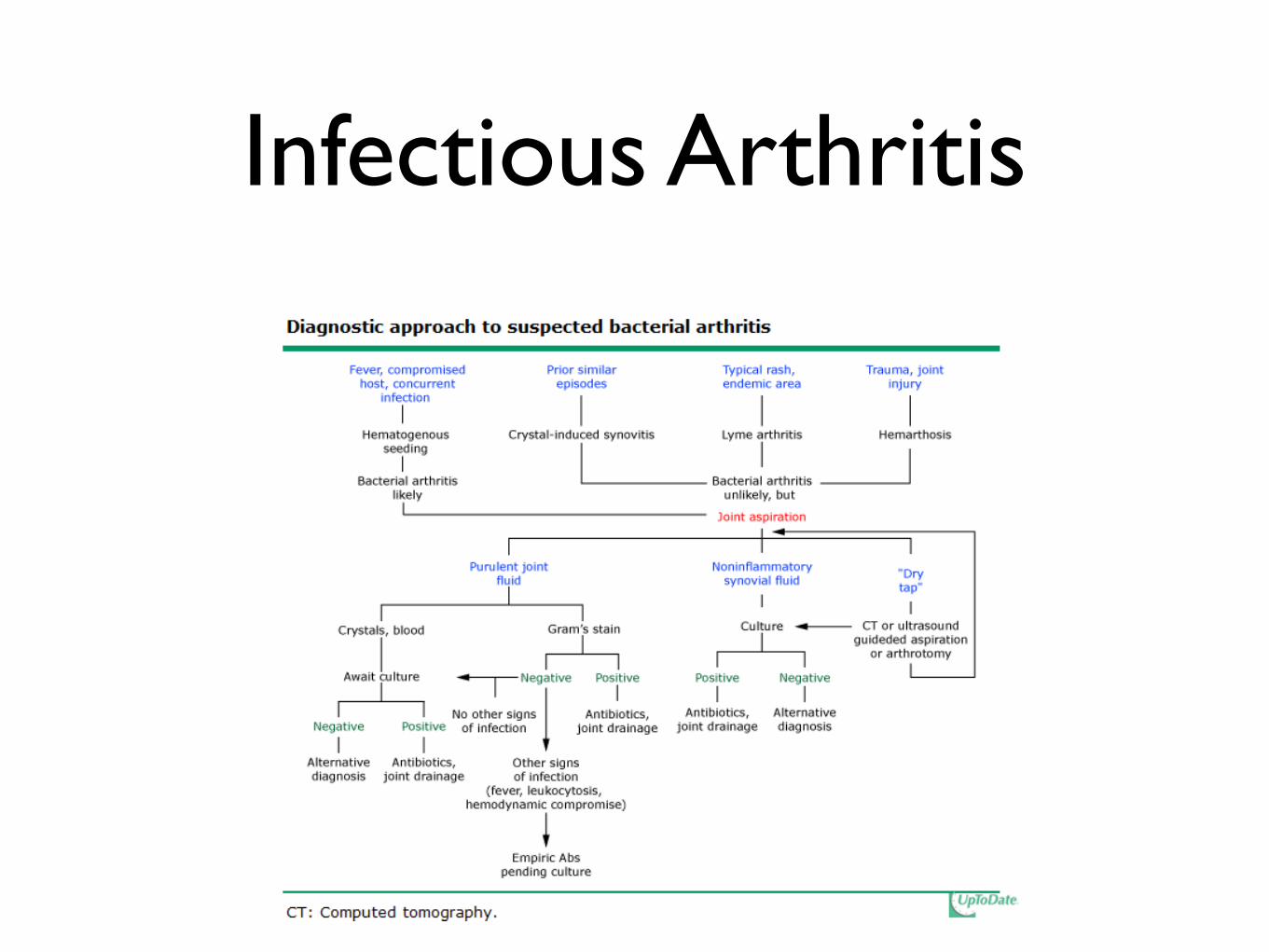
Infectious Arthritis
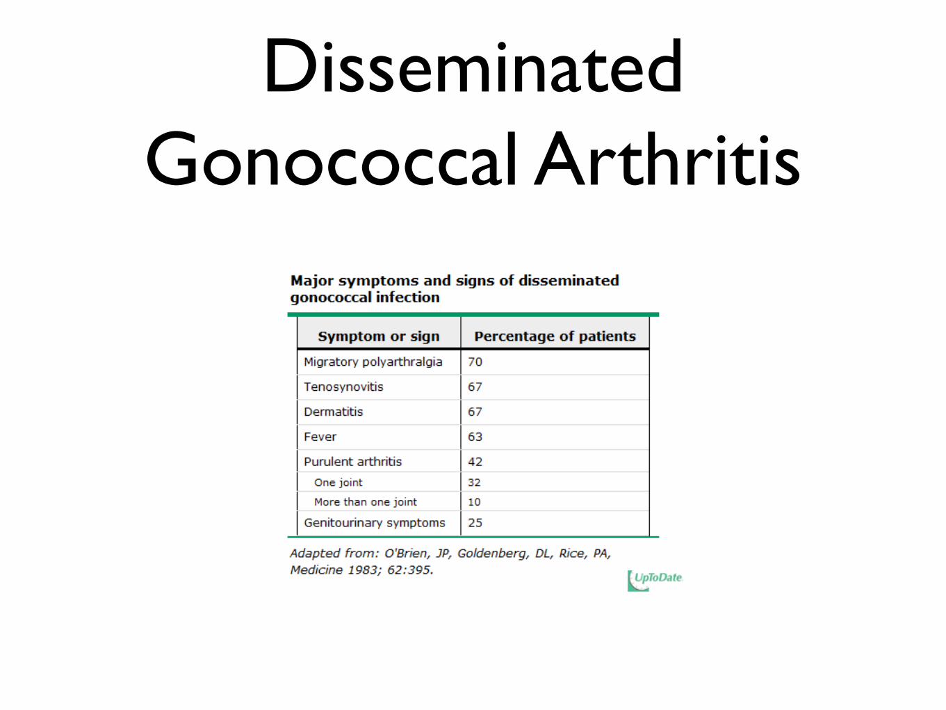
Disseminated Gonococcal Arthritis
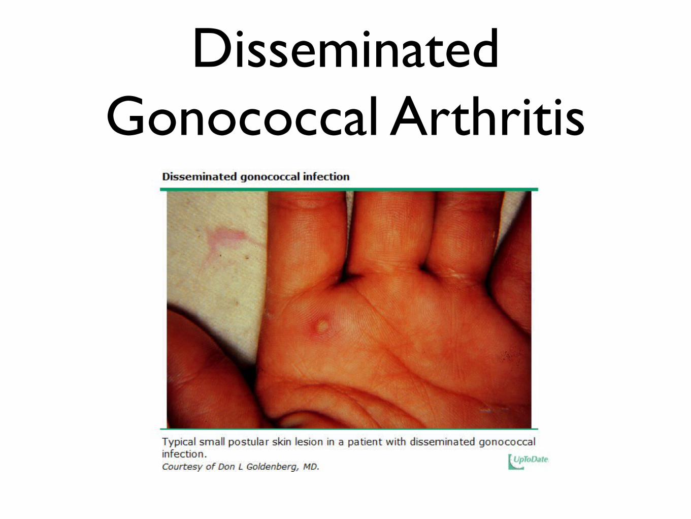
Disseminated Gonococcal Arthritis
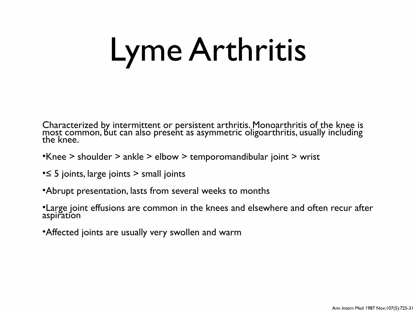
Lyme Arthritis
Characterized by intermittent or persistent arthritis. Monoarthritis of the knee is most common, but can also present as asymmetric oligoarthritis, usually including the knee.
•Knee > shoulder > ankle > elbow > temporomandibular joint > wrist
•≤ 5 joints, large joints > small joints
•Abrupt presentation, lasts from several weeks to months
•Large joint effusions are common in the knees and elsewhere and often recur after aspiration
•Affected joints are usually very swollen and warm
Ann Intern Med 1987 Nov;107(5):725-31
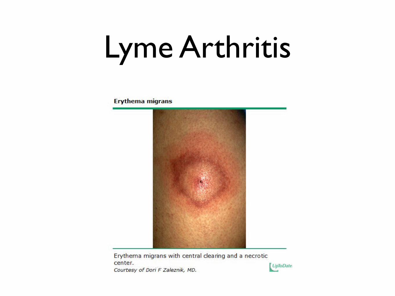
Lyme Arthritis
Reactive Arthritis
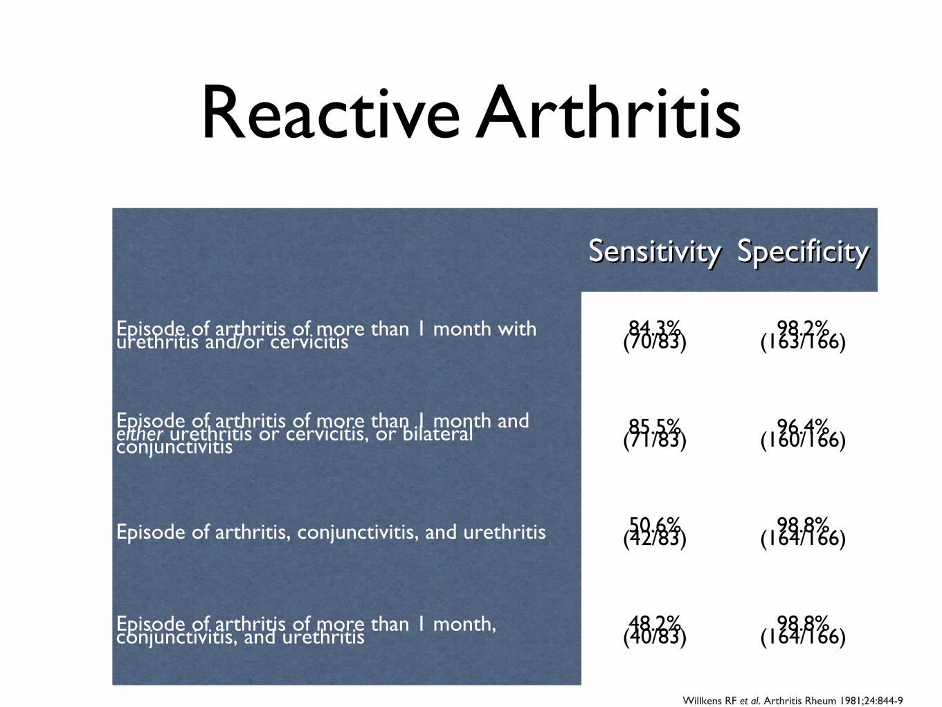
Willkens RF et al. Arthritis Rheum 1981;24:844-9
SensitivitySensitivity SpecificitySpecificity
Episode of arthritis of more than 1 month with urethritis and/or cervicitis 84.3%(70/83) 98.2%(163/166)
Episode of arthritis of more than 1 month and either urethritis or cervicitis, or bilateral conjunctivitis85.5%(71/83) 96.4%(160/166)
Episode of arthritis, conjunctivitis, and urethritis 50.6%(42/83) 98.8%(164/166)
Episode of arthritis of more than 1 month, conjunctivitis, and urethritis 48.2%(40/83) 98.8%(164/166)
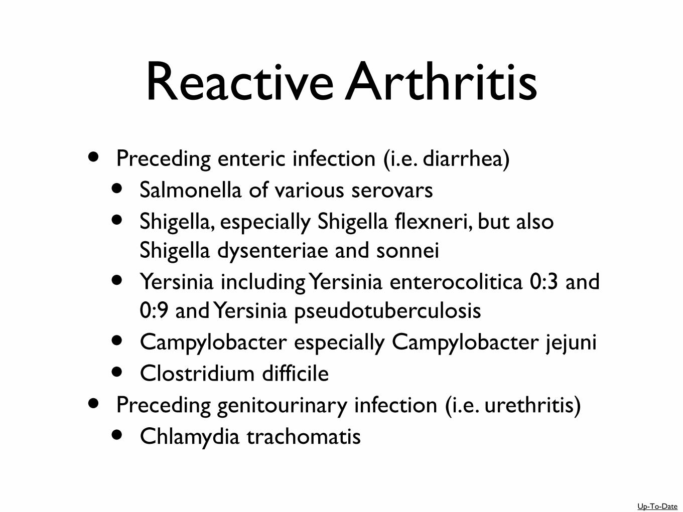
Reactive Arthritis• Preceding enteric infection (i.e. diarrhea)
• Salmonella of various serovars
• Shigella, especially Shigella flexneri, but also Shigella dysenteriae and sonnei
• Yersinia including Yersinia enterocolitica 0:3 and 0:9 and Yersinia pseudotuberculosis
• Campylobacter especially Campylobacter jejuni
• Clostridium difficile
• Preceding genitourinary infection (i.e. urethritis)
• Chlamydia trachomatis
Up-To-Date
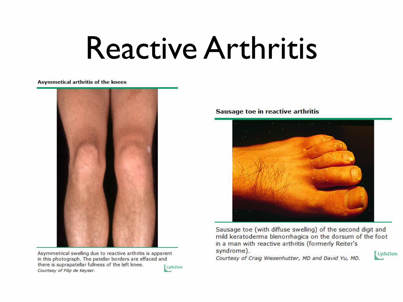
Reactive Arthritis

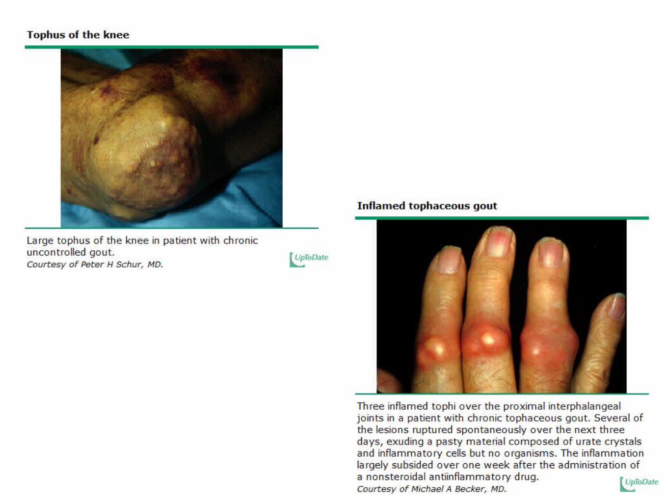
Gout1977 American College of 1977 American College of
Rheumatology CriteriaRheumatology Criteria New York CriteriaNew York Criteria Rome CriteriaRome Criteria
Monosodium urate monohydrate microcrystals in joint
fluid during attack or ≥ 6 of the following
MSU microcrystals in joint fluid or tissue or tophus or
≥ 2 of the following≥ 2 of the following
> 1 attack of acute arthritis 2 attacks of painful limb joint swelling
Maximum inflammation developed within 1 day Abrupt onset and remission in 1-2 weeks initially Abrupt, painful joint swelling, clearing in 1-2 weeks initially
Monoarthritis attack
Redness observed over joints
First metatarsophalangeal joint painful or swollen First metatarsophalangeal joint attack
Unilateral first metatarsophalangeal joint attack
Unilateral tarsal joint attack
Tophus (proven or suspected) Presence of a tophus Presence of tophi
Hyperuricemia Serum uric acid > 7 in males and > 6 in females
Asymmetric swelling within a joint on x-ray
Subcortical cysts without erosions on x-ray
Joint fluid culture negative for organisms during attackResponse to colchicine—major reduction in
inflammation within 48 hours MSU microcrystals in joint fluid or tissueWallace SL, et al. Arthritis Rheum 1977;20:895-900 and J Clin Rheumatol. 2009 Feb;15(1):22-4

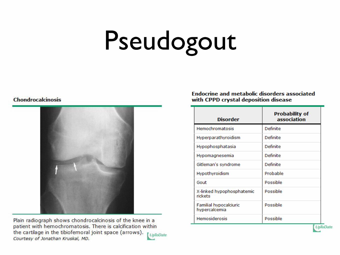
Pseudogout
Definite diagnosis of pseudogout requires either:
• The demonstration of CPPD crystals in tissue or synovial fluid by definitive means (eg, x-ray diffraction, etc) or
• The presence of both positively (but weakly) birefringent crystals by compensated polarized light microscopy and typical cartilage or joint capsule calcification on x-ray examination
Probable diagnosis of pseudogout occurs with either:
• The identification of positively (but weakly) birefringent crystals by compensated polarized light microscopy or
• The presence of typical cartilage or joint capsule calcification on radiographic examination.
Up-To-Date
Pseudogout
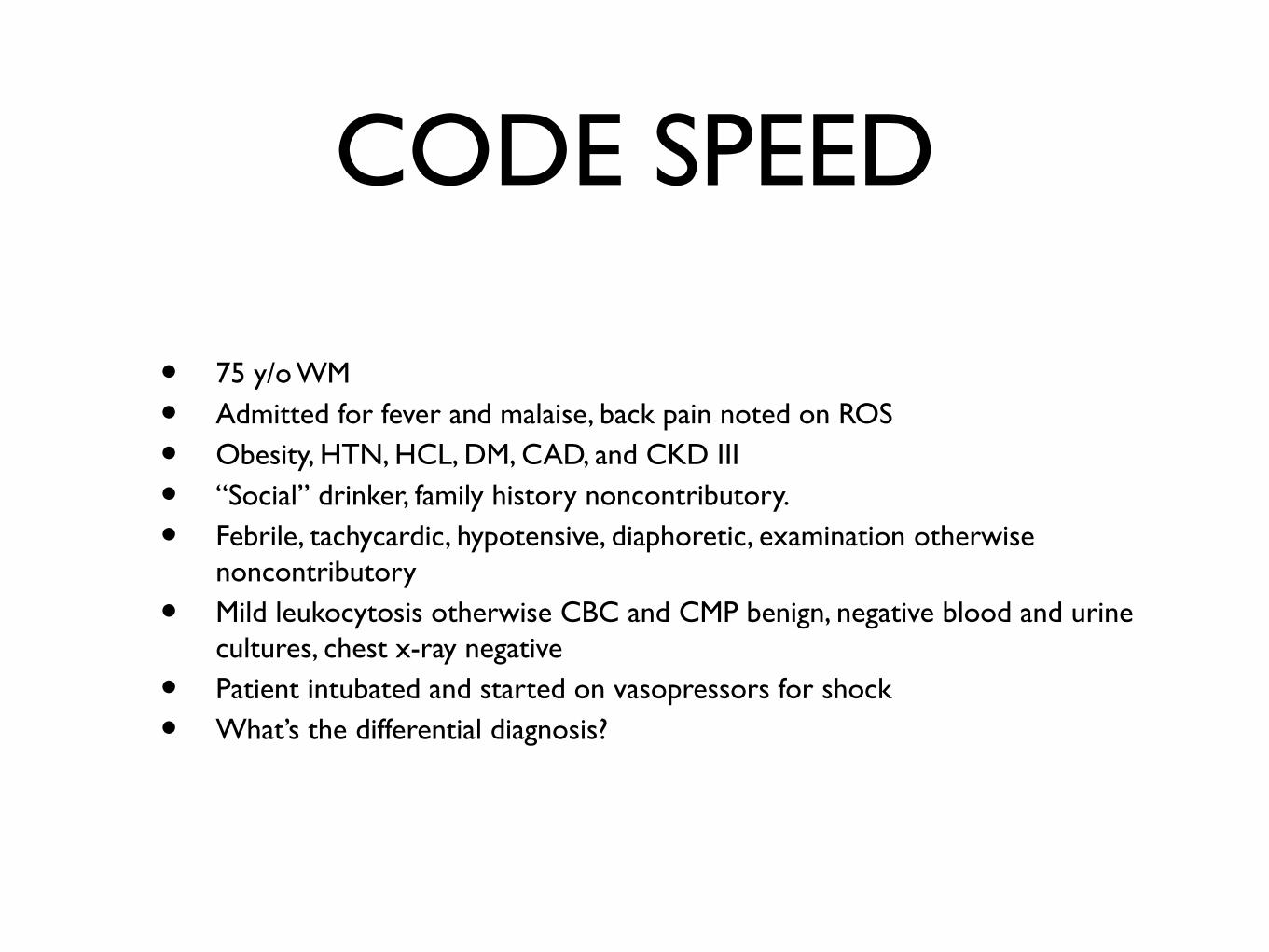
CODE SPEED
• 75 y/o WM
• Admitted for fever and malaise, back pain noted on ROS
• Obesity, HTN, HCL, DM, CAD, and CKD III
• “Social” drinker, family history noncontributory.
• Febrile, tachycardic, hypotensive, diaphoretic, examination otherwise noncontributory
• Mild leukocytosis otherwise CBC and CMP benign, negative blood and urine cultures, chest x-ray negative
• Patient intubated and started on vasopressors for shock
• What’s the differential diagnosis?


![Inflammatory Arthritis Care Path Toolkit · (AAC) in response to ... An 11-question self-administered questionnaire (Early Inflammatory Arthritis [EIA] ... Inflammatory Arthritis](https://img.pdfslide.us/doc/110x75/5ae82c107f8b9a6d4f8f2276/inflammatory-arthritis-care-path-aac-in-response-to-an-11-question-self-administered.jpg)