Embed Size (px)
Citation preview

GENITO – URINARY TUBERCULOSIS
Dr Althaf Hussain DNB Urology MMHRC
young to middle-aged adults.
M/F ratio= 5:3
Uncommon in children
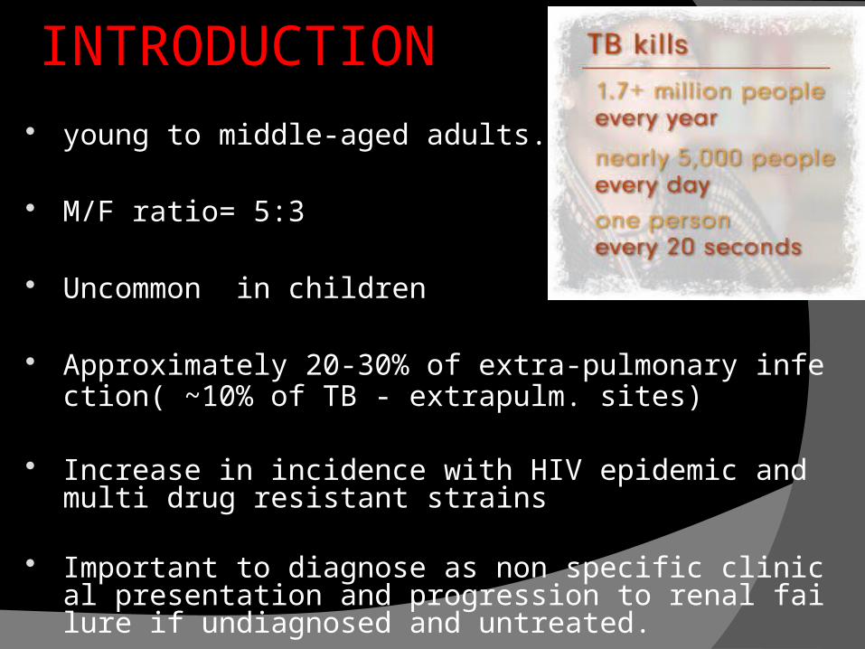
Approximately 20-30% of extra-pulmonary infection( ~10% of TB - extrapulm. sites)
Increase in incidence with HIV epidemic and multi drug resistant strains
Important to diagnose as non specific clinical presentation and progression to renal failure if undiagnosed and untreated.
INTRODUCTION

The kidneys are the most common site of GUTB
Causative organism : Mycobacterium Tuberculosis.
history of previous clinical TB (25%) with a lag time
of 2- 20 years
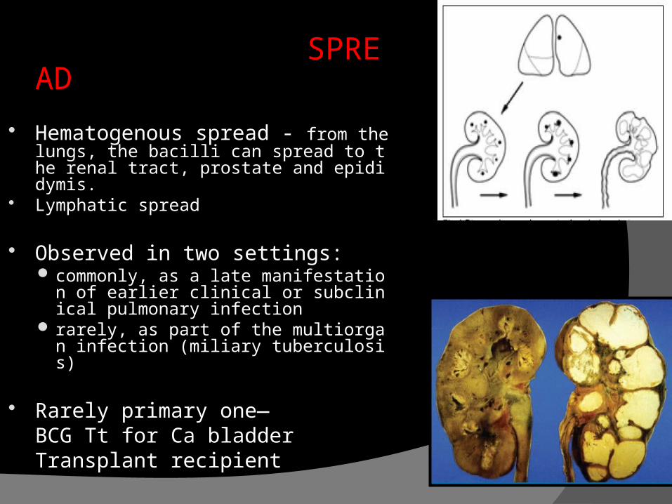
SPREAD Hematogenous spread - from the
lungs, the bacilli can spread to the renal tract, prostate and epididymis.
Lymphatic spread
Observed in two settings: commonly, as a late manifestation of
earlier clinical or subclinical pulmonary infection
rarely, as part of the multiorgan infection (miliary tuberculosis)
Rarely primary one—BCG Tt for Ca bladderTransplant recipient
CLINICAL FEATURES gross / microscopic hematuria sterile pyuria� Mild proteinuria urinary frequency, dysuria, ‘intractable’ UTI frequency, urgency, dysuria with involvement of bladder back, flank, or abdominal pain. : => extensive renal
disease Constitutional symptoms such as fever, weight loss,
fatigue, and anorexia are less common haemospermia ‘acute epididymo-orchitis’ Hydrocele,discharging scrotal/perineal sinuses Infertility,spontaneous abortion,ectopic pregnancy. Menstrual irregularities
Three other major complications of renal tuberculosis:
hypertension (RAS axis mediated)
super-infection (12 to 50%)nephrolithiasis (7 to 18%)
OTHER COMPLICATIONS: Perinephric inflammation Abscess formation :including psoas abscess Fistulae Sinus tract into adjacent tissues or viscera.

PATHOGENESIS
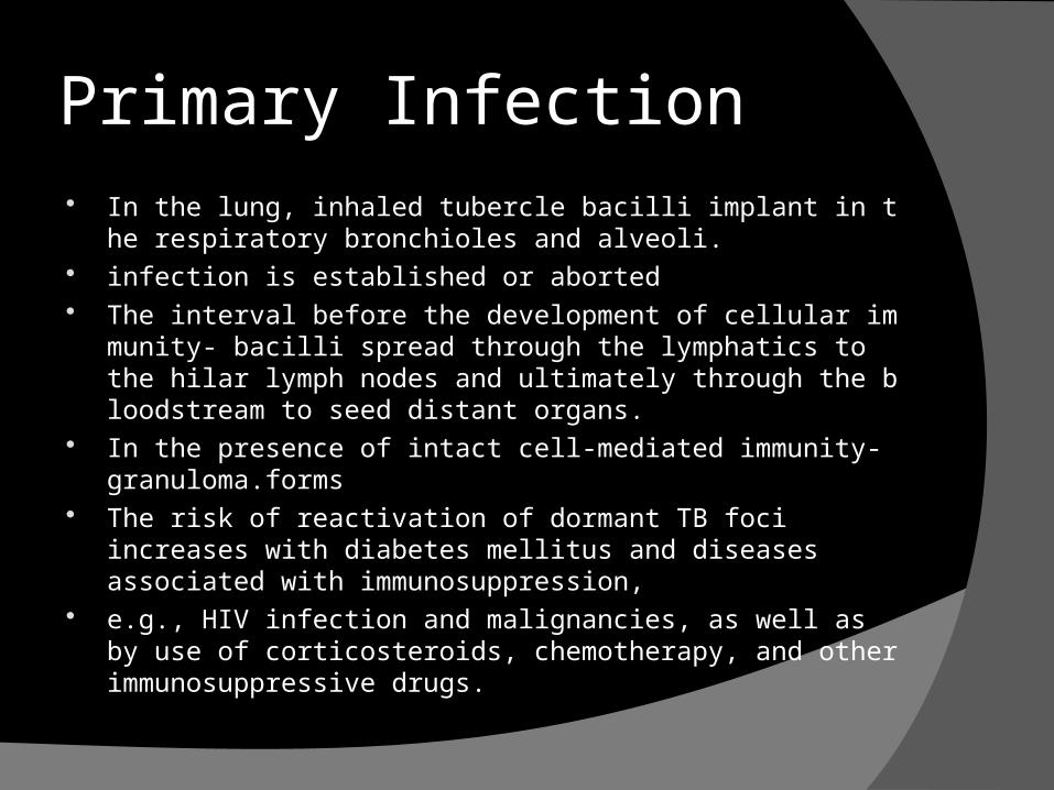
Primary Infection In the lung, inhaled tubercle bacilli implant in the respiratory br
onchioles and alveoli. infection is established or aborted The interval before the development of cellular immunity-
bacilli spread through the lymphatics to the hilar lymph nodes and ultimately through the bloodstream to seed distant organs.
In the presence of intact cell-mediated immunity- granuloma.forms
The risk of reactivation of dormant TB foci increases with diabetes mellitus and diseases associated with immunosuppression,
e.g., HIV infection and malignancies, as well as by use of corticosteroids, chemotherapy, and other immunosuppressive drugs.

Pathologic Features Kidney : The kidneys are the primary site of hematogenous spread of
TB. cortex being favored due to its greater blood supply and higher oxygen tension
Upon activation of the disease, a chronic inflammatory process arises with the subsequent development of characteristic granulomata,
Central caseous necrosis builds up within the tubercles, and neighboring tuberculous foci coalesce to form confluent areas of caseation.
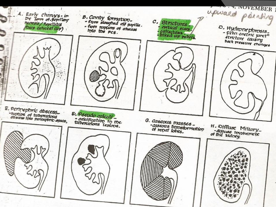
inflammatory changes extend into the renal tubules and medulla Renal papilla involvement results in sloughing and caseous
material gaining access to the collecting system by calyceal ulceration
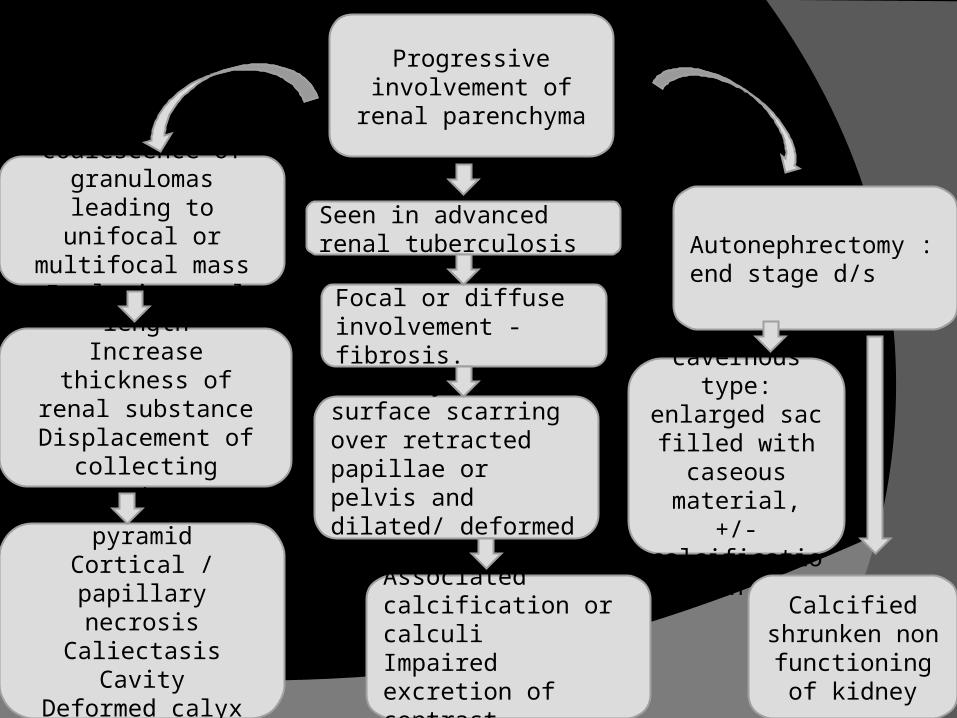
Progressive involvement of renal
parenchyma
coalescence of granulomas leading to unifocal or multifocal
mass lesions
Seen in advanced renal tuberculosis
Increase renal lengthIncrease thickness of
renal substanceDisplacement of
collecting system.
Parenchymal surface scarring over retracted papillae or pelvis and dilated/ deformed calyces.
Associated calcification or calculiImpaired excretion of contrast
Erosion of pyramidCortical / papillary
necrosisCaliectasis
CavityDeformed calyx
Caseo – cavernous type:
enlarged sac filled with caseous
material, +/- calcification
Calcified shrunken non functioning of
kidney
Autonephrectomy : end stage d/s
Focal or diffuse involvement - fibrosis.
12
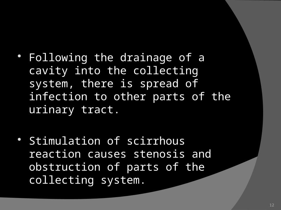
Following the drainage of a cavity into the collecting system, there is spread of infection to other parts of the urinary tract.
Stimulation of scirrhous reaction causes stenosis and obstruction of parts of the collecting system.
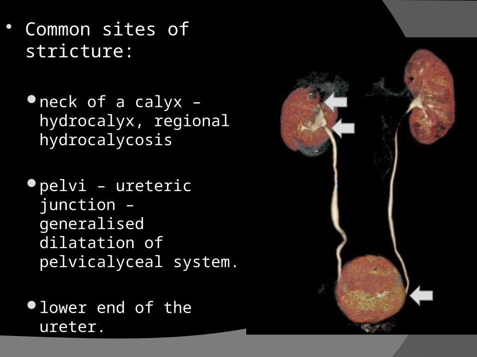
Common sites of stricture:
neck of a calyx – hydrocalyx, regional hydrocalycosis
pelvi – ureteric junction – generalised dilatation of pelvicalyceal system.
lower end of the ureter.
Adrenal tuberculosis
seen in less than 6% of active TB cases usually bilateral The glands are enlarged, surrounded by a thickened capsule, and have irregul
ar nodular surfaces with infrequent calcifications.
Ureter Almost always secondry to renal tuberculosis
Spread of infection by bacilluria.
ureteral involvement is usually unilateral, bilateral changes are asymmetric when they occur.
The most common site of involvement is the lower third of the ureter.

URETER
Tubercle formation is soon followed by ulceration of the mucosa and subsequent fibrosis and scarring, leading to
ureteric stricture disease and obstruction Renal damage secondry to ureteral strictures may be mo
re severe than the effect of original parenchymal involvement.
Dilatation and stenting of the ureter may restore ureteral patency and salvage a kidney.
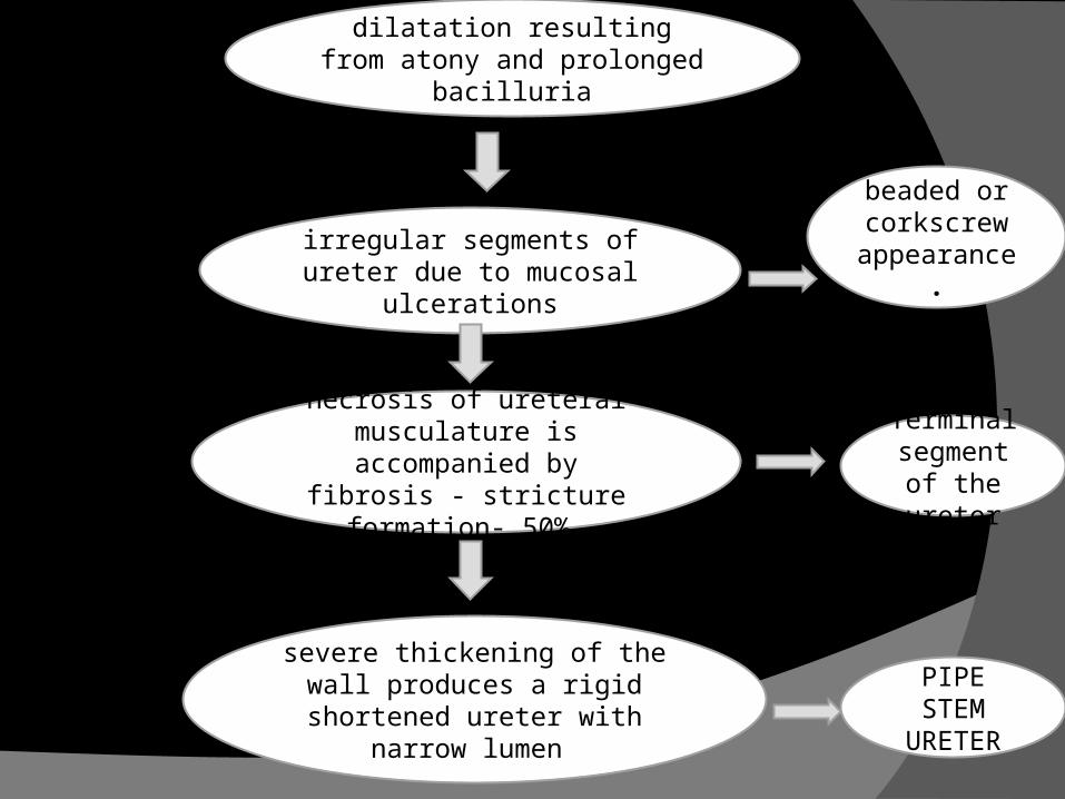
dilatation resulting from atony and prolonged bacilluria
PIPE STEM
URETER
irregular segments of ureter due to mucosal ulcerations
necrosis of ureteral musculature is accompanied
by fibrosis - stricture formation- 50%.
severe thickening of the wall produces a rigid shortened ureter with narrow lumen
beaded or corkscrew
appearance.
Terminal segment of the ureter

Bladder occurs secondary to TB of the kidney. The bladder urothelium is very resistant to infection by TB ba
cilli. Involved in later course of d/s in 1/3 rd cases The most common sites affected by TB are the areas surrou
nding the ureteric orifices and the trigone. The urothelium is initially swollen and inflamed, following the
formation of tubercles within the bladder mucosa. The ureteric orifice may be completely obscured by the swoll
en mucosa. progression to the stage of mucosal ulceration is rare.

Genital tuberculosis
Epididymis, Vas, and Testis
Tubercle bacilli reach the epididymis by hematogenous spread.
The disease initially affects the more vascular globus minor.
subsequently leads to fibrous narrowing and possible obliteration of the lumen beaded vas as a result of tubercles even
tually inducing dense fibrosis.
Prostate and Seminal Vesicles
The prostate is rarely affected, it is however one of the sites of
hematogenous spread of TB. caseous destruction of prostatic tissue e
nsues that may be significant enough to cause a noticeable reduction in semen volume
Densely fibrotic nodules may form and are indistinguishable from cancer

Penis and Urethra Tuberculosis of the penis in adults is very rare
and is secondary to infection of the kidney and bladder.
infected granulation tissue, which gradually infiltrates the glanular and cavernous tissues a
nd may invade the whole thickness of the penis.
pea-sized masses can be felt in the cavernous bodies and urethra,
Urethral tuberculosis is rare, often only seen at the meatus.

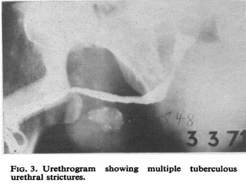
Urethral tuberculosis Male urethra – uncommon, occurs secondry to
renal infection.
The periurethral glands of Littre may become distended with bacteria and leukocytes and may lead to abscess formation.
Associated with prostatic abscess or fistula formation.
Result in non specific stricture in bulbo-membranous urethra.
Female genital tract - TB
Hematogenous spread. Associated wet or dry peritonitis strongly associated with infertility in
women, rates of successful pregnancy remain low even after treatment.
Salpingitis (94%): mostly bilateral
Tuboovarian abscess: extension into extraperitoneal compartment
Diagnosis
Urinalysis and Culture Ziehl-Neelsen staining of concentrated urine samples for acid-fast bacilli is often
negative. Urine c/s - may take upto 6 weeks BACTEC systems - 2 weeks
Purified Protein Derivative–TuberculinTest—Mantoux Test
standard dose of 5 tuberculin units (0.1 mL) is injected intradermal and read 48-72 hrs later
T-cell–mediated delayed-type hypersensitivity - principle of the test.


Nucleic Acid Amplification (NAA) Testing—PCR The PCR test has high sensitivity, specif
icity, and rapid results. sensitivity ranging from 87% to 95% (us
ually >90%) and specificity from 92% to 99.8% (usua
lly >95%) caveats exist in the interpretation of NA
AT results- dead org. after chemo yield positive results - hence use only for diagnosis not for follow up.

GeneXpert MTB/RIF
automated diagnostic test that can identify Mycobacterium tuberculosis (MTB)DNA and resistance to rifampicin (RIF)by nucleic acid amplification technique(NAAT).
Results are obtained from unprocessed sputum samples in 90 minutes
QuantiFERON- TB GOLD test for tuberculosis infection or latent tubercul
osis. QFT is an interferon-γ release assay (IGRA) IGRAs are not affected by Bacille Calmette-G
uérin (BCG) vaccination status negative IGRA result rules out the possibility of
both active and latent tuberculosis. The test is based on the quantification of interf
eron-gamma (IFN-γ) released from sensitized lymphocytes in whole blood incubated overnight with purified protein derivative (PPD) from M. tuberculosis and control antigens.
Imaging
High dose IVU – traditional gold standard
CT – new standard Pyelography (ante/retrograde) – limited
use Plain radiographs – important CXR,spine X-Ray,X-Ray KUB US – limited value Nuclear Perfusion Scan – function MRI – little application
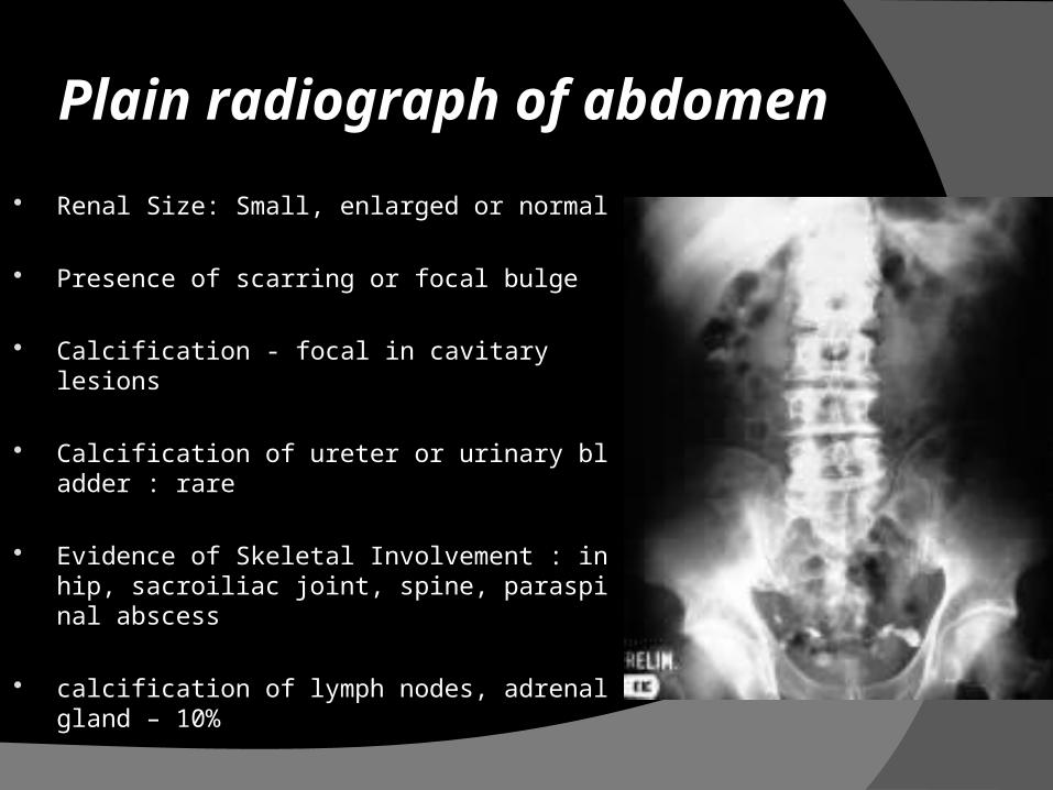
Plain radiograph of abdomen
Renal Size: Small, enlarged or normal
Presence of scarring or focal bulge
Calcification - focal in cavitary lesions
Calcification of ureter or urinary bladder : rare
Evidence of Skeletal Involvement : in hip, sacroiliac joint, spine, paraspinal abscess
calcification of lymph nodes, adrenal gland – 10%
33
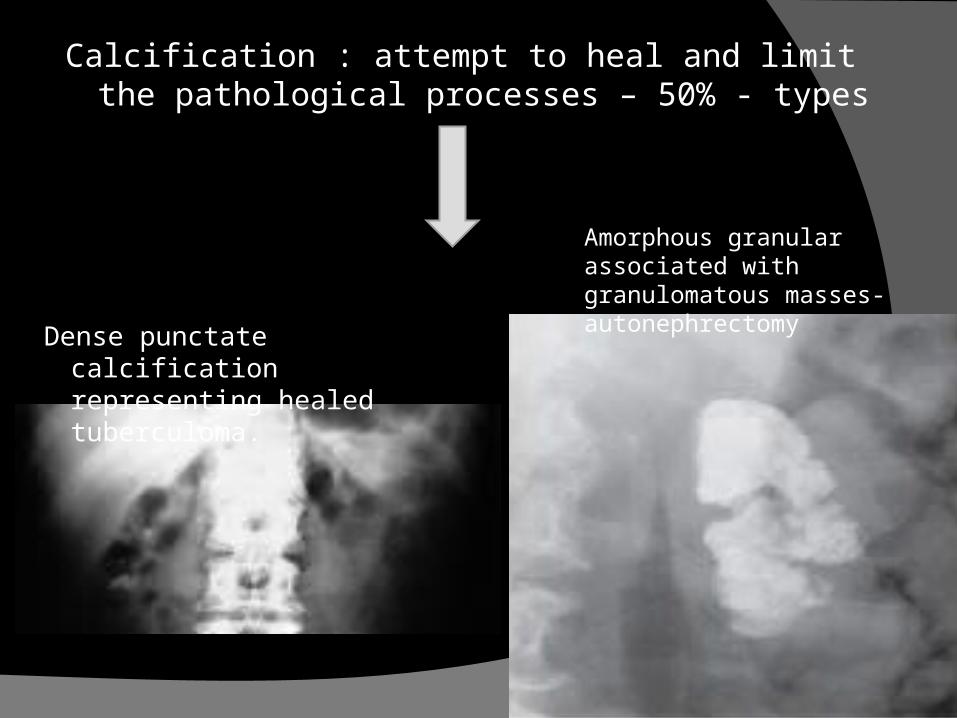
Calcification : attempt to heal and limit the pathological processes – 50% - types
Dense punctate calcification representing healed tuberculoma.
Amorphous granular associated with granulomatous masses- autonephrectomy
34
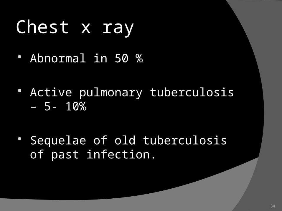
Chest x ray
Abnormal in 50 %
Active pulmonary tuberculosis – 5- 10%
Sequelae of old tuberculosis of past infection.
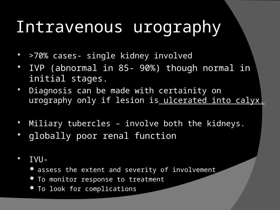
Intravenous urography
>70% cases- single kidney involved IVP (abnormal in 85- 90%) though normal in initial stages. Diagnosis can be made with certainity on urography only if
lesion is ulcerated into calyx.
Miliary tubercles – involve both the kidneys. globally poor renal function
IVU- assess the extent and severity of involvement To monitor response to treatment To look for complications
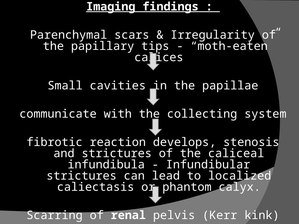
Imaging findings :
Parenchymal scars & Irregularity of the papillary tips - “moth-eaten” calices
Small cavities in the papillae
communicate with the collecting system
fibrotic reaction develops, stenosis and strictures of the caliceal infundibula -
Infundibular strictures can lead to localized caliectasis or phantom calyx.
Scarring of renal pelvis (Kerr kink)
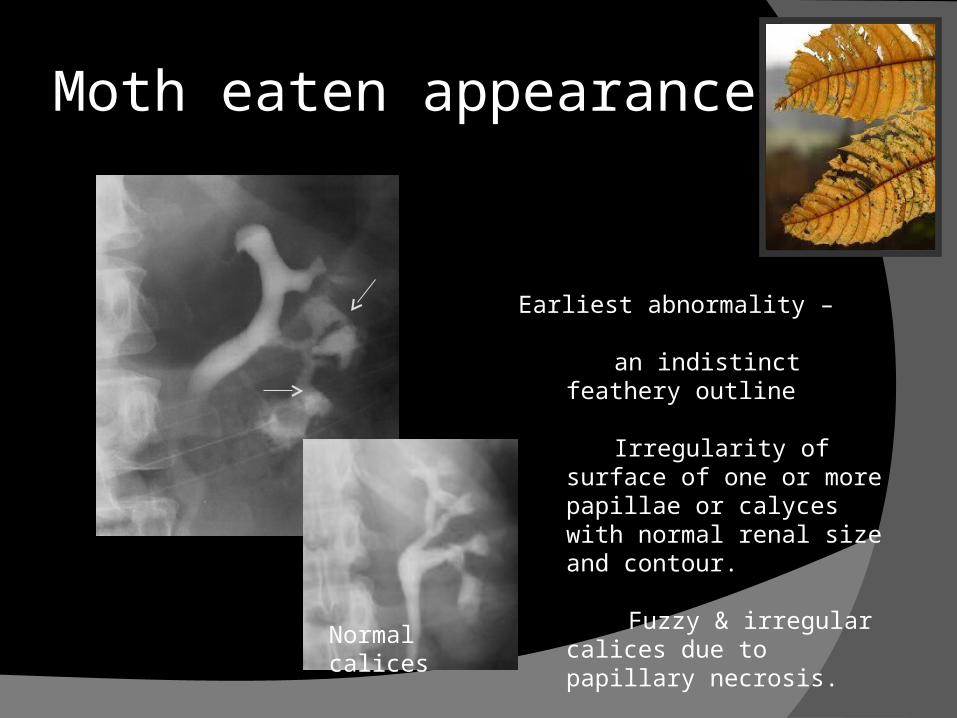
Moth eaten appearance
Normal calices
Earliest abnormality –
an indistinct feathery outline
Irregularity of surface of one or more papillae or calyces with normal renal size and contour.
Fuzzy & irregular calices due to papillary necrosis.
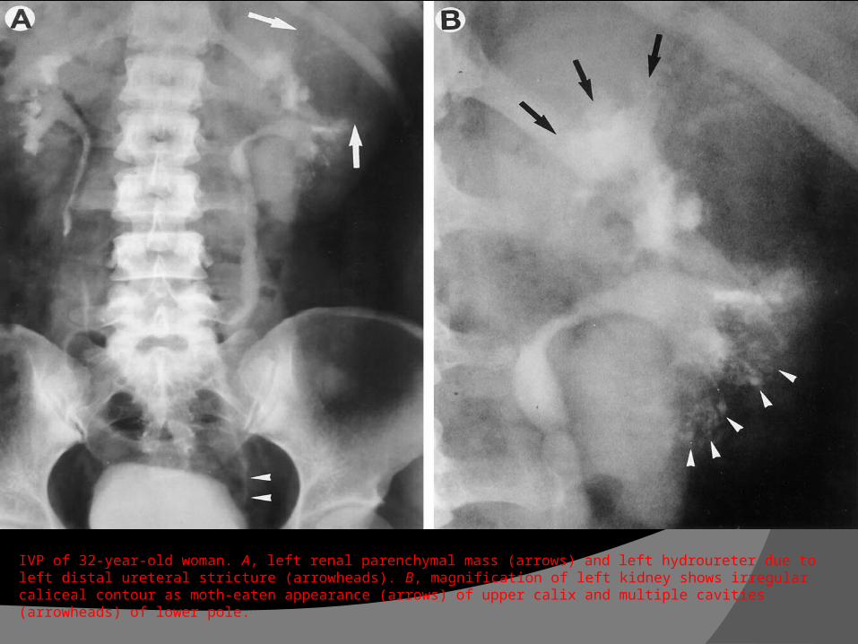
IVP of 32-year-old woman. A, left renal parenchymal mass (arrows) and left hydroureter due to left distal ureteral stricture (arrowheads). B, magnification of left kidney shows irregular caliceal contour as moth-eaten appearance (arrows) of upper calix and multiple cavities (arrowheads) of lower pole.
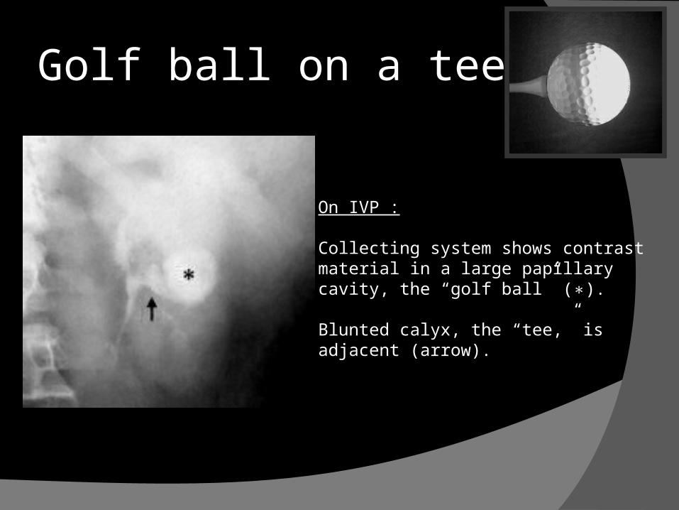
Golf ball on a tee
On IVP :
Collecting system shows contrast material in a large papillary cavity, the “golf ball” ( ).∗
Blunted calyx, the “tee,” is adjacent (arrow).
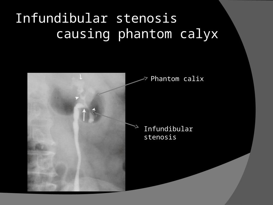
Infundibular stenosis causing phantom calyx
Phantom calix
Infundibular stenosis
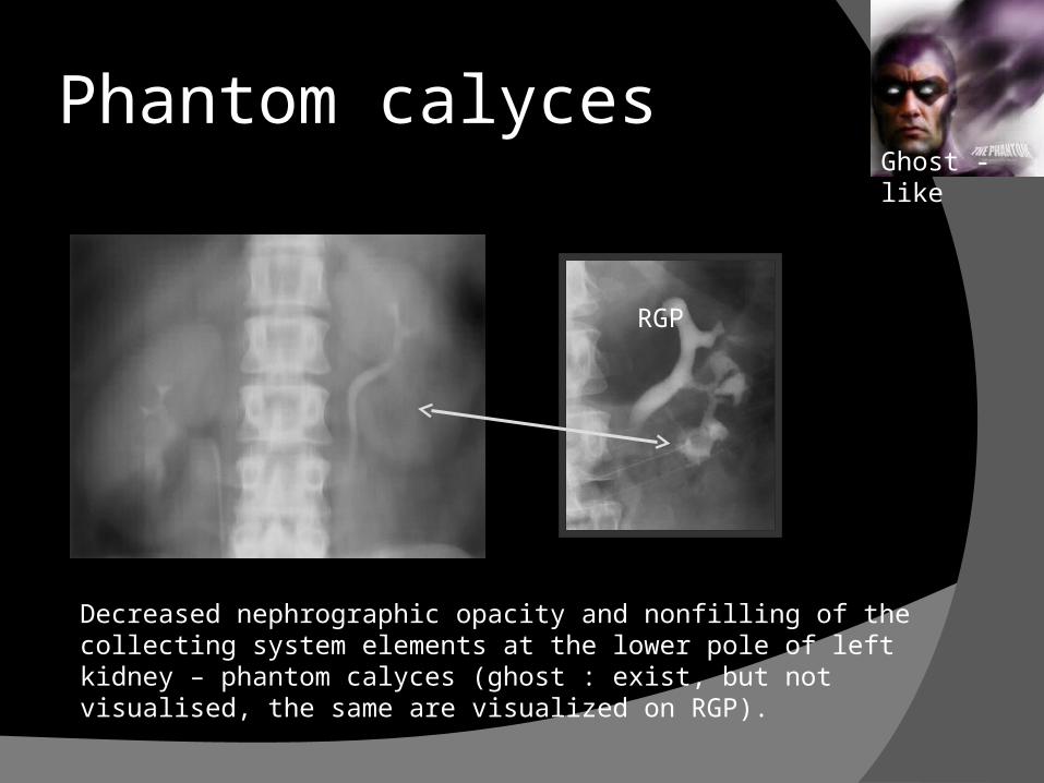
Phantom calyces
Decreased nephrographic opacity and nonfilling of the collecting system elements at the lower pole of left kidney – phantom calyces (ghost : exist, but not visualised, the same are visualized on RGP).
Ghost - like
RGP
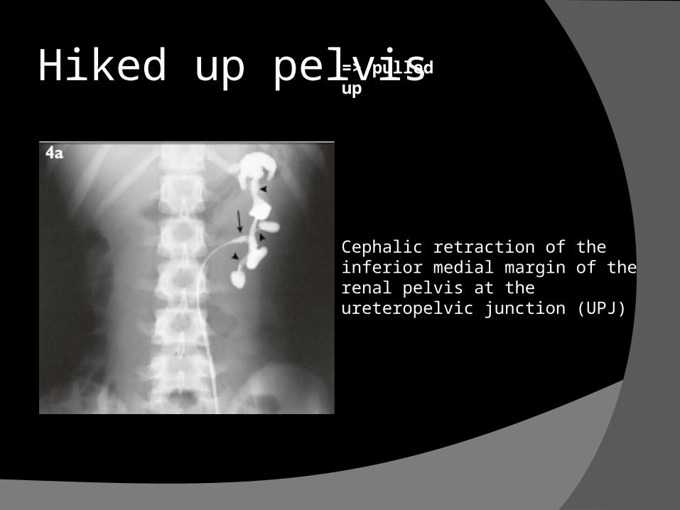
Hiked up pelvis=> pulled up
Cephalic retraction of the inferior medial margin of the renal pelvis at the ureteropelvic junction (UPJ)
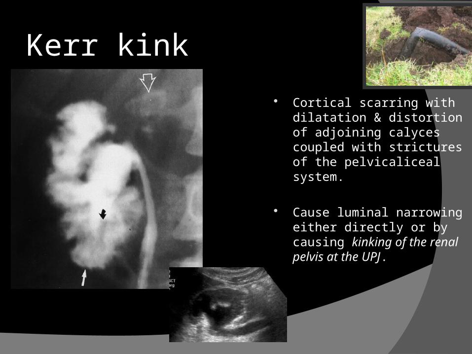
Cortical scarring with dilatation & distortion of adjoining calyces coupled with strictures of the pelvicaliceal system.
Cause luminal narrowing either directly or by causing kinking of the renal pelvis at the UPJ.
Kerr kink
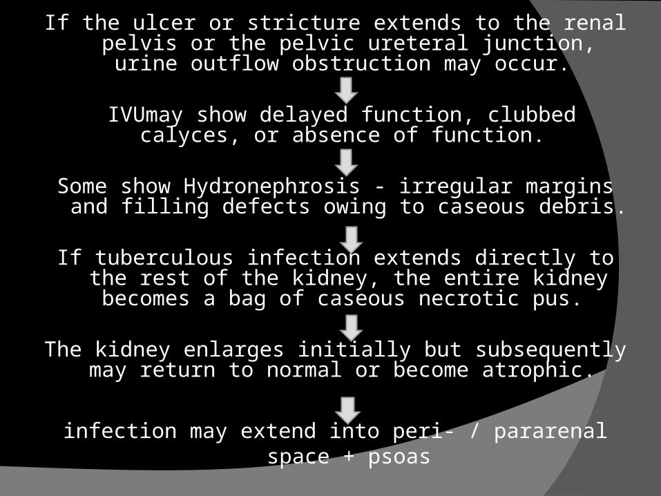
If the ulcer or stricture extends to the renal pelvis or the pelvic ureteral junction, urine outflow obstruction may
occur.
IVUmay show delayed function, clubbed calyces, or absence of function.
Some show Hydronephrosis - irregular margins and filling defects owing to caseous debris.
If tuberculous infection extends directly to the rest of the kidney, the entire kidney becomes a bag of caseous
necrotic pus.
The kidney enlarges initially but subsequently may return to normal or become atrophic.
infection may extend into peri- / pararenal space + psoas
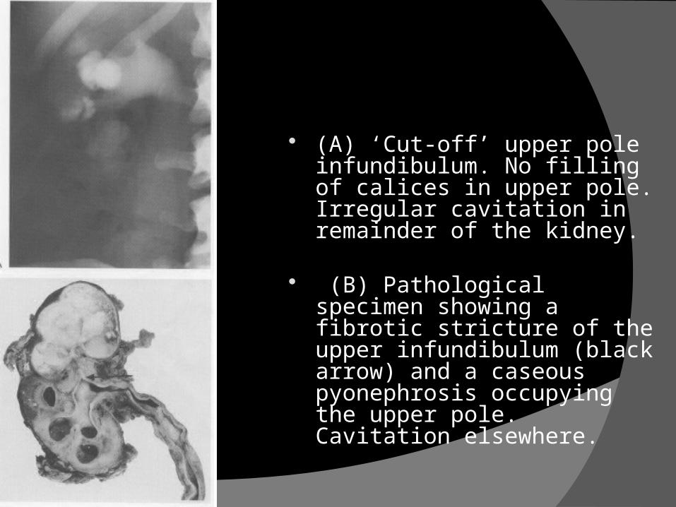
(A) ‘Cut-off’ upper pole infundibulum. No filling of calices in upper pole. Irregular cavitation in remainder of the kidney.
(B) Pathological specimen showing a fibrotic stricture of the upper infundibulum (black arrow) and a caseous pyonephrosis occupying the upper pole. Cavitation elsewhere.
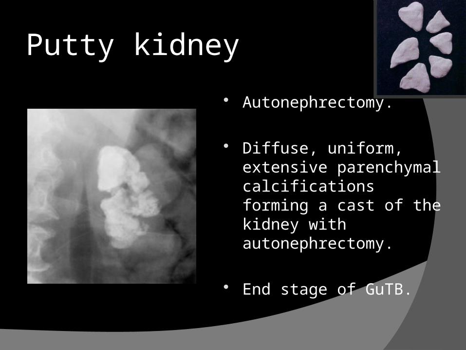
Autonephrectomy.
Diffuse, uniform, extensive parenchymalcalcifications forming a cast of the kidney with autonephrectomy.
End stage of GuTB.
Putty kidney
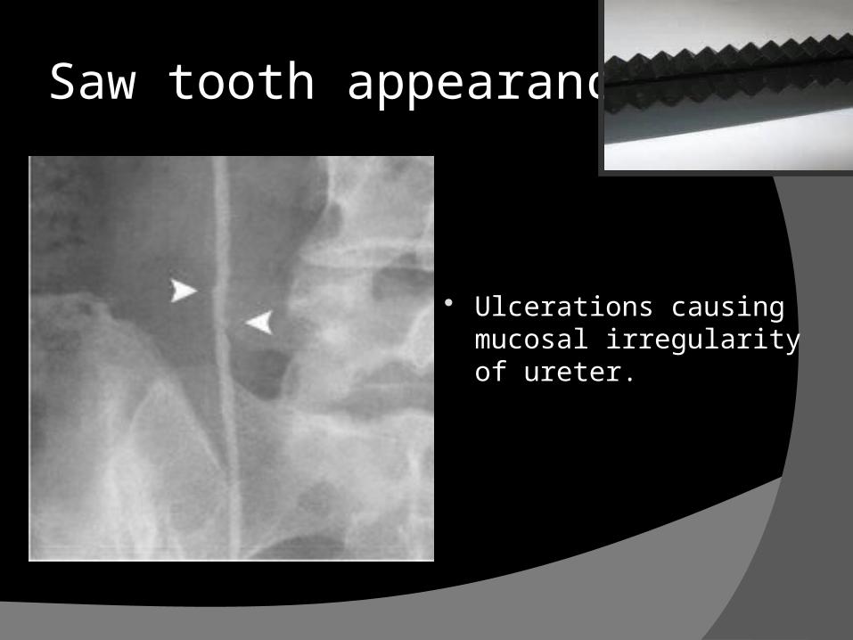
Ulcerations causingmucosal irregularity of ureter.
Saw tooth appearance
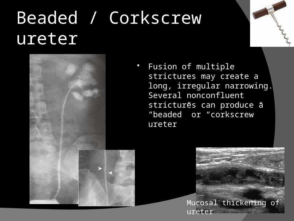
Fusion of multiple strictures may create a long, irregular narrowing. Several nonconfluent strictures can produce a “beaded” or “corkscrew” ureter
Beaded / Corkscrew ureter
Mucosal thickening of ureter
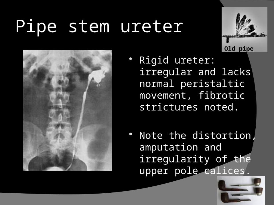
Rigid ureter: irregular and lacks normal peristaltic movement, fibrotic strictures noted.
Note the distortion, amputation and irregularity of the upper pole calices.
Pipe stem ureterOld pipe stem
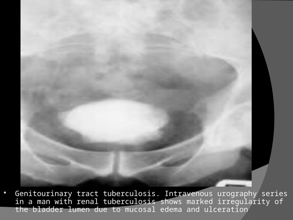
Genitourinary tract tuberculosis. Intravenous urography series in a man with renal tuberculosis shows marked irregularity of the bladder lumen due to mucosal edema and ulceration
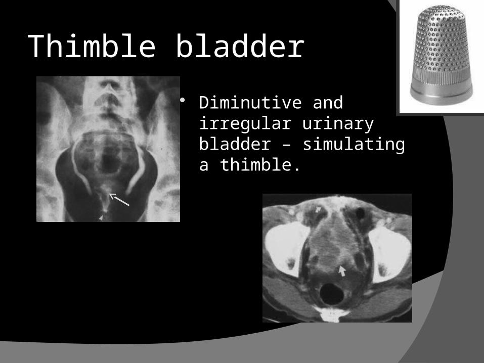
Diminutive and irregular urinary bladder – simulating a thimble.
Thimble bladder
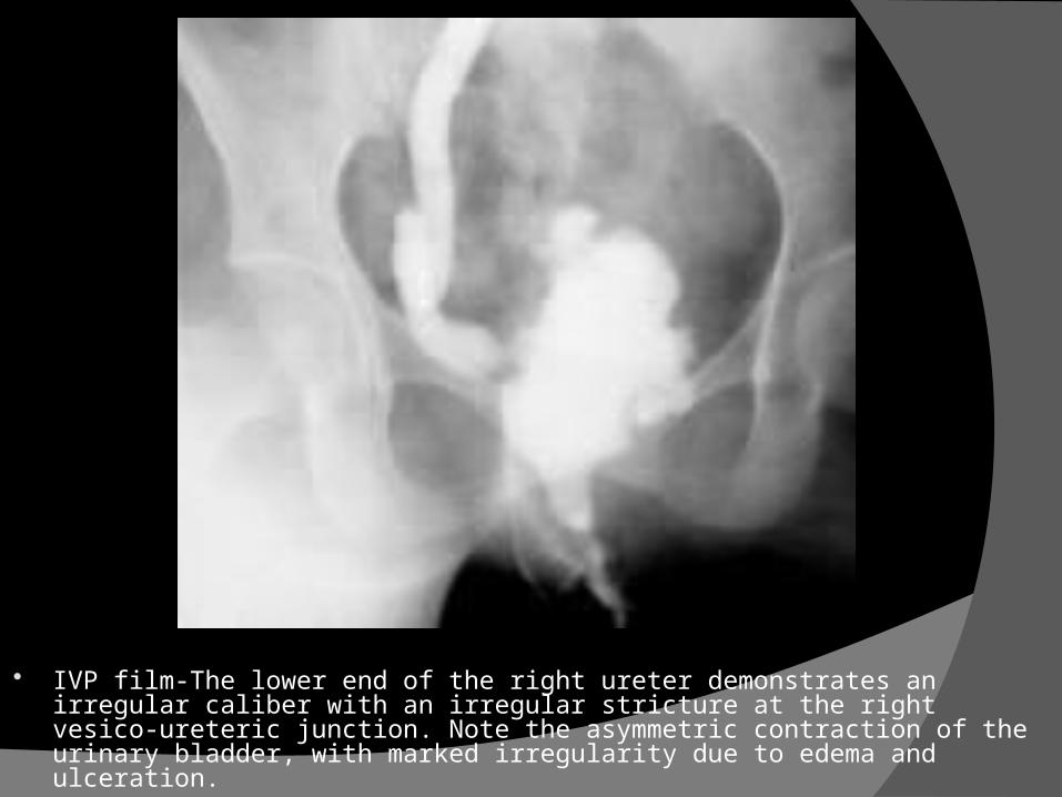
IVP film-The lower end of the right ureter demonstrates an irregular caliber with an irregular stricture at the right vesico-ureteric junction. Note the asymmetric contraction of the urinary bladder, with marked irregularity due to edema and ulceration.

Retrograde pyelography
Indicated in patients with non functioning kidney to demonstrate ureteric obstruction and cavitation in kidney.
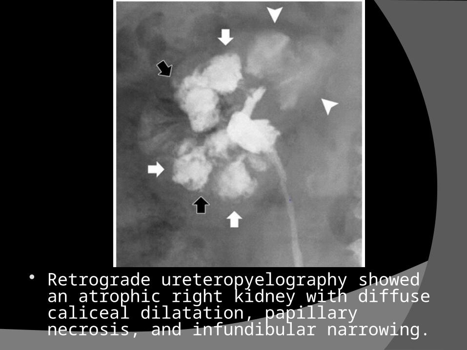
Retrograde ureteropyelography showed an atrophic right kidney with diffuse caliceal dilatation, papillary necrosis, and infundibular narrowing.
57
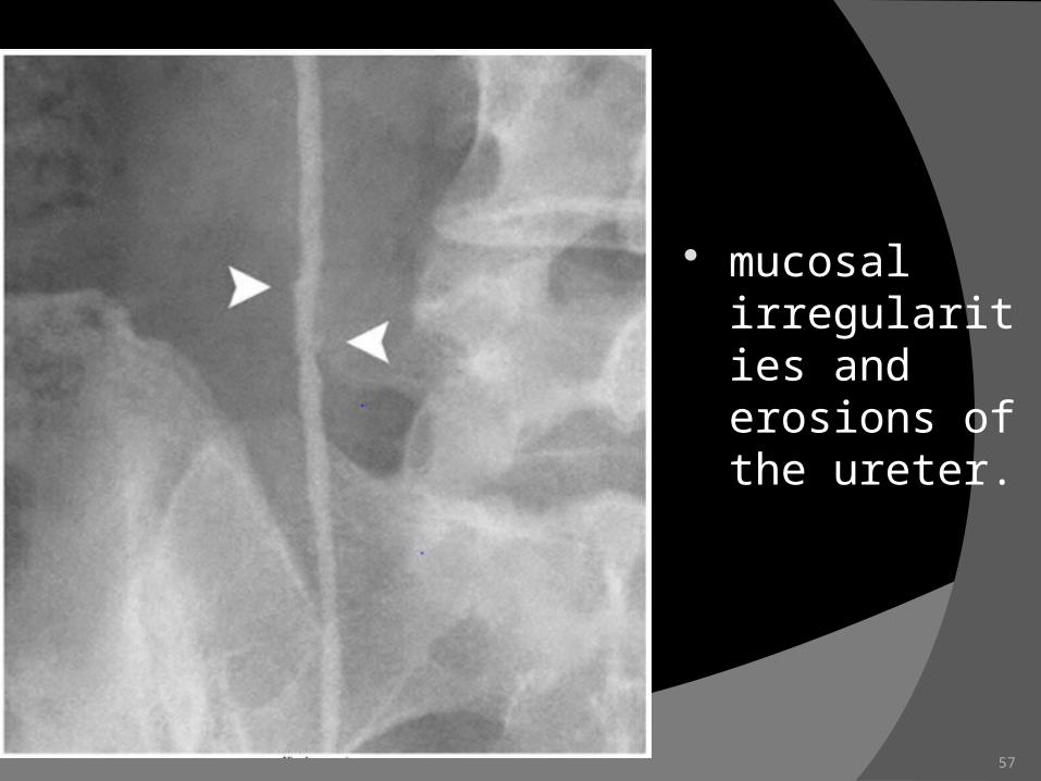
mucosal irregularities and erosions of the ureter.
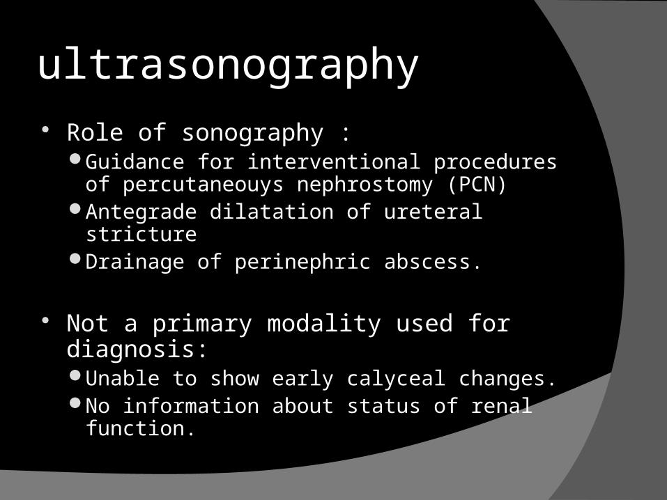
ultrasonography Role of sonography :
Guidance for interventional procedures of percutaneouys nephrostomy (PCN)
Antegrade dilatation of ureteral strictureDrainage of perinephric abscess.
Not a primary modality used for diagnosis:Unable to show early calyceal changes.No information about status of renal function.
Usg is poor in assessing ureter but shows back pressure changes and adjacent retroperitoneal disease.
UB- focal irregular thickening with reduced capacity.
Deformed shape and focal abnormalities better appreciated following distension.

Computed tomography
Has replaced IVU in current era
Uses :MDCT:Renal and extra renal spread of disease.Length of ureteric strictureAdjoining retroperitoneal diseaseAssociated spinal or solid organ involvement.
CT identifying renalcalcifications, Coalesced cortical granulomas containing either caseous or calcified
material Calices that are dilated and filled with fluid have an attenuation between 0
and 10 HU; debris and caseation, between 10 and 30 HU; putty-like calcification, between 50 and 120 HU; and calculi, greater than 120 HU. Cortical thinning is a common CT finding and may be either focal or global. Parenchymal scarring is readily apparent at CT. Fibrotic strictures of the infundibula, renal pelvis, and ureters may be seen
at contrast-enhanced CT and are highly suggestive of tuberculosis.
Ureter : thickening of ureteral wall or pelvis with periureteric inflammation
Bladder Tuberculosis thickened bladder wall (= muscle
hypertrophy + inflammatory tuberculomas) filling defects (due to multiple granulomas) bladder wall ulcerations shrunken bladder - scarred bladder with
diminished capacity - thimble bladder.� bladder wall calcifications (rare)
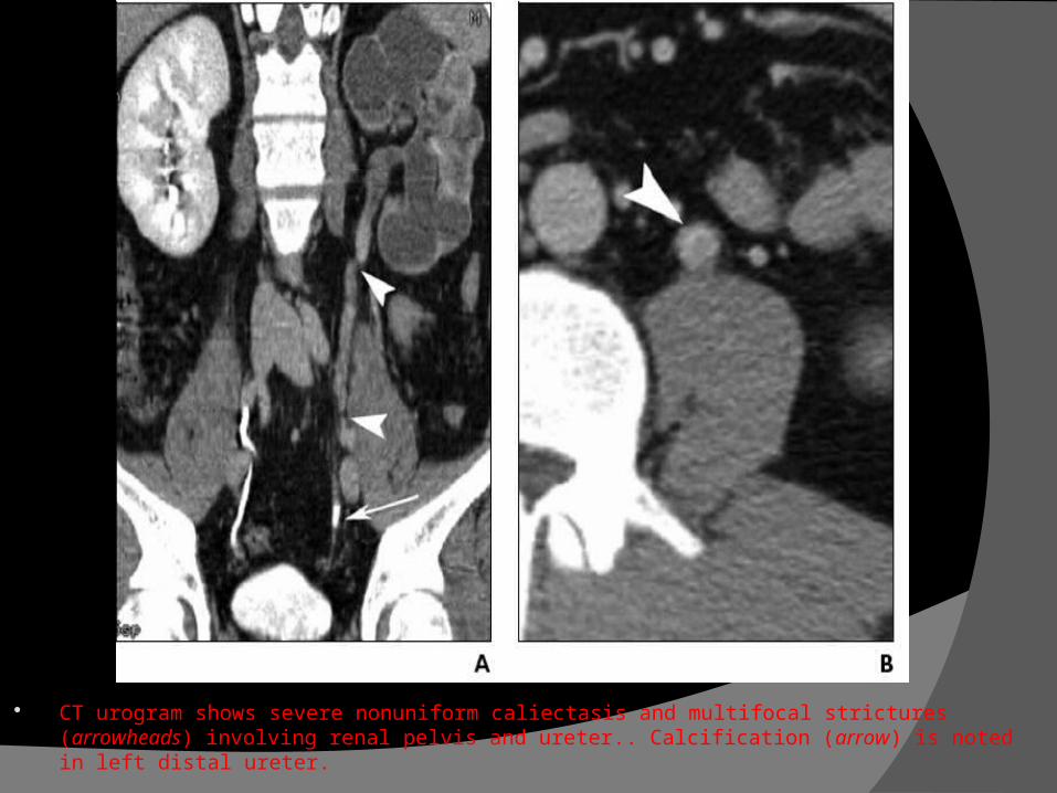
CT urogram shows severe nonuniform caliectasis and multifocal strictures (arrowheads) involving renal pelvis and ureter.. Calcification (arrow) is noted in left distal ureter.
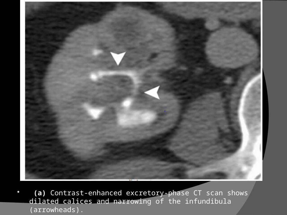
(a) Contrast-enhanced excretory-phase CT scan shows dilated calices and narrowing of the infundibula (arrowheads).
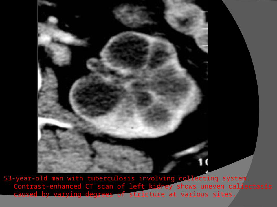
53-year-old man with tuberculosis involving collecting system. Contrast-enhanced CT scan of left kidney shows uneven caliectasis caused by varying degrees of stricture at various sites.
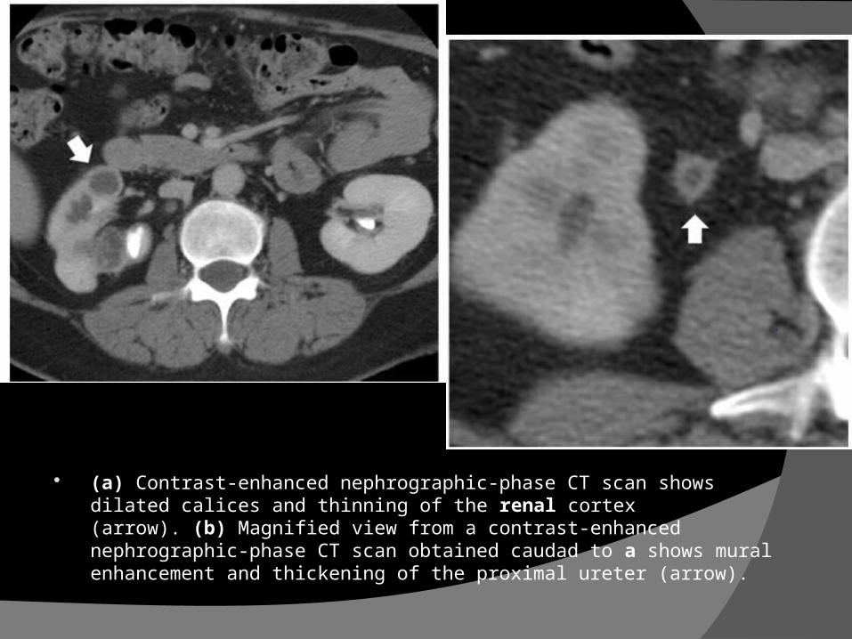
(a) Contrast-enhanced nephrographic-phase CT scan shows dilated calices and thinning of the renal cortex (arrow). (b) Magnified view from a contrast-enhanced nephrographic-phase CT scan obtained caudad to a shows mural enhancement and thickening of the proximal ureter (arrow).
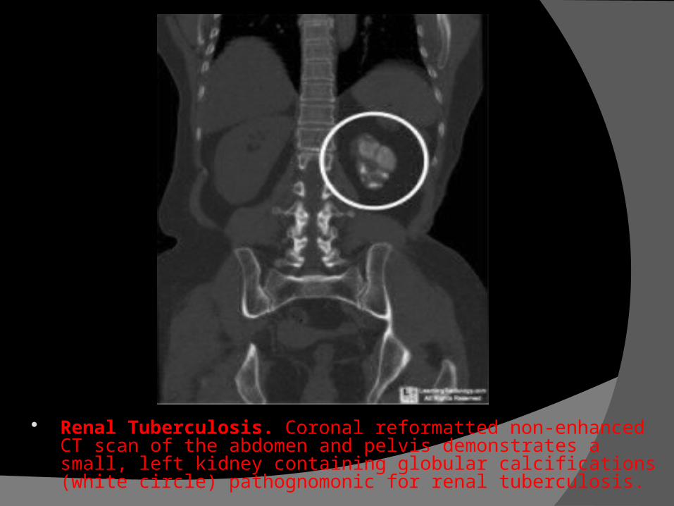
Renal Tuberculosis. Coronal reformatted non-enhanced CT scan of the abdomen and pelvis demonstrates a small, left kidney containing globular calcifications (white circle) pathognomonic for renal tuberculosis.
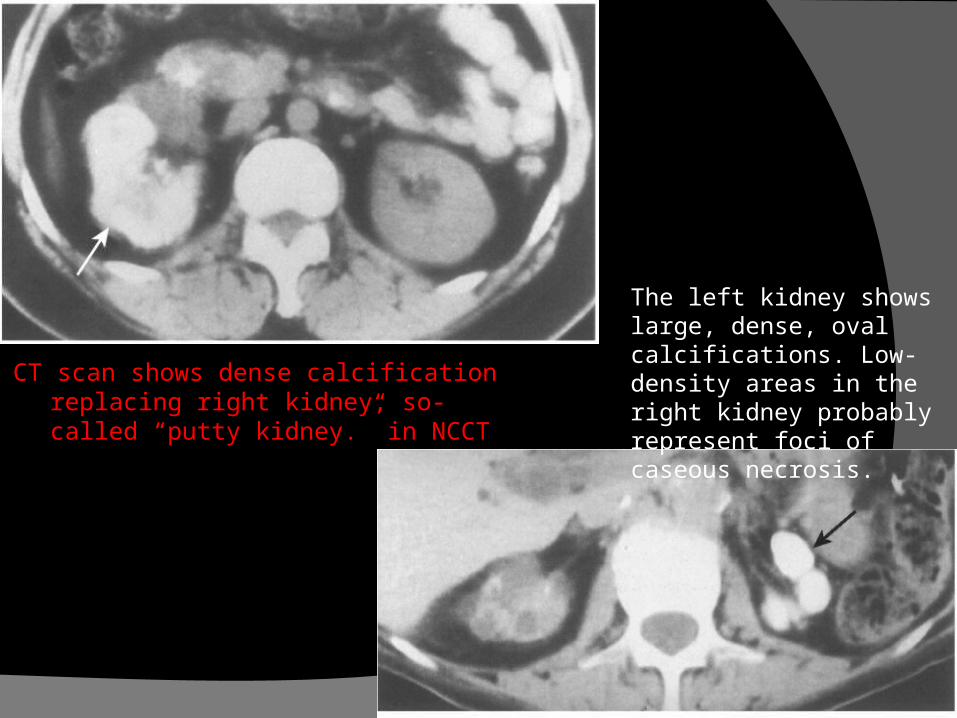
CT scan shows dense calcification replacing right kidney, so-called “putty kidney.” in NCCT
The left kidney shows large, dense, oval calcifications. Low-density areas in the right kidney probably represent foci of caseous necrosis.
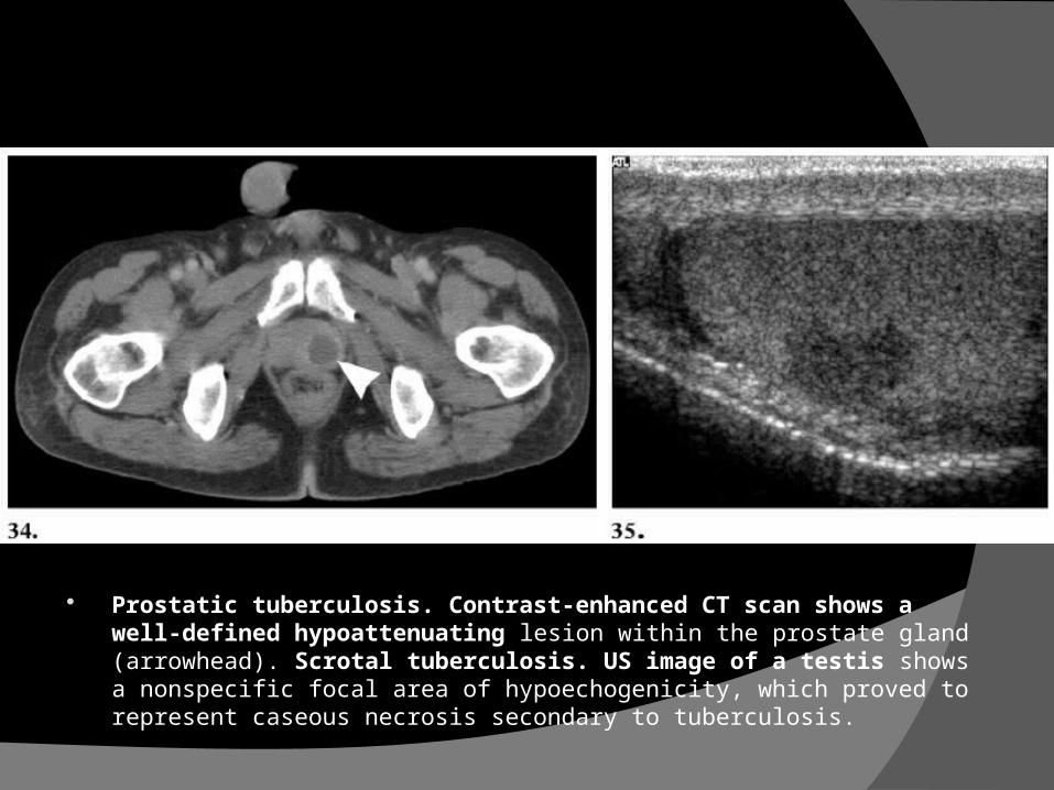
Prostatic tuberculosis. Contrast-enhanced CT scan shows a well-defined hypoattenuating lesion within the prostate gland (arrowhead). Scrotal tuberculosis. US image of a testis shows a nonspecific focal area of hypoechogenicity, which proved to represent caseous necrosis secondary to tuberculosis.

Prostatic abscess, T2-weighted MRI shows a peripheral enhancing cystic mass with radiating, streaky areas of low signalintensity.
Watermelon skin
MRI
MR urography: evaluate poorly or non functioning kidney specially obstructive form for demonstration of ureteric involvement.
MR – renal parenchymal changes and details of PCS

HSG - GTB obstruction and multiple constrictions of the fallopian
tubes. Rigid pipe-stem tubes A clubbed ampula with retort-shaped
hydrosalpingx Vascular or lymphatic intravasation of contrast Small shrunken uterine cavity with filling defects
with adhesions Long and dilated cervical canal & dye in
cervical crypts Bilateral cornual block Punctate opacification of crypts and diverticulae
in lumen of tubes
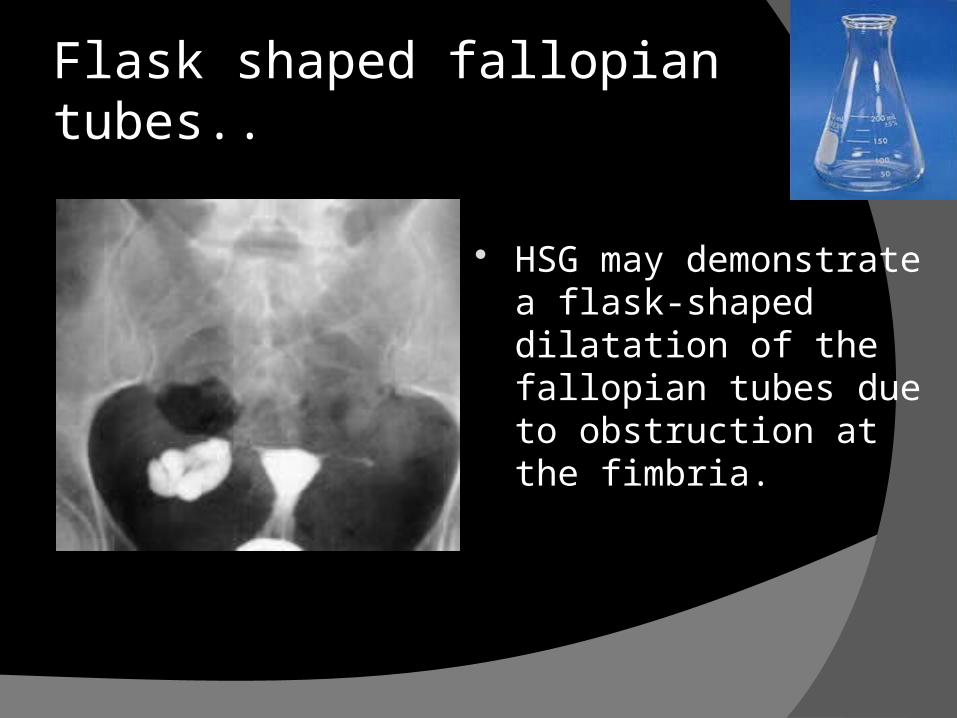
HSG may demonstrate a flask-shaped dilatation of the fallopian tubes due to obstruction at the fimbria.
Flask shaped fallopian tubes..
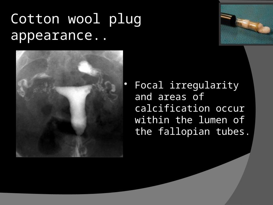
Focal irregularity and areas of calcification occur within the lumen of the fallopian tubes.
Cotton wool plug appearance..
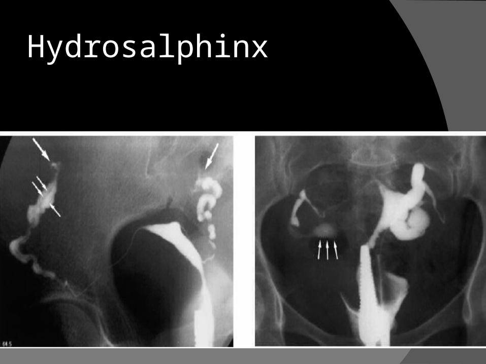
Hydrosalphinx

Bilateral T.O.masses even after ATT
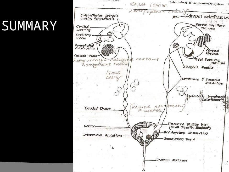
SUMMARY
Cystoscopy and Ureteroscopy
limited role in the diagnosis of TB Despite direct visualization of lesions, there are
no pathognomonic findings that are specific for tuberculosis. A “golf-hole” ureteric orifice is
suggestive of tuberculosis,
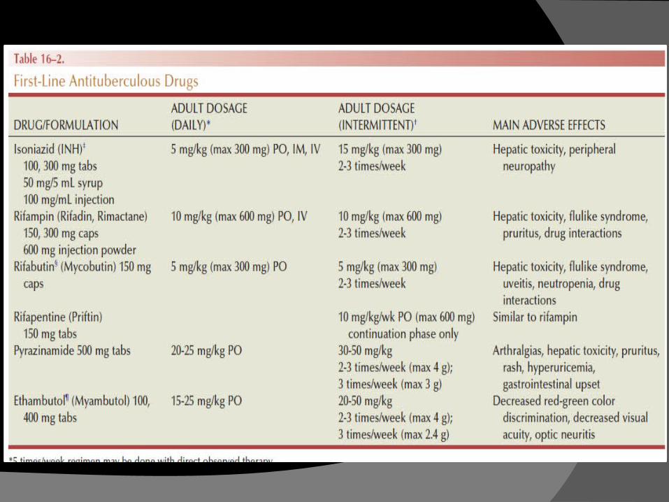
Treatment Medical Therapy isoniazid, rifampicin, and pyrazinamide attain high concentrations in urine A standard treatment regimen for tuberculosis requires 6 mo
nths of therapy. The first 2 months involve three to four drugs: Rifampicin, isoniazid, and pyrazinamide are administered daily; ethambu
tol is added if drug resistance to isoniazid is suspected. An additional 4 months of rifampicin and isoniazid daily, twic
e per week, or three times per week are used During treatment, all patients should be monitored for advers
e effects on a monthly basis. Routine follow-up of liver enzymes is important to detect hepatotoxicity of rifampicin.

Corticosteroids limited role in management of genitourin
ary TB. lowering the host immune response that
is responsible for the tissue destruction and subsequent scarring
eg:severe acute tuberculous cystitis

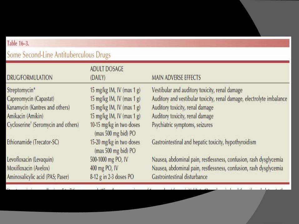
Multidrug-Resistant TB Drug-resistant tuberculosis is defined as
M. tuberculosis that is resistant to one of the first-line antituberculosis
drugs (isoniazid,rifampin, pyrazinamide, or ethambutol)
Multidrug-resistant tuberculosis (MDR-TB) refers to M. tuberculosis
that is resistant to at least isoniazid and rifampin, and possibly
additional chemotherapeutic agents.

Extensively drug-resistant tuberculosis (XDR-TB) refers to M. tuberculosis that is resistant to at least isoniazid and rifampin, and is additionally resistant to fluoroquinolones and either aminoglycosides (amikacin, kanamycin) or capreomycin, or both

Surgical Therapy About 55% of patients with genitourinary TB wi
ll require surgical intervention Currently, more than half of surgeries perform
ed for TB are reconstructive surgical treatment is best carried out after an initial 3 to 6 weeks of medical
treatment(allows intense inflammatory changes to resolve and lesions to stabilize)
Procedures to Relieve Obstruction definitive local treatment, upper urinary tract reconstruction, lower urinary tract reconstruction, and surgery for genital TB.
Procedures to Relieve Obstruction emergently required in cases of uremia or sepsis Early ureteral stenting or percutaneous nephrostomy
(PCN) for tuberculous ureteral strictures In segmental hydronephrosis, more than one PCN may
be required PCN placement must be followed by correcting the cause of obstruction. A tuberculous cutaneous fistula invariably develops if
the PCN is simply removed.
Nephrectomy decision for a nephrectomy is based upo
n the extent of renal parenchymal destruction and, more importantly,
the function of the kidney Coexisting renal carcinoma also mandat
es nephrectomy Dense fibrosis may be encountered
during dissection
Partial Nephrectomy
only where parenchymal destruction is clearly localized, such as a calcified polar cavitary
lesion or localized lesions that progress to calcification despite 6 weeks of adequate chemotherapy.
Ureteropelvic and Ureteral Surgery Upper and midureteric strictures are rare may be amenable to endourologic treat
ment Lower ureteric strictures are the commonest form and will often r
equire surgical intervention.
Endoscopic Management. Generally, short, passable strictures, with good ren
al function yield the best endoscopic outcome. Strictures forming during medical treatment and m
anaged by early stenting Balloon dilatation by retrograde or antegrade acce
ss has been described for TB strictures of the ureter, UPJ, and calyceal infundibula( high failure rates - repeat procedures required)
Failure to respond or progression after 6 weeks is an indication for definitive management.
Surgical Options. Long, complex strictures require surgical repair Dismembered pyeloplasty is feasible for extrarenal pelvis with s
hort segment scarring. When anatomic reconstruction is not possible, ureterocalicosto
my (ureter to the lower pole calyx) is an option. Upper and middle ureteric strictures- excision and
ureteroureterostomy. Lower ureter strictures- excision and ureterocystostomy( upto 5
cm ) If >5 cm - Usage of Boari flap.-- Only if good bladder capacity.
Otherwise - ileal interposition (ileal ureteric replacement) can be done
Bladder Surgery
Augmentation cystoplasty- management of the tuberculous contracted bladder
Extremely contracted bladders (thimble bladders of 20 mL capacity)
are best managed by orthotopic bladder substitution
Prostate and Urethra
Bladder neck contracture or severe granulomatous prostatitis- BNI /TURP
Urethral strictures- high recurrence . start ATT and do SPC. Then reconstructive surgery

Thank You.