Embed Size (px)
Citation preview
Evaluation &
management of comaDR ABDUL RUB
2ND YEAR PG MEM

COMA
Coma is prolonged Unconsciousness Or Unarousable Unresponsiveness

DISORDERS OF CONSCIOUNESS

Really Simple Neuroanatomy
Arousal: where is it localized?
Ascending Reticular Activating System (ARAS) ‘core of the brainstem’
receives input from numerous somatic afferents
projects to midline thalamic nuclei (which are in a circuit with cortical structures)
and the limbic system
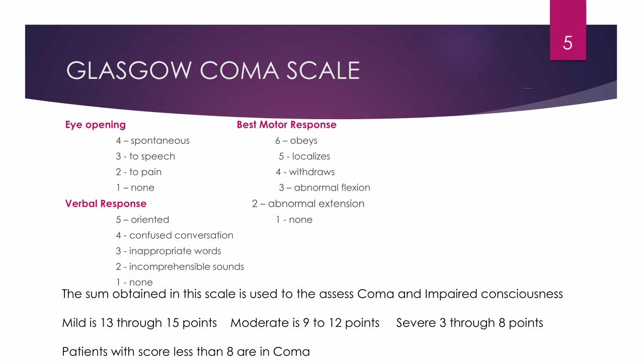
GLASGOW COMA SCALE
Eye opening Best Motor Response
4 – spontaneous 6 – obeys
3 - to speech 5 - localizes
2 - to pain 4 - withdraws
1 – none 3 – abnormal flexion
Verbal Response 2 – abnormal extension
5 – oriented 1 - none
4 - confused conversation
3 - inappropriate words
2 - incomprehensible sounds
1 - none
5
The sum obtained in this scale is used to the assess Coma and Impaired consciousness
Mild is 13 through 15 points Moderate is 9 to 12 points Severe 3 through 8 points
Patients with score less than 8 are in Coma
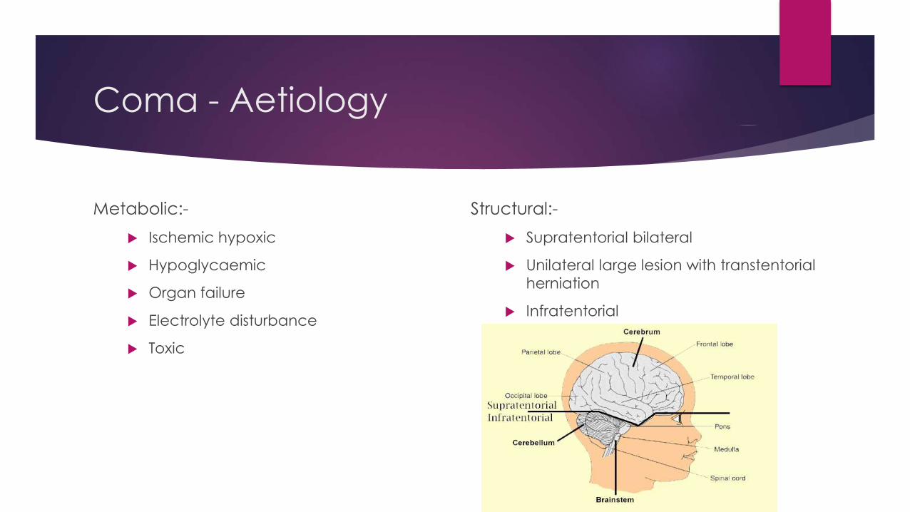
Coma - Aetiology
Metabolic:-
Ischemic hypoxic
Hypoglycaemic
Organ failure
Electrolyte disturbance
Toxic
Structural:-
Supratentorial bilateral
Unilateral large lesion with transtentorial
herniation
Infratentorial

Supratentorial Lesions
Epidural or Subdural Hematoma
Intraparenchymalhaemorrhage
Large Ischemic Infarction
Tumour
Trauma
Abscess
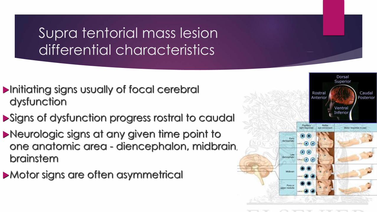
Supra tentorial mass lesion
differential characteristics
Initiating signs usually of focal cerebral
dysfunction
Signs of dysfunction progress rostral to caudal
Neurologic signs at any given time point to
one anatomic area - diencephalon, midbrain,
brainstem
Motor signs are often asymmetrical
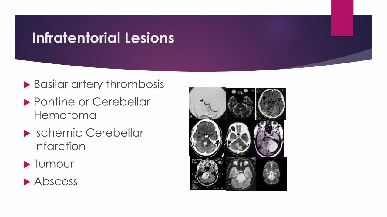
Infratentorial Lesions
Basilar artery thrombosis
Pontine or Cerebellar
Hematoma
Ischemic Cerebellar
Infarction
Tumour
Abscess
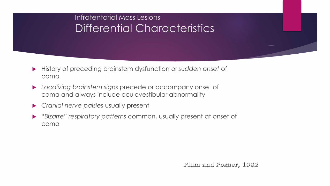
Infratentorial Mass Lesions
Differential Characteristics
History of preceding brainstem dysfunction or sudden onset of
coma
Localizing brainstem signs precede or accompany onset of
coma and always include oculovestibular abnormality
Cranial nerve palsies usually present
“Bizarre” respiratory patterns common, usually present at onset of
coma
Plum and Posner, 1982

Why coma management?
Common medical emergency 3-5%
Large proportion of comatose patient recover
Untreated coma may lead to further brain damage

Emergency treatment
Maintain ventilation oxygenation
Maintain circulation
Control seizure
Reduce icp
Maintain temperature
Control hypoglycemia

Maintain ventilation
Insert oral airway
Clean oropharyngeal secretion
Insert cuffed endotracheal tube if apnea, hypoventilation or liable to
aspirate
Mechanical ventilation if apnea or raised intracranial pressure

Maintain circulation
If hypotenstion ( <90mmHg systolic)
Replace fluid:
Saline if hyperglycemia or suspected stroke, diabetes
Dextrose saline or isolyte if undiagnosed
Vasopressor if low systolic pressure inspite of fluid
Hypertension: Betablocker, Nitroglycerine or Nitropruside

Reduce intracranial pressure
Inj Mannitol 20% 1gm/kg IV fast
Hyperventilatin to bring pCO2 25-30mmHg

Control Seizure
Inj Lorazepam 4mg or Midazolam 5mg IV slowly
Inj Diazepam 10-20mg iv slowly
Inj Phenytoin 15-20mg/Kg 50mg/min IV
Inj Phenobarb 15-20mg/Kg 50mg/min IV
Inj Sodium valproate 200-400mg IV

Maintain Temperature
Hperthermia: tapid sponging, paracetamol
Hypothermia: heating blanket
HISTORY
Inquire about-
i. History of diabetes
ii. Hypertension
iii. Head injury
iv. Convulsions
v. Alcohol or drug use
vi. Circumstances in which patient was found
vii. Medications in hospitalized patient like anesthetics, sedatives, antiepileptic, opiates, antidepressants, antipsychotics.
18
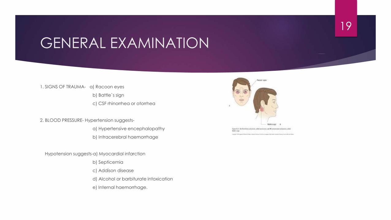
GENERAL EXAMINATION
1. SIGNS OF TRAUMA- a) Racoon eyes
b) Battle`s sign
c) CSF rhinorrhea or otorrhea
2. BLOOD PRESSURE- Hypertension suggests-
a) Hypertensive encephalopathy
b) Intracerebral haemorrhage
Hypotension suggests-a) Myocardial infarction
b) Septicemia
c) Addison disease
d) Alcohol or barbiturate intoxication
e) Internal haemorrhage.
19

3. PULSE- Bradycardia with periodic breathing and hypertension (CUSHING REFLEX) suggests raised ICP.
4. TEMPERATURE- Hypothermia suggests-
a) Alcohol or barbiturate intoxication
b) Myxedema
c) Advanced tubercular meningitis
d) Peripheral circulatory failure
Hyperthemia suggests- a) Systemic infection
b) Meningoencephalitis
c) Heat stroke
d) Anticholinergic drugs abuse
20

5. SIGNS OF MENINGEAL IRRITATION-
a) Meningitis
b) SAH
6. FUNDUS- a) Raised ICP (Papilledema)
b) SAH ( retinal haemorrhages,disc swelling)
c) Hypertensive encephalopathy
7. SKIN INSPECTION- a) Rash suggests menigococcemia, staphylocoocalendocarditis, typhus, RMSF
b) Excessive sweating suggest hypoglycemia or shock
c) Diffuse petechiae suggest TTP, DIC, fat embolism
21
NEUROLOGICAL ASSESMENT
Observation first without examiner intervention.
Simply watch posture of limbs and body, position of head and eyes, presence or absence of spontaneous movements on one side, rate and depth of respiration.
Yawning and spontaneous shifting of body position indicates minimal degree of unresponsiveness.
Multifocal myoclonus almost always indicate metabolic disorder.
Assess responsiveness by noting patient`s reaction to calling his name, or to noxious stimuli such as supraorbital or sternal pressure.
Glasgow coma scale allows rapid assessment and allows to track neurological changes over time.
22

POSTURE IN COMATOSE PATIENT
Decerebrate rigidity: consists of opisthotonus, clenching of jaws, stiff
extension of limbs with internal rotation of arms & plantar flexion of feet.
Precise correlation between extensor posturing & level of lesion is rarely
possible because extensor posturing arises in variety of settings: midbrain
compression, cerebellar lesions, metabolic, drug intoxication etc.
Decorticate rigidity: arms in flexion and adduction and legs extended signify lesion rostral to midbrain.
Extensor posture of arms with weak flexor responses of legs is seen with
lesions at level of vestibular nuclei (medulla)
23
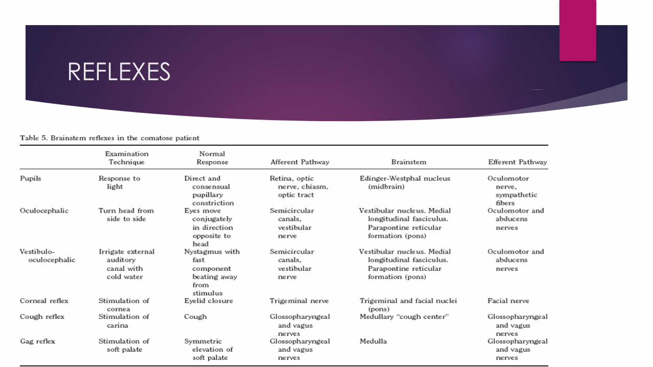
REFLEXES

BRAINSTEM REFLEXES
Pupillary size and reactivity
Ocular movements
Corneal responses
Ocular-vestibular reflexes
Pattern of breathing
25
AS A RULE, WHEN THESE BRAINSTEM ACTIVITIES ARE PRESERVED,
PARTICULARLY THE PUPIL REACTIONS AND EYE MOVEMENTS, COMA MUST BE
ASCRIBED TO BILATERAL HEMISPHERAL DISEASE. THE CONVERSE, HOWEVER,
IS NOT ALWAYS TRUE, AS A MASS IN THE HEMISPHERES MAY BE THE
UNDERLYING CAUSE OF COMA BUT NONETHELESS PRODUCE BRAINSTEM
SIGNS BY INDUCING TRANSTENTORIAL HERNIATION.
26
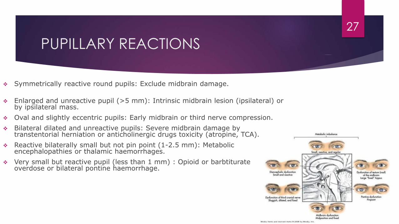
PUPILLARY REACTIONS
Symmetrically reactive round pupils: Exclude midbrain damage.
Enlarged and unreactive pupil (>5 mm): Intrinsic midbrain lesion (ipsilateral) or by ipsilateral mass.
Oval and slightly eccentric pupils: Early midbrain or third nerve compression.
Bilateral dilated and unreactive pupils: Severe midbrain damage by transtentorial herniation or anticholinergic drugs toxicity (atropine, TCA).
Reactive bilaterally small but not pin point (1-2.5 mm): Metabolic encephalopathies or thalamic haemorrhages.
Very small but reactive pupil (less than 1 mm) : Opioid or barbtiturateoverdose or bilateral pontine haemorrhage.
27

EYE MOVEMENTS
In light coma of metabolic origin, eyes rove conjugately from side to side in
random fashion. These movements disappear as coma deepens.
Adducted eye at rest: 6th nerve palsy. If it is bilateral it is due to raised ICT.
Abducted eye at rest: 3rd nerve palsy.
Downward and inward deviation of eyes: Lesions of thalamus and upper
midbrain.
Eyes turn toward convulsing side in focal seizures.
28

OCULAR BOBBING: Brisk downward and slow upward movements of the
eyes associated with loss of horizontal eye movements is diagnostic of
lesions in midbrain and pons.
OCULAR DIPPING: Slow downward followed by faster upward movement
in patients with normal horizontal gaze and it indicates diffuse cortical
anoxic damage and drug intoxication.
29

OCULO-CEPHALIC REFLEX
Also called Doll`s-eye movement.
Elicited by briskly turning or tilting the head.
Response in coma of metabolic origin or that due to bihemispheral structural lesions consist of conjugate movements of eyes in the opposite direction.
Positive response indicates-
i. Oculomotor, abducent, midbrain and pons are intact.
ii. There is loss of cortical inhibition on brainstem that normally holds these movements in check.
Absent reflex indicates damage within brainstem but also can be due to
profound overdose of sedatives or anticonvulsants.
30

RESPIRATORY PATTERNS
Slow, shallow, regular breathing: metabolic or drug depression.
Cheyne-Stokes respiration: Massive supratentorial lesions, B/L
cerebral lesions & mild metabolic disturbance.
Central neurogenic hyperventilation: Lesions of lower midbrain &
upper pons either primary or secondary to transtentorial herniation.
Apneustic breathing: Lower pontine lesions.
Biot`s or ataxic breathing: Lesions of dorsomedial part of medulla.
Agonal gasps: B/L lower brainstem damage & terminal respiratory
pattern.
31
PATHOLOGICAL ANATOMY OF COMA
Only if cerebral lesions are bilateral and extensive, then consciousness will
be impaired.
Unilateral mass lesions like infarct or hemorrhage if are causing coma, it
means compression of midbrain and subthalamic region of RAS has
occurred.
Either lateral displacement or herniation of temporal lobe can cause their
compression.
Even small lesions in upper brainstem and thalamus are sufficient to cause
coma.
32
Onset of coma-
i. Sudden onset- vascular origin especially brainstem stoke or SAH.
ii. Rapid progression from hemispheric signs to coma- intracerebralhaemorrhage.
iii. Protracted course- tumor, abscess, chronic SDH.
iv. Coma preceded by confusional or agitated state & without lateralizing signs-metabolic cause.
v. REMEMBER FRONTAL AND OCCIPITAL HEMORRHAGES ARE LESS LIKELY TO DISPLACE DEEP STRUCTURES AND TO CAUSE COMA THAN ARE CLOTS OF EQUIVALENT SIZE IN THE PARIETAL OR TEMPORAL LOBES
33

LABORATORY STUDIES AND IMAGING
Complete blood count
Random blood sugar
RFT, LFT
Serum electrolytes
Urine examination for specific gravity, glucose, acetone & protein content.
ABG analysis
Chest X-Ray
ECG
CT or MRI Scan
Lumbar Puncture
EEG
34

TAKE HOME MESSAGE
Remember clinical analysis of comatose patient is urgency.
Evaluate for airway, breathing & circulation.
History & systematic general and neurological assessment will help a lot.
Presence or absence of brainstem reflexes helps to localize the lesion.
Evaluate for imminent herniation.
Implement rapid & systematic investigation and take prompt therapeutic
action.
35

THANKS 36