Embed Size (px)
Citation preview
EVALUATION OF COMATOSE PATIENT
Prof. G. ZulianiProf. G. Zuliani

Consciousness
• Two components of conscious behavior:– Vigilance (arousal): appearance of
wakefulness– Awareness (content): the sum of cognitive – Awareness (content): the sum of cognitive
and affective function
• Awareness depends on arousal, but normal arousal does not guarantee normal awareness

Neuroanatomy
• Where is Vigilance localized?
– Ascending Reticular Activating System (ARAS) in pons and midbrain (mesencefalo)(ARAS) in pons and midbrain (mesencefalo)
– receives input from numerous somatic afferents
– projects to midline thalamic nuclei (which are in circuit with cortical structures) and the limbic system

• Consciousness requires:• An intact pontine ARAS• An intact cerebral hemisphere, or at least
Consciousness
• An intact cerebral hemisphere, or at least part of a hemisphere
• Coma requires dysfunction of either the:• Pontine ARAS or• Bihemispheric cerebral dysfunction

• Definition of Coma: unarousable and unresponsiveness in which the subjects lie with eyes closed
COMA
with eyes closed
• Other older terms: obtundation, stupor– fallen out of favour because of imprecision– descriptive methods favoured

What is Coma?
• No awareness or vigilance• Lasts > 1-6 h (differential diagnosis with syncope and
concussion)• No spontaneous speech or movement, eyes shut• No eye opening to verbal command • Noxious stimuli: vocalisation absent or limited• Noxious stimuli: vocalisation absent or limited• Noxious stimuli: motor activity absent or abnormal or
reflexive (not purposeful or defensive)

States of altered consciousness

AW
AR
EN
ES
S
VIGILANCE
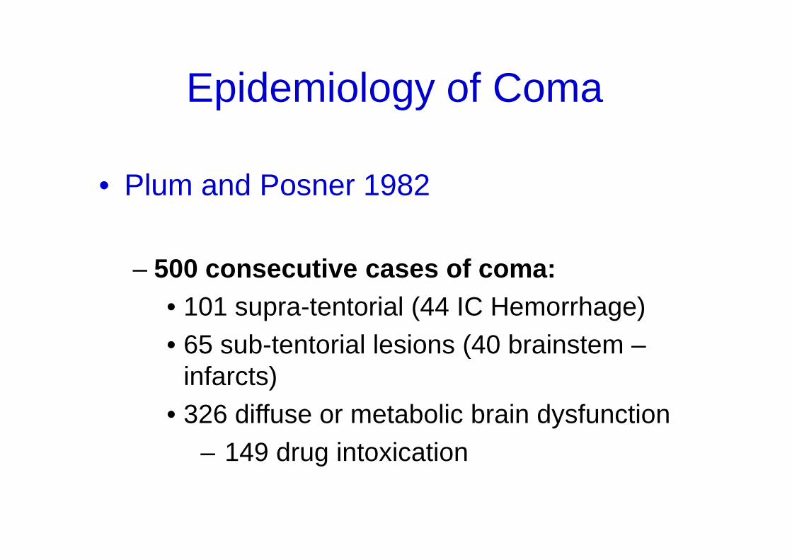
Epidemiology of Coma
• Plum and Posner 1982
– 500 consecutive cases of coma:– 500 consecutive cases of coma:• 101 supra-tentorial (44 IC Hemorrhage)• 65 sub-tentorial lesions (40 brainstem –
infarcts)• 326 diffuse or metabolic brain dysfunction
– 149 drug intoxication

1. Supratentorial lesions : cause coma by either widespread bilateral disease, increased intracranial pressure or herniation.
2. Infratentorial lesions : involve the ARAS,
Pathogenesis of Coma
2. Infratentorial lesions : involve the ARAS, usually with associated brainstem signs
3. Metabolic coma : causes diffuse hemispheric involvement and depression of ARAS, usually without focal findings
4. Psychogenic

Pathogenesis of Coma

Pathogenesis of Coma

1. Supratentorial Mass Lesions
a. Hematomab. Neoplasmc. Abscessd. Contusione. Vascular Accidents

Subdural Hematoma
• Subdural hematoma is a traumatic brain injury in whichblood (usually of vein origin) gathers between the DuraMater and the Arachnoid . Subdural hemorrhages maycause an increase in intracranial pressure, which cancause compression of and damage to brain tissue (masseffect).effect).
• Subdural hematomas are divided into: ACUTE,SUBACUTE, and CHRONIC, depending on their speed ofonset. Acute subdural hematomas that are due to traumaare the most lethal of all head injuries and have a highmortality if they are not rapidly treated with surgicaldecompression.

Subdural Hematoma
• Factors increasing the risk of a subduralhematoma include:
- very young age- very old age (search for previous falls in olders )- very old age (search for previous falls in olders )- anticoagulants (warfarin)- aspirin and other antiplatelet drugs- alcohol abuse- dementia
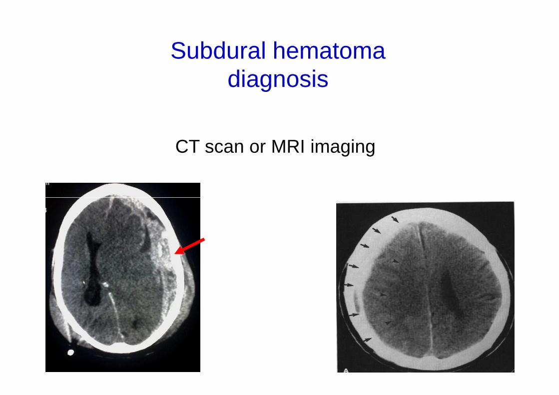
Subdural hematomadiagnosis
CT scan or MRI imaging

Acute Epidural Hematoma
• Epidural or extradural hematoma is a type of traumatic brain injury in which a buildup of blood occurs between the Dura Mater and the Skull. • Epidural bleeding is rapid because it is usually from arteries, which are high pressure. Epidural bleeds from arteries can grow until they reach their peak size at six to arteries can grow until they reach their peak size at six to eight hours post injury, spilling from 25 to 75 cc of blood into the intracranial space. • As the hematoma expands, it strips the dura from the inside of the skull, causing an intense headache.

Acute Epidural Hematoma
CT scan or MRI imaging

Cerebral Abscess
• Brain abscess is an abscess within the brain tissuecaused by collection of infected material, coming fromlocal (e.g. ear infection, dental abscess, infection ofparanasal sinuses, infection of the mastoid air cells of theparanasal sinuses, infection of the mastoid air cells of thetemporal bone, epidural asbcess) or remote (lung, heart,kidney, etc.) infectious sources.
• The infection may also be introduced through a skullfracture following a head trauma or surgical procedures.

Cerebral Abscess
• The symptoms of brain abscess are caused by increasedintracranial pressure due to a space-occupying lesion:
• confusion, Coma• Headache, vomiting• focal neurologic signs• focal neurologic signs
plus• infection: fever, fatigue
The most frequent presenting symptoms are: headache,drowsiness, confusion, seizures, hemiparesis or speechdifficulties together with fever with a rapidly progressivecourse.

Supratentorial Mass LesionsPathophysiology
• The altered consciousness is based on:
• Increased intracranial pressure• Herniation • Diffuse bilateral lesions

2. Infratentorial Lesions
• Cause coma by affecting ascending reticular activating system (ARAS) in pons.
• Brainstem nuclei and tracts usually are involved with resultant focal brainstem findings.

Infratentorial Lesions
a. Vascular accidentsb. Traumac. Neoplasmc. Neoplasmd. Cerebellar hemorrhagee. Demyelinating diseasef. Central pontine myelinolysis (e.g. too
rapid correction of hyponatremia)

• History of preceding brainstem dysfunctionor sudden onset of coma
• Localizing brainstem signs precede or accompany onset of coma and always
Infratentorial Lesions:Differentiating Features
accompany onset of coma and always include oculo-vestibular abnormality
• Cranial nerve palsies usually present• “Bizarre” respiratory patterns are common,
usually present at onset of coma

• Drugs• Anoxia• Epilepsy: may be sub-
• Sodium ↑ ↓• Liver ko• Kidneys ko
3. Metabolic Coma
clinical• CO• SIRS• Hypoxia• Hypercapnia• Glucose ↓ or ↑↑
• Hypothermia• Hypertension • Hypotension• Calcaemia ↑• Wernicke’s• Thyroid, adrenal,
pituitary ko

Drug poisoning and Coma

Metabolic Coma:Differentiating Features
• Confusion and coma commonly precede motor signs
• Motor signs are usually symmetrical• Motor signs are usually symmetrical• Pupillary reactions are usually preserved• Asterixis, myoclonus, tremor, and seizures
are common• Acid -base imbalance with hyper- or
hypoventilation is frequent

Metabolic Coma

Coma “Mimics”
• Akinetic mutism• ‘Locked-in’ syndrome• ‘Locked-in’ syndrome• Catatonia• Conversion reactions

Coma “Mimics”

Akinetic Mutism
• Silent, immobile but alert appearing• Usually due to lesion in bilateral mesial
frontal lobes, bilateral thalamic lesions or lesions in peri-aqueductal grey (brainstem)

“Locked-In” Syndrome
• Infarction of basis pontis (all descending motor fibers to body and face)
• Patient aware but cant’ move• May spare eye-movements• May spare eye-movements• Often spare eye-opening• EEG is normal or shows alpha activity
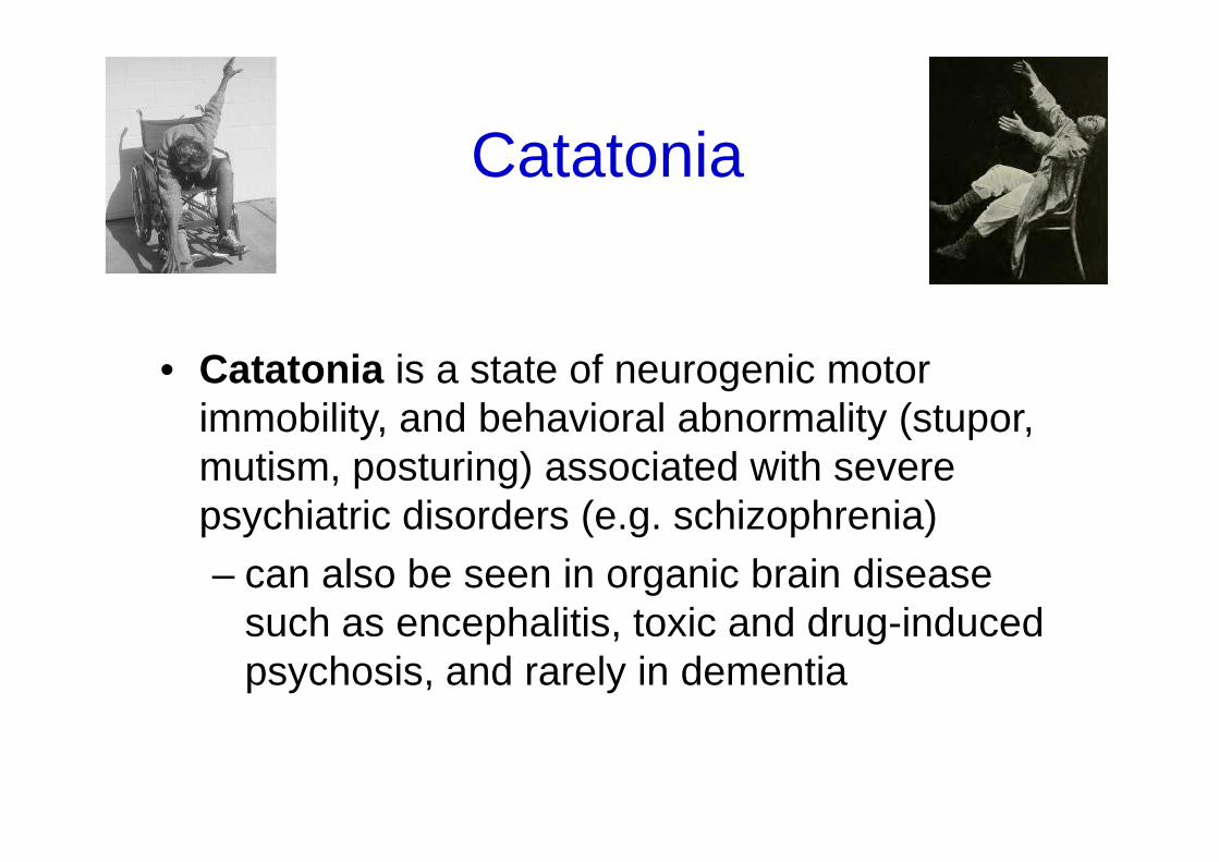
Catatonia
• Catatonia is a state of neurogenic motor immobility, and behavioral abnormality (stupor, immobility, and behavioral abnormality (stupor, mutism, posturing) associated with severe psychiatric disorders (e.g. schizophrenia)– can also be seen in organic brain disease
such as encephalitis, toxic and drug-induced psychosis, and rarely in dementia

Conversion reactions
• Fairly rare (former called hysteria)• The presence of nystagmus with cold
water calorics indicates the patient is water calorics indicates the patient is physiologically awake
• EEG used to confirm normal activity

Coma “Mimics”

Coma: Clues from History
• Onset of symptoms (important):– sudden onset– slow onset– slow onset– fluctuations
• Associated with neurologic symptoms• Medications review

Approach to the Comatose PatientPriorities
• Airway• Breathing• Circulation• Circulation• Identify and address life threatening
inadequacies• Evaluate for intracranial hypertension and
imminent herniation and treat

Glasgow Coma Scale

Glasgow Coma Scale
• Eye opening• 4 - spontaneous• 3 - to speech• 2 - to pain• 1 - none
• Best Motor Response• 6 - obeys• 5 - localizes• 4 - withdraws• 3 - abnormal flexion• 1 - none
• Verbal Response• 5 - oriented• 4 - confused conversation• 3 - inappropriate words• 2 - incomprehensible sounds• 1 - none
• 3 - abnormal flexion• 2 - abnormal extension• 1 - none
Max score: 15
(Coma <9)

• During ABC and secondary survey:– Start IV and obtain labs:
• ABG (EGA)• Whole clinical chemistry, ammonia, coagulation
Approach to the Comatose PatientPriorities
• Whole clinical chemistry, ammonia, coagulation• Glucose stick• Toxin screens
– As soon as IV in and labs drawn, give:• Glucose • Consider Thiamin (vit. B1) (alcoholism)

Approach to the Comatose PatientPriorities

• If CT scan / MRI is normal: probably metabolic coma
• Emergent causes of metabolic coma (even after ABC):
Approach to the Comatose PatientPriorities
after ABC):– Hypoglycemia: give glucose– Infections-sepsis: consider antibiotics and acyclovir
If diagnostic studies delayed, treat first– Certain toxins: antidepressants, salicylates,
theophylline, alcohol (methanol) and ethylene glycol– Subclinical status epilepticus (EEG)

General Neurological Examination
• Tone, power, reflexes• Identifies lateralising signs: hemispheric
lesionslesions• In general plantars do not help; signifies
hemispheric lesions but may be old• Decerebrate or decorticate

General Neurological Examination
Decerebrate Upper brain stem
If a patient moves from abnormal flexion (Decorticate) to abnormal extension (Decerebrate) it may be sign that damage has progressed from a supra-tentorial position to infra-tentorial position (e.g. herniation of
brainstem through foramen magnum).
Decorticate
Upper brain stem
Corticospinal tract

Meningism
Meningism is present in 3 conditions:
• Sub Arachnoidal Haemorrhage (SAH) • Sub Arachnoidal Haemorrhage (SAH) • Meningitis• Meningoencephalitis
Respiratory pattern
• Hyperventilation: pons or midbrain injury (shock, fever, acidosis, psychiatric disease)
• Cheyne-Stokes: bilateral diencephalic or hemispheric injury (CHF, COPD, drugs depressing response to CO2, injury (CHF, COPD, drugs depressing response to CO2, ventilatory over- support)
• Apneustic breathing: pons
• Ataxic: medulla, usually preterminal

Respiratory pattern

Pupillary responses
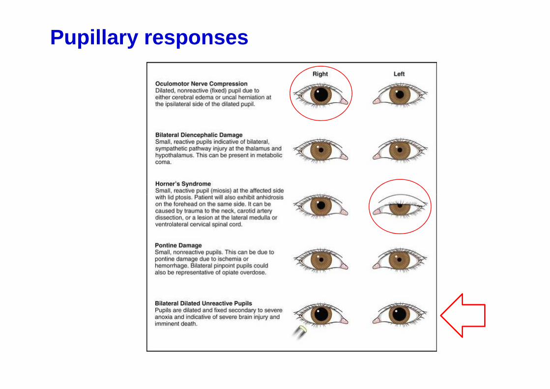
Pupillary responses

Fundus oculi
Edema della papilla

Wernicke-Korsakoff syndrome
Wernicke-Korsakoff syndrome (also called Korsakoff's psychosis, alcoholic encephalopathy, Wernicke's disease, and alcoholic encephalopathy) is a manifestation of thiamine (vitamin B1) deficiency.
This is usually secondary to alcohol abuse. This is usually secondary to alcohol abuse. It is generally agreed that:- Wernicke's encephalopathy results from severe acutedeficiency of thiamine, while - Korsakoff's psychosis is a chronicneurologic sequela after Wernicke's encephalopathy.

Wernicke-Korsakoff syndrome
Wernicke's encephalopathy is characterized by:- confusion - nystagmus - ophthalmoplegia (impaired eye movement) - anisocoria - ataxia - ataxia - sluggish pupillary reflexes - coma and death if untreated
Korsakoff's psychosis is characterized by:- anterograde amnesia (inability to form new memories) - retrograde amnesia (loss of existing memories) - confabulation (false perceptions or memories) - hallucinations

Wernicke-Korsakoff syndrome
� Thiamine diphosphate plays a major role as a cofactor or coenzyme in glucose metabolism . The enzymes dependent on thiamine diphosphate are associated with the Kreb’s Cycle . Thus, anything that encourages glucose metabolism will exacerbate an existing clinical or sub-clinical thiamine deficiency.
� Treatment consists of reversing the thiamine deficiency by giving supplemental thiamine (IV or IM). Some authors think it is important to start thiamine BEFORE giving glucose, as it is important to start thiamine BEFORE giving glucose, as encephalopathy would be worsened by glucose (glucose administration promotes decarboxylation of pyruvate, a biochemical reaction which requires thiamine). However, this is based on case reports.
� By the time amnesia and psychosis have occurred, complete recovery is extremely unlikely.