Embed Size (px)
Citation preview
EPITHELIUM
PRESENTED BY:
DR. KALPAJYOTI BHATTACHARJEE
DEPT. OF ORAL PATHOLOGY AND MICROBIOLOGY
CONTENTS Definition
Development
Characteristics
Classification
Functions
Cell polarity
Membrane specialization of epithelia
Glands
Epithelial cell renewal
Structure of the oral epithelium
DEFINITION
Epithelium is an avascular tissue composed of cells
that cover the exterior body surfaces and line internal
closed cavities (including the vascular system) and
body tubes that communicate with the exterior (the
alimentary, respiratory, and genitourinary tracts).
Epithelium also forms the secretory portion
(parenchyma) of glands and their ducts. In addition,
specialized epithelial cells function as receptors for
the special senses (smell, taste, hearing, and vision).
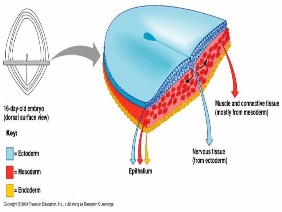
DEVELOPMENT
EPITHELIUM IS DERIVED FROM 3 GEREM LAYERS
ECTODERM MESODERM ENDODERM
ALTHOUGH MOST OF THE EPITHELIA ARE DERIVED FROM ECTODERM AND ENDODEREM
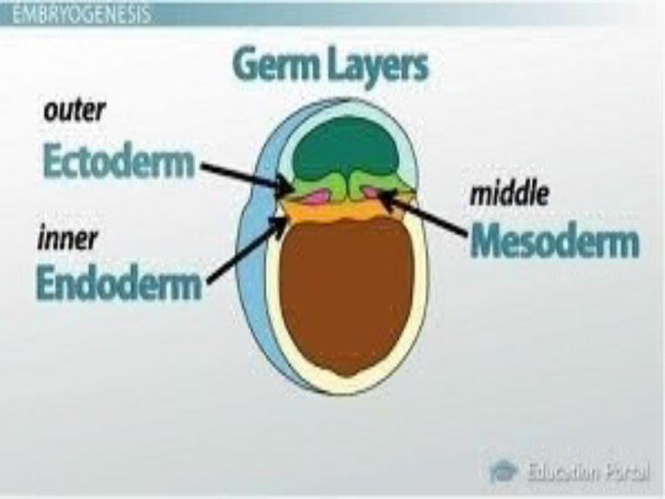

ECTODERM: Oral and nasal mucosa, cornea, epidermis of the skin & glands of the skin & the mammary glands.
ENDODERM: The liver, the pancreas & the lining of the respiratory and GIT.
MESODERM: Uriniferous tubules of the kidney, the lining of the male and female reproductive systems, the endothelial lining of the circulatory system and the mesothelium of the body cavity.
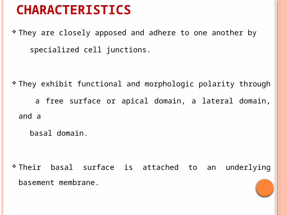
CHARACTERISTICS
They are closely apposed and adhere to one another by
specialized cell junctions.
They exhibit functional and morphologic polarity through
a free surface or apical domain, a lateral domain, and a
basal domain.
Their basal surface is attached to an underlying
basement membrane.
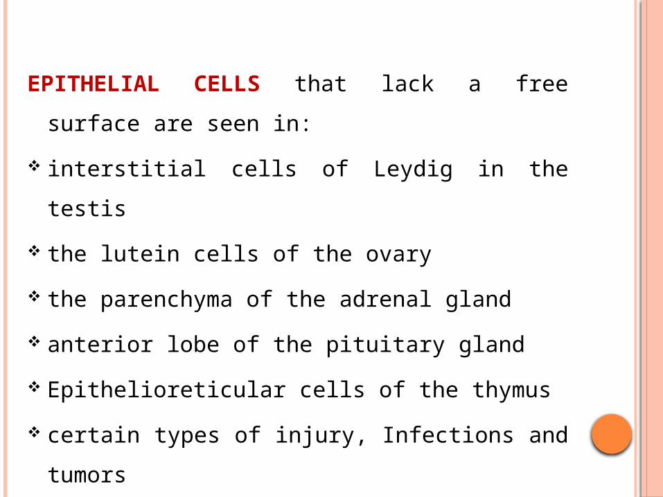
EPITHELIAL CELLS that lack a free surface are
seen in:
interstitial cells of Leydig in the testis
the lutein cells of the ovary
the parenchyma of the adrenal gland
anterior lobe of the pituitary gland
Epithelioreticular cells of the thymus
certain types of injury, Infections and tumors

EPITHELIUM creates a selective barrier between the external environment and the underlying connective tissue seen in
Blood
lymph
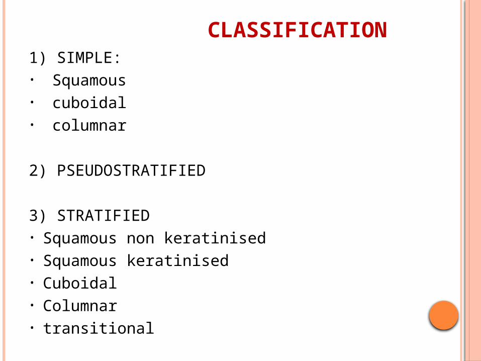
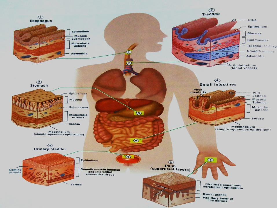
CLASSIFICATION1) SIMPLE:• Squamous• cuboidal• columnar
2) PSEUDOSTRATIFIED
3) STRATIFIED• Squamous non keratinised• Squamous keratinised• Cuboidal• Columnar• transitional
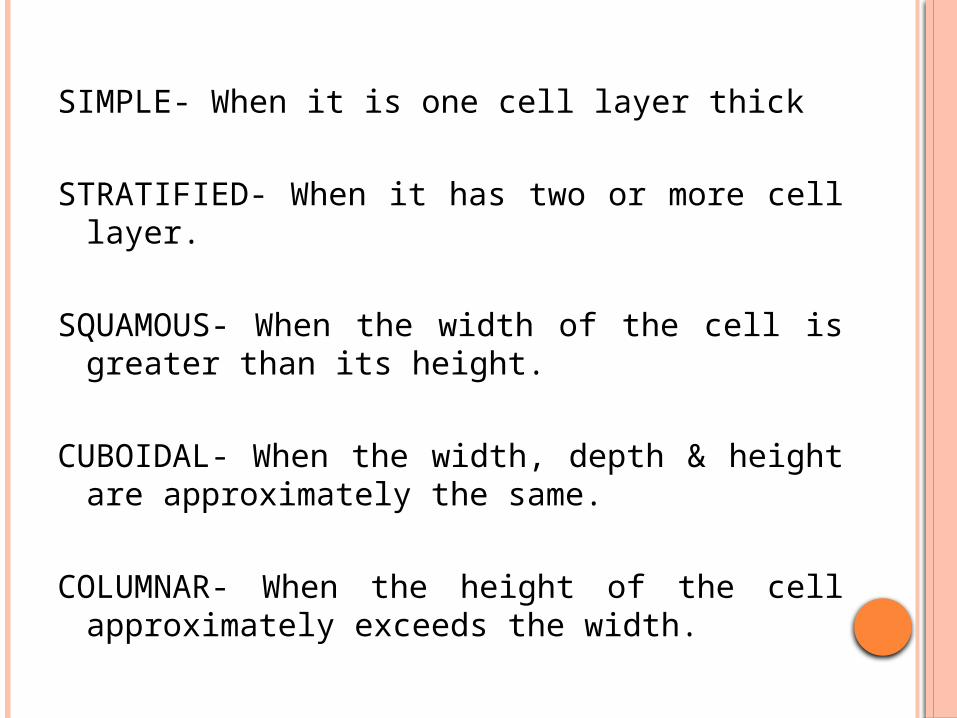
SIMPLE- When it is one cell layer thick
STRATIFIED- When it has two or more cell layer.
SQUAMOUS- When the width of the cell is greater than its height.
CUBOIDAL- When the width, depth & height are approximately the same.
COLUMNAR- When the height of the cell approximately exceeds the width.

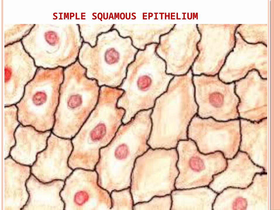
SIMPLE SQUAMOUS EPITHELIUM
Composed of flattened, irregularly shaped cells forming a continuous surface which may be reffered to as pavemented epithelium.
Term ‘squamous’ derives from the comparison of the cells to the scales of a fish.
Supported by an underlying delicate membrane.
Involved in passive transport of either gases or fluids.
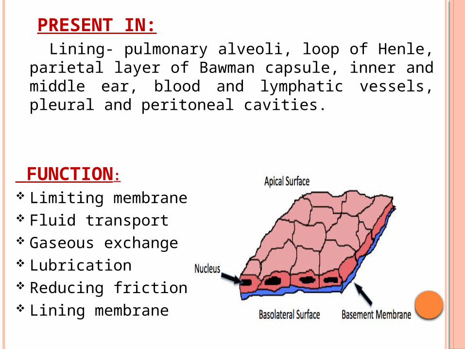
PRESENT IN: Lining- pulmonary alveoli, loop of Henle, parietal
layer of Bawman capsule, inner and middle ear, blood and lymphatic vessels, pleural and peritoneal cavities.
FUNCTION:
Limiting membrane Fluid transport Gaseous exchange Lubrication Reducing friction Lining membrane
SIMPLE SQUAMOUS EPITHELIUM

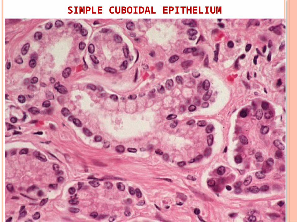
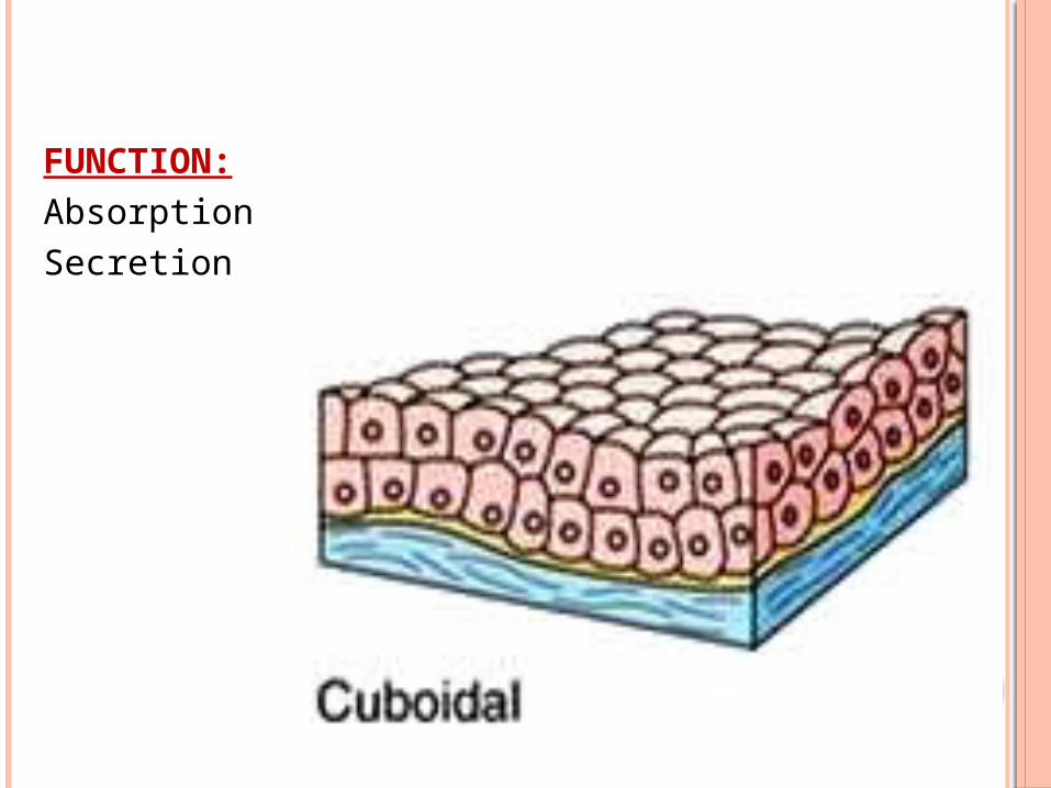
SIMPLE CUBOIDAL EPITHELIUM
Intermediate form between simple squamous and simple columnar epithelium.
Nucleus is round and located in the centre of the cell.
Present in: Ducts of exocrine glands Surface of ovary Kidney tubules Thyroid follicles
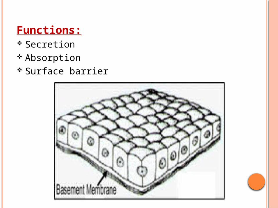
Functions: Secretion Absorption Surface barrier
SIMPLE CUBOIDAL EPITHELIUM
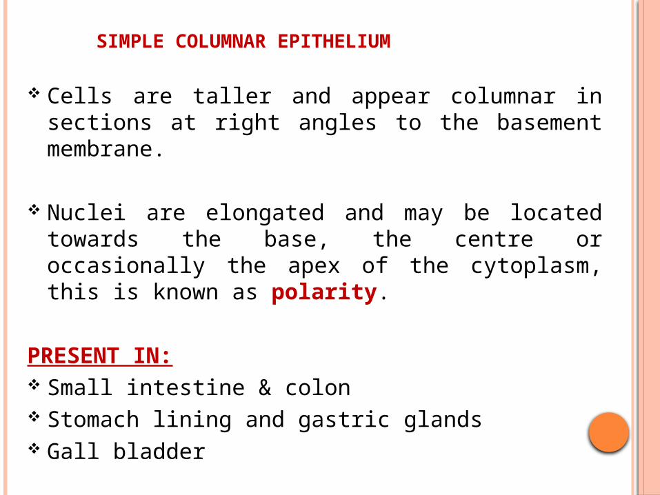
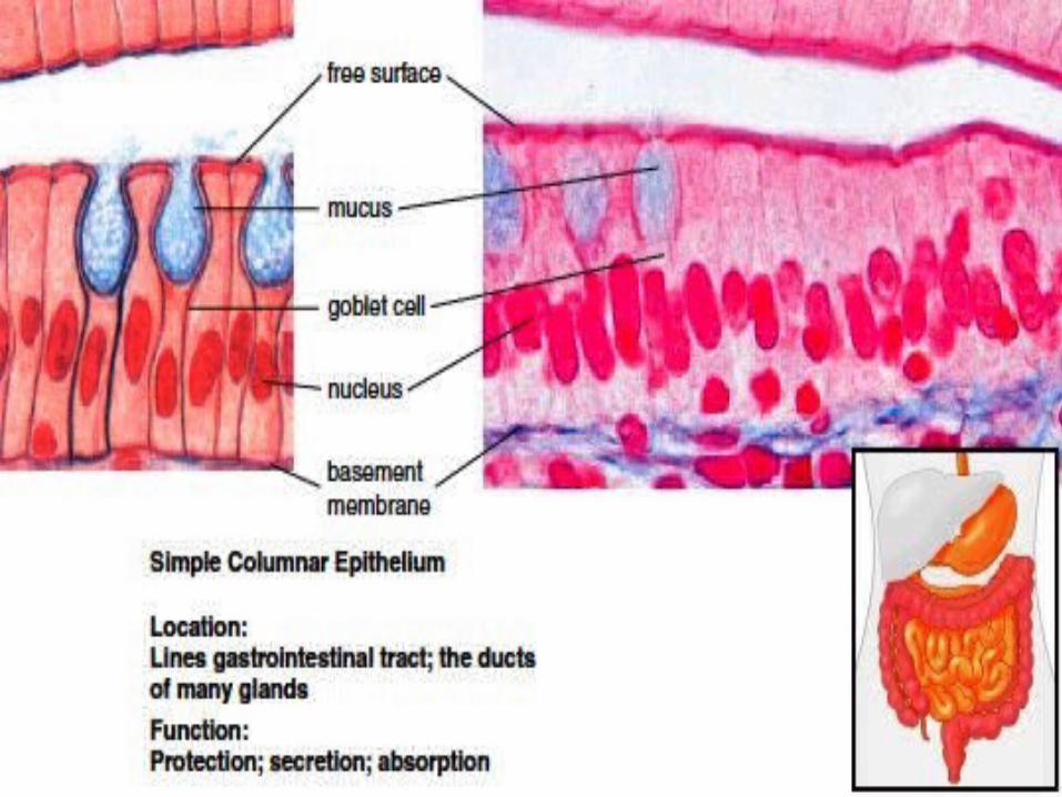
SIMPLE COLUMNAR EPITHELIUM
Cells are taller and appear columnar in sections at right angles to the basement membrane.
Nuclei are elongated and may be located towards the base, the centre or occasionally the apex of the cytoplasm, this is known as polarity.
PRESENT IN: Small intestine & colon Stomach lining and gastric glands Gall bladder
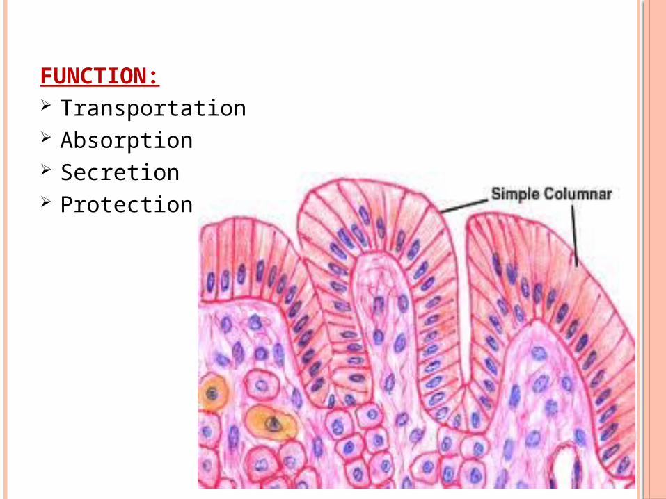
FUNCTION: Transportation Absorption Secretion Protection

SIMPLE COLUMNAR CILIATED EPITHELIUM
Described as a special entity because of the presence of surface specialisation called cilia.
Each cilia consists of a finger like projection of the plasma membrane.
Not common in humans except in the female reproductive tract.
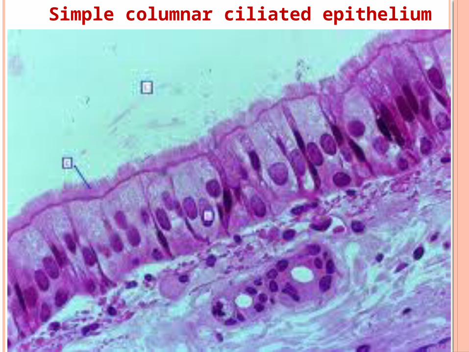
Simple columnar ciliated epithelium
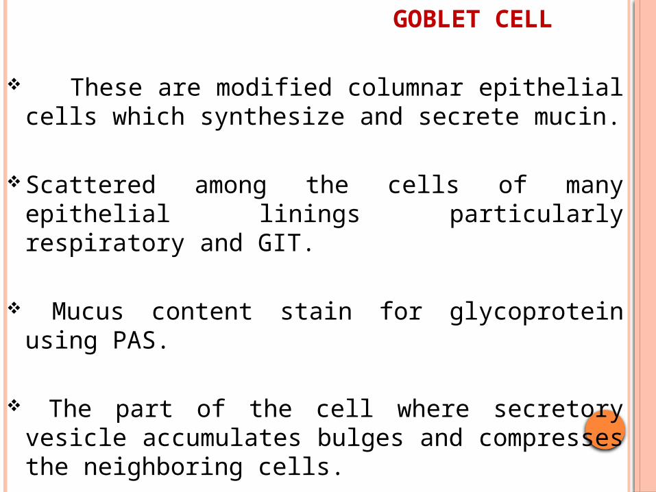
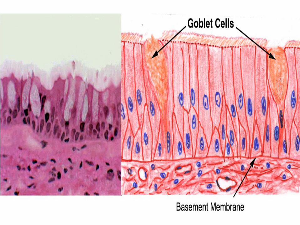
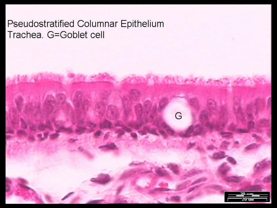
GOBLET CELL These are modified columnar epithelial cells
which synthesize and secrete mucin. Scattered among the cells of many epithelial
linings particularly respiratory and GIT. Mucus content stain for glycoprotein using
PAS. The part of the cell where secretory vesicle
accumulates bulges and compresses the neighboring cells.
PSEUDOSTRATIFIED COLUMNAR EPITHELIUM
The term pseudostratified is derived from the appearance of this epithelium in section which conveys the erroneous impression that there is more than one layer of cells.
True simple epithelium since all the cells rest on the basement membrane.
Nuclei are disposed at different levels thus creating the illusion of cellular stratification.
Exhibit polarity with nuclei confined to the basal two-third of the epithelium, cilia are never present on stratified epithelium
PRESENT IN:Trachea & bronchial treeDuctus deferensAuditory tube and tympanic cavityNasal cavity & lacrimal sacMale urethraLarge excretory ducts
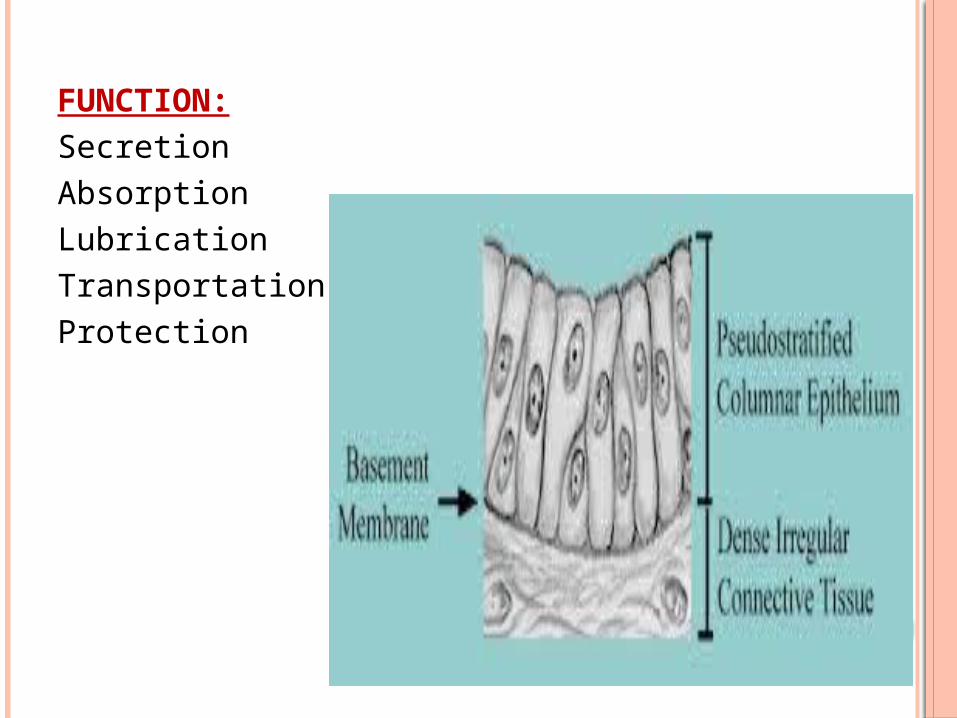
FUNCTION:SecretionAbsorptionLubricationTransportationProtection
STRATIFIED SQUAMOUS EPITHELIUM
It consists of a variable number of cells layer which exhibit transition from a cuboidal basal layer to a flattened surface.
Basal layer divide continuously.
Well adapted to withstand abrasion since loss of surface cells does not compromise the underlying tissue.
Nuclei become progressively condensed (pyknotic) and flattened, before ultimately disintegrating.

STRATIFIED SQUAMOUS EPITHELIUM

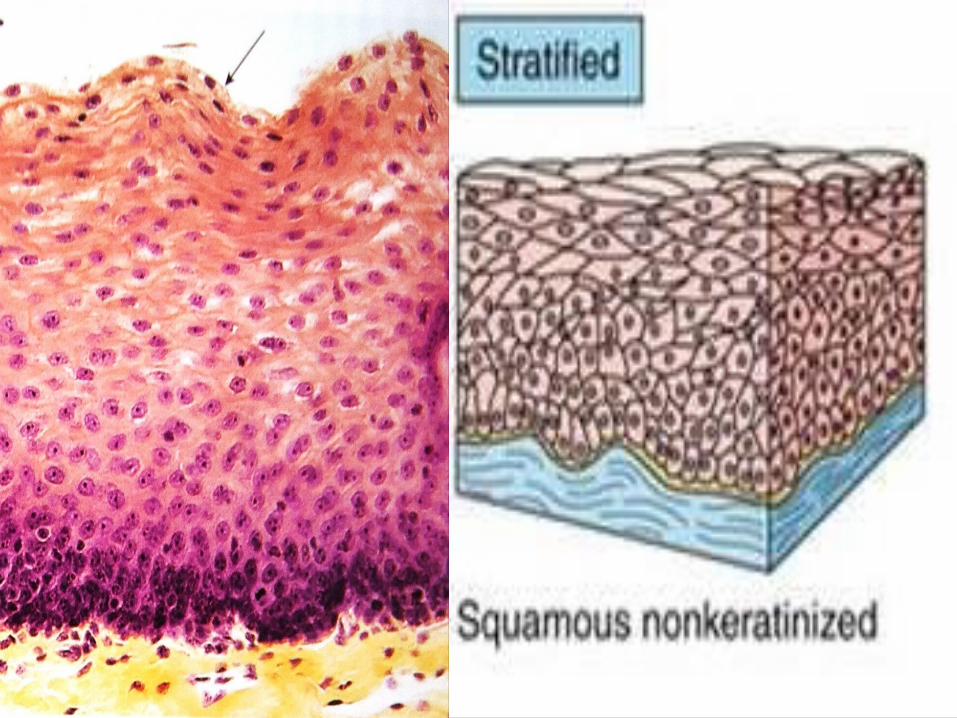
STRATIFIED SQUAMOUS NONKERATINISED EPITHELIUM
Flattened with nuclei.Moist superficial cells are living.
PRESENT IN:MouthEpiglottisEsophagusVocal foldsVagina
FUNCTION:ProtectionSecretion
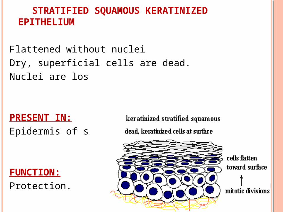
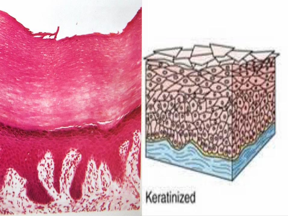
STRATIFIED SQUAMOUS KERATINIZED EPITHELIUM
Flattened without nucleiDry, superficial cells are dead.Nuclei are lost.
PRESENT IN:Epidermis of skin.
FUNCTION:Protection.
STRATIFIED CUBOIDAL EPITHELIUM
Thin, stratified epithelium which usually consists of only two or three layers of cuboidal or low columnar cells.
Not involved in significant absorptive or secretory activity
PRESENT IN:Ducts of sweat glandsLarge ducts of exocrine glandsAnorectal junction
FUNCTION:AbsorptionSecretion


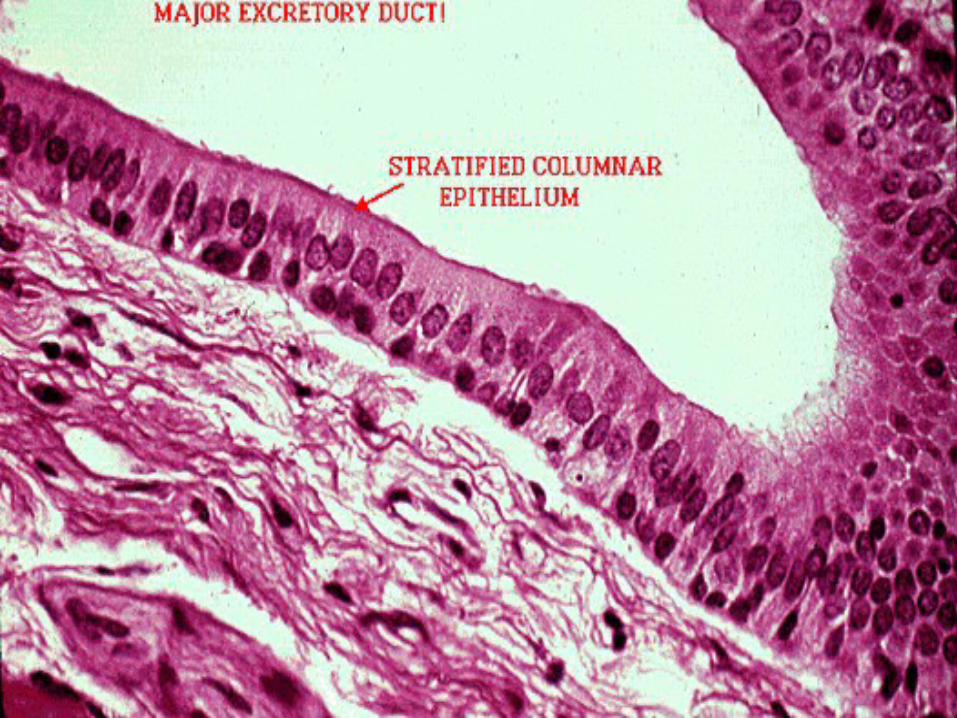
STRATIFIED COLUMNAR EPITHELIUM
PRESENT IN:Conjunctiva of eyeSome large excretory ductsPortions of male urethra
FUNCTION:SecretionAbsorptionProtection
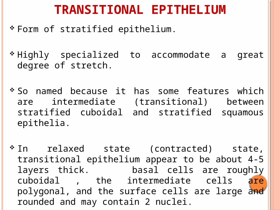
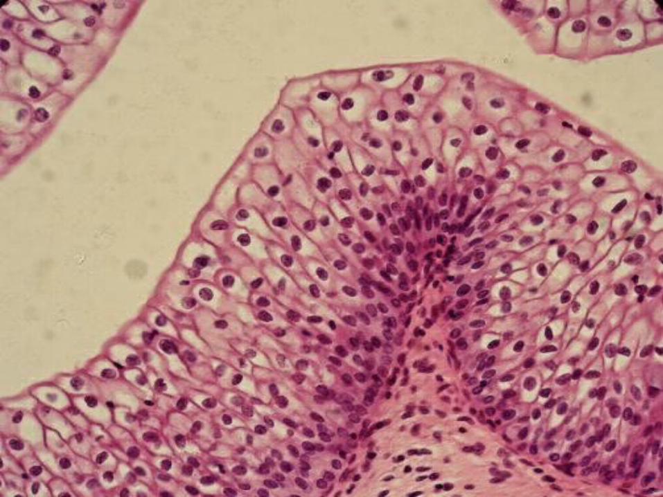
TRANSITIONAL EPITHELIUM Form of stratified epithelium.
Highly specialized to accommodate a great degree of stretch.
So named because it has some features which are intermediate (transitional) between stratified cuboidal and stratified squamous epithelia.
In relaxed state (contracted) state, transitional epithelium appear to be about 4-5 layers thick. basal cells are roughly cuboidal , the intermediate cells are polygonal, and the surface cells are large and rounded and may contain 2 nuclei.
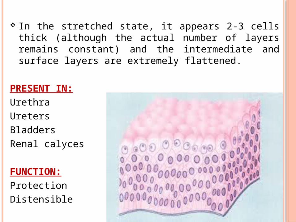
In the stretched state, it appears 2-3 cells thick (although the actual number of layers remains constant) and the intermediate and surface layers are extremely flattened.
PRESENT IN:UrethraUretersBladdersRenal calyces
FUNCTION:ProtectionDistensible
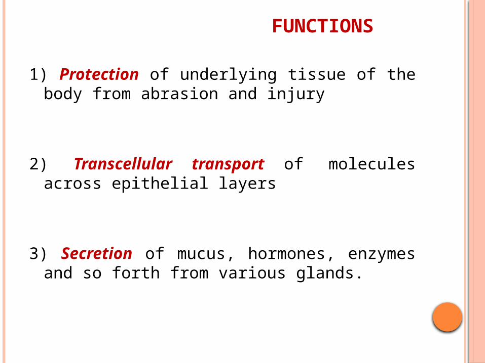
FUNCTIONS
1) Protection of underlying tissue of the body from abrasion and injury
2) Transcellular transport of molecules across epithelial layers
3) Secretion of mucus, hormones, enzymes and so forth from various glands.

4) Absorption of materials from a lumen
5) Control of movement of materials between body compartments via selective permeability of intracellular junctions between epithelial cells.
6) Detection of sensations via taste buds, retina of the eye and specialized hair cells in the ear.
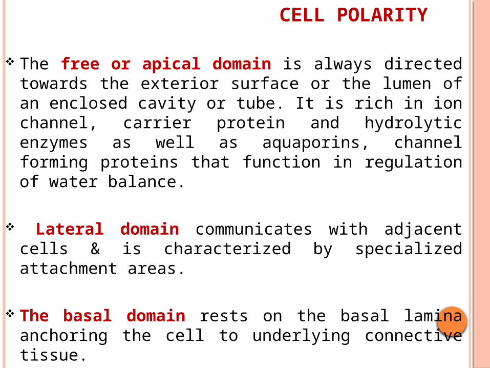
CELL POLARITY
The free or apical domain is always directed towards the exterior surface or the lumen of an enclosed cavity or tube. It is rich in ion channel, carrier protein and hydrolytic enzymes as well as aquaporins, channel forming proteins that function in regulation of water balance.
Lateral domain communicates with adjacent cells & is characterized by specialized attachment areas.
The basal domain rests on the basal lamina
anchoring the cell to underlying connective tissue.
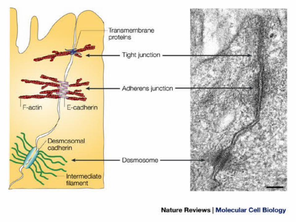
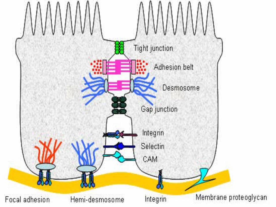
MEMBRANE SPECIALIZATION OF EPITHELIA
The intercellular, luminal and basal surface of epithelial cells exhibit a variety of specialization.
1) INTERCELLULAR SURFACE: The apposed surface of epithelial cells are lined
by several different types of membrane and cytoskeletal specialization. Cell junctions are:
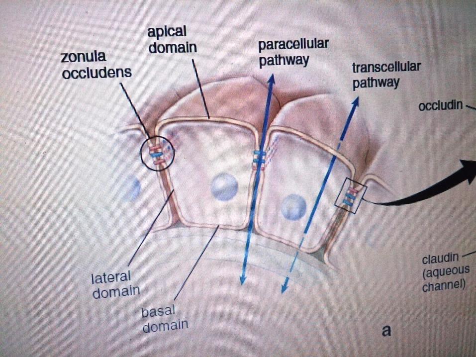
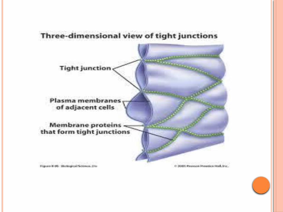
a) Occluding or tight junctions: Located immediately behind the luminal surface
of simple columnar epithelium Intercellular spaces are oblitereted Transmembrane adhesive protein- occludin,
claudin, junctional adhesive molecule
Each tight junction forms a continuous circumferential band or zonules around the cell and are thus known as zonula occludens.
FUNCTIONS: Seal adjacent cells together Involved in cell signaling Defines apical and basolateral domain of
plasma membrane.
Tightness of the junction is related to the claudins present
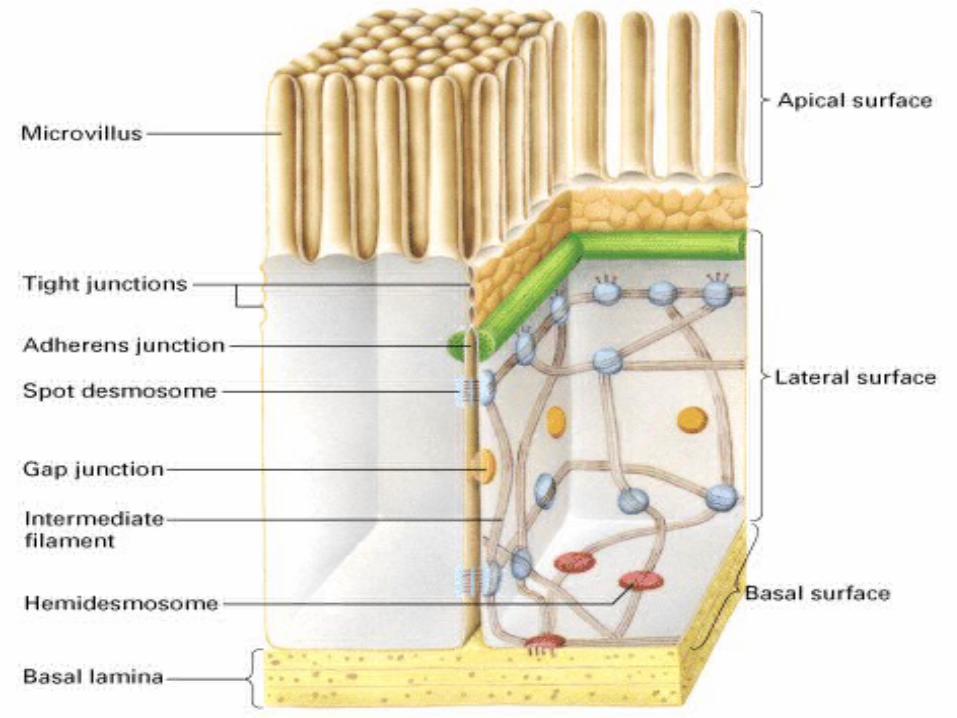
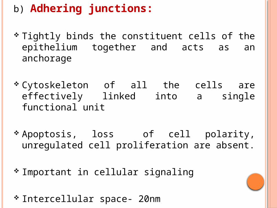
b) Adhering junctions:
Tightly binds the constituent cells of the epithelium together and acts as an anchorage
Cytoskeleton of all the cells are effectively linked into a single functional unit
Apoptosis, loss of cell polarity, unregulated cell proliferation are absent.
Important in cellular signaling
Intercellular space- 20nm
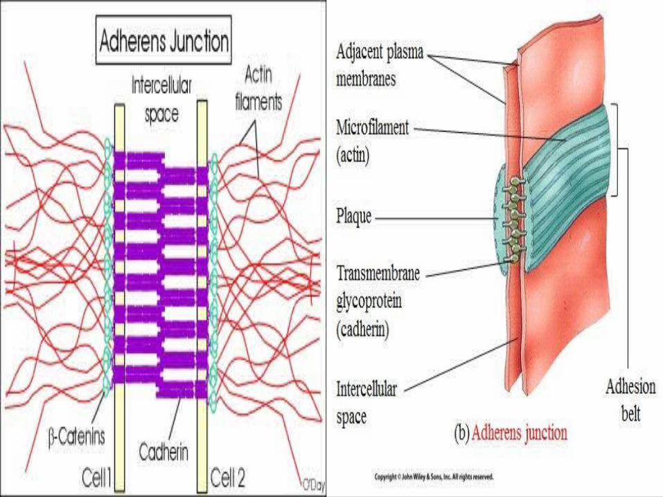
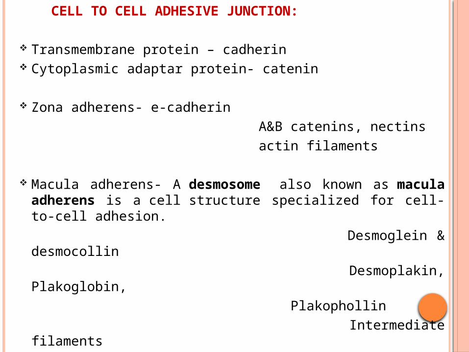
CELL TO CELL ADHESIVE JUNCTION:
Transmembrane protein – cadherin Cytoplasmic adaptar protein- catenin
Zona adherens- e-cadherin A&B catenins, nectins actin filaments
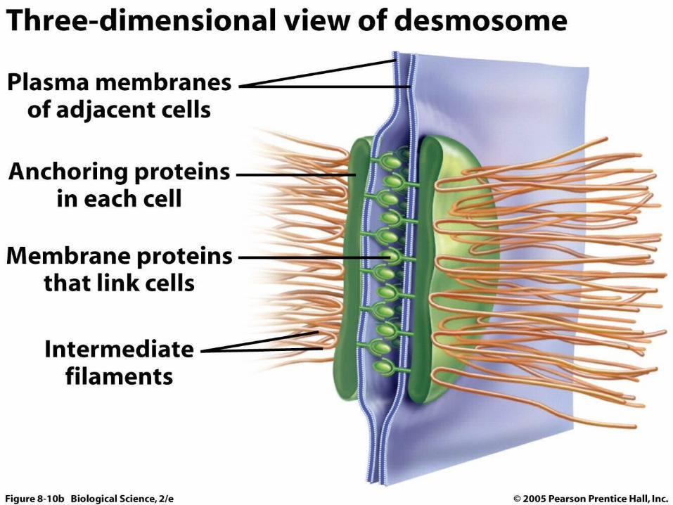
Macula adherens- A desmosome also known as macula adherens is a cell structure specialized for cell-to-cell adhesion.
Desmoglein & desmocollin Desmoplakin, Plakoglobin, Plakophollin Intermediate filaments

CELL TO CELL MATRIX JUNCTION:
Focal adhesion which anchor actin filaments of the cytoskeleton into the basement membrane
Integrin, A-actinin, vinculin, talin, actin filaments, remodelling of actin filaments.
Hemidesmosomes which anchor the intermediate filaments of the cytoskeleton into the basement membrane.
Hemidesmosomes are asymmetrical and are found in epithelial cells connecting the basal face of the cell to basal lamina. Similar in form to desmosomes when visualized by electron microscopy
Integrin, A6B4, BP230, Plectin, intermediate filaments links the cells to the basal lamina.
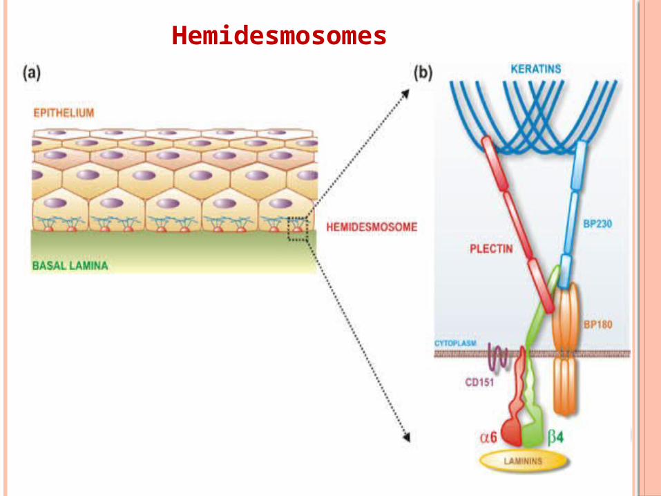
Hemidesmosomes
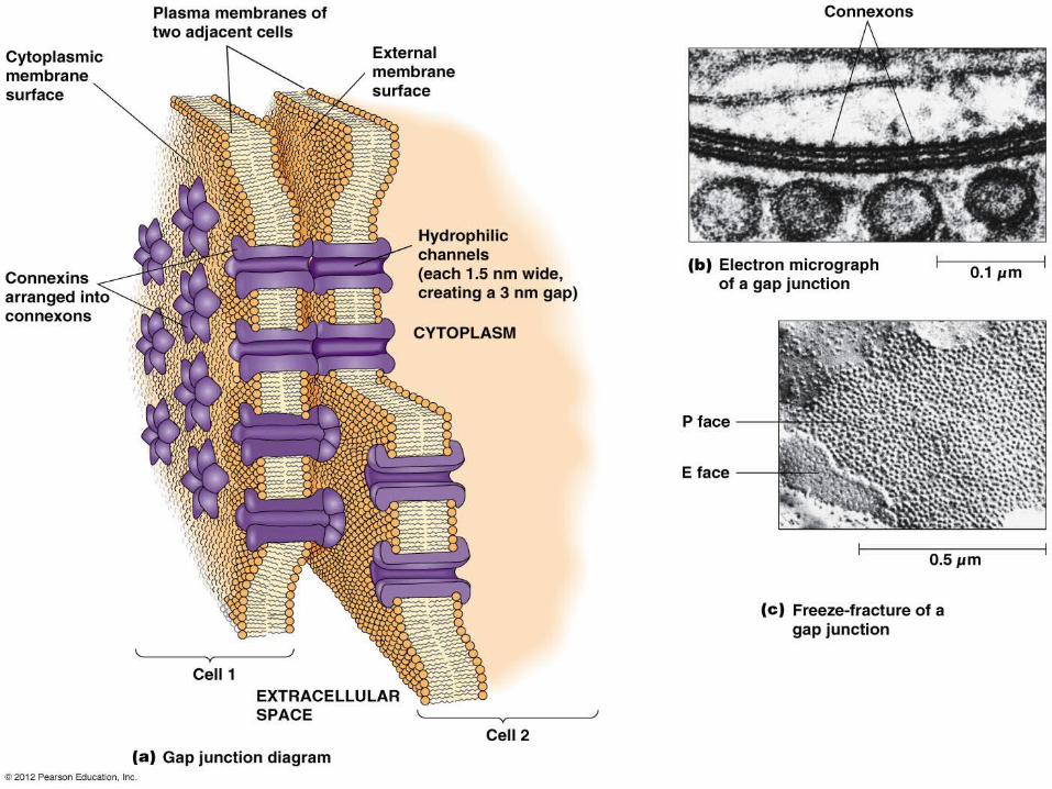
c) Gap junction:
Circular intercellular contacts areas containing hundreds of tiny pores which permit passage of small molecules between adjacent cells.
Intercellular space- 2-3 nm Transmembrane protein- connexin (form aqueous
channels) Function-:-
Creates a (nexus) adjacent cell conduct
between two adjacent cells for passage of small
ions and informational micromolecules.
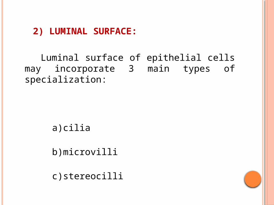
2) LUMINAL SURFACE:
Luminal surface of epithelial cells may incorporate 3 main types of specialization:
a)cilia b)microvilli c)stereocilli
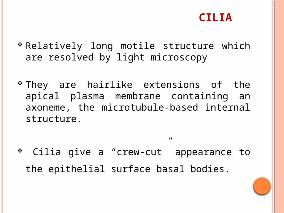
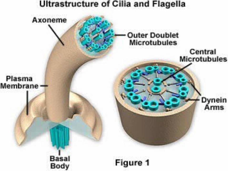
CILIA
Relatively long motile structure which are resolved by light microscopy
They are hairlike extensions of the apical plasma membrane containing an axoneme, the microtubule-based internal structure.
Cilia give a “crew-cut” appearance to the
epithelial surface basal bodies.
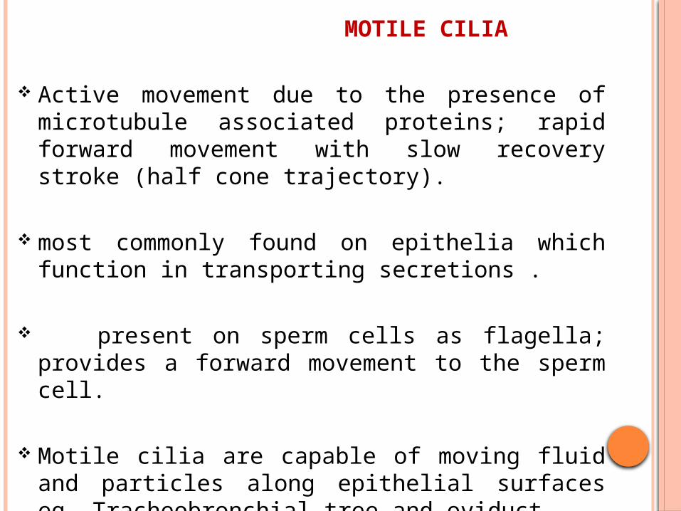
MOTILE CILIA
Active movement due to the presence of microtubule associated proteins; rapid forward movement with slow recovery stroke (half cone trajectory).
most commonly found on epithelia which function in transporting secretions .
present on sperm cells as flagella; provides a forward movement to the sperm cell.
Motile cilia are capable of moving fluid and
particles along epithelial surfaces eg. Tracheobronchial tree and oviduct.
PRIMARY CILIA
found in almost all cells in the body .
transmit signals from extracellular space into the cell.
No active movement; passively bend due to flow of fluid.
Function: chemosensors osmosensors mechanosensors.
NODAL CILIA
Structure similar to primary cilia except they have an ability for active transport, active rotational movement
Found in the embryo during gastrulation on the bilaminar disc near the area of primitive node.
Essential in developing left-right asymmetry of internal organs.
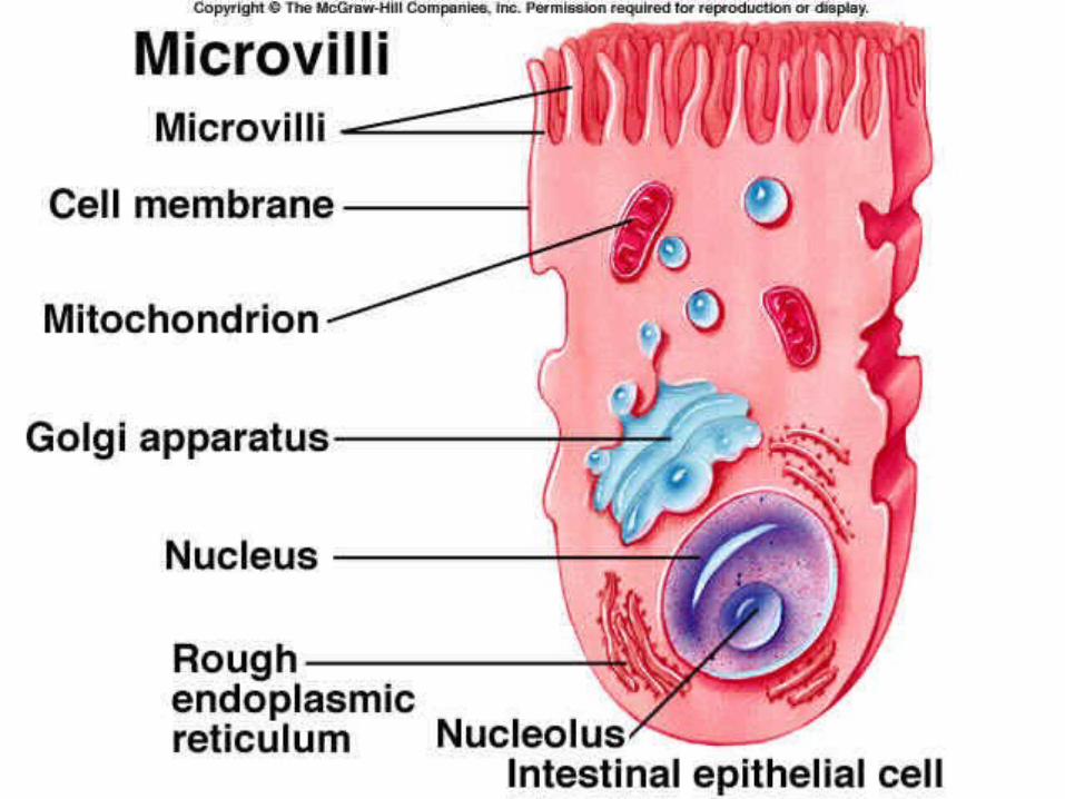
MICROVILLI
Microvilli are fingerlike cytoplasmic projections on the apical surface of most epithelial cells .
In intestinal absorptive cell this surface structure was originally called the striated border; in the kidney tubule cells, it is called the brush border.
Can not be individually resolved with the microscope
Internal structure contain a core of actin filament that are cross linked by several actin binding protein
Increase absorptive capacity
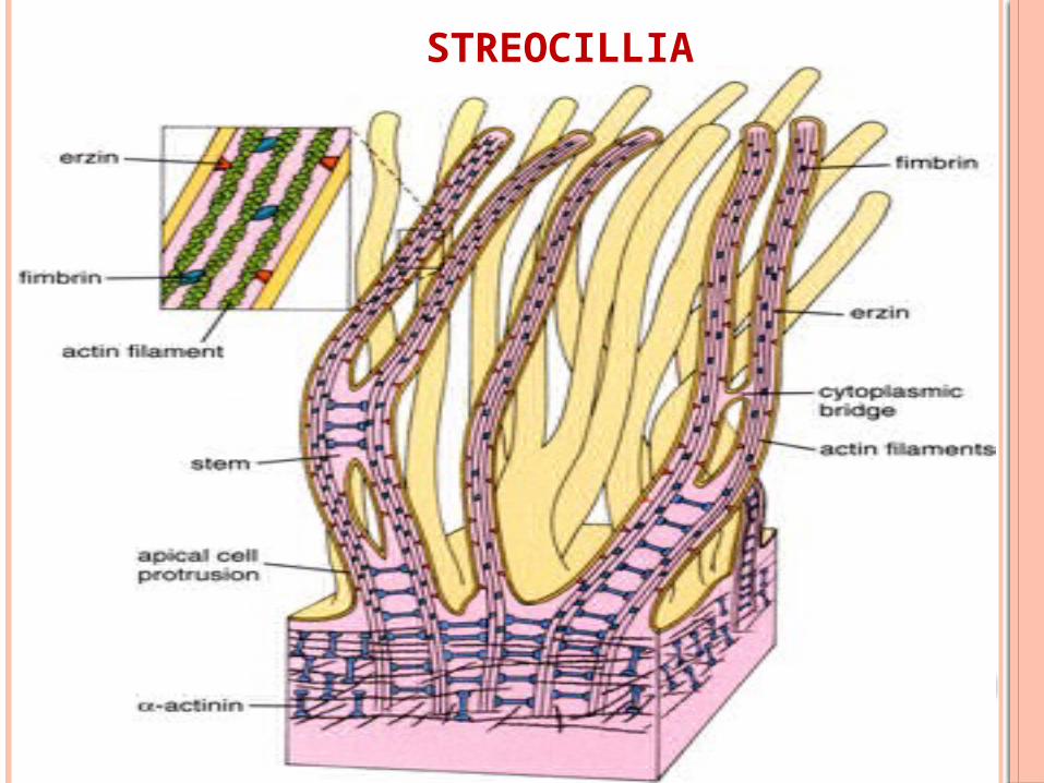
STREOCILIA
Stereocilia are unusually long, immotile microvilli.
Found only singly or in small number in odd sites such as the male reproductive tracts.
Contains ezrin and A-actinin.
Treadmilling effect- structure renewal process
STREOCILLIA
3) BASAL SURFACE
The interface between all epithelia and underlying supporting structures is marked by a noncellular structure known as the basement membrane.
It provides structural support for epithelia and constitute a selective barrier to the passage of material between epithelium and supporting tissue.
Hemidesmosomes provides a mean of anchorage of the cells via its cytoskeleton to the basement membrane and underlying supporting tissue
Consists of 3 zones: lamina lucida lamina densa lamina fibroreticularis or sublamina densa
LAMINA DENSA
The lamina densa is a component of the basement membrane zone between the epidermis and dermis of the skin, and is an electron-dense zone between the lamina lucida and dermis.
Synthesized by the basal cells of the epidermis
Electron dense matrix 50nm thick between the epithelium and the adjacent connective tissue
Exhibit a network of fine, 3-4nm filaments composed of laminins, a type iv collagen molecule (chicken-wire) and proteoglycans and glycoprotein.
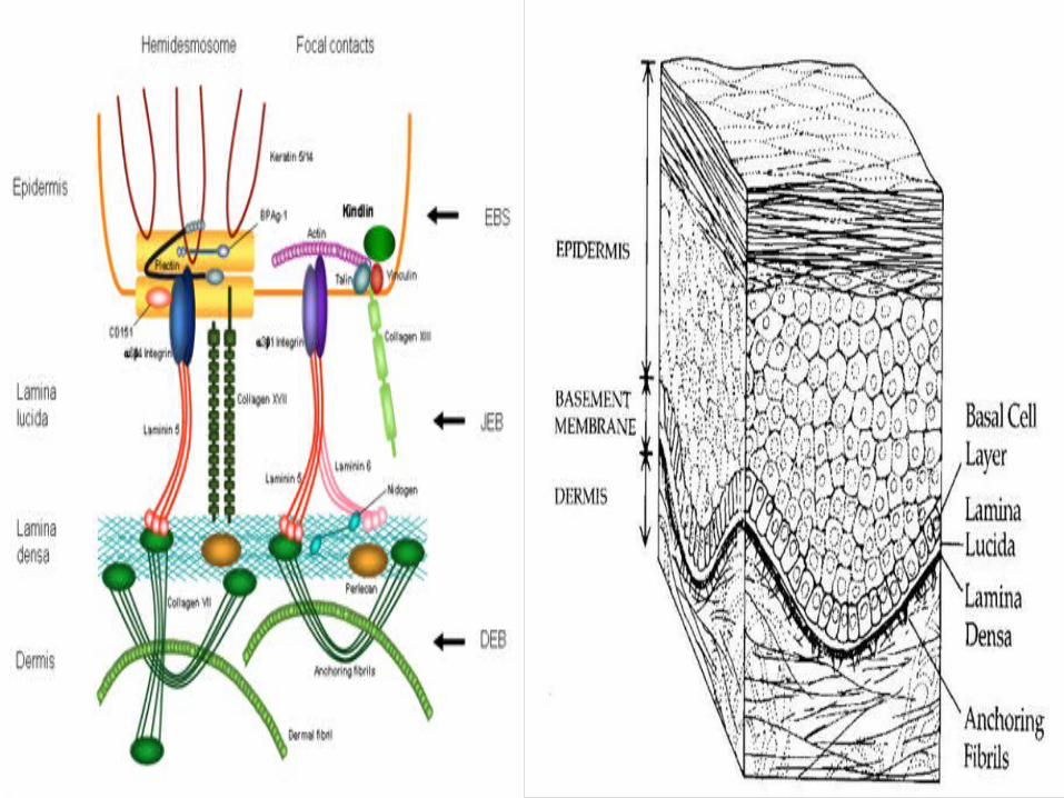
LAMINA LUCIDA The lamina lucida is a component of the
basement membrane which is found between the epithelium and underlying connective tissue.
Clear zone 40nm thick that attach the cells to the basal lamina
Contain- collagen type xvii, integrins, laminin v
Anchoring fibrils consists of collagen type vii attach basal lamina to connective tissue.
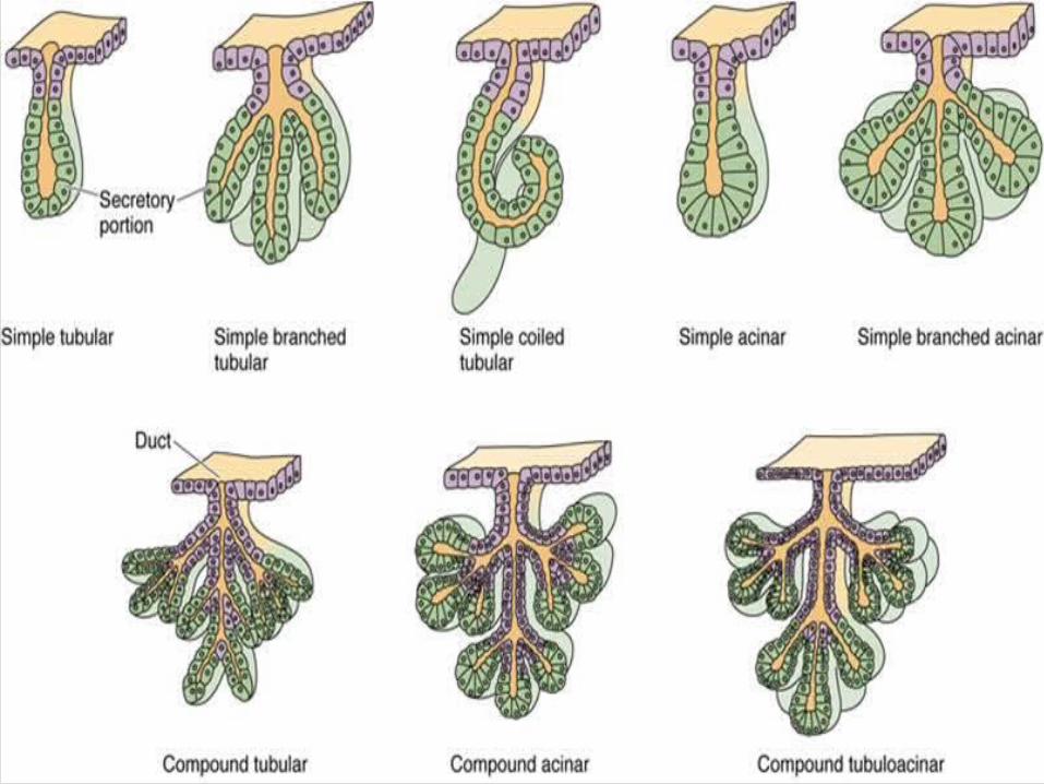
GLANDS
Typically glands are classified into:
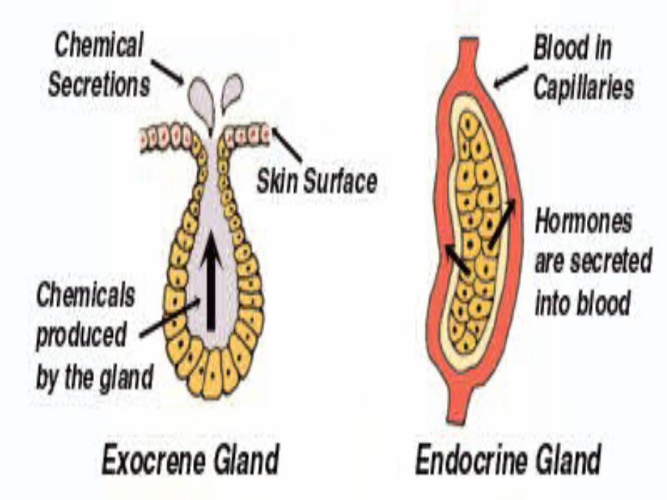
1)Exocrine glands discharge their secretory product via a duct onto an epithelial surface. Cells of which are composed of highly specialized epithelial cells, the internal structure of the cells reflecting the nature of the secretory product and the mode of secretion.
Morphology: a)simple: single, unbranched duct. b)compound: branched duct
system.
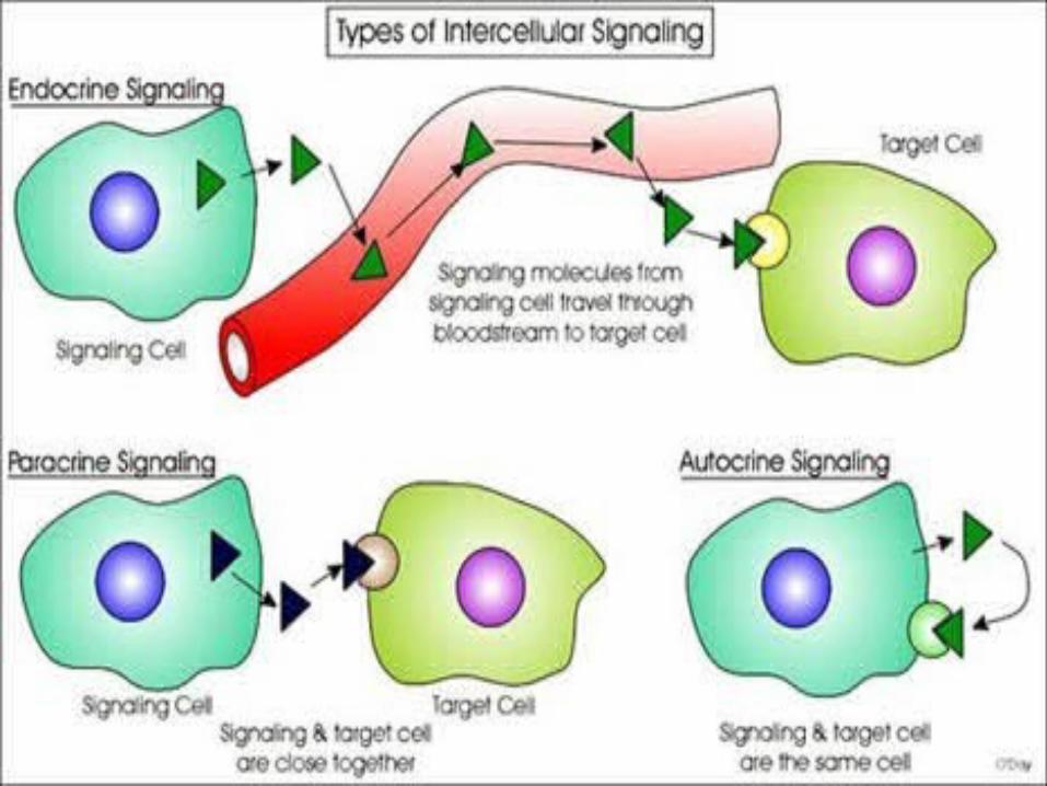
2)Endocrine glands are ductless. Secrete their product into the connective tissue where they enter the blood stream to reach the target cells. The products of endocrine glands are called hormone.
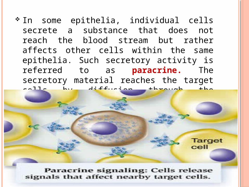
In some epithelia, individual cells secrete a substance that does not reach the blood stream but rather affects other cells within the same epithelia. Such secretory activity is referred to as paracrine. The secretory material reaches the target cells by diffusion through the extracellular space or immediately subjacent connective tissue.
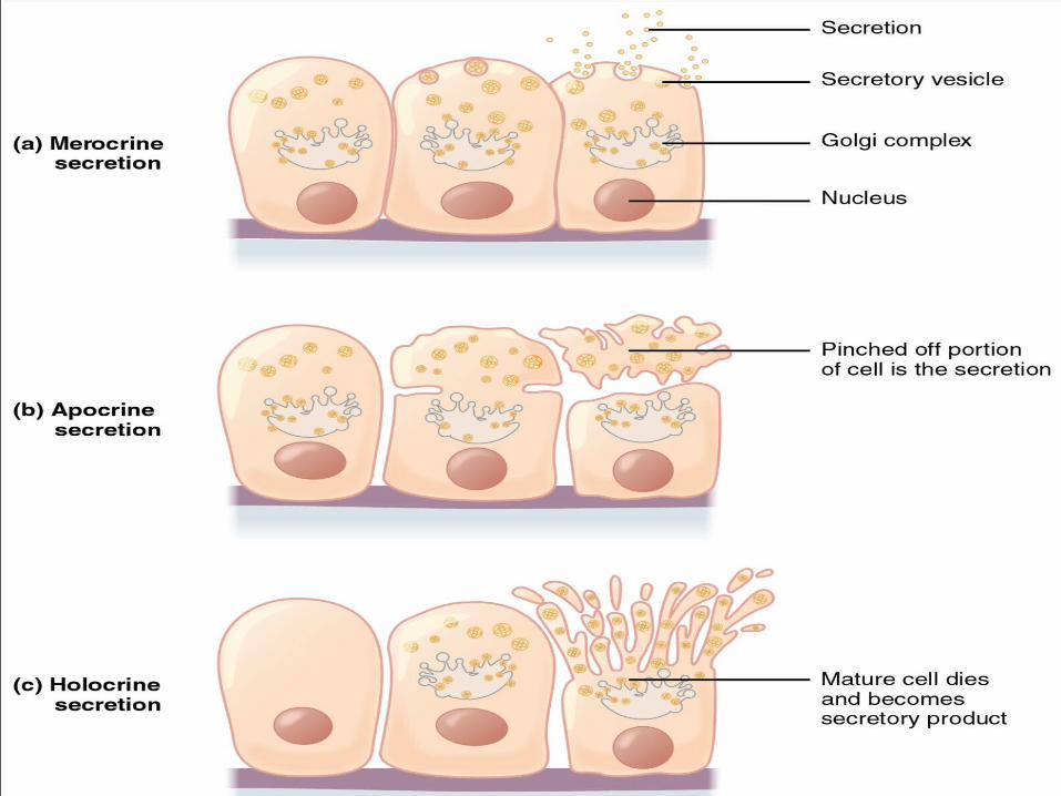
Cells of exocrine glands exhibit different mechanism of secretion:
Merocrine secretion- involves the process of exocytosis and is the most common form of secretion, protein are usually the major secretory product
Apocrine secretion- involves discharge of free, unbroken, membrane bound vesicles containing secretory product. This is an unusual mode of secretion and appears to lipid secretory products in the breasts and some sweat glands
Holocrine secretion- involves discharge of whole secretory cells with subsequent disintegration of the cells to release the secretory product. Occurs principally on the sebaceous glands.
EPITHELIAL CELL RENEWAL
The stratified squamous epithelium of skin is replaced in approximately 28 days.
Cells in the stratum basale undergo mitosis to provide for cell renewal.
As these cells differentiate they are pushed toward the surface by new cells in the basal layer.
Ultimately, the cells become keratinized and slough off.
Thus a steady state is maintained within the epithelium, with new cells normally replacing exfoliated cells at the same rate.
Cells arising by division in the basal layer may remain in the progenitor cell population or undergo a process of maturation as they move to surface.
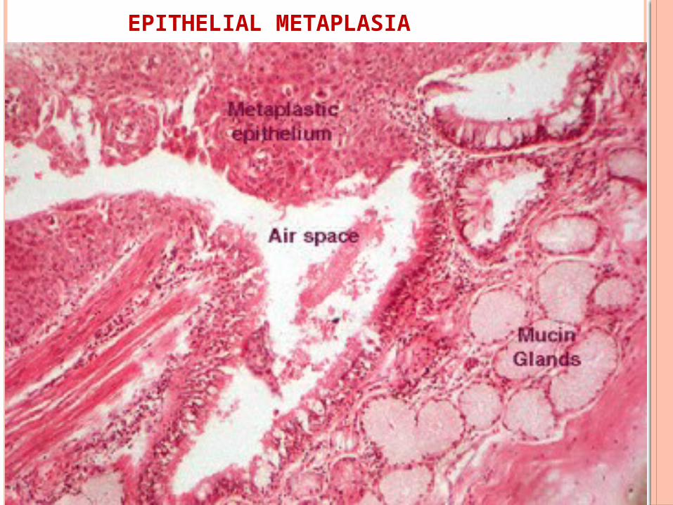
EPITHELIAL METAPLASIA
Epithelial metaplasia is a reversible
conversion of one mature epithelial cell type
to another mature epithelial cell type.
Metaplasia is generally an adaptive response
to stress, chronic inflammation, or other
abnormal stimuli.
EPITHELIAL METAPLASIA
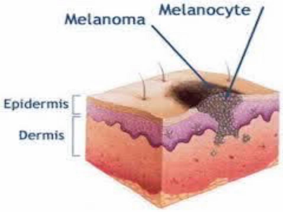
TUMOURS ARISING FROM EPITHELIA
A tumour can arise from any tissue if there is uncontrolled growth of cells.
A malignant tumour arising from an epithelia is a carcinoma.
If it arises from squamous epithelium it is a squamous cell carcinoma
If tumour arising from glandular epithelium it is called adenoma.
Diagnosis can be made by Immuno histochemical technique.
STRUCTURE OF THE ORAL EPITHELIUM
Stratified squamous variety.
May be keratinized (ortho or parakeratinized) or nonkeratinized depending on location.
Keratinized: gingiva and hard palate (masticatory mocosa). In many gingival epithelium is parakeratinized.
Non keratinized: cheeks, faucial and sublingual tissue.
Both keratinized and nonkeratinized contains 2 groups of cells- keratinocytes and nonkeratinocytes.
TURNOVER TIME OF THE EPITHELIUM
Turnover time- time taken for a cell to divide and pass through the entire epithelium.
E.g. – * skin - 52 to 75 days . * gut - 4 to 14 days. * gingiva - 41 to 57 days. * cheek - 25 days.
Nonkeratinised buccal epithelium turns over faster than keratinized gingival epithelium.
Keratinized epithelium: 1) stratum basale 2) stratum spinosum 3) stratum
granulosum 4) stratum corneum
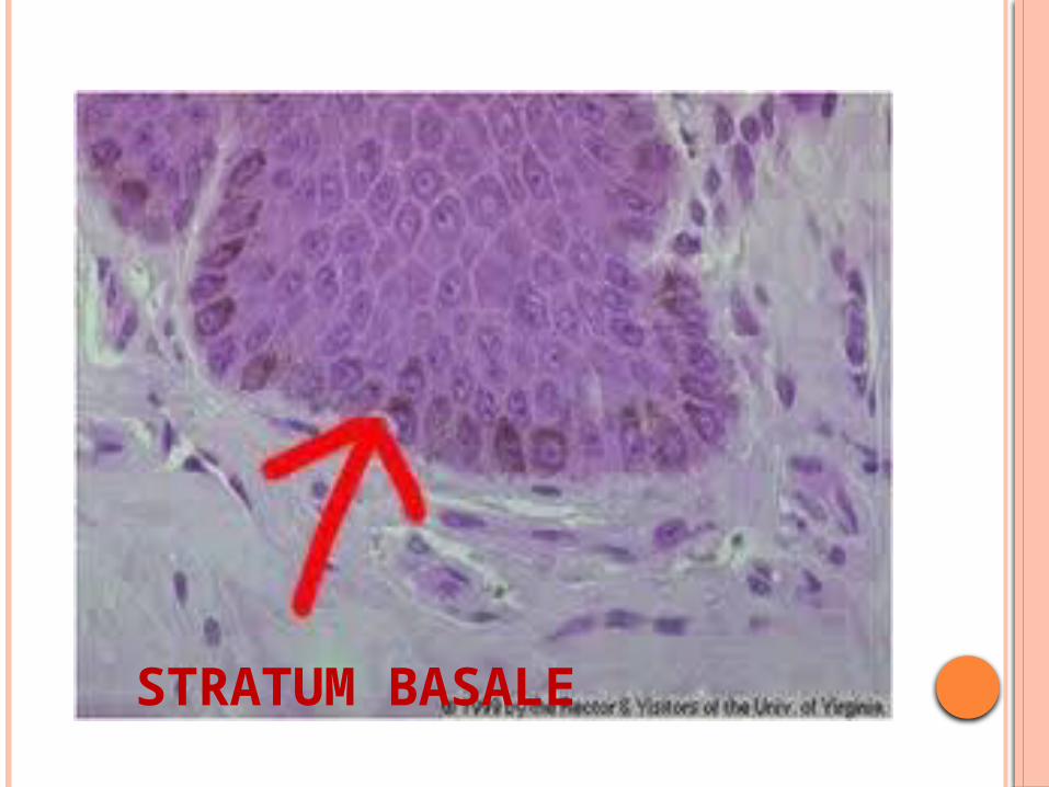
STRATUM BASALE
Single layer of cuboidal cells
Made up of cells that synthesize DNA and undergo mitosis thus providing new cells
Basal cells and parabasal cells are referred to as stratum germinativum but only basal cells can divide.
Basal cells synthesize proteins
Hemidesmosomes are found in basal layer.
Lateral borders of the adjacent cells are closely apposed and connected by desmosomes.
The basal cells contain tonofilaments and are attached to the attachment plaque
Desmosomes consists of 2 principal proteins: transmembranous protein and proteins within the cells and related to attachment plaque.
STRATUM BASALE
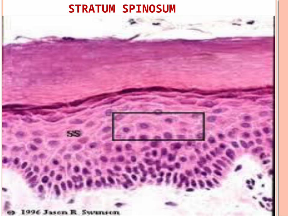
STRATUM SPINOSUM
Irregular polyhedral cells larger than basal cells.
In light microscopy, it appears these are joined by “intercellular bridges”
Tonofilaments seems to course from cell to cell across the bridge.
Electron microscopy revels- intercellular bridges are desmosomes and tonofibrils are bundles of tonofilament.
Desmosome attachment plaques contain the polypeptides desmoplakin and plakoglobin.
Intercellular space contains glycoprotein, glycosaminoglycan and fibronectin.
Prickle cell layer- shrinks away from each other remaining in contact at the desmosomes.
Most active layer in protein synthesis.
STRATUM SPINOSUM
STRATUM GRANULOSUM
Flatter and wider cells larger than spinous cells
Contains basophilic keratohyalin granules
Nucleus show degeration and pyknosis.
Tonofilaments are more dense in quantity and are often seen associated with keratohyalin granules.
Cell surface are more regular and more closely attached to adjacent cell surface.
Lamellar granules: keratinosome or odland
body- membrane coating acts as permeability barrier.
Involucrin (keratolin)- protein present at the upper half.
Membrane coating granules are glycoprotein.

STRATUM GRANULOSUM
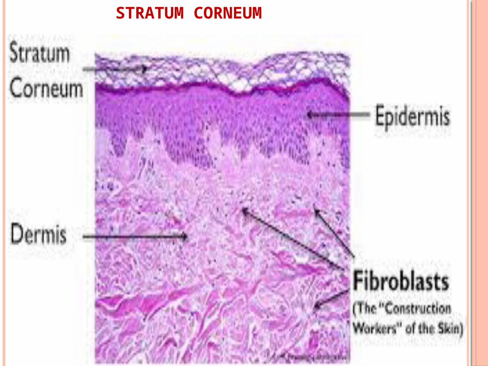
STRATUM CORNEUM
keratinized squamae which are larger and flatter
than granular cells.
Nuclei and organelles have disappeared.
Acidophilic and histologically amorphous layer.
Keratohyalin granules have disappeared.
Cells are composed of densely packed filaments
coated by basic protein of keratohyaline granules,
filaggrin.
STRATUM CORNEUM
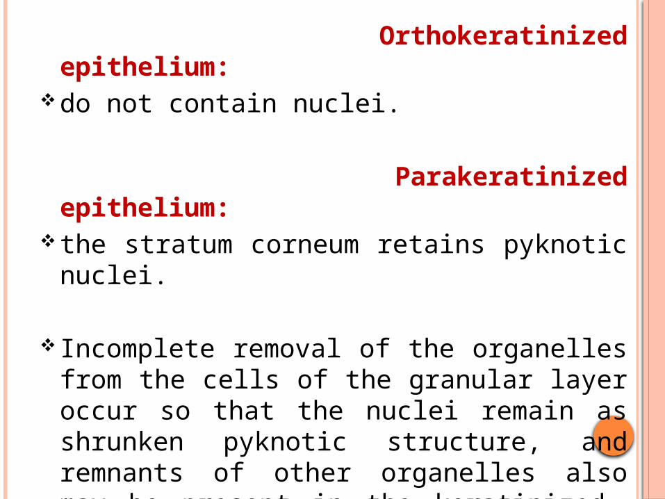
Orthokeratinized epithelium: do not contain nuclei.
Parakeratinized epithelium: the stratum corneum retains pyknotic
nuclei.
Incomplete removal of the organelles from the cells of the granular layer occur so that the nuclei remain as shrunken pyknotic structure, and remnants of other organelles also may be present in the keratinized layer
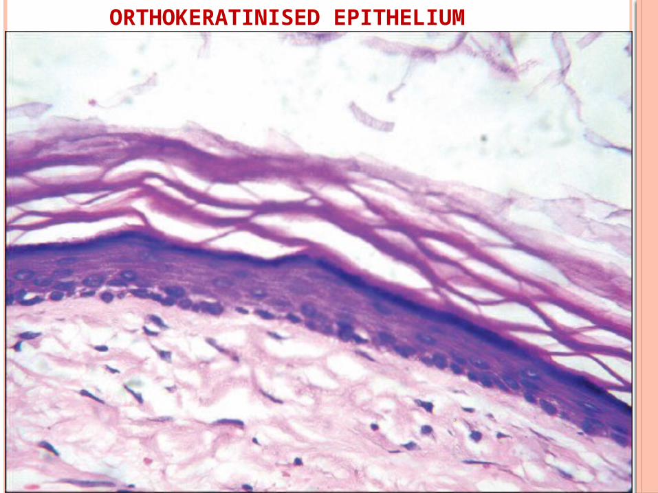
ORTHOKERATINISED EPITHELIUM
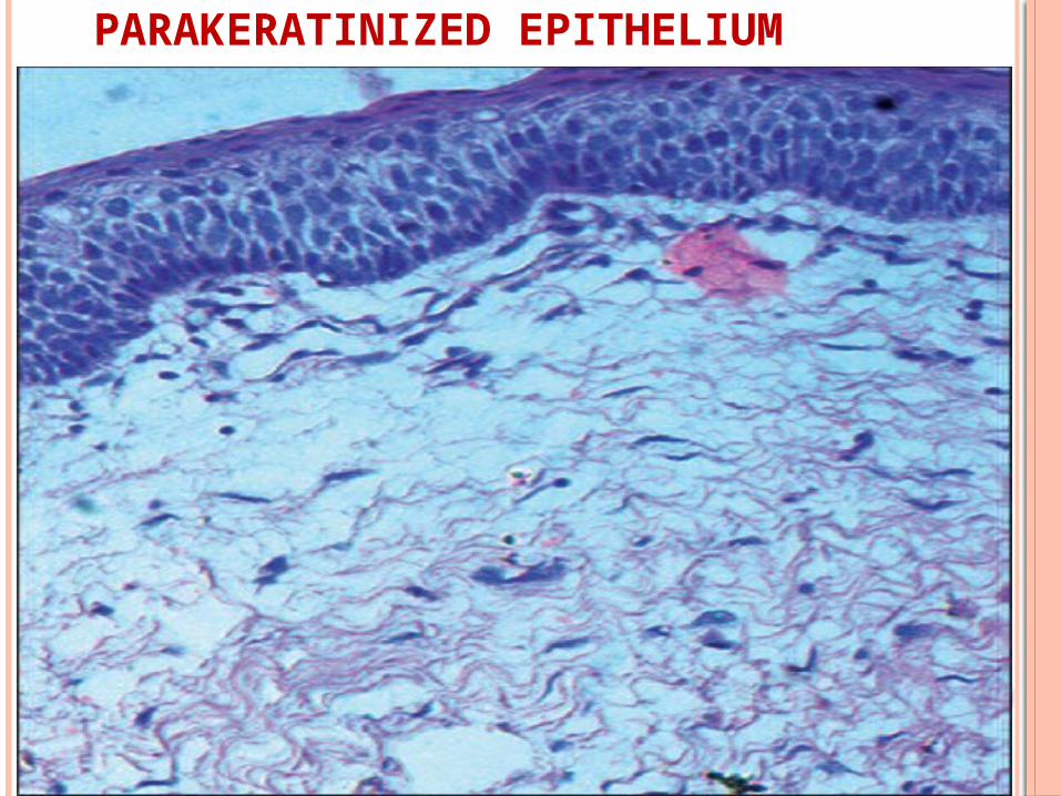
PARAKERATINIZED EPITHELIUM
NONKERATINIZED EPITHELIUM
Layers Basal - (Stratum Basale) Intermediate - (Stratum Intermedium) Superficial - (Stratum Superficiale)
Basal cells are similar.
Cells of stratum intermedium are larger than
spinosum and are attached by desmosomes
and other junction.
More closely attached than spinous cells.
No Stratum Granulosum
No Stratum Corneum.
Stratum Superficiale – nucleated cells
Less number of tonofilaments
Lack keratohyaline granules.
Have higher rate of mitosis than keratinized epithelium.
Parakeratosis –physiologic normally keratinizing tissue becomes
parakeratinized.
Keratosis- Pathologic keratinization occurs in anormally
nonkeratinized tissue.
KERATINOCYTE
Epidermal/epithelial cells that synthesize keratin.
Characteristic intermediate filament protein is cytokeratin.
Show cell division, undergo maturation and finally desquamate
Increase in volume in each successive from basal to superficial.
NONKERATINOCYTES
Donot possess cytokeratin filament
Do not show mitotic activity undergo maturation and finally desquamate
Usually dendritic and appears unstained or clear in routine H&E stains
Identified by special stain or Imunohistochemical technique
Migrate to oral epithelium from neural crest or bone marrow.
MELANOCYTES
Present in basal layer.
Arise from neural crest ectoderm.
Staining reaction- dopa oxidase- tyrosinase,
silver stains.
Stained by : Mason-Fontana stain
Dendritic, no desmosomes and tonofilaments.
Premelanosomes and melanosomes are present.
Function- synthesis of melanin pigment granules
(melanosomes) and transfer to surrounding
keratinocytes.
LANGERHANS CELL
Present in suprabasal layer.
Arise from bone marrow.
Dendritic or clear cells with no desmosomes or
tonofilaments.
Characteristic langerhans granule- Birbeck
granules
Staining reactions- cell surface antigen markers
Stains by: gold chloride, ATPase &
immunofluorescent markers.
Function-
antigen trapping and processing.
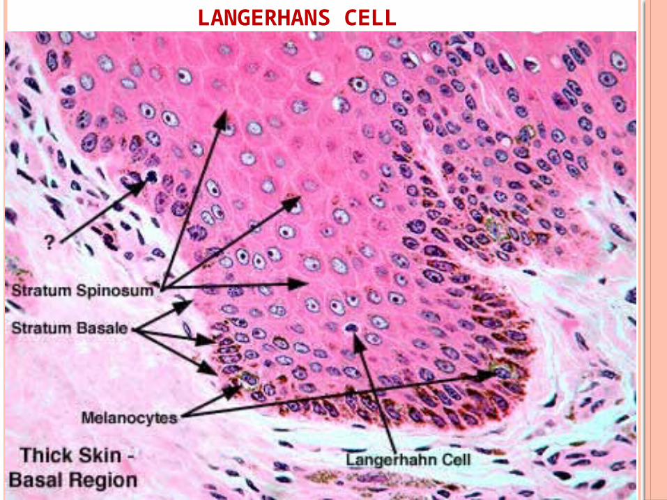
LANGERHANS CELL

MERKEL CELLS
Present in basal layer.
Arise from division of epithelial cell.
Staining reaction- PAS positive.
Seen in masticatory mucosa but are absent in
lining mucosa
Non-dendritic with less desmosomes and
tonofilaments.
sensory and respond to touch.
Characteristic electron-dense vesicles and
associated nerve axon.
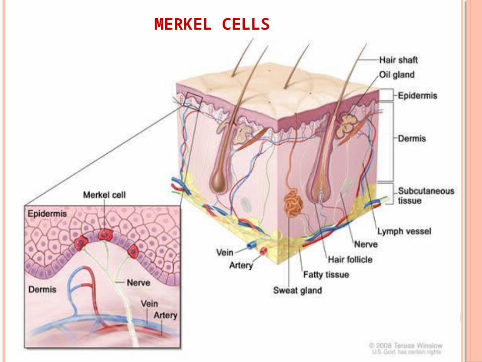
MERKEL CELLS
ULTRASTRUCTURE OF EPITHELIAL CELLS
Intracellular filaments- tonofilaments
Intracellular proteins- cytokeratins
Low mol wt keratin(40)- glandular & simple
Intermediate wt- stratified epithelia
Highest(67) – keratinized
stratified
Stratified oral epithelim - keratin 5 & 14
Keratinized epithelium - keratin 1, 6, 10, 16
Non-keratinized - keratin 4, 13, 19
REFERENCES
Michael H. Ross and Wojciech Pawlina; Histology A Text & Atlas; 6th edition; p.105-146
Kumar GS , Orban’s Oral Histology and Embryology, 12th Ed,2009,Elsevier,New Delhi, p.210-226.
Nanci A , Ten Cate’s Oral Histology Development structure and function, 7th Ed,2008,Mosby,New Delhi,p.320-336.

Singh.I,Histology of Human Histology Colour Atlas,5th Ed , Jaypee brothers , 2009,New Delhi, p.45-53.
Wheaters, functional histology, a text and colour atlas, 4th edition, page 80-96
BKB Berkovitz, oral anatomy, histology and embryology, 3rd edition, page 220-224
Leslic P. Gartner, colour textbook of histology, 3rd edition, page 85-109