Embed Size (px)
DESCRIPTION
Glandular epithelium. describes epithelial tissue found in glands and specialized for exocrine or endocrine secretion. . Endocrines release their secretory product (typically hormones) into the spaces between the secretory cells (extracellular space) from which it enters the bloodstream. - PowerPoint PPT Presentation
Citation preview
Glandular epithelium
• describes epithelial tissue found in glands and specialized for exocrine or endocrine secretion.

• Endocrines release their secretory product (typically hormones) into the spaces between the secretory cells (extracellular space) from which it enters the bloodstream.
• Both exocrines and most of enodcrines are developmentally derived from epithelia, which form a down-growth into the underlying connective tissue. The cells forming this down-growth then develop the special characteristics of the mature gland.

• Classification of Glands according to the number of cells:
• Unicellular gland e.g. goblet cell• Multicellular gland

unicellular glands (Goblet cells):In mammals, the only example of unicellular glands are goblet cells
• The name "goblet" refers to the cell's shape, narrow at the base and bulging apically. Goblet cells secrete the glycoprotein mucin, which by the uptake of water is converted into a slimy substance,mucus.
are scattered among the absorptive cells in the epithelium of the small intestine and colon and the respiratory tract and the reproductive female tract.
These epithelial cells are specialized for secretion of mucus, which facilitates passage of material through the bowel.

The simplest form of a multicellular gland is • a secretory epithelial sheath - a surface
epithelium consisting entirely of secretory cells • e.g. the epithelium lining the inner surface of the
stomach, where the mucous secretion protects the stomach wall from the acidic contents of the stomach).
• Other multicellular glands have their secretory portion embedded in the connective tissue underlying the epithelium.
• The secretion is either discharged directly from the secretory portion onto the epithelium or reaches the epithelium via a duct system that consists of non-secretory cells.

Classification of the glands according to the presence of duct:• Endocrines (without duct)• Exocrines (have duct opens upon one of
the surfaces of the body e.g. skin, gastrointestinal tract etc).
• Mixocrines
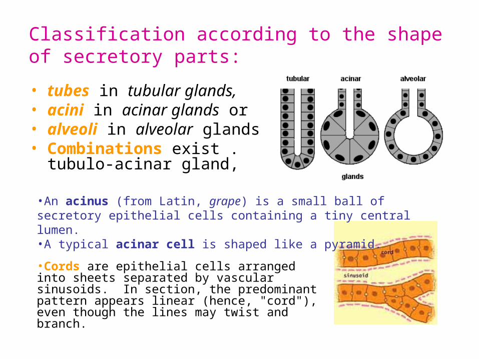
Classification according to the shape of secretory parts:
• tubes in tubular glands, • acini in acinar glands or • alveoli in alveolar glands• Combinations exist . tubulo-
acinar gland,
•Cords are epithelial cells arranged into sheets separated by vascular sinusoids. In section, the predominant pattern appears linear (hence, "cord"), even though the lines may twist and branch.
•An acinus (from Latin, grape) is a small ball of secretory epithelial cells containing a tiny central lumen. •A typical acinar cell is shaped like a pyramid.
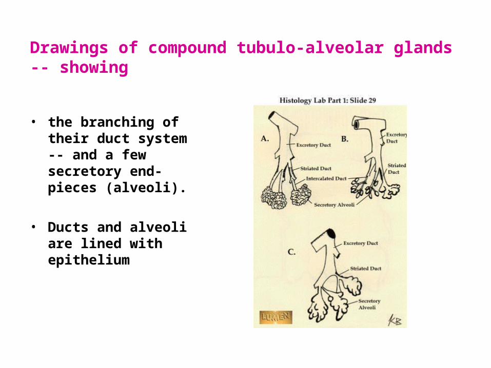
Drawings of compound tubulo-alveolar glands -- showing
• the branching of their duct system -- and a few secretory end-pieces (alveoli).
• Ducts and alveoli are lined with epithelium

Classification exocrines according to the branching pattern of their duct:
• Simple gland: with an unbranched excretory duct. There is only a single secretory unit.
• compound gland: when the excretory duct is branched. these glands are typically fairly bulky and contain very many individual secretory units.
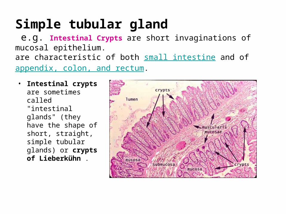
Simple tubular gland e.g. Intestinal Crypts are short invaginations of mucosal epithelium. are characteristic of both small intestine and of appendix, colon, and rectum.
• Intestinal crypts are sometimes called "intestinal glands" (they have the shape of short, straight, simple tubular glands) or crypts of Lieberkühn .
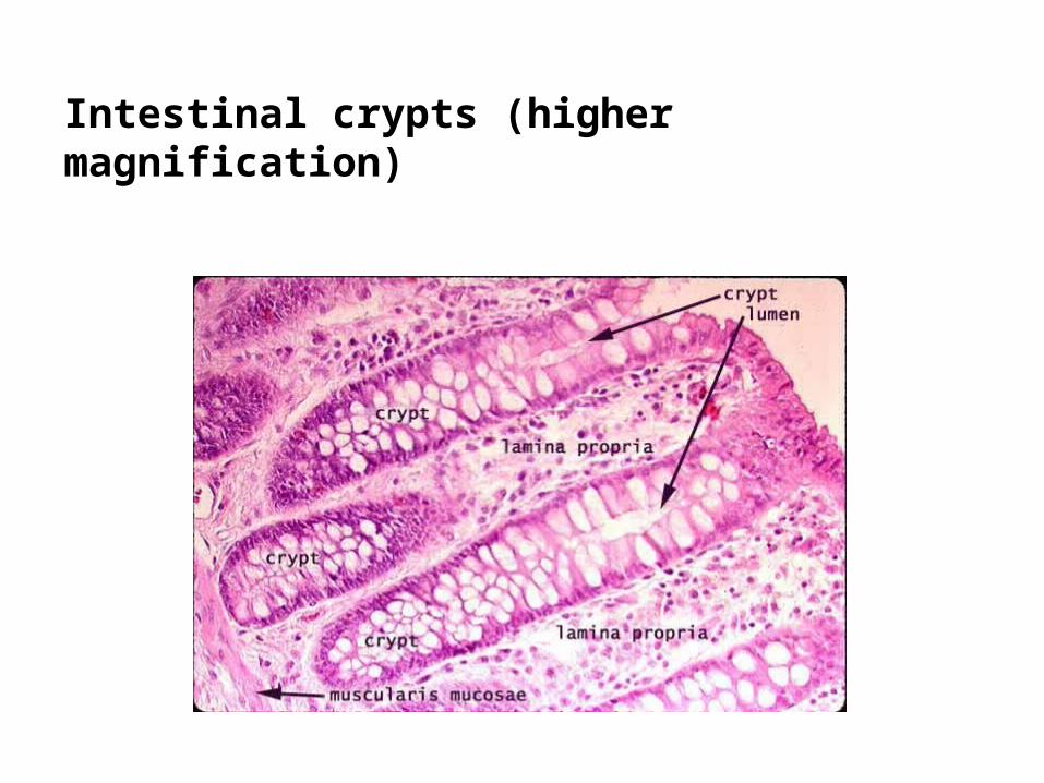
Intestinal crypts (higher magnification)

Coiled simple tubular gland:e.g. Sweat Gland in Skin
Both the duct and the secretory portion of the gland are formed from cuboidal epithelium, with round nuclei centrally placed within boxy cells.
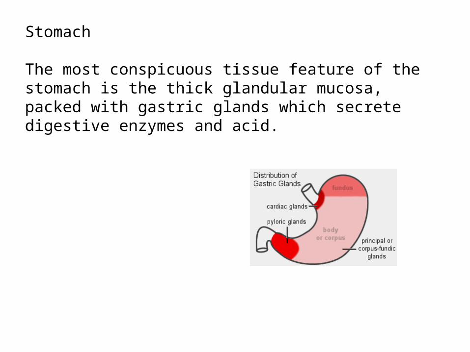
Stomach
The most conspicuous tissue feature of the stomach is the thick glandular mucosa, packed with gastric glands which secrete digestive enzymes and acid.
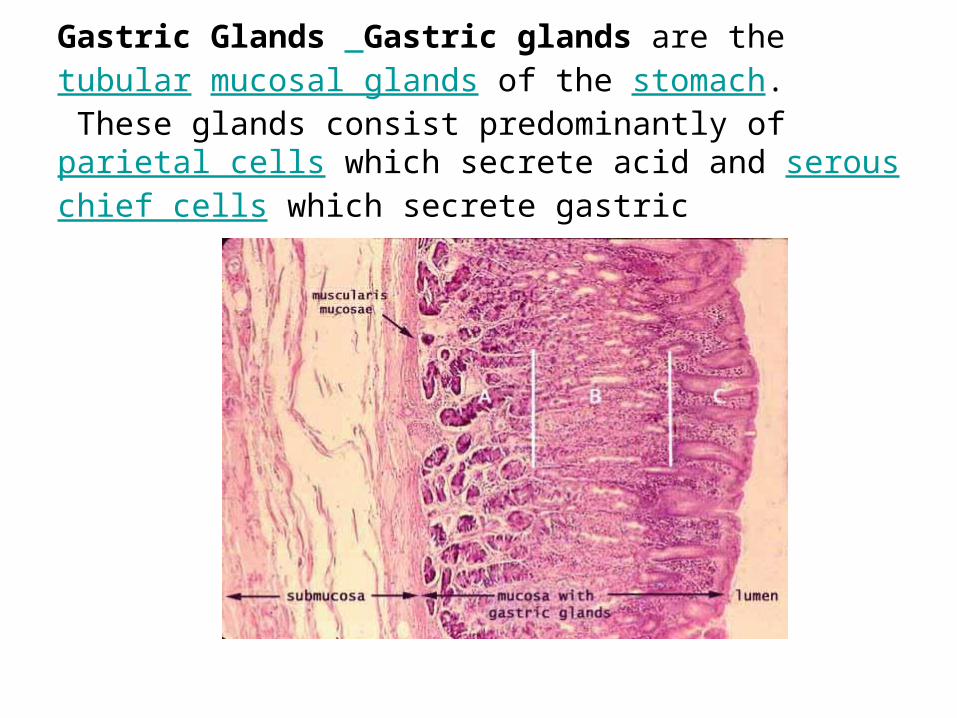
Gastric Glands Gastric glands are the tubular mucosal glands of the stomach. These glands consist predominantly of parietal cells which secrete acid and serous chief cells which secrete gastric

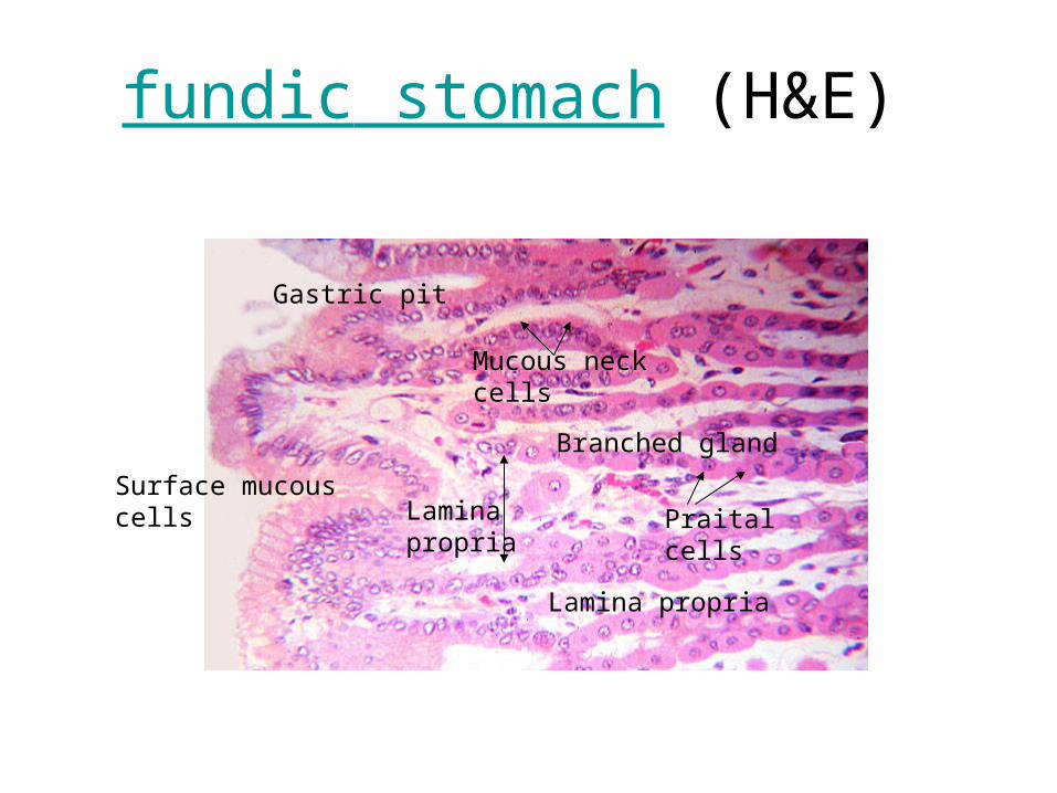
fundic stomach (H&E)
Gastric pitlumen
Gastric glands
submucosaMuscularis mucosa
Muscularis externa
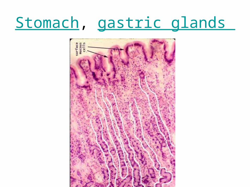
Stomach, gastric glands
fundic stomach (H&E)
Lamina propria
Lamina propria
Gastric pit
Surface mucous cells
Branched gland
Praital cells
Mucous neck cells
Lamina propria
Lamina propria
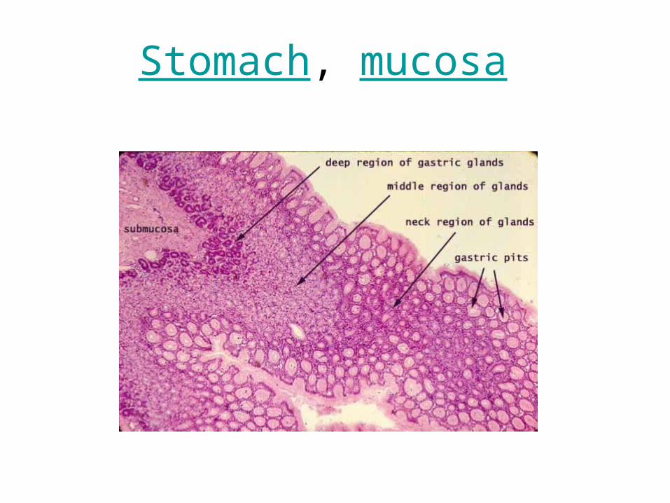
Stomach, mucosa
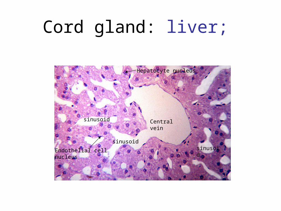
Cord gland: liver;
Central veinsinusoid
sinusoid
Hepatocyte nucleus
Endothelial cell nucleus sinusoid
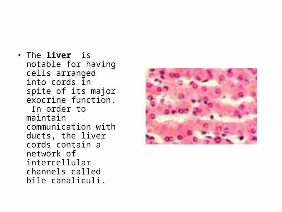
• The liver is notable for having cells arranged into cords in spite of its major exocrine function. In order to maintain communication with ducts, the liver cords contain a network of intercellular channels called bile canaliculi.
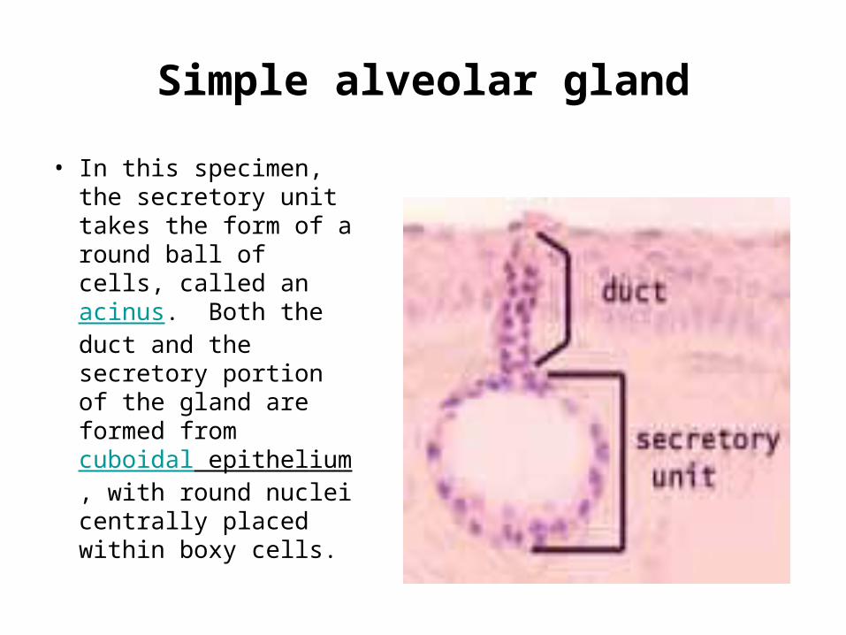
Simple alveolar gland
• In this specimen, the secretory unit takes the form of a round ball of cells, called an acinus. Both the duct and the secretory portion of the gland are formed from cuboidal epithelium, with round nuclei centrally placed within boxy cells.
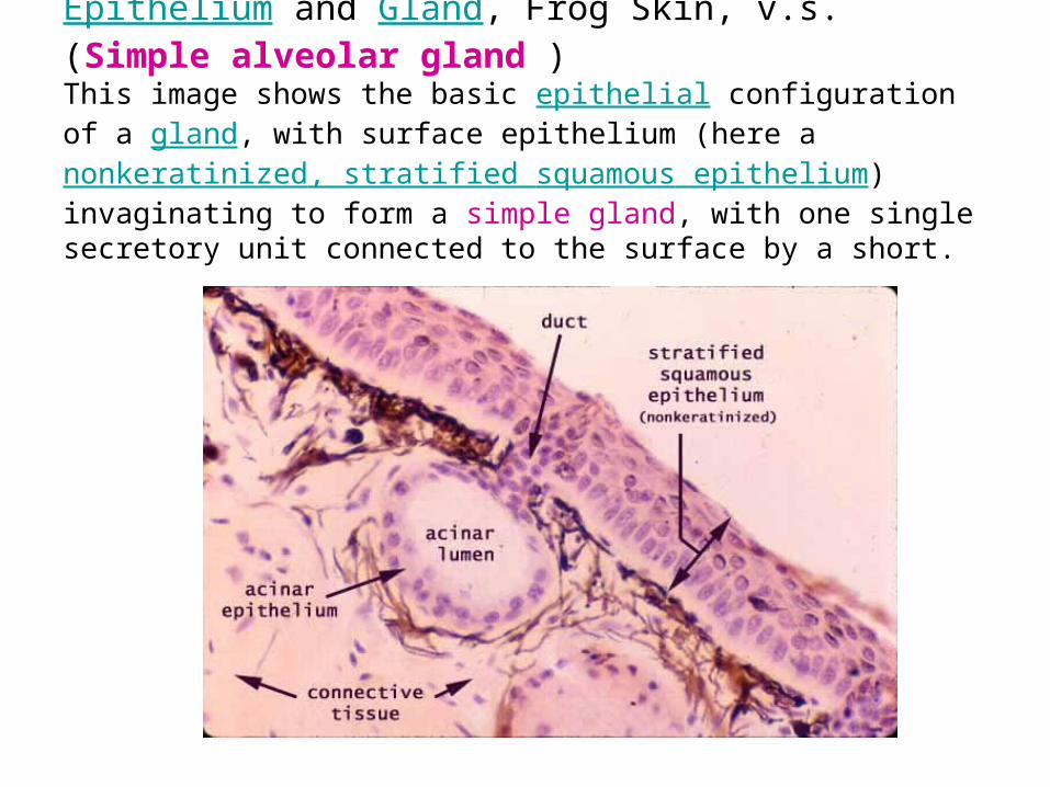
Epithelium and Gland, Frog Skin, v.s.(Simple alveolar gland )This image shows the basic epithelial configuration of a gland, with surface epithelium (here a nonkeratinized, stratified squamous epithelium) invaginating to form a simple gland, with one single secretory unit connected to the surface by a short.

Scalp, H &E
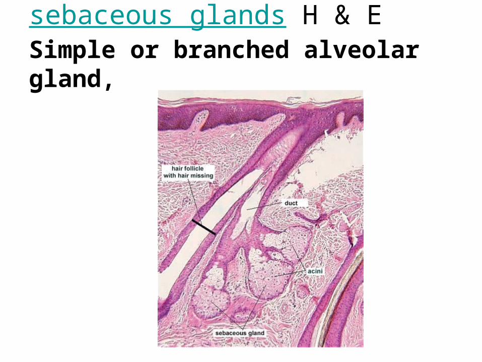
sebaceous glands H & E Simple or branched alveolar gland,

Parotid Salivary Gland The parotid salivary gland is a compound, acinar, serous gland. Unlike all other salivary glands, the parotid includes no mucous cells.

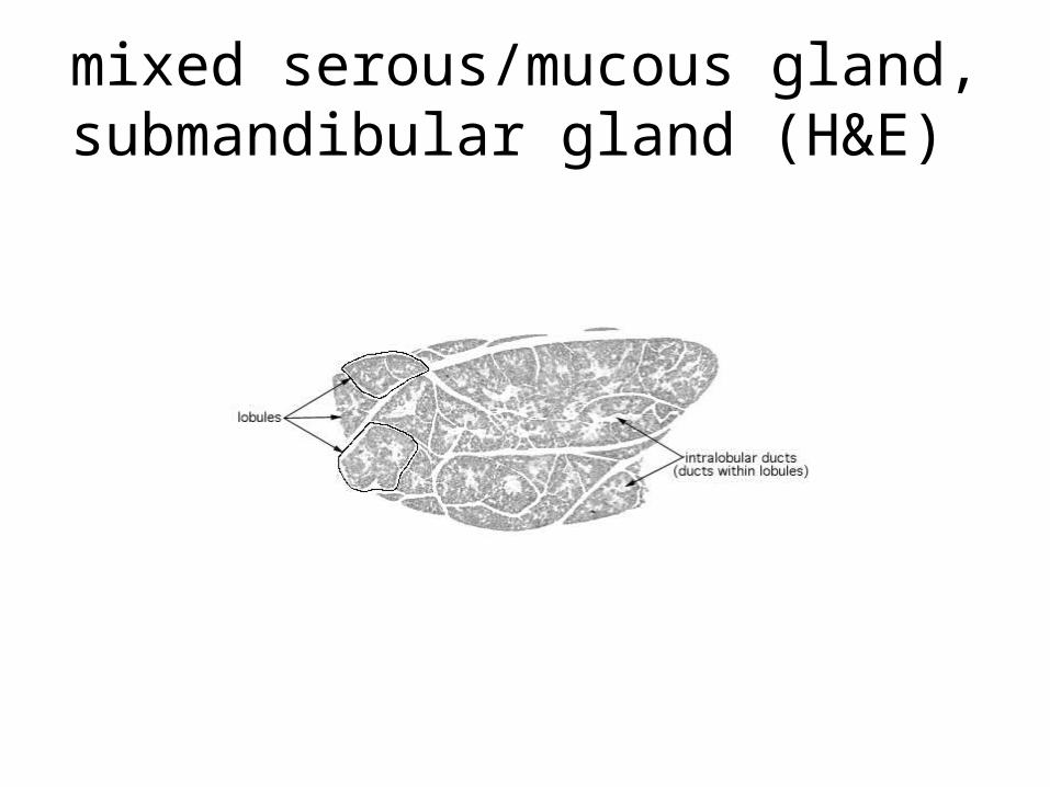
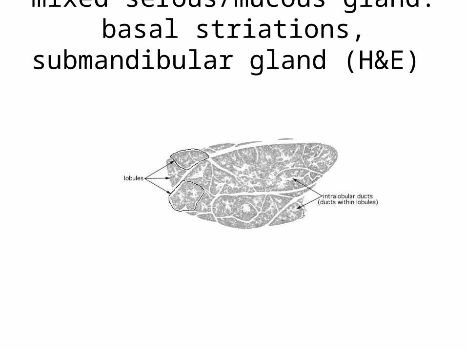
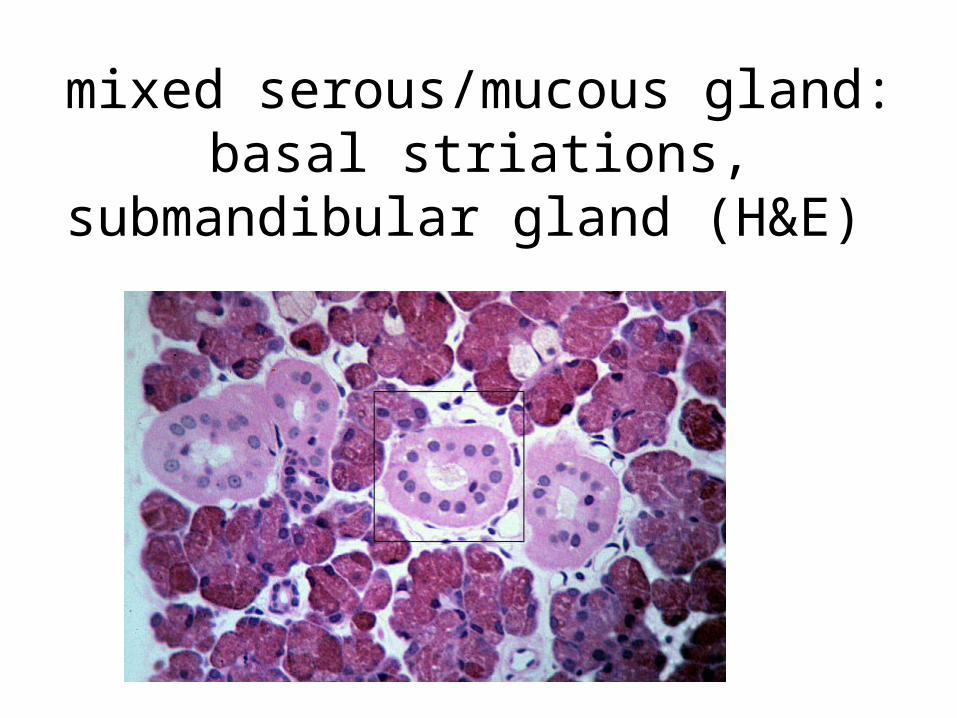
Mixed salivary glandThis image of minor, mixed salivary glands contrasts the appearance of serous cells and mucous cells. In routine preparations such as this one, serous cells often appear darker appearance than mucous cells. Serous cells are usually arranged into acini. Mucous cells are usually arranged into tubules. Occasionally, individual serous cells will occur at the ends of a mucous tubules. In section, these have a crescent-moon appearance. Several of these serous demilunes appear in the above illustration.
Serous acinus
Mucous tubu;educt
Mucous tubule with Serous demilune
Mucous tubule and duct
arteriol

mixed serous/mucous gland, submandibular gland (H&E)
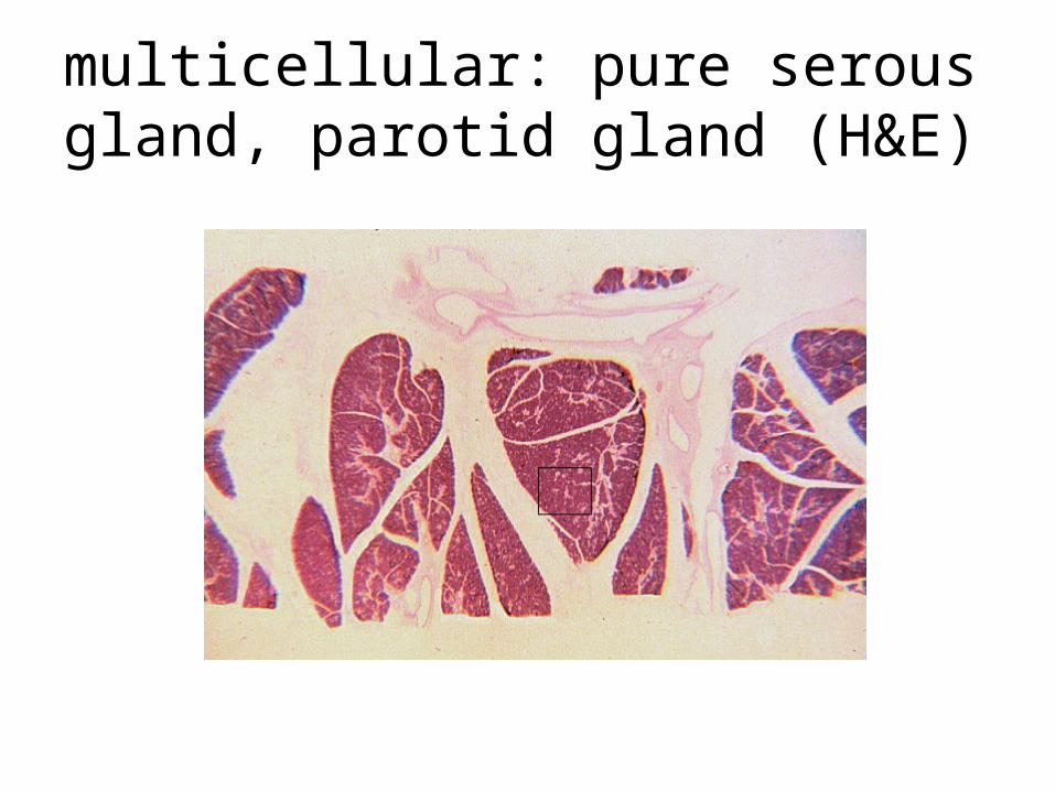
multicellular: pure serous gland, parotid gland (H&E)
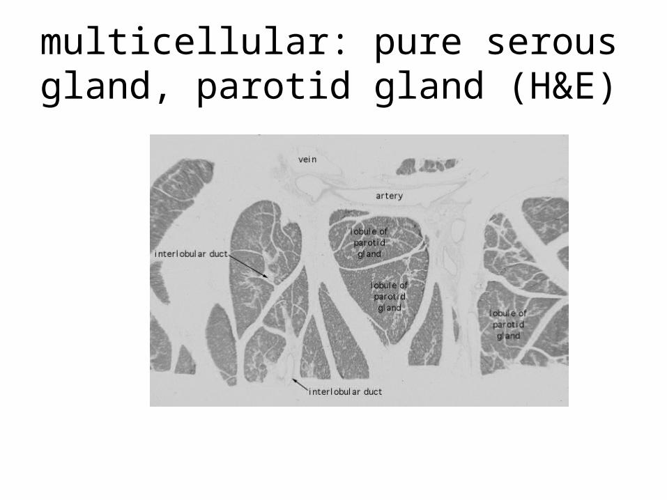
multicellular: pure serous gland, parotid gland (H&E)

mixed serous/mucous gland, submandibular gland (H&E)
mixed serous/mucous gland, submandibular gland (H&E)

mixed serous/mucous gland, submandibular gland (H&E)

mixed serous/mucous gland: basal striations, submandibular gland
(H&E)
mixed serous/mucous gland: basal striations, submandibular gland
(H&E)
mixed serous/mucous gland: basal striations, submandibular gland
(H&E)

mixed serous/mucous gland: basal striations, submandibular gland
(H&E)
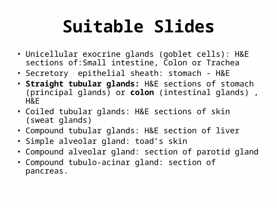
Suitable Slides• Unicellular exocrine glands (goblet cells): H&E sections
of:Small intestine, Colon or Trachea• Secretory epithelial sheath: stomach - H&E • Straight tubular glands: H&E sections of stomach
(principal glands) or colon (intestinal glands) , H&E• Coiled tubular glands: H&E sections of skin (sweat
glands)• Compound tubular glands: H&E section of liver• Simple alveolar gland: toad’s skin• Compound alveolar gland: section of parotid gland• Compound tubulo-acinar gland: section of pancreas.


![Case Report Malignant triton tumor of the prostate: a case …glandular epithelium, adipose tissue, or even squamous cells [1]. Tumors with rhabdomyo-blastic differentiation and malignant](https://img.pdfslide.us/doc/110x75/60f7b7a2384f7b44a45f45b5/case-report-malignant-triton-tumor-of-the-prostate-a-case-glandular-epithelium.jpg)