Embed Size (px)
Citation preview
Dr. Anurag Yadav
Post-graduate, Biochemistry
Father Muller Medical college
ELECTROPHORESIS
1 Dr Anurag yadav,Bio-FMMC
CONTENT
Dr Anurag yadav,Bio-FMMC 2
Introduction
Principle
Factors affecting
Conventional electrophoresis
General operation
Technical and practical Consideration
Types of electrophoresis
INTRODUCTION
Dr Anurag yadav,Bio-FMMC 3
Electrophoresis is the migration of charged particles or
molecules in a medium under the influence of an applied
electric field.
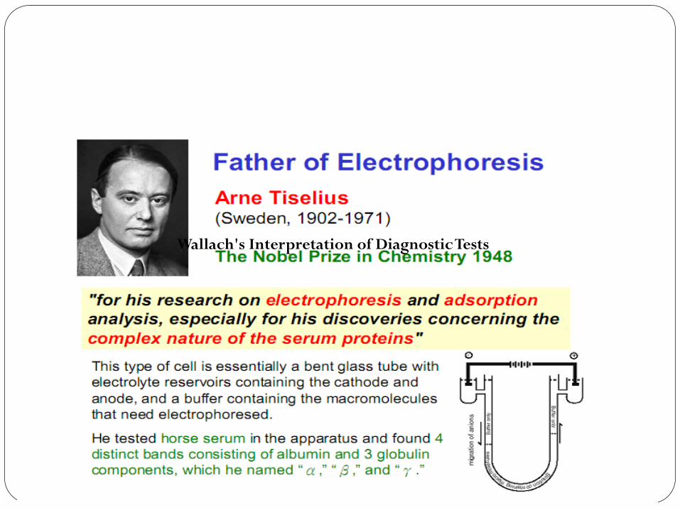
Wallach's Interpretation of Diagnostic Tests
Electrophoresis
Dr Anurag yadav,Bio-FMMC 5
a separation technique
Simple, rapid and highly sensitive
used in clinical laboratories to separate charged molecules from each
other in presence of electric field
– Proteins in body fluids: serum, urine, CSF
– Proteins in erythrocytes: hemoglobin
– Nucleic acids: DNA, RNA
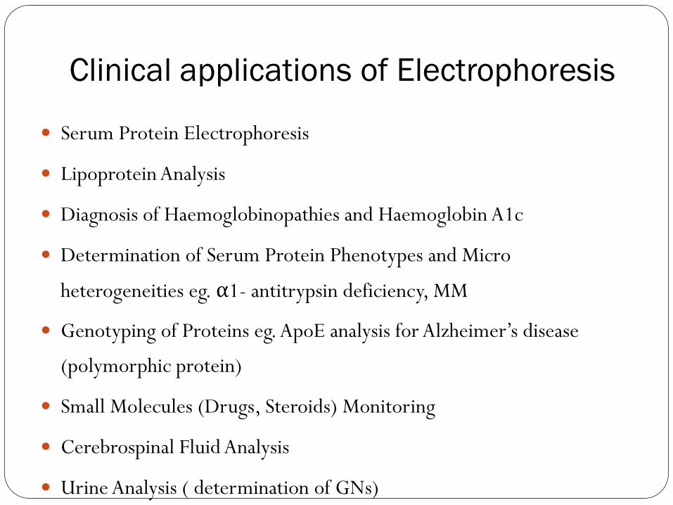
Clinical applications of Electrophoresis
Serum Protein Electrophoresis
Lipoprotein Analysis
Diagnosis of Haemoglobinopathies and Haemoglobin A1c
Determination of Serum Protein Phenotypes and Micro
heterogeneities eg. α1- antitrypsin deficiency, MM
Genotyping of Proteins eg. ApoE analysis for Alzheimer’s disease
(polymorphic protein)
Small Molecules (Drugs, Steroids) Monitoring
Cerebrospinal Fluid Analysis
Urine Analysis ( determination of GNs)
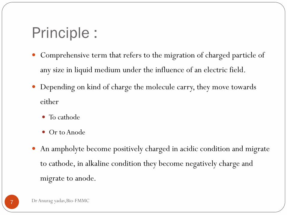
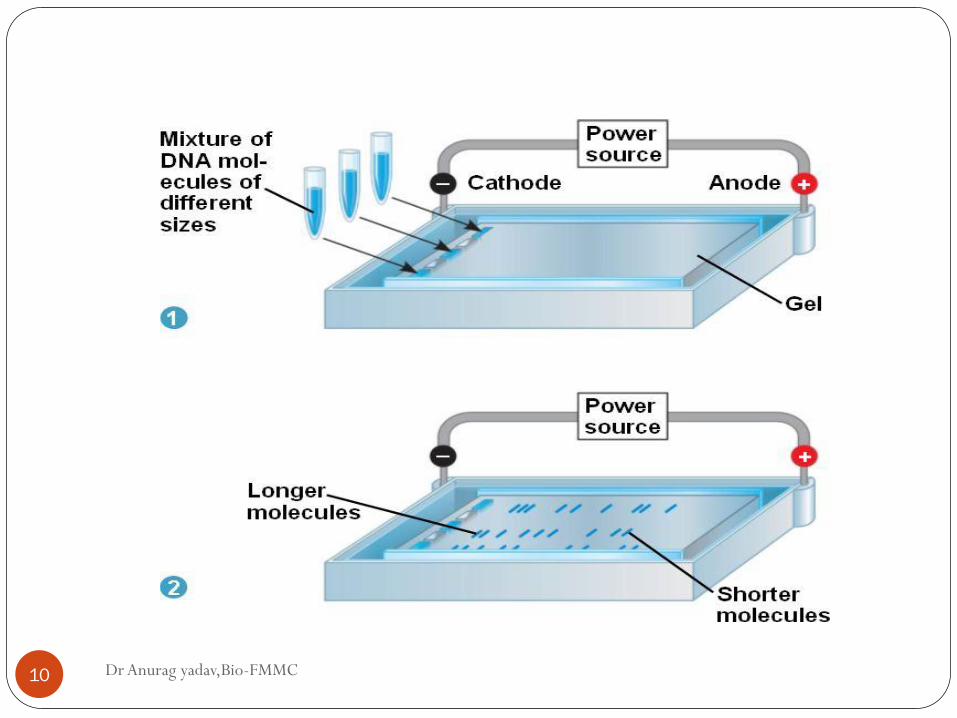
Principle :
Dr Anurag yadav,Bio-FMMC 7
Comprehensive term that refers to the migration of charged particle of
any size in liquid medium under the influence of an electric field.
Depending on kind of charge the molecule carry, they move towards
either
To cathode
Or to Anode
An ampholyte become positively charged in acidic condition and migrate
to cathode, in alkaline condition they become negatively charge and
migrate to anode.
Dr Anurag yadav,Bio-FMMC 8
Eg: as protein contain the ionizable amino and carboxyl
group.
The rate of migration of an ion in electrical field depend on
factors,
1. Net charge of molecule
2. Size and shape of particle
3. Strength of electrical field
4. Properties of supporting medium
5. Temperature of operation
1. Mobility
Dr Anurag yadav,Bio-FMMC 9
Under the electrical field, the mobility of the particle is
determined by two factors:
Its charge
Frictional coefficient
Size and shape of the particle decide the velocity with which the
particle will migrate under the given electrical field and the
medium.
Dr Anurag yadav,Bio-FMMC 10
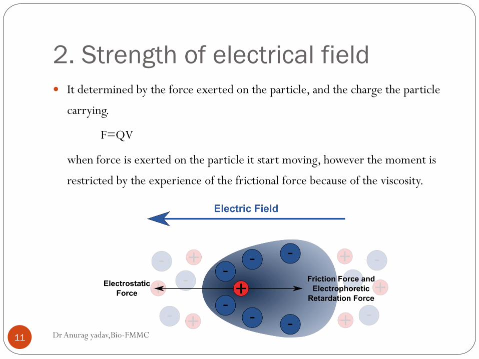
2. Strength of electrical field
Dr Anurag yadav,Bio-FMMC 11
It determined by the force exerted on the particle, and the charge the particle
carrying.
F=QV
when force is exerted on the particle it start moving, however the moment is
restricted by the experience of the frictional force because of the viscosity.
Effect of pH on Mobility
Dr Anurag yadav,Bio-FMMC 12
As the molecule exist as amphoteric , they will carry the
charges based on the solvent pH.
Their overall net charge is NEUTRAL when it is at zwitter
ion state. And hence the mobility is retarded to zero.
Mobility is directly proportional to the magnitude of the
charge, which is functional of the pH of solvent.
The pH is maintained by the use of Buffers of different pH.
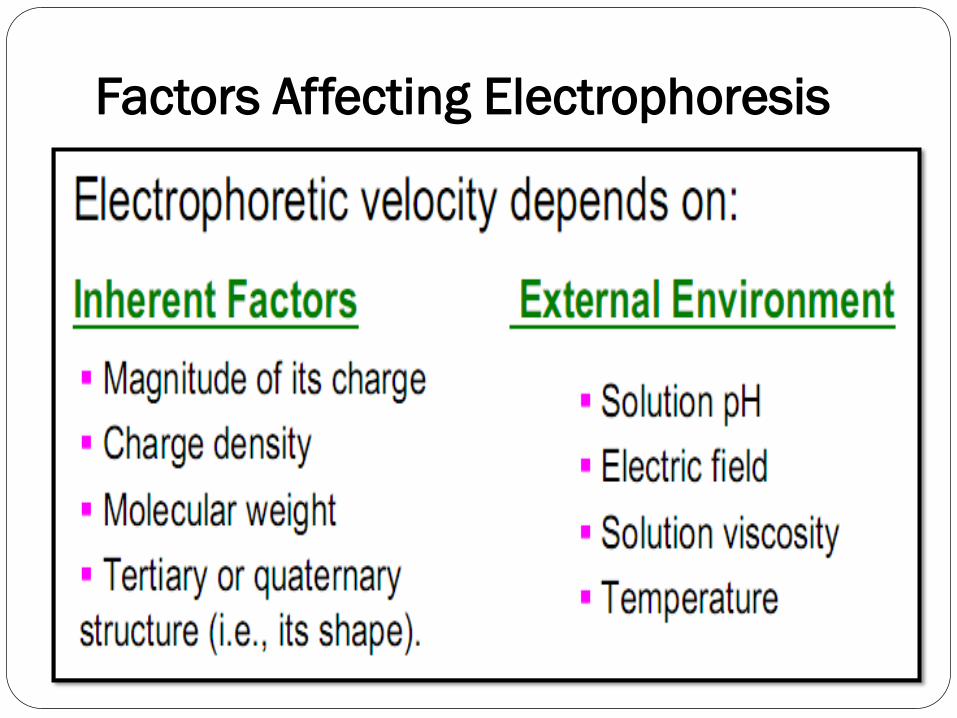
Factors Affecting Electrophoresis
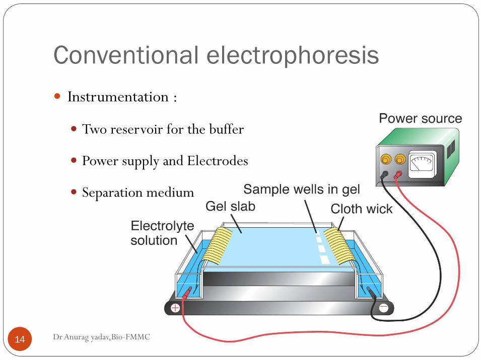
Conventional electrophoresis
Dr Anurag yadav,Bio-FMMC 14
Instrumentation :
Two reservoir for the buffer
Power supply and Electrodes
Separation medium
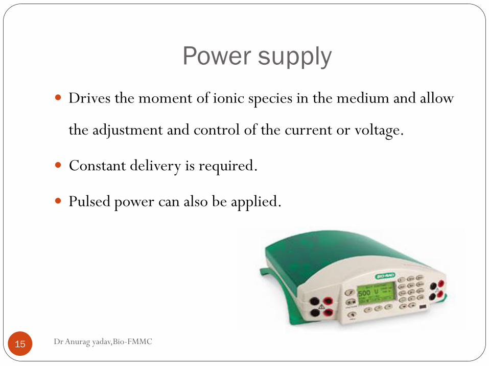
Power supply
Dr Anurag yadav,Bio-FMMC 15
Drives the moment of ionic species in the medium and allow
the adjustment and control of the current or voltage.
Constant delivery is required.
Pulsed power can also be applied.
Buffer
Dr Anurag yadav,Bio-FMMC 16
The buffer in electrophoresis has twofold purpose:
Carry applied electrical current
They set the pH as which electrophoresis is carried out.
Thus they determine;
Type of charge on solute.
Extent of ionization of solute
Electrode towards which the solute will migrate.
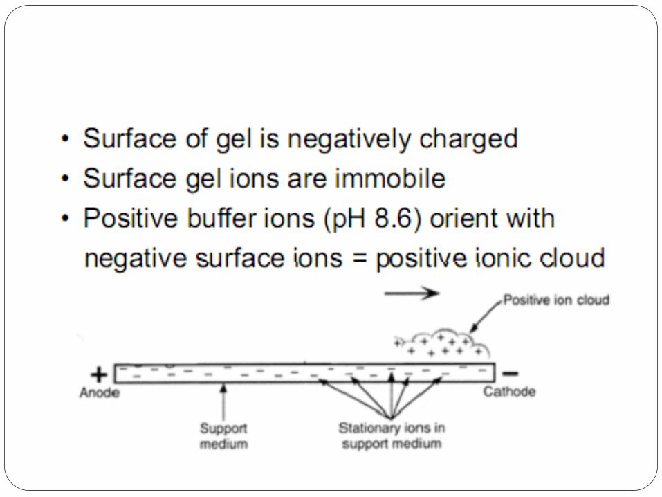
The buffer ionic strength will determine the thickness of the ionic
cloud.
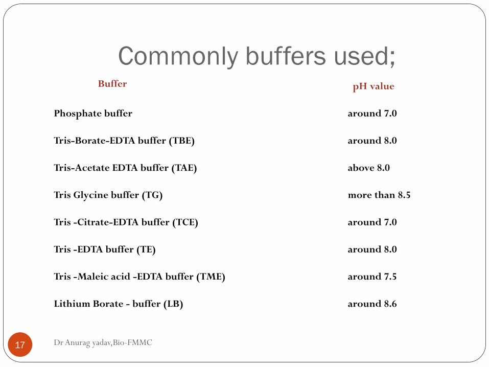
Commonly buffers used;
Dr Anurag yadav,Bio-FMMC 17
Buffer
pH value
Phosphate buffer
around 7.0
Tris-Borate-EDTA buffer (TBE)
around 8.0
Tris-Acetate EDTA buffer (TAE)
above 8.0
Tris Glycine buffer (TG)
more than 8.5
Tris -Citrate-EDTA buffer (TCE)
around 7.0
Tris -EDTA buffer (TE)
around 8.0
Tris -Maleic acid -EDTA buffer (TME)
around 7.5
Lithium Borate - buffer (LB)
around 8.6
Supporting medium
Dr Anurag yadav,Bio-FMMC 18
Supporting medium is an matrix in which the protein
separation takes place.
Various type has been used for the separation either on slab
or capillary form.
Separation is based on to the charge to mass ratio of protein
depending on the pore size of the medium, possibly the
molecular size.
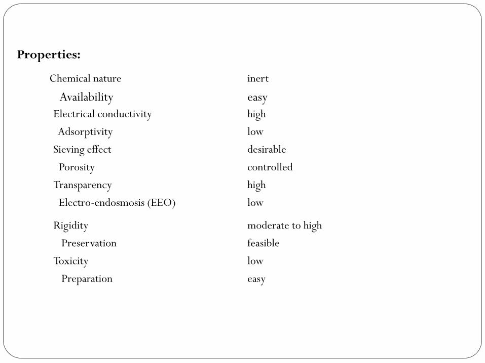
Chemical nature
inert
Availability
easy
Electrical conductivity
high
Adsorptivity
low
Sieving effect
desirable
Porosity
controlled
Transparency
high
Electro-endosmosis (EEO)
low
Rigidity
moderate to high
Preservation
feasible
Toxicity
low
Preparation
easy
Properties:
Dr Anurag yadav,Bio-FMMC 20
- Starch gel
- Cellulose acetate
- Agarose
- Polyacrylamide gel
Agarose Gel
Dr Anurag yadav,Bio-FMMC 21
A linear polysaccharide (made-up of repeat unit of agarobiose-alternating
unit of galactose and 3,6-anhydrogalactose).
Used in conc as 1% and 3%.
The gelling property are attributed to both inter- and intramolecular
hydrogen bonding
Pore size is controlled by the % of agarose used.
Large pore size are formed with lower conc and vice versa.
Purity of the agarose is based on the number of sulphate conc, lower the
conc of sulphate higher is the purity of agarose.
Dr Anurag yadav,Bio-FMMC 22
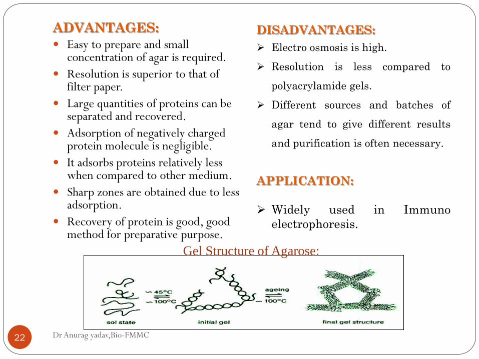
ADVANTAGES:
Easy to prepare and small concentration of agar is required.
Resolution is superior to that of filter paper.
Large quantities of proteins can be separated and recovered.
Adsorption of negatively charged protein molecule is negligible.
It adsorbs proteins relatively less when compared to other medium.
Sharp zones are obtained due to less adsorption.
Recovery of protein is good, good method for preparative purpose.
DISADVANTAGES:
Electro osmosis is high.
Resolution is less compared to
polyacrylamide gels.
Different sources and batches of
agar tend to give different results
and purification is often necessary.
APPLICATION:
Widely used in Immuno
electrophoresis.
Gel Structure of Agarose:
Cellulose acetate
Dr Anurag yadav,Bio-FMMC 23
Thermoplastic resin made by treating cellulose with acetic
anhydride to acetylate the hydroxyl group.
When dry, membrane contain about 80% air space within fibers
and brittle film.
As the film is soak in buffer, the space are filled.
Because of their opacity, the film has to be made transparent by
soaking in 95:5 methanol:glacial acetic acid.
It can be stored for longer duration.
Polyacrylamide
Dr Anurag yadav,Bio-FMMC 24
Frequently referred to as PAGE.
Cross-linked polyacrylamide gel are formed from the polymerization of
the monomer in presence of small amount of N,N”-methylene-
bisacrylamide.
Bisacrylamide – two acrylamide linked by the methylene group.
The polymerization of the acrylamide is an example for free radical
catalysis.
They are defined in terms of total percentage of acrylamide present, and
pore size vary with conc.
Dr Anurag yadav,Bio-FMMC 25
Made in conc between 3-30% acrylamide.
Thus low % has large pore size and vice versa.
Proteins are separated on the basis of charge to mass ratio and
molecular size, a phenomenon called Molecular sieving.
ADVANTAGES:
Gels are stable over wide range of pH and temperature.
Gels of different pore size can be formed.
Simple and separation speed is good comparatively.
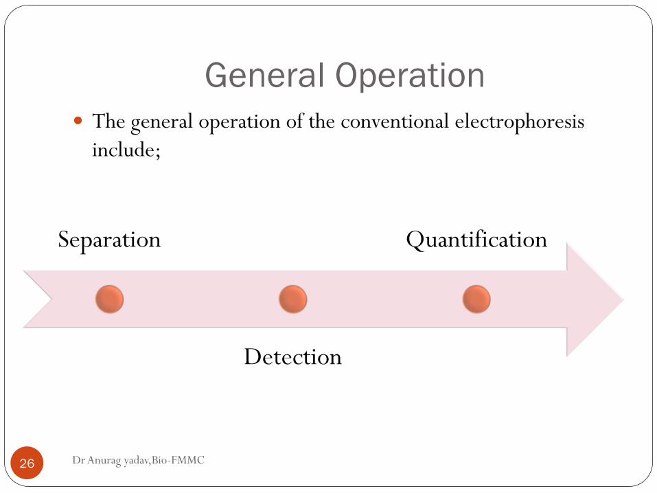
General Operation
Dr Anurag yadav,Bio-FMMC 26
The general operation of the conventional electrophoresis
include;
Separation
Detection
Quantification
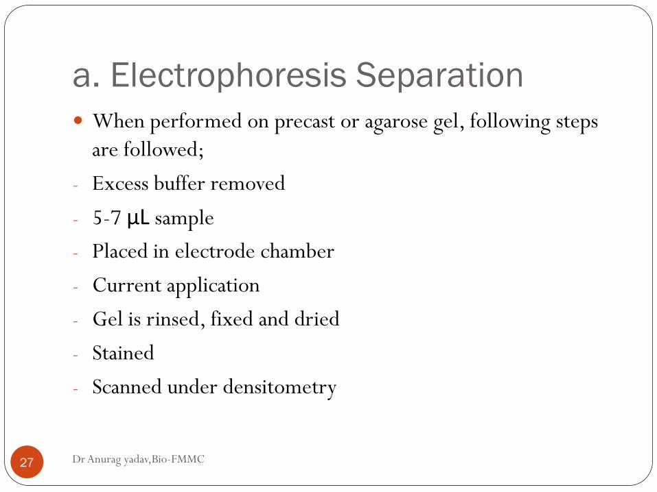
a. Electrophoresis Separation
Dr Anurag yadav,Bio-FMMC 27
When performed on precast or agarose gel, following steps
are followed;
- Excess buffer removed
- 5-7 μL sample
- Placed in electrode chamber
- Current application
- Gel is rinsed, fixed and dried
- Stained
- Scanned under densitometry
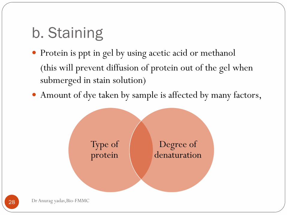
b. Staining
Dr Anurag yadav,Bio-FMMC 28
Protein is ppt in gel by using acetic acid or methanol
(this will prevent diffusion of protein out of the gel when
submerged in stain solution)
Amount of dye taken by sample is affected by many factors,
Type of protein
Degree of denaturation
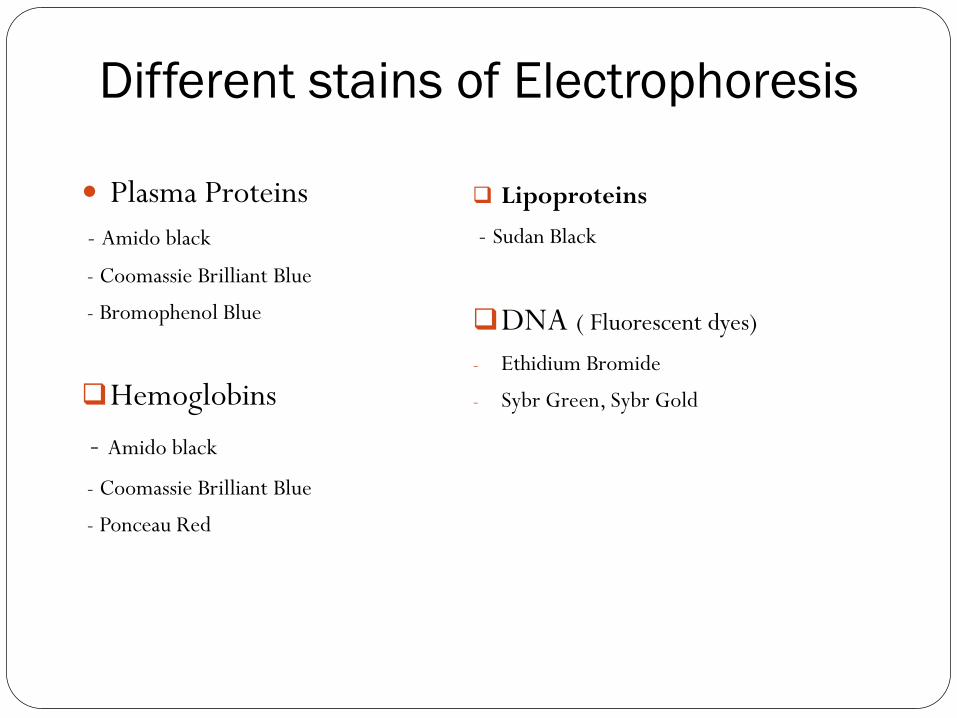
Different stains of Electrophoresis
Plasma Proteins
- Amido black
- Coomassie Brilliant Blue
- Bromophenol Blue
Hemoglobins
- Amido black
- Coomassie Brilliant Blue
- Ponceau Red
Lipoproteins
- Sudan Black
DNA ( Fluorescent dyes)
- Ethidium Bromide
- Sybr Green, Sybr Gold
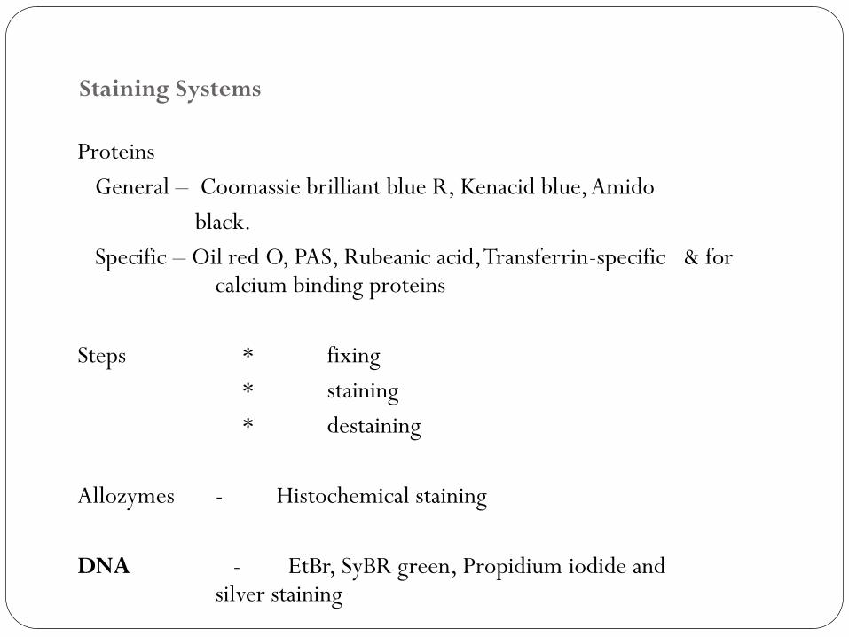
Staining Systems
Proteins
General – Coomassie brilliant blue R, Kenacid blue, Amido
black.
Specific – Oil red O, PAS, Rubeanic acid, Transferrin-specific & for calcium binding proteins
Steps * fixing
* staining
* destaining
Allozymes - Histochemical staining
DNA - EtBr, SyBR green, Propidium iodide and silver staining
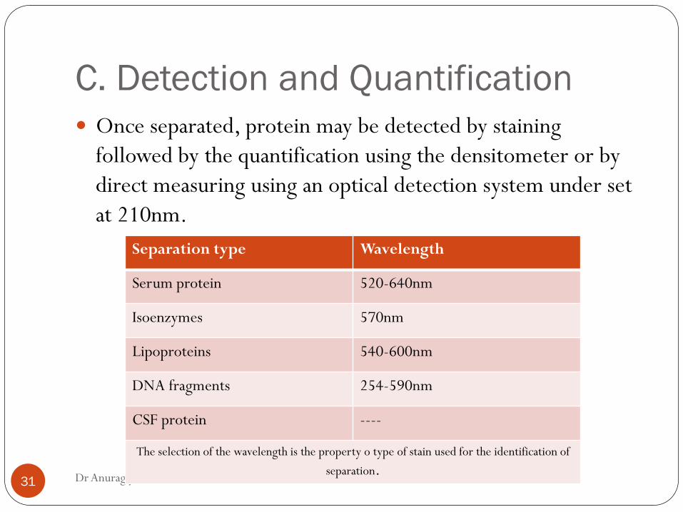
C. Detection and Quantification
Dr Anurag yadav,Bio-FMMC 31
Once separated, protein may be detected by staining
followed by the quantification using the densitometer or by
direct measuring using an optical detection system under set
at 210nm.
Separation type Wavelength
Serum protein 520-640nm
Isoenzymes 570nm
Lipoproteins 540-600nm
DNA fragments 254-590nm
CSF protein ----
The selection of the wavelength is the property o type of stain used for the identification of
separation.
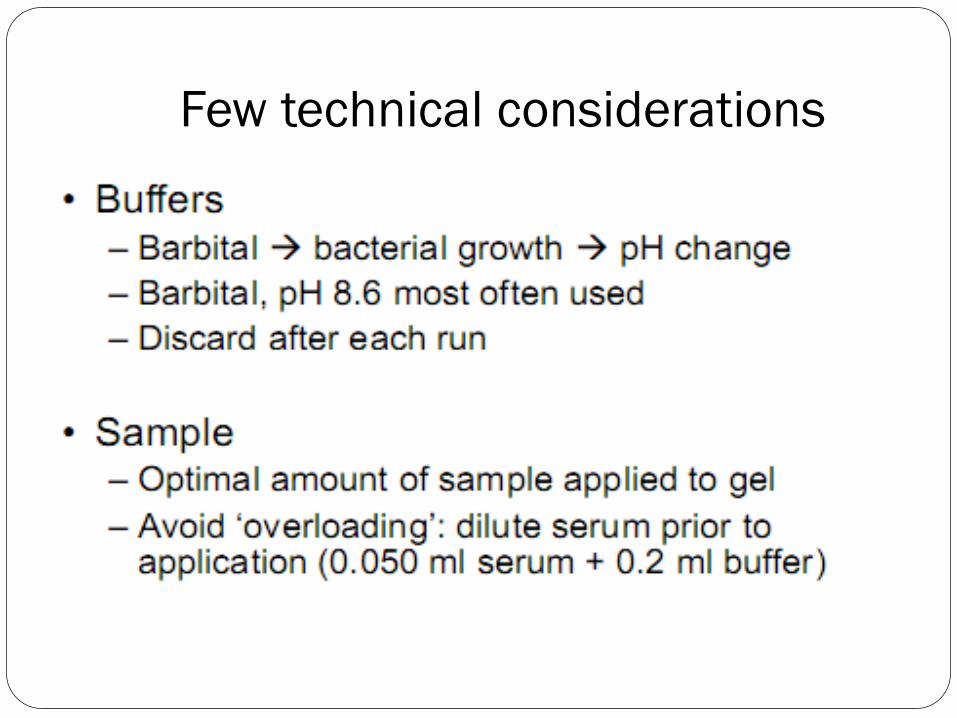
Few technical considerations
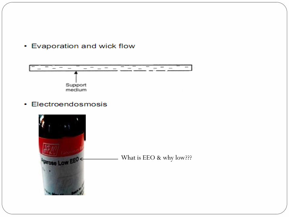
What is EEO & why low???
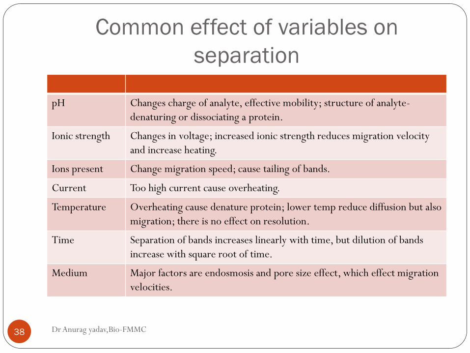
Common effect of variables on
separation
Dr Anurag yadav,Bio-FMMC 38
pH Changes charge of analyte, effective mobility; structure of analyte-
denaturing or dissociating a protein.
Ionic strength Changes in voltage; increased ionic strength reduces migration velocity
and increase heating.
Ions present Change migration speed; cause tailing of bands.
Current Too high current cause overheating.
Temperature Overheating cause denature protein; lower temp reduce diffusion but also
migration; there is no effect on resolution.
Time Separation of bands increases linearly with time, but dilution of bands
increase with square root of time.
Medium Major factors are endosmosis and pore size effect, which effect migration
velocities.
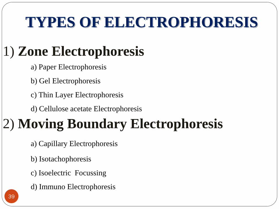
TYPES OF ELECTROPHORESIS
1) Zone Electrophoresis a) Paper Electrophoresis
b) Gel Electrophoresis
c) Thin Layer Electrophoresis
d) Cellulose acetate Electrophoresis
2) Moving Boundary Electrophoresis
a) Capillary Electrophoresis
b) Isotachophoresis
c) Isoelectric Focussing
d) Immuno Electrophoresis
39
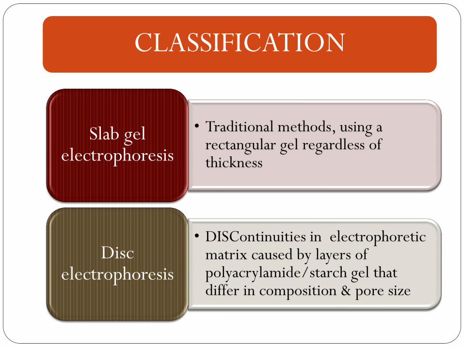
CLASSIFICATION
• Traditional methods, using a rectangular gel regardless of thickness
Slab gel electrophoresis
• DISContinuities in electrophoretic matrix caused by layers of polyacrylamide/starch gel that differ in composition & pore size
Disc electrophoresis
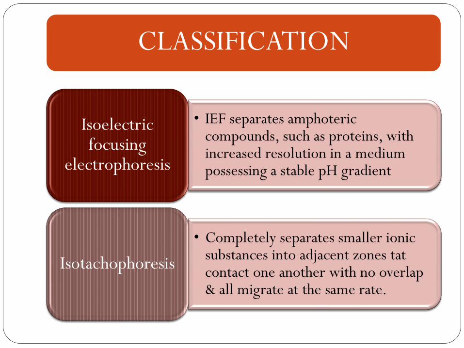
CLASSIFICATION
• IEF separates amphoteric compounds, such as proteins, with increased resolution in a medium possessing a stable pH gradient
Isoelectric focusing
electrophoresis
• Completely separates smaller ionic substances into adjacent zones tat contact one another with no overlap & all migrate at the same rate.
Isotachophoresis
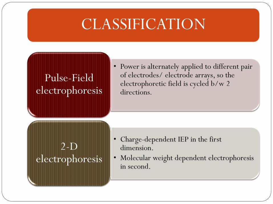
CLASSIFICATION
• Power is alternately applied to different pair of electrodes/ electrode arrays, so the electrophoretic field is cycled b/w 2 directions.
Pulse-Field electrophoresis
• Charge-dependent IEP in the first dimension.
• Molecular weight dependent electrophoresis in second.
2-D electrophoresis
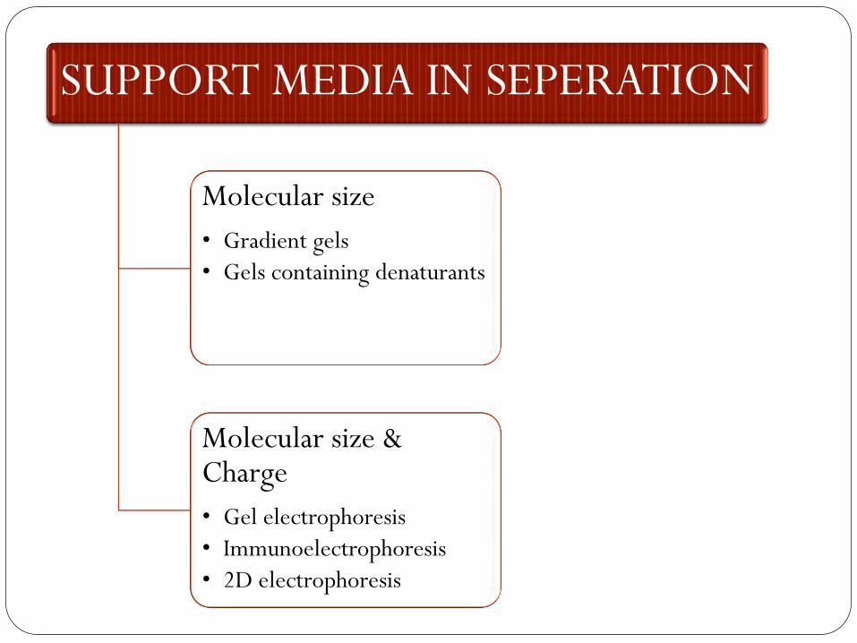
SUPPORT MEDIA IN SEPERATION
Molecular size
• Gradient gels
• Gels containing denaturants
Molecular size & Charge
• Gel electrophoresis
• Immunoelectrophoresis
• 2D electrophoresis
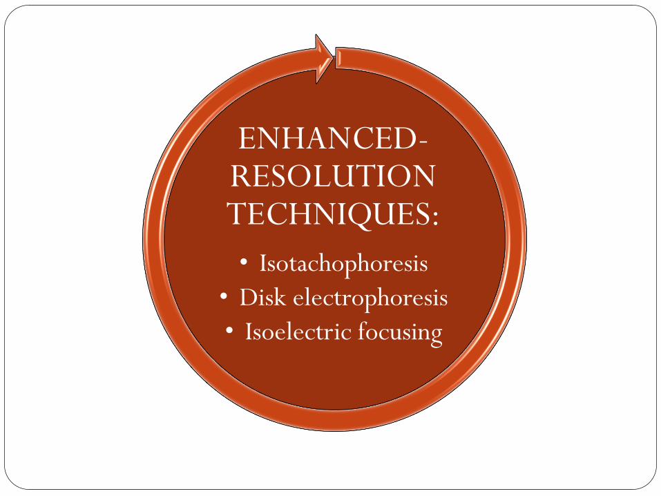
ENHANCED-RESOLUTION TECHNIQUES:
• Isotachophoresis
• Disk electrophoresis
• Isoelectric focusing
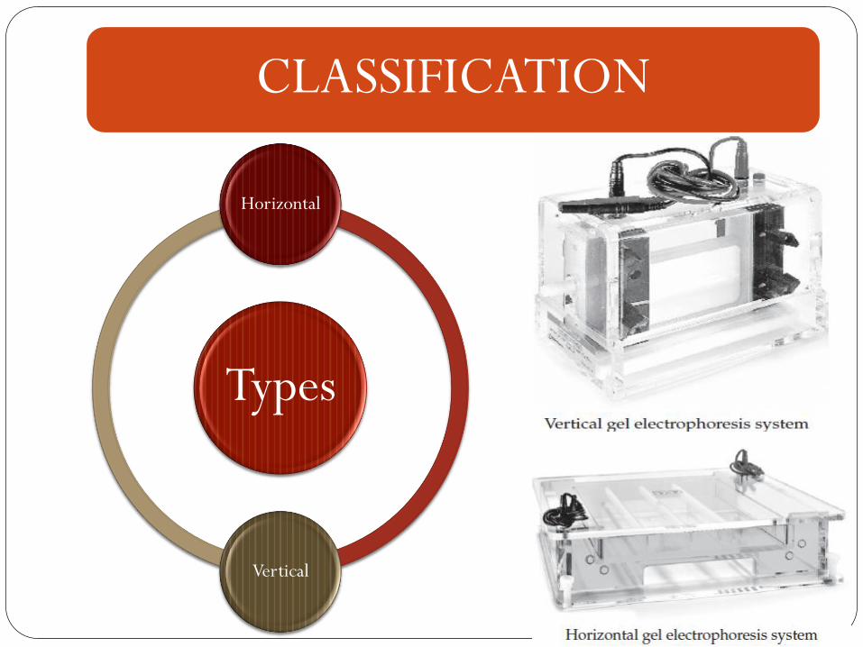
CLASSIFICATION
Types
Horizontal
Vertical
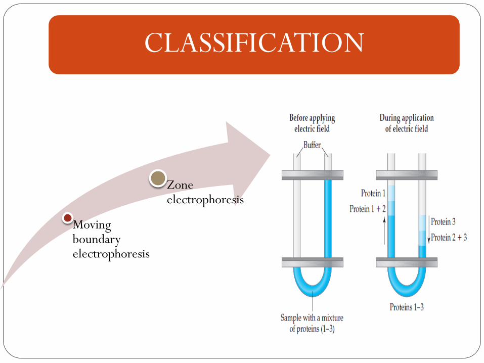
CLASSIFICATION
Moving boundary electrophoresis
Zone electrophoresis
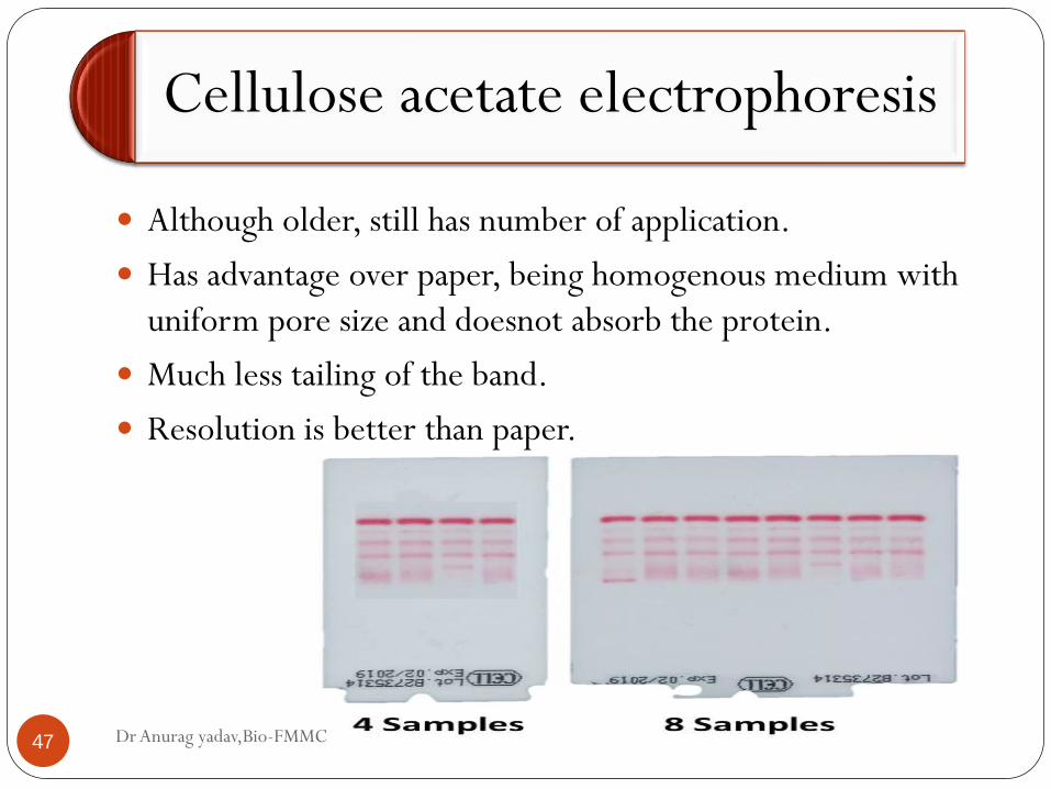
Cellulose acetate electrophoresis
Dr Anurag yadav,Bio-FMMC 47
Although older, still has number of application.
Has advantage over paper, being homogenous medium with
uniform pore size and doesnot absorb the protein.
Much less tailing of the band.
Resolution is better than paper.
Dr Anurag yadav,Bio-FMMC 48
Much simpler to run. Can be used as single sample or
multiple sample run.
Acetate paper is first wetted in the buffer, and the sample is
loaded.
The strip is kept for the electrophoretic run.
6-8 V/cm for about 3 hr.
The protein separation is stained, for better visualization.
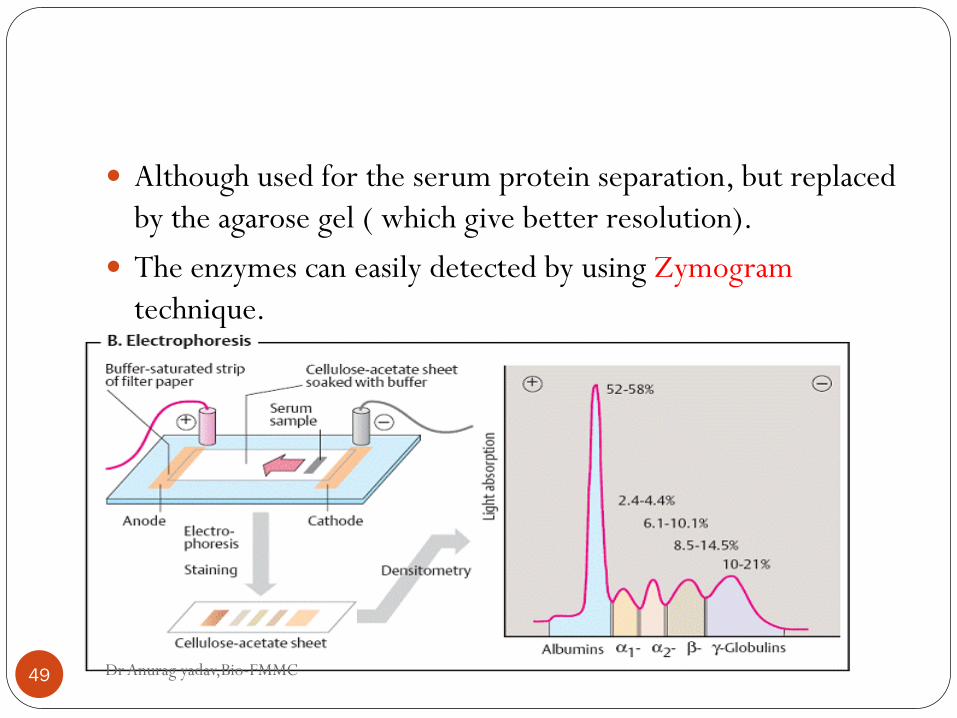
Dr Anurag yadav,Bio-FMMC 49
Although used for the serum protein separation, but replaced
by the agarose gel ( which give better resolution).
The enzymes can easily detected by using Zymogram
technique.
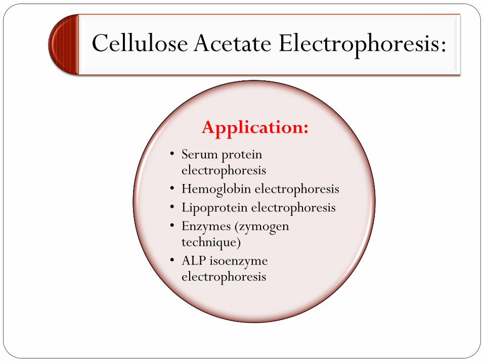
Cellulose Acetate Electrophoresis:
Application:
• Serum protein electrophoresis
• Hemoglobin electrophoresis
• Lipoprotein electrophoresis
• Enzymes (zymogen technique)
• ALP isoenzyme electrophoresis
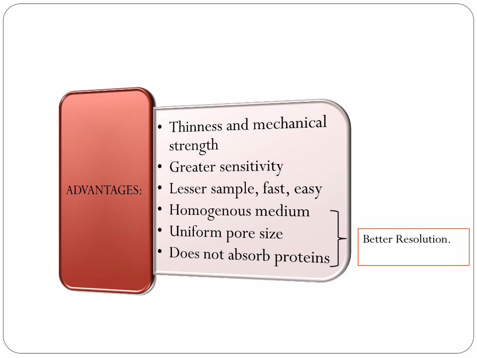
Cellulose Acetate Electrophoresis:
Better Resolution.
Cellulose Acetate Electrophoresis:
• Resolution less as compared to PAGE
• 8-9 serum fractions as compared to 30 with disk/PAGE
DISADVANTAGES
SDS-PAGE
Dr Anurag yadav,Bio-FMMC 53
Sodium dodecyl sulphate- polyacrylamide gel
electrophoresis.
Most widely used method for analysing protein mixture
qualitatively.
Useful for monitoring protein purification – as separation of
protein is based on the size of the particle.
Can also be used for determining the relative molecular mass
of a protein.
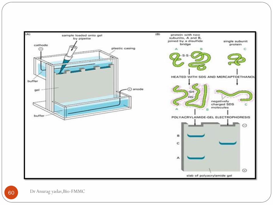
Dr Anurag yadav,Bio-FMMC 54
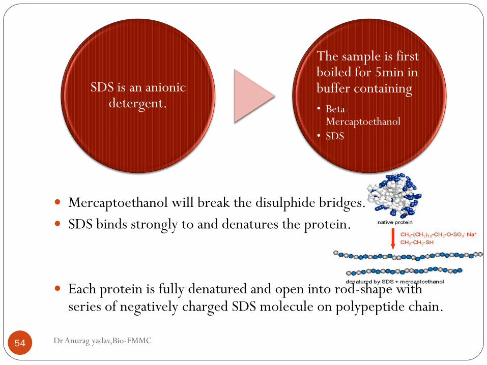
Mercaptoethanol will break the disulphide bridges.
SDS binds strongly to and denatures the protein.
Each protein is fully denatured and open into rod-shape with series of negatively charged SDS molecule on polypeptide chain.
SDS is an anionic detergent.
The sample is first boiled for 5min in buffer containing
• Beta-Mercaptoethanol
• SDS
Dr Anurag yadav,Bio-FMMC 55
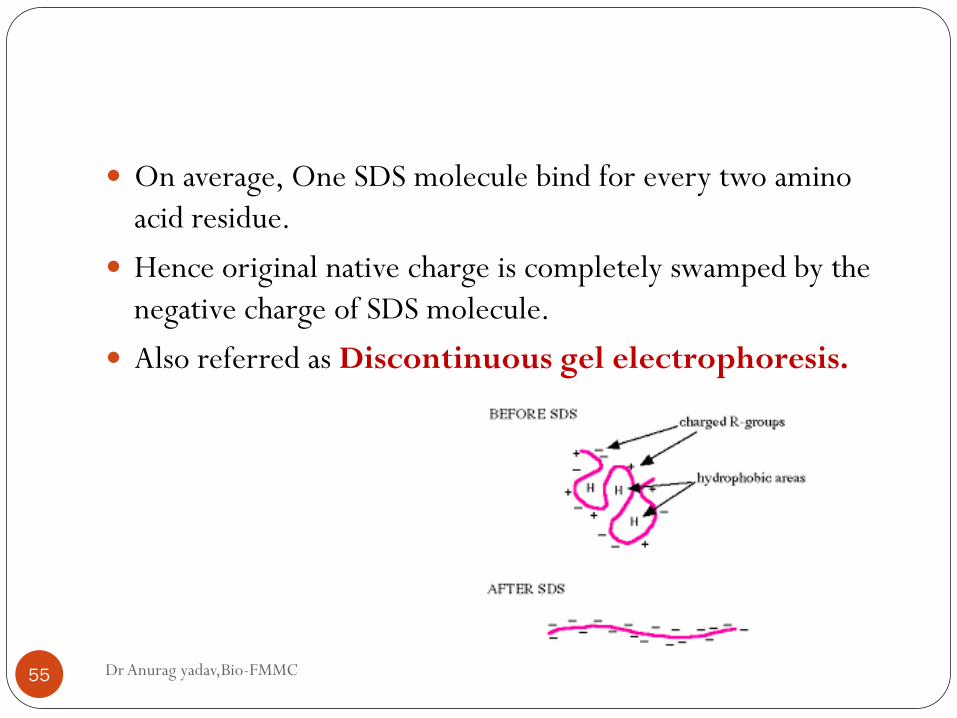
On average, One SDS molecule bind for every two amino
acid residue.
Hence original native charge is completely swamped by the
negative charge of SDS molecule.
Also referred as Discontinuous gel electrophoresis.
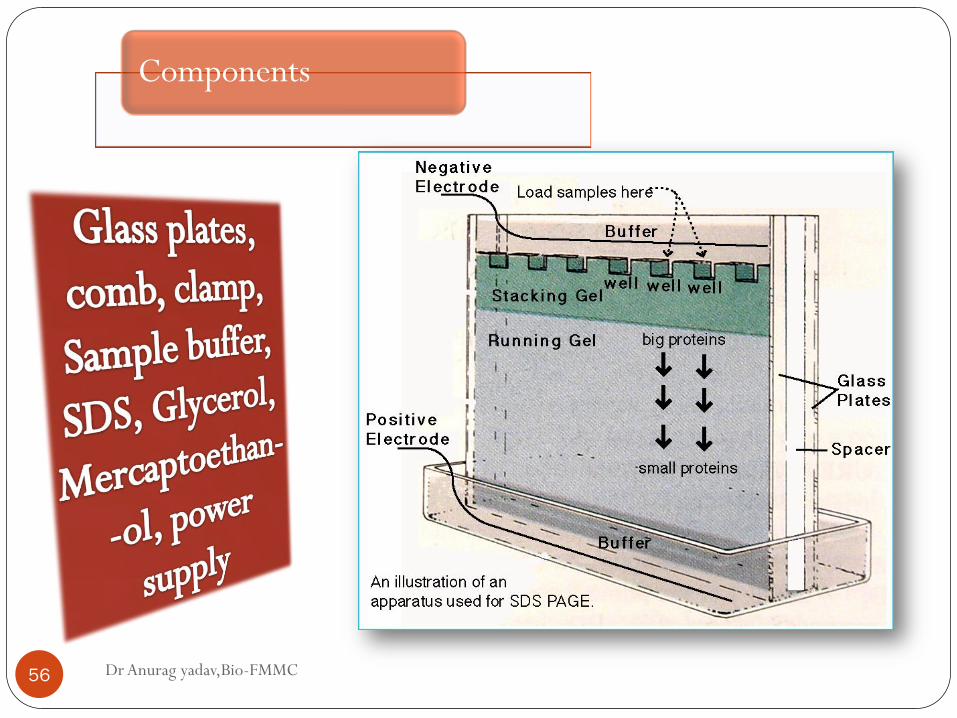
Components
Dr Anurag yadav,Bio-FMMC 56
Dr Anurag yadav,Bio-FMMC 57
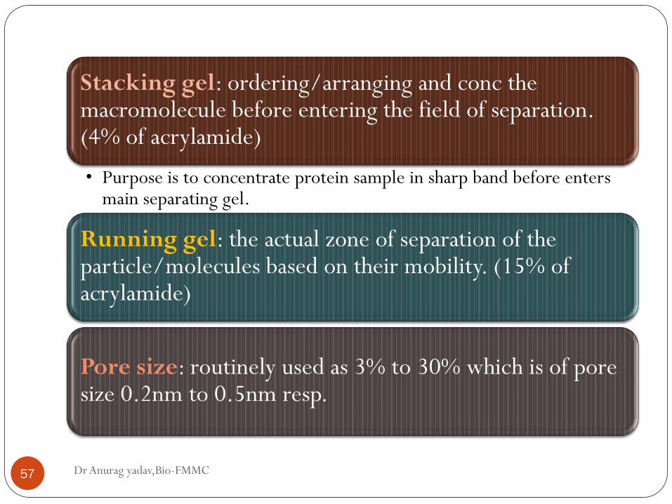
Stacking gel: ordering/arranging and conc the macromolecule before entering the field of separation. (4% of acrylamide)
• Purpose is to concentrate protein sample in sharp band before enters main separating gel.
Running gel: the actual zone of separation of the particle/molecules based on their mobility. (15% of acrylamide)
Pore size: routinely used as 3% to 30% which is of pore size 0.2nm to 0.5nm resp.
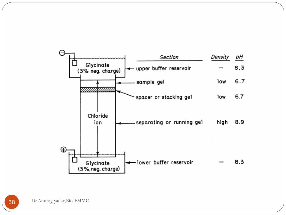
Dr Anurag yadav,Bio-FMMC 58
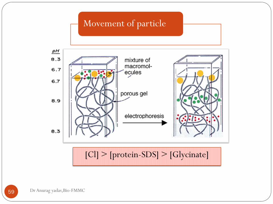
Movement of particle
Dr Anurag yadav,Bio-FMMC 59
[Cl] > [protein-SDS] > [Glycinate]
Dr Anurag yadav,Bio-FMMC 60
Dr Anurag yadav,Bio-FMMC 61
In separating gel, protein separate owing to molecular sieving
properties.
Smaller proteins pass more easily, larger one retarded by
friction.
- Research tool
- Measuring molecular weight
- Peptide mapping
- Protein identification
- Determination of sample purity
- Identifying disulfide bonds
- Separation of proteins and establishing size
- Blotting
- Smaller fragments of DNA
- Separation of nucleic acids
- Major clinical use – ALP separation
APPLICATION:
ADVANTAGES:
- Clear, fairly easy to prepare
- Exhibit reasonable mechanical strength over acrylamide conc
- Low endosmosis effect
DISADVANTAGES
- Gel preparation and casting- exacting n time-consuming
- Complete reproducibility of gel preparation not possible
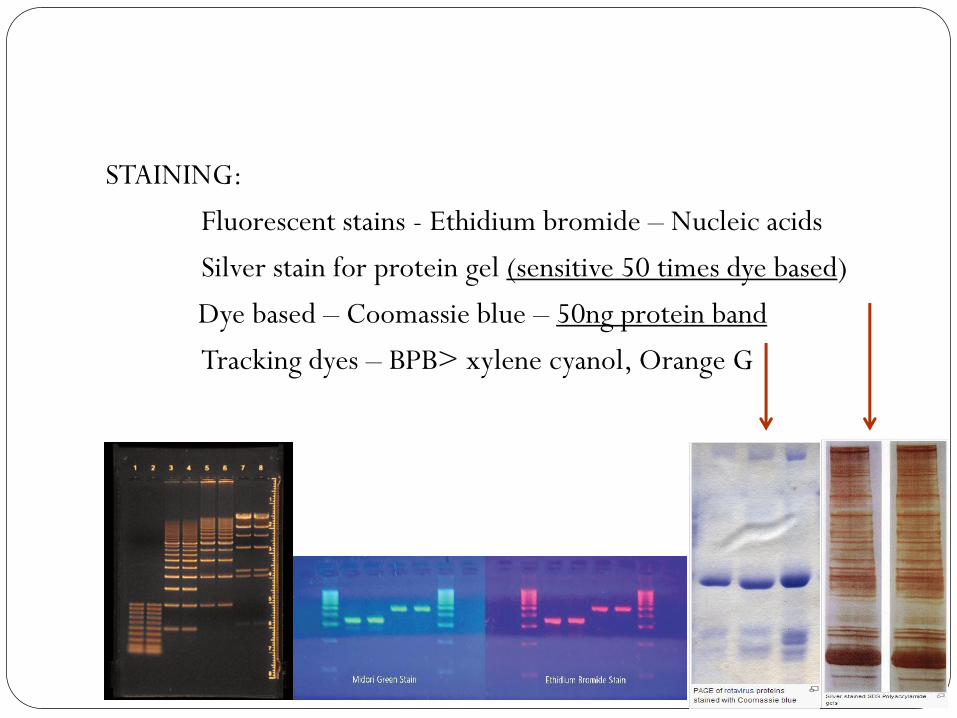
STAINING:
Fluorescent stains - Ethidium bromide – Nucleic acids
Silver stain for protein gel (sensitive 50 times dye based)
Dye based – Coomassie blue – 50ng protein band
Tracking dyes – BPB> xylene cyanol, Orange G
Native (buffer) gel
Dr Anurag yadav,Bio-FMMC 65
Done by using the polyacrylamide gel (7.5%).
As used for the enzyme separation, the denaturing agent is
not added - hence SDS is absent.
pH of 8.7
Proteins are separated according to the electrophoretic
mobility & Sieving effect of the gel.

Dr Anurag yadav,Bio-FMMC 66
Alternative approach for enzyme detection is to include the
substrate on agarose gel, which is poured over acrylamide gel.
The diffusion and interaction of the substrate and the enzyme
results in color formation.
This can be cut and used for
Total protein estimation
Enzyme activity.
Gradient gel
Dr Anurag yadav,Bio-FMMC 67
This is again an polyacrylamide gel system.
Instead of running a slab of uniform pore size, a gradient gel
is formed.
Uniformly from 5% to 25% acrylamide from top to bottom.
The highest conc gradient is layed first and than decreasing
gradient is poured.
But the sample move down, were the pore size reduces along
the path.
Dr Anurag yadav,Bio-FMMC 68
Normally run with the stacking gel at the top.
Advantage :
Greater range of protein can be separated. (Complex mixtures
can be run.)
Protein with similar molecular range may be resolved.
Protein moves till the pore size become smaller n limit its
descend further.
Proteins separated will have a distinct sharp bands.
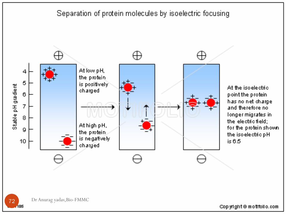
Isoelectric focussing gels
Dr Anurag yadav,Bio-FMMC 69
First described by- H.Svensson in Sweden.
Method is ideal for the separation of the amphoteric
substances.
Method has high resolution.
Able to separate the protein which differ in isoelectric point
by little 0.01 of pH unit.
Most widely used as the horizontal gel slab.
Dr Anurag yadav,Bio-FMMC 70
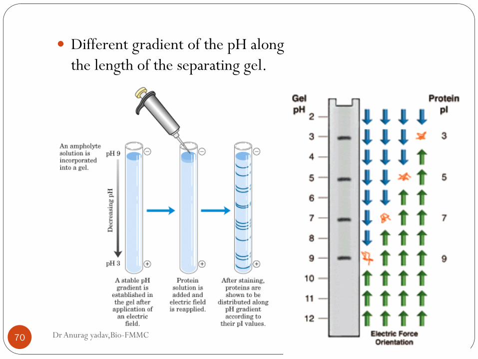
Different gradient of the pH along
the length of the separating gel.
Establishment of ph gradient:
Dr Anurag yadav,Bio-FMMC 71
This is achieved by the ampholyte & must have following prop:
Must dictate pH course (buffering capacity at their Ip)
Should have conductance at their Ip.
Low molecular weight
Soluble in water
Low light absorbance at 280nm.
Available commercially with pH band (3-11)
Eg: Ampholine, Pharmalyte and Bio-lyte.
Dr Anurag yadav,Bio-FMMC 72
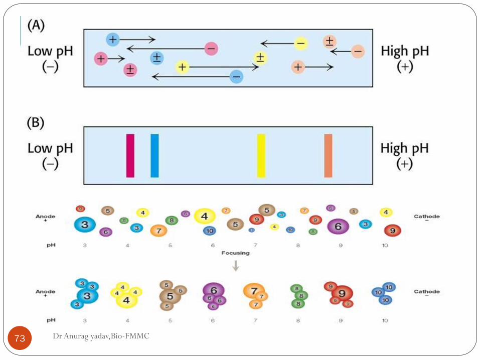
Movement
Dr Anurag yadav,Bio-FMMC 73
Dr Anurag yadav,Bio-FMMC 74
Duration : 2-3h
High voltage : 2500V
Cooling plates : 100C
Stable power pack
Fixing (trichloroacetic acid) and Staining (Coomassie
Brilliant blue)
Dr Anurag yadav,Bio-FMMC 75
Application:
- Highly sensitive for studying the microheterogeneity of
proteins
- Useful for separating the isoenzymes.
- Human genetic lab
- Research in enzymology, immunology,
- Forensic, food and agriculture industry,
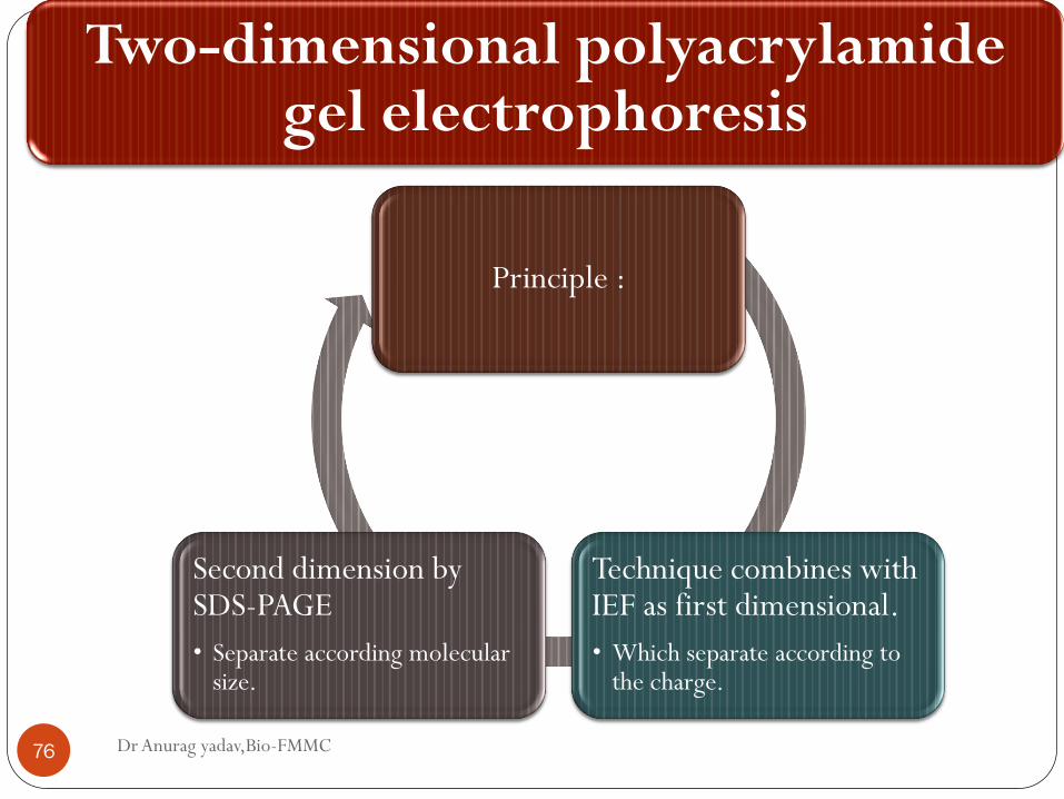
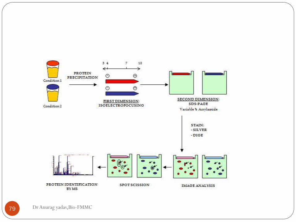
Two-dimensional polyacrylamide gel electrophoresis
Dr Anurag yadav,Bio-FMMC 76
Principle :
Technique combines with IEF as first dimensional.
• Which separate according to the charge.
Second dimension by SDS-PAGE
• Separate according molecular size.
Dr Anurag yadav,Bio-FMMC 77
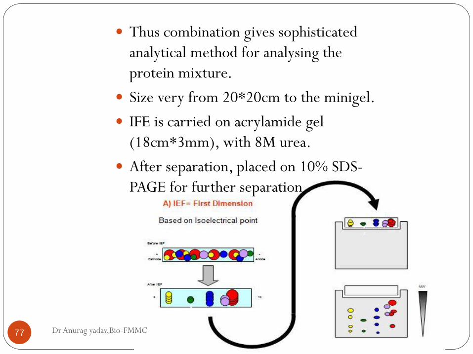
Thus combination gives sophisticated
analytical method for analysing the
protein mixture.
Size very from 20*20cm to the minigel.
IFE is carried on acrylamide gel
(18cm*3mm), with 8M urea.
After separation, placed on 10% SDS-
PAGE for further separation .
Dr Anurag yadav,Bio-FMMC 78
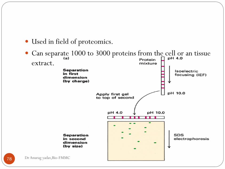
Used in field of proteomics.
Can separate 1000 to 3000 proteins from the cell or an tissue
extract.
Dr Anurag yadav,Bio-FMMC 79
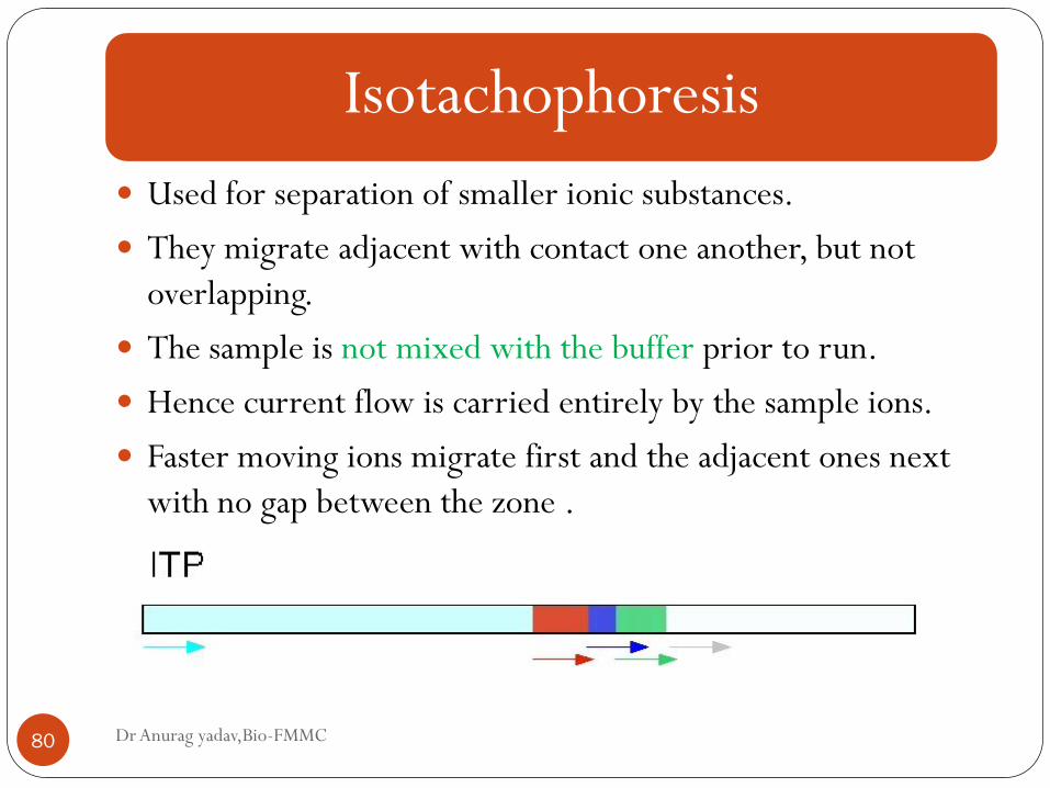
Isotachophoresis
Dr Anurag yadav,Bio-FMMC 80
Used for separation of smaller ionic substances.
They migrate adjacent with contact one another, but not
overlapping.
The sample is not mixed with the buffer prior to run.
Hence current flow is carried entirely by the sample ions.
Faster moving ions migrate first and the adjacent ones next
with no gap between the zone .
Dr Anurag yadav,Bio-FMMC 81
All ions migrate at the rate of fastest ion in zones.
Then it is measured by UV absorbance.
Application-
Separation of small anions and cations
Amino acids
Peptides
Nucleotides
Nucleosides
Proteins.
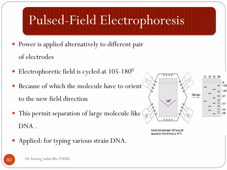
Pulsed-Field Electrophoresis
Dr Anurag yadav,Bio-FMMC 82
Power is applied alternatively to different pair
of electrodes
Electrophoretic field is cycled at 105-1800
Because of which the molecule have to orient
to the new field direction
This permit separation of large molecule like
DNA .
Applied: for typing various strain DNA.
High voltage electrophoresis
Dr Anurag yadav,Bio-FMMC 83
First described by Michl.
As the name describe, the electrophoresis is carried under
the very high voltage.
This is required for the substances of lower molecular weight
which will have considerable high diffusion rate.
Eg: amino acids, peptides.
Dr Anurag yadav,Bio-FMMC 84
The voltage applied was ranging from
2500-10000 V or
50-200V/cm, 500mA.
This resulted in better resolution and even very rapid
separation.
And even with tremendous amount of heat generation.
To tackle this, it need a good cooling system.
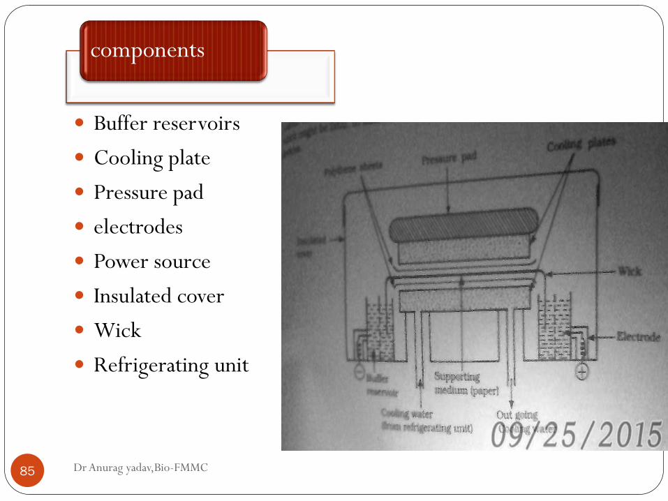
components
Dr Anurag yadav,Bio-FMMC 85
Buffer reservoirs
Cooling plate
Pressure pad
electrodes
Power source
Insulated cover
Wick
Refrigerating unit
Dr Anurag yadav,Bio-FMMC 86
Precautions :Temperature of the system has to be maintained
constant.
Plate dimension 50*50cm
The HVE one direction can be combined with the
chromatography- which is right angle to first.
Possible even to run in two direction at two different pH.
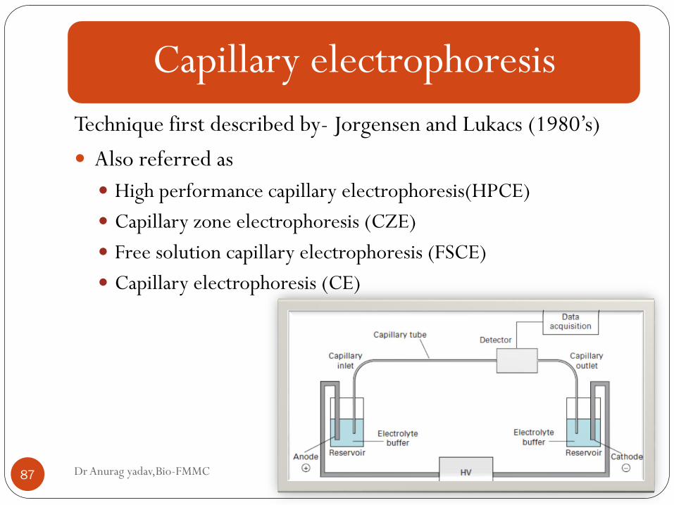
Capillary electrophoresis
Dr Anurag yadav,Bio-FMMC 87
Technique first described by- Jorgensen and Lukacs (1980’s)
Also referred as
High performance capillary electrophoresis(HPCE)
Capillary zone electrophoresis (CZE)
Free solution capillary electrophoresis (FSCE)
Capillary electrophoresis (CE)
Dr Anurag yadav,Bio-FMMC 88
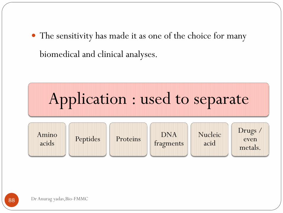
The sensitivity has made it as one of the choice for many
biomedical and clinical analyses.
Application : used to separate
Amino acids
Peptides Proteins DNA
fragments Nucleic
acid
Drugs / even
metals.
Dr Anurag yadav,Bio-FMMC 89
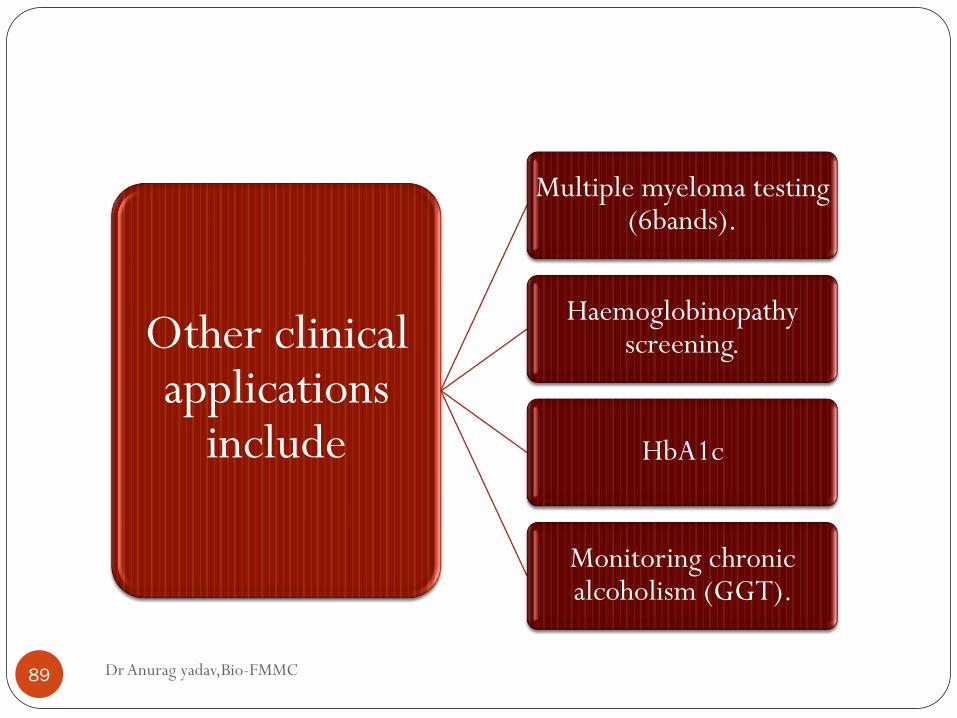
Other clinical applications
include
Multiple myeloma testing (6bands).
Haemoglobinopathy screening.
HbA1c
Monitoring chronic alcoholism (GGT).
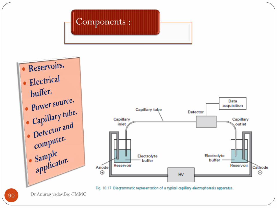
Components :
Dr Anurag yadav,Bio-FMMC 90
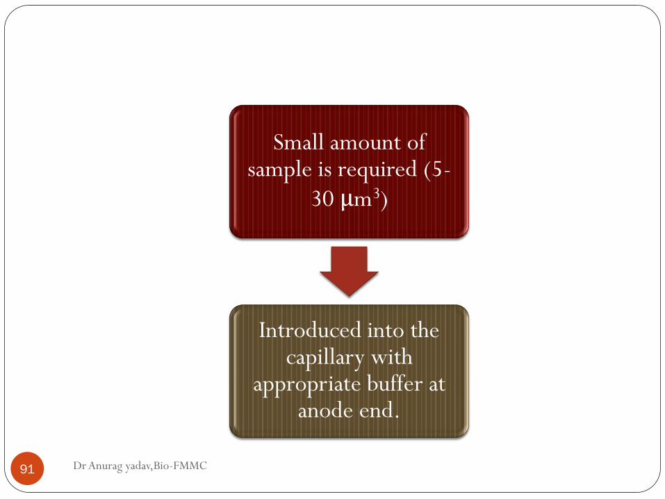
Dr Anurag yadav,Bio-FMMC 91
Small amount of sample is required (5-
30 μm3)
Introduced into the capillary with
appropriate buffer at anode end.
High voltage injection Pressure injection
Dr Anurag yadav,Bio-FMMC 92
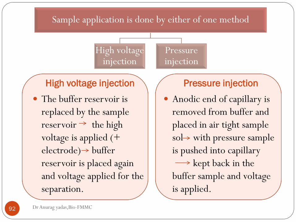
The buffer reservoir is
replaced by the sample
reservoir the high
voltage is applied (+
electrode) buffer
reservoir is placed again
and voltage applied for the
separation.
Anodic end of capillary is
removed from buffer and
placed in air tight sample
sol with pressure sample
is pushed into capillary
kept back in the
buffer sample and voltage
is applied.
Sample application is done by either of one method
High voltage injection
Pressure injection
Dr Anurag yadav,Bio-FMMC 93
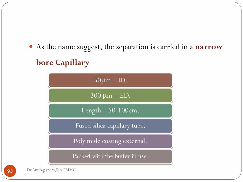
50μm – ID.
300 μm – ED.
Length – 50-100cm.
Fused silica capillary tube.
Polyimide coating external.
Packed with the buffer in use.
As the name suggest, the separation is carried in a narrow
bore Capillary
Dr Anurag yadav,Bio-FMMC 94
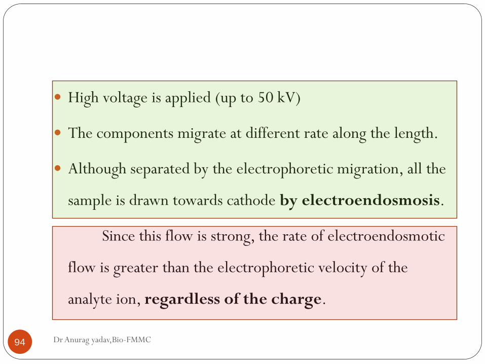
High voltage is applied (up to 50 kV)
The components migrate at different rate along the length.
Although separated by the electrophoretic migration, all the
sample is drawn towards cathode by electroendosmosis.
Since this flow is strong, the rate of electroendosmotic
flow is greater than the electrophoretic velocity of the
analyte ion, regardless of the charge.
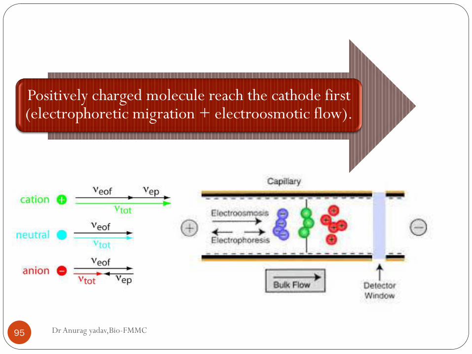
Dr Anurag yadav,Bio-FMMC 95
Positively charged molecule reach the cathode first (electrophoretic migration + electroosmotic flow).
Dr Anurag yadav,Bio-FMMC 96
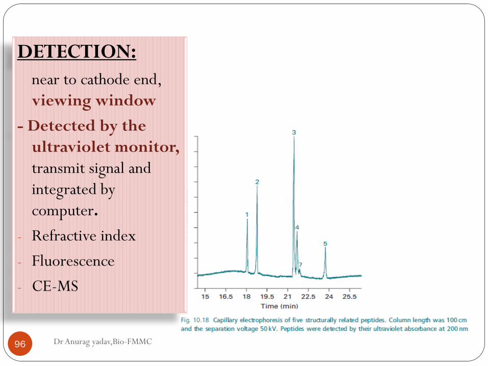
DETECTION:
near to cathode end,
viewing window
- Detected by the
ultraviolet monitor,
transmit signal and
integrated by
computer.
- Refractive index
- Fluorescence
- CE-MS
Dr Anurag yadav,Bio-FMMC 97
Troubleshooting :
Adsorption of protein to the wall of capillary – leading to
smearing of protein – viewed as peak broadening – or complete
loss of protein.
- Use of neutral coating group to the inner surface of the capillary.
Dr Anurag yadav,Bio-FMMC 98
Advantage over slab type:
Reduce the problem of heating effect.
Large surface to volume ratio.
Less diffusion of the separated bands.
Dr Anurag yadav,Bio-FMMC 99
Variations in technique:
Add of surfactant to buffer i.e., SDS (for Neutral molecules).
Micellar formation In MECC- electrophoresis + chromatography.
Different modes of operation
Dr Anurag yadav,Bio-FMMC 100
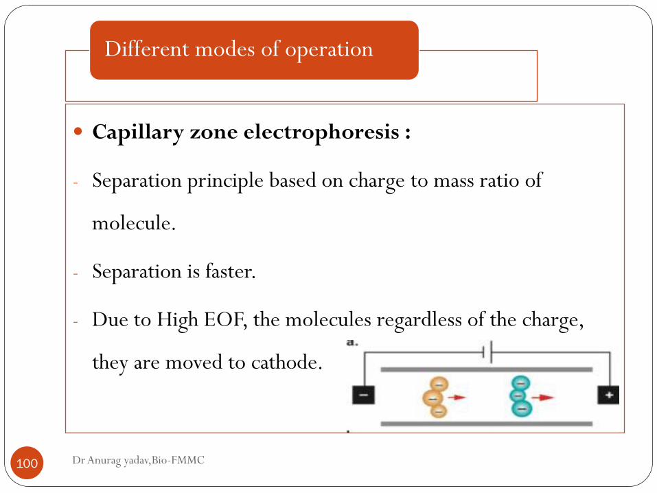
Capillary zone electrophoresis :
- Separation principle based on charge to mass ratio of
molecule.
- Separation is faster.
- Due to High EOF, the molecules regardless of the charge,
they are moved to cathode.
Different modes of operation
Dr Anurag yadav,Bio-FMMC 101
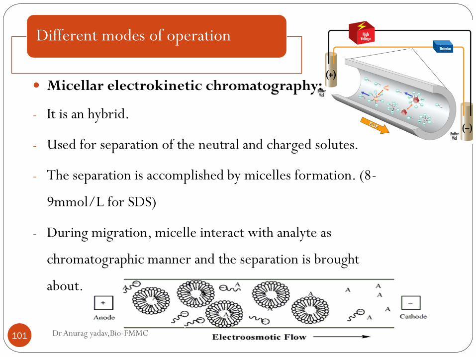
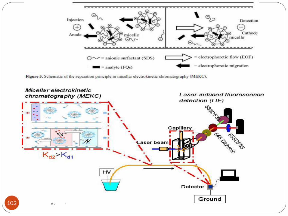
Micellar electrokinetic chromatography:
- It is an hybrid.
- Used for separation of the neutral and charged solutes.
- The separation is accomplished by micelles formation. (8-
9mmol/L for SDS)
- During migration, micelle interact with analyte as
chromatographic manner and the separation is brought
about.
Dr Anurag yadav,Bio-FMMC 102
Different modes of operation
Dr Anurag yadav,Bio-FMMC 103
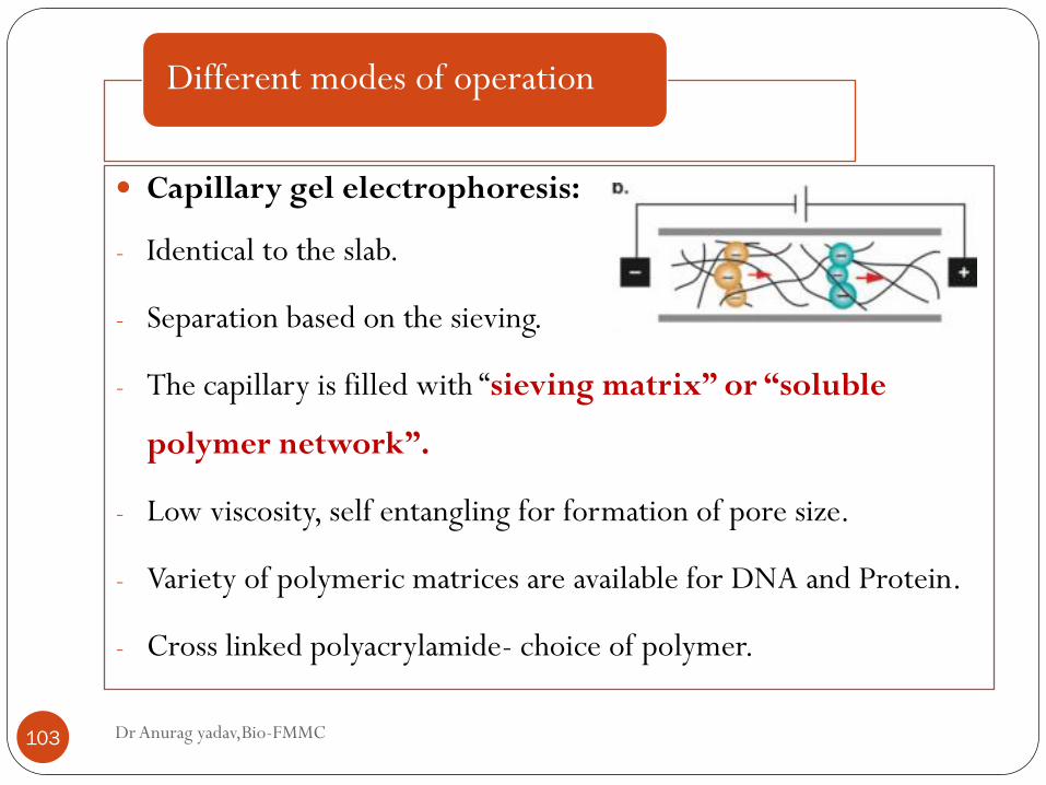
Capillary gel electrophoresis:
- Identical to the slab.
- Separation based on the sieving.
- The capillary is filled with “sieving matrix” or “soluble
polymer network”.
- Low viscosity, self entangling for formation of pore size.
- Variety of polymeric matrices are available for DNA and Protein.
- Cross linked polyacrylamide- choice of polymer.
Dr Anurag yadav,Bio-FMMC 104
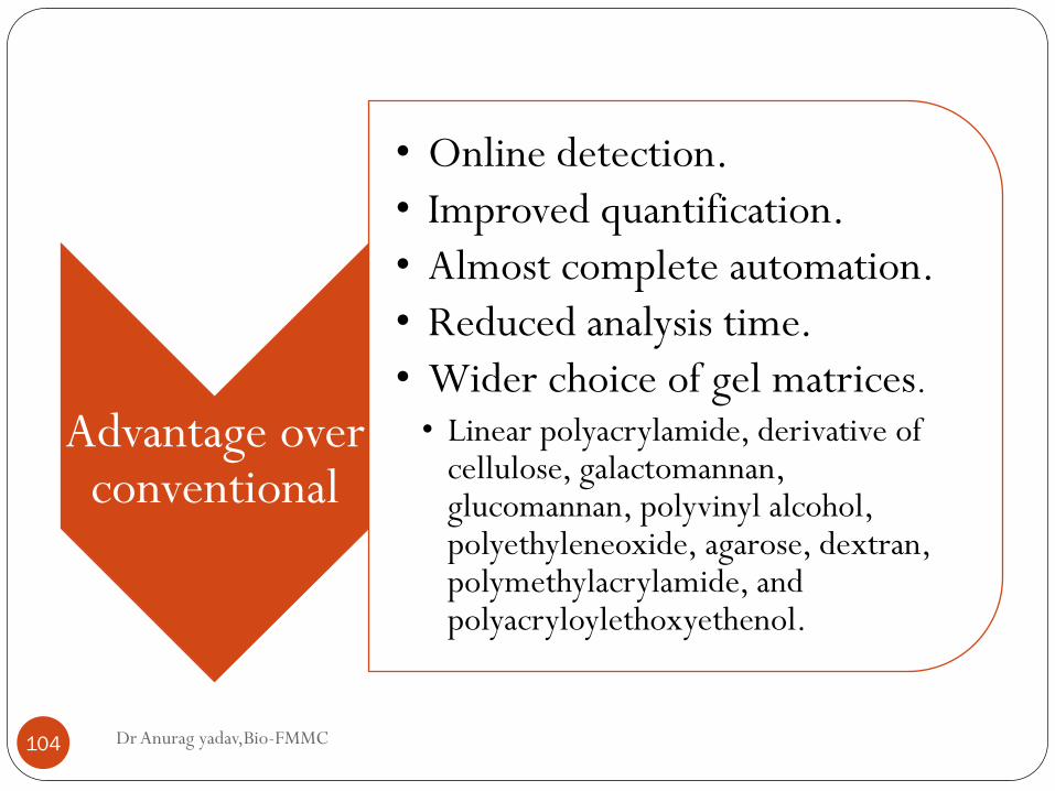
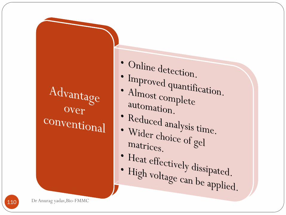
Advantage over conventional
• Online detection.
• Improved quantification.
• Almost complete automation.
• Reduced analysis time.
• Wider choice of gel matrices.
• Linear polyacrylamide, derivative of cellulose, galactomannan, glucomannan, polyvinyl alcohol, polyethyleneoxide, agarose, dextran, polymethylacrylamide, and polyacryloylethoxyethenol.
Different modes of operation
Dr Anurag yadav,Bio-FMMC 105
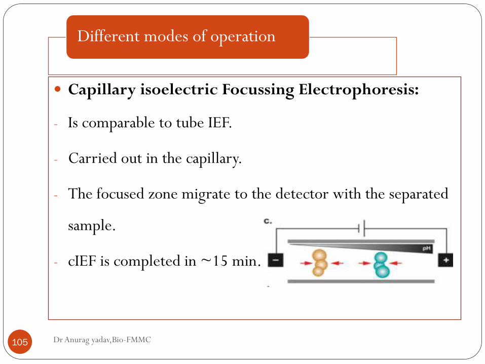
Capillary isoelectric Focussing Electrophoresis:
- Is comparable to tube IEF.
- Carried out in the capillary.
- The focused zone migrate to the detector with the separated
sample.
- cIEF is completed in ~15 min.
Different modes of operation
Dr Anurag yadav,Bio-FMMC 106
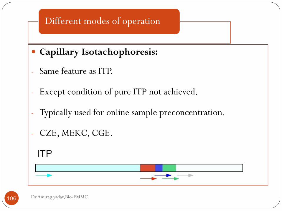
Capillary Isotachophoresis:
- Same feature as ITP.
- Except condition of pure ITP not achieved.
- Typically used for online sample preconcentration.
- CZE, MEKC, CGE.
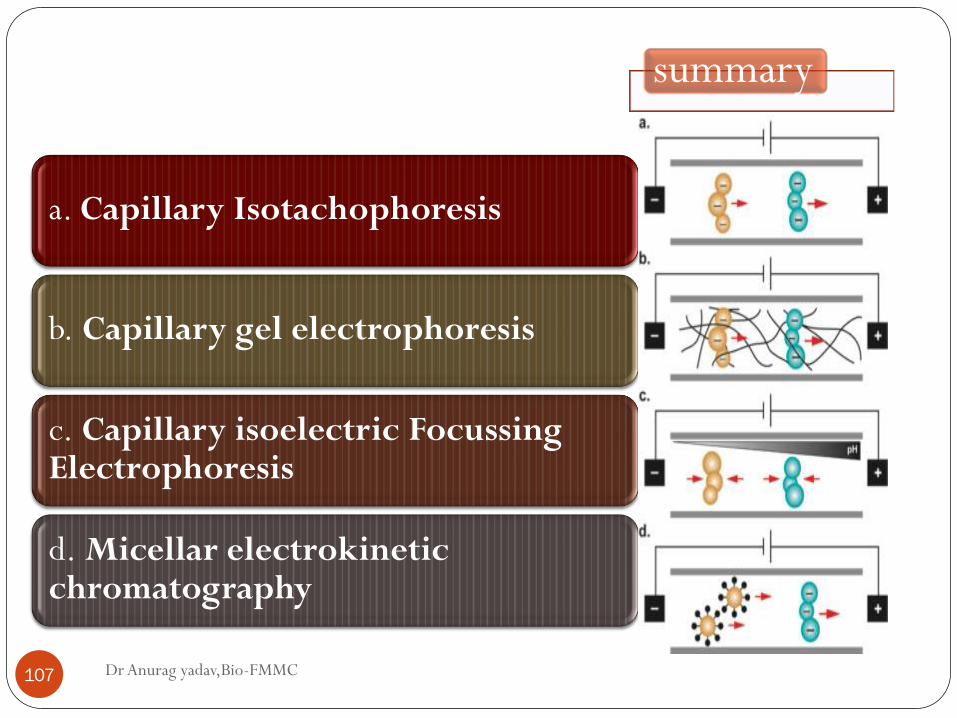
Dr Anurag yadav,Bio-FMMC 107
a. Capillary Isotachophoresis
b. Capillary gel electrophoresis
c. Capillary isoelectric Focussing Electrophoresis
d. Micellar electrokinetic chromatography
summary
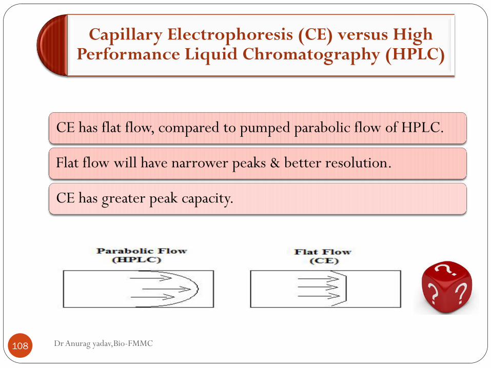
Capillary Electrophoresis (CE) versus High Performance Liquid Chromatography (HPLC)
Dr Anurag yadav,Bio-FMMC 108
CE has flat flow, compared to pumped parabolic flow of HPLC.
Flat flow will have narrower peaks & better resolution.
CE has greater peak capacity.
Dr Anurag yadav,Bio-FMMC 109
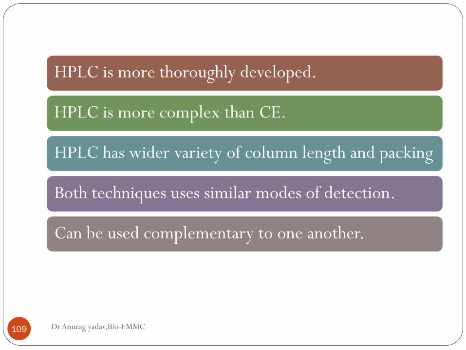
HPLC is more thoroughly developed.
HPLC is more complex than CE.
HPLC has wider variety of column length and packing
Both techniques uses similar modes of detection.
Can be used complementary to one another.
Dr Anurag yadav,Bio-FMMC 110
Microchip electrophoresis
Dr Anurag yadav,Bio-FMMC 111
Current advanced method.
Development in technique include
Integrated microchip design
Advanced detection system
New application
Protein and DNA separation can be done
Instrumentation
Dr Anurag yadav,Bio-FMMC 112
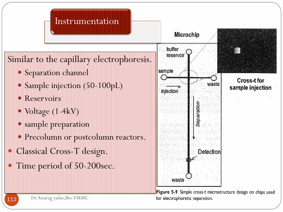
Similar to the capillary electrophoresis.
Separation channel
Sample injection (50-100pL)
Reservoirs
Voltage (1-4kV)
sample preparation
Precolumn or postcolumn reactors.
Classical Cross-T design.
Time period of 50-200sec.
Dr Anurag yadav,Bio-FMMC 113
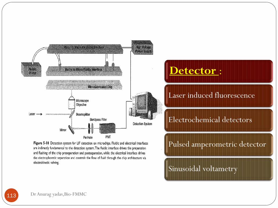
Detector :
Laser induced fluorescence
Electrochemical detectors
Pulsed amperometric detector
Sinusoidal voltametry
Application
Dr Anurag yadav,Bio-FMMC 114
An alternative for the DNA analysis.
Herpes simplex virus DNA in CSF for diagnosing encephalitis.
Gene rearrangement correlative with lymphoproliferative
disorders.
Polymorphisms in gene.
Tetranucleotide associated with hypercholesterolemia.
Diagnosing fragile X syndrome.
Muscular dystrophy.
Anthracis specific PCR product.

Dr Anurag yadav,Bio-FMMC 115
References
Dr Anurag yadav,Bio-FMMC 116
Keith Wilson- Principles and techniques of biochemistry
and molecular biology.
Upadhyay- biophysical chemistry.
Tietz- Text book of clinical chemistry.
Kaplan- clinical chemistry.
YouTube and Google images.