Embed Size (px)
Citation preview
Prepared By:
M. Kaleem Iqbal
Roll No: 309
M.Phil Biotechnology 2012-14
Principle Got its name from electromotive force. Charged
particles move under the influence of electric field from one electrode to the other.
If they are passed through a medium they can be separated on the basis of their sizes.
DNA is negatively charged, its fragments moves towards anode.

Agarose gel is a polymer meshwork
++++POSITIVEELECTRODE
------NEGATIVEELECTRODE
Power source
Sample well with DNA
Movement of DNA
General scheme of Agarose Gel Electrophoresis
Small DNA moves faster Large DNA moves slow
Types of Gels used for Electrophoresis Depending on the size of fragments different materials
can be used for electrophoresis.
Agarose: Chains of branched and unbranchedcarbohydrates. Mostly used for larger DNA fragments. Contains no cross links.
Polyacrylamide: Amides of acrylic acid. Used for resolving smaller fragments. It forms cross links with bisacrylamide.

Agarose Gel Electrophoresis Agarose in solid form is mixed with buffer and heated
for sometime.
Fix the combs according to required wells in the casting apparatus.
Poured in gel casters and allowed to solidify at room temperature.
Polyacrylamide Gel Acryamide and bisacrylamide are dissolved in buffer.
Ammonium persulfate and TEMED are used to polymerize the gel.
Gel is poured in casters (plates), fixed the combs and allowed to stay for sometime to polymerize.
Sample preparation DNA samples to be run can be generated by the action
of restriction enzymes (as in RFLPs) or it may be a PCRproduct.
Add adequate amount of loading dye. It increases the density of samples and help in visualization while the gel is running.

Detection Ethidium Bromide is added
into the gel prior to solidification.
Gel is visualized under UV.
EtBr. Intercalates with DNA and gives fluorescence under UV.
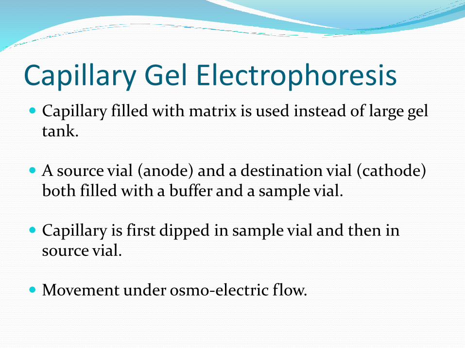
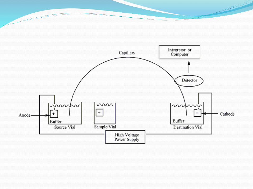
Capillary Gel Electrophoresis Capillary filled with matrix is used instead of large gel
tank.
A source vial (anode) and a destination vial (cathode) both filled with a buffer and a sample vial.
Capillary is first dipped in sample vial and then in source vial.
Movement under osmo-electric flow.

Pulsed-Field Gel Electrophoresis Specialized to separate larger fragments ranging from
10Kb to 10Mb.
Current is not passed in one direction, instead alternatively at different angles and it is not continuous but in the form of pulses.

Schematic diagram of pulse field gel electrophoresis
Applications Diseases.
Sequencing.
Family.
Animals.
Forensics.

Thanks