Embed Size (px)
Citation preview
Diagnosis and Treatment of Ascites
Clinical practice guidelines 2015
Mohamed El Sayed Sarhan (M.Sc)
Assistant lecturer of Internal Medicine
Tanta University Hospital
Ascites means pathologic fluid collection within
the abdominal cavity.
80% hepatic causes.
The development of ascites is associated with a
poor prognosis and impaired quality of life in
patients with cirrhosis.
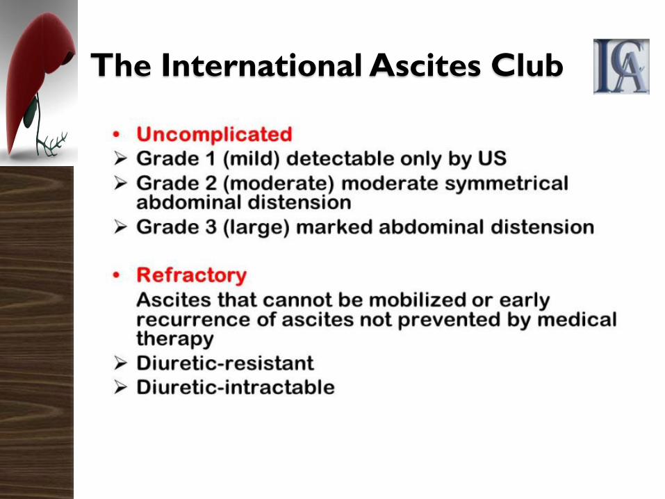
The International Ascites Club
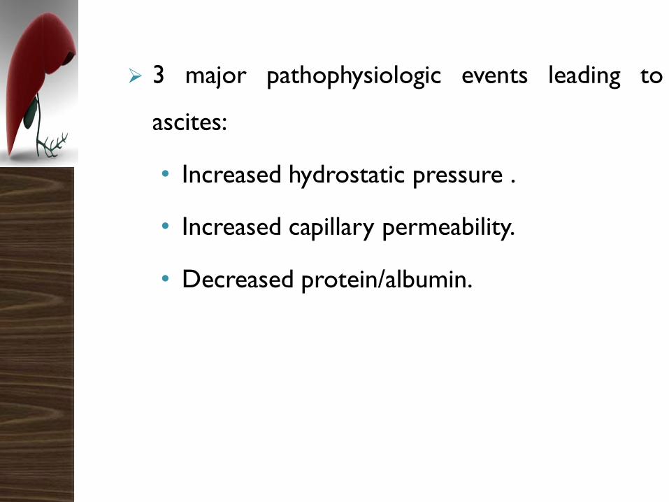
3 major pathophysiologic events leading to
ascites:
• Increased hydrostatic pressure .
• Increased capillary permeability.
• Decreased protein/albumin.
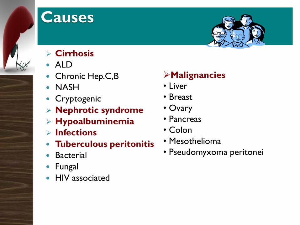
Causes
Cirrhosis
ALD
Chronic Hep.C,B
NASH
Cryptogenic
Nephrotic syndrome
Hypoalbuminemia
Infections
Tuberculous peritonitis
Bacterial
Fungal
HIV associated
Malignancies
• Liver
• Breast
• Ovary
• Pancreas
• Colon
• Mesothelioma
• Pseudomyxoma peritonei
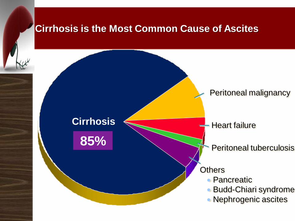
Cirrhosis Heart failure
Peritoneal tuberculosis
Cirrhosis is the Most Common Cause of Ascites
Others
Pancreatic
Budd-Chiari syndrome
Nephrogenic ascites
Peritoneal malignancy
85%
About 85% of patients with ascites have cirrhosis, Past
history of cancer, heart failure,or TB.
Symptoms:
◦ Abdominal distension:
Painless or with abdominal discomfort
Course of days (eg, bloody ascites due to trauma) or
months (eg, malignant ascites)
◦ Weight gain, shortness of breath, early satiety,
and dyspnea due to fluid accumulation and
increased abdominal pressure.
◦ Spontaneous bacterial peritonitis fever,
abdominal tenderness, and altered mental
status.
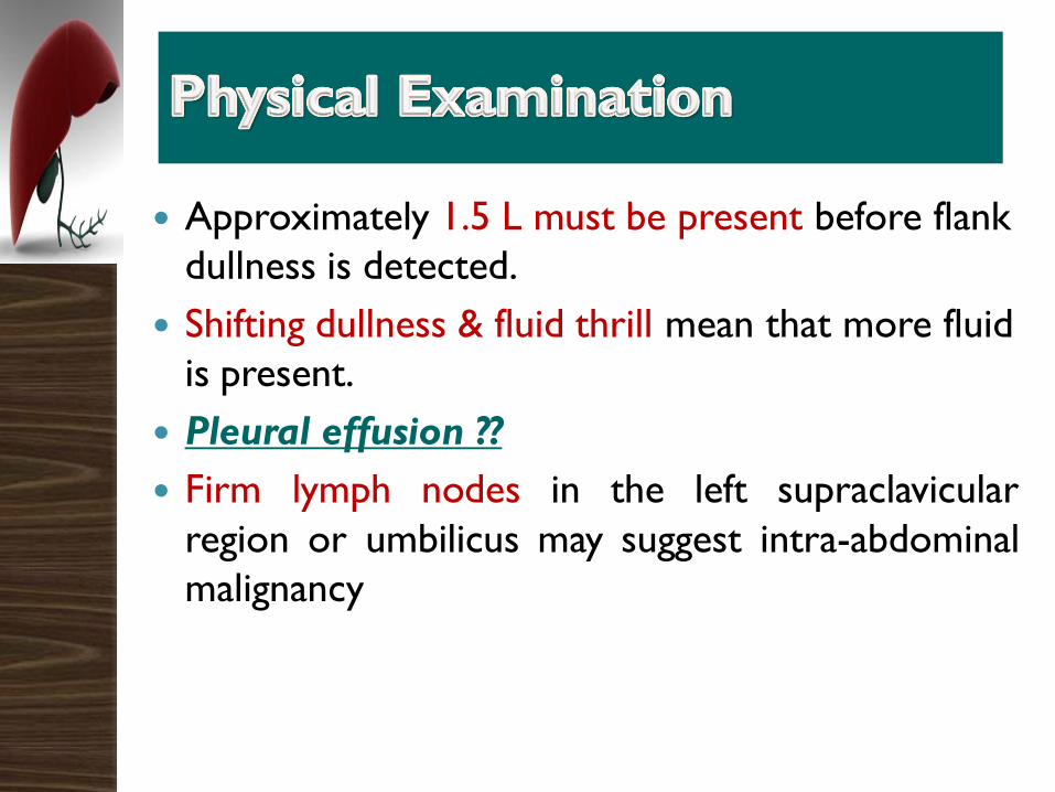
Approximately 1.5 L must be present before flank
dullness is detected.
Shifting dullness & fluid thrill mean that more fluid
is present.
Pleural effusion ??
Firm lymph nodes in the left supraclavicular
region or umbilicus may suggest intra-abdominal
malignancy

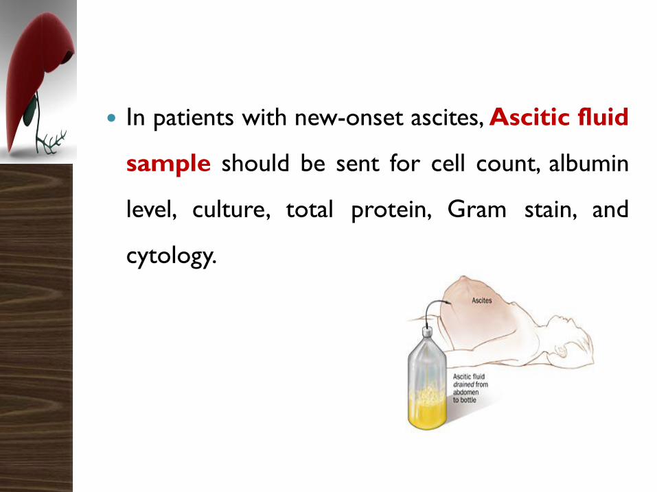
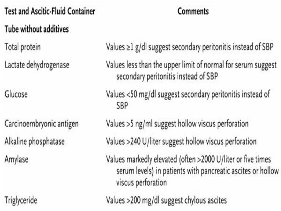
In patients with new-onset ascites, Ascitic fluid
sample should be sent for cell count, albumin
level, culture, total protein, Gram stain, and
cytology.
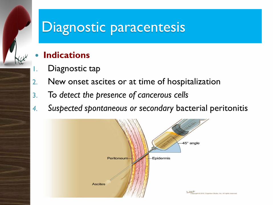
Diagnostic paracentesis
Indications
1. Diagnostic tap
2. New onset ascites or at time of hospitalization
3. To detect the presence of cancerous cells
4. Suspected spontaneous or secondary bacterial peritonitis
Contraindications
Absolute contraindication
Acute abdomen that requires surgery.
Relative contraindications
Severe thrombocytopenia (platelet count < 20 X 103/μL),
coagulopathy.
Pregnancy.
Distended urinary bladder.
Abdominal wall cellulitis.
Ascitic fluid sample
Inspection:
Most ascitic fluid is transparent and tinged
yellow Blood-tinged fluid. This may result from
either a traumatic tap or malignancy.
Bloody fluid from a traumatic tap is
heterogeneously bloody, nontraumatic bloody fluid
is homogeneously red. (Ruptered hepatoma)?
CT with contrast.
Cloudy ascitic fluid with a purulent consistency
indicates infection.
Green bilious, or deep jaundice/ upper GI
perforation
White Chylous
Cell count:
A PMN count of greater than 250 cells/µL
is highly suggestive of bacterial peritonitis
(neutrocytic ascites) either primary or
secondary bacterial peritonitis .
In tuberculous peritonitis and peritoneal
carcinomatosis, lymphocytes usually
predominate.
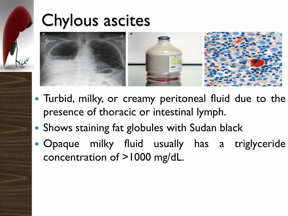
Chylous ascites
Turbid, milky, or creamy peritoneal fluid due to the
presence of thoracic or intestinal lymph.
Shows staining fat globules with Sudan black
Opaque milky fluid usually has a triglyceride
concentration of >1000 mg/dL.
Chylous ascites
Is most often the result of lymphatic obstruction
from ;
Trauma / surgeries
Tumor
Tuberculosis
Filariasis
Congenital abnormalities
Nephrotic syndrome

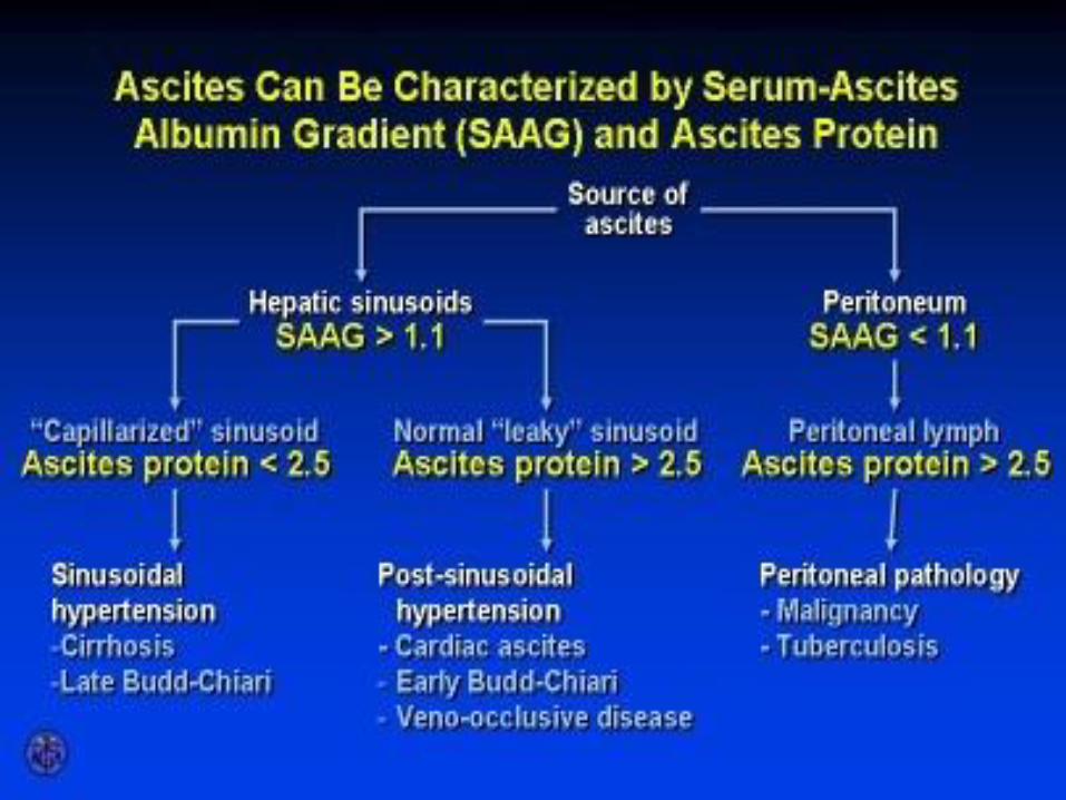
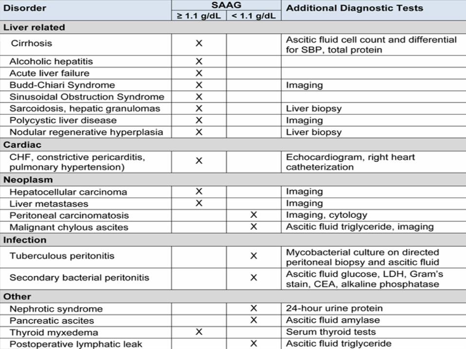
SAAGSerum Ascites Albumin Gradient
The SAAG is the best single test for
classifying ascites into portal hypertensive
(SAAG >1.1 g/dL) and non–portal
hypertensive (SAAG < 1.1 g/dL) causes.
The accuracy of the SAAG results is
approximately 97% in classifying ascites.
Calculated by substracting the ascitic fluid
albumin from serum albumin.
Albumin Serum – Albumin Ascites(g/dL) (g/dL)
in the same day
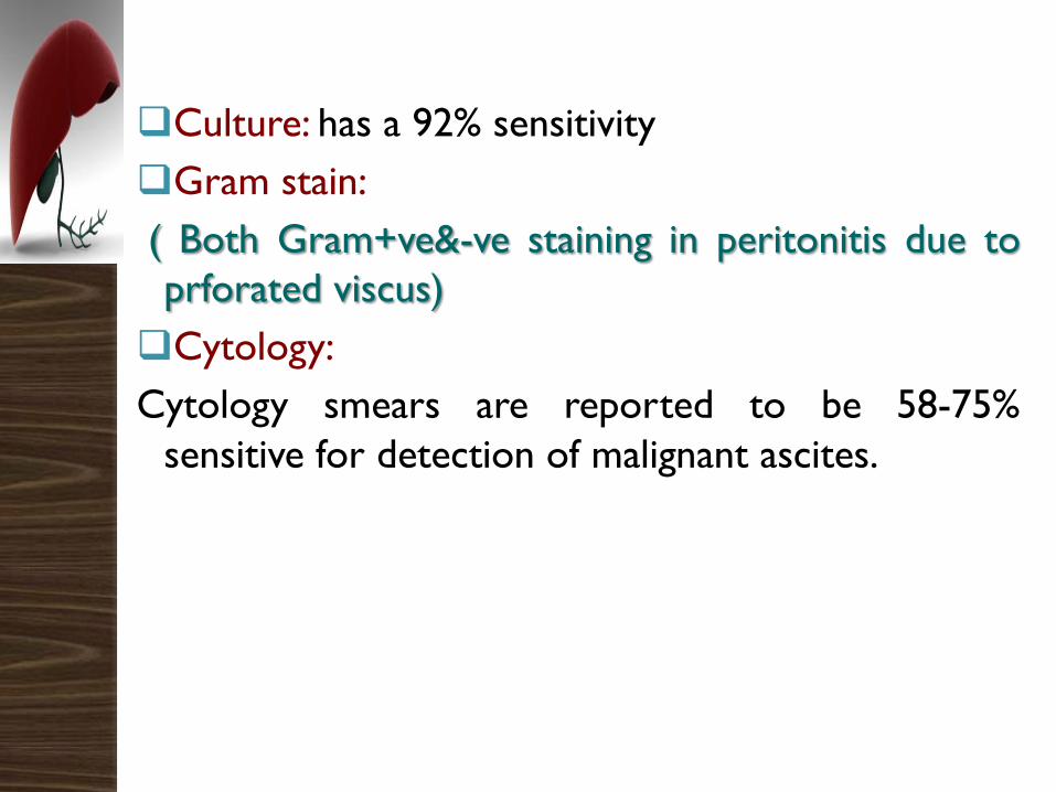
Culture: has a 92% sensitivity
Gram stain:
( Both Gram+ve&-ve staining in peritonitis due to
prforated viscus)
Cytology:
Cytology smears are reported to be 58-75%
sensitive for detection of malignant ascites.
Recommendations
A diagnostic paracentesis should be performed
in all patients with new onset grade 2 or 3
ascites, and in all patients hospitalized for
worsening of ascites or any complication of
cirrhosis (Level A1).
Recommendations
It is important to measure ascitic total protein
concentration, since patients with an ascitic
protein concentration of less than 15 g/L have
an increased risk of developing spontaneous
bacterial peritonitis (Level A1) and may benefit
from antibiotic prophylaxis (Level A1).
Recommendations
Measurement of the serum–ascites albumin
gradient may be useful when the diagnosis of
cirrhosis is not clinically evident or in patients
with cirrhosis in whom a cause of ascites
different than cirrhosis is suspected (Level A2).
Radiological features
Abdominal ultrasound is useful in confirming
the presence of ascites and in the guidance of
paracentesis.
Both ultrasound and CT imaging are useful in
distinguishing between causes of portal and
nonportal hypertensive ascites.
Doppler ultrasound and CT can detect
thrombosis of the hepatic veins (Budd-Chiari
syndrome) or portal veins.
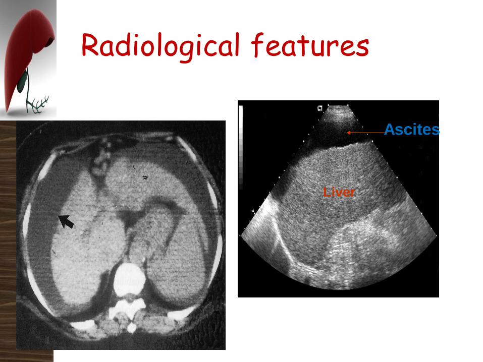
Radiological features
Ascites
Liver
Laparoscopy
Laparoscopy is an important test in the
evaluation of some patients with nonportal
hypertensive ascites (low SAAG) or mixed
ascites.
It permits direct visualization and biopsy of the
peritoneum, liver, and some intra-abdominal
lymph nodes.
Cases of suspected peritoneal tuberculosis or
suspected malignancy with nondiagnostic CT
imaging and ascitic fluid cytology are best
evaluated by this method.

Ascites: Treatment
Goals:
◦ Minimize ascitic volume and peripheral edema
◦ Avoid intravascular volume depletion
Benefits:
◦ Patient comfort
◦ Reduced risk of hernia formation
◦ Possible reduction in SBP
◦ Improve nutrition
Management of uncomplicated ascites
Patients with cirrhosis and ascites are at high
risk for other complications of liver disease,
including:
1. Refractory ascites,
2. SBP,
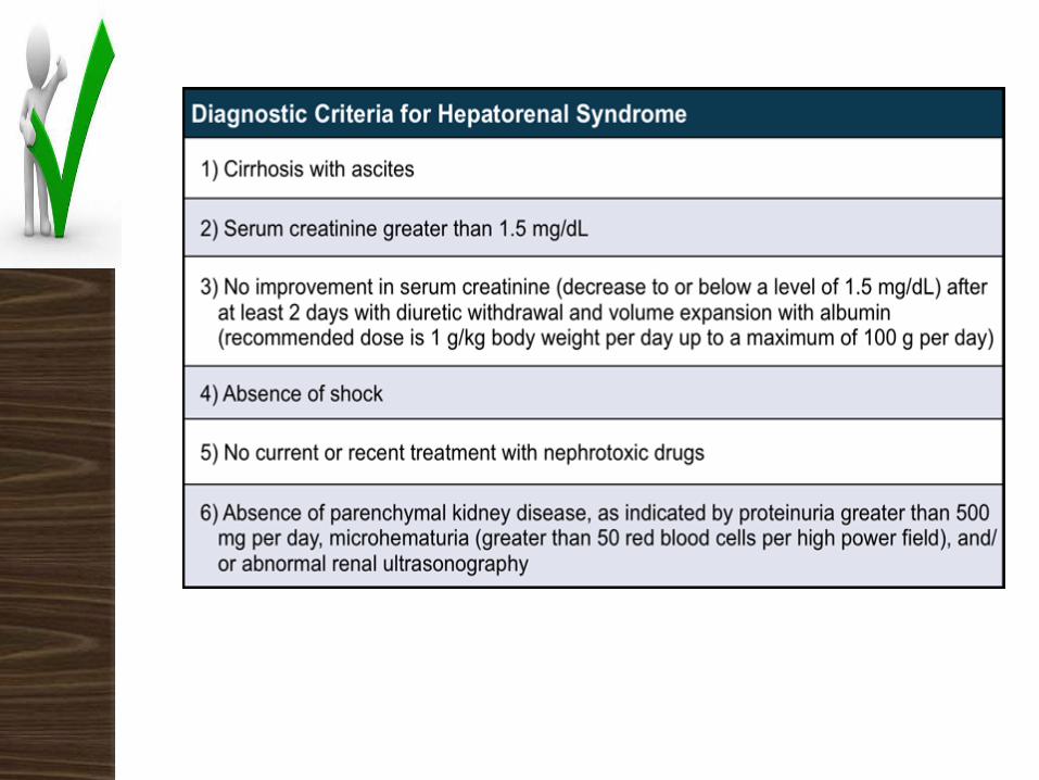
3. Hepatorenal syndrome (HRS).
The absence of these ascites-related
complications qualifies ascites as uncomplicated.
Patients with moderate ascites can be
treated as outpatients and do not
require hospitalization unless they
have other complications of cirrhosis.
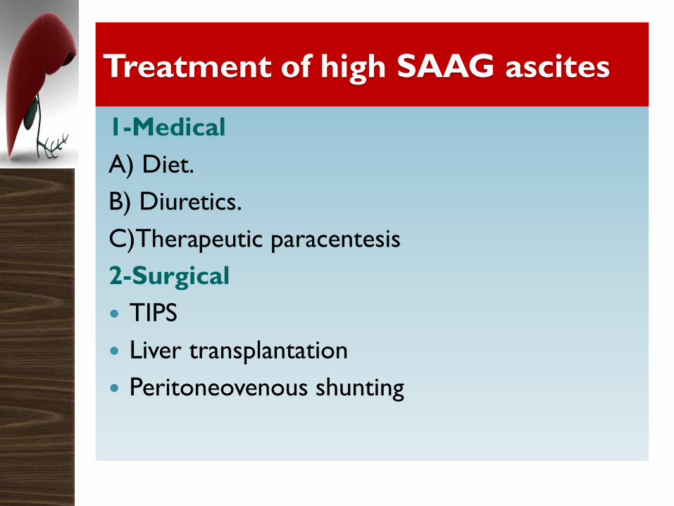
Treatment of high SAAG ascites
1-Medical
A) Diet.
B) Diuretics.
C)Therapeutic paracentesis
2-Surgical
TIPS
Liver transplantation
Peritoneovenous shunting
Recommendations
Since the development of grade 2 or 3
ascites in patients with cirrhosis is associated
with reduced survival, liver transplantation
should be considered as a potential treatment
option (Level B1).
Recommendations
Moderate restriction of salt intake is an
important component of the management of
ascites (intake of sodium of 80–120 mmol/day,
which corresponds to 4.6– 6.9 g of salt/day)
(Level B1).
This is generally equivalent to a no added salt
diet with avoidance of pre-prepared meals.
Many low – sodium foods are now available.
Recommendations There is insufficient evidence to recommend forced
bed rest as part of the treatment of ascites. There
are no data to support the use of fluid restriction
in patients with ascites with normal serum
sodium concentration (Level B1). AASLD
Fluid restriction is not necessary unless serum sodium
is less than 120-125 mmol/L.)
Recommendations
There are no data to support the
prophylactic use of salt restriction in
patients who have never had ascites.
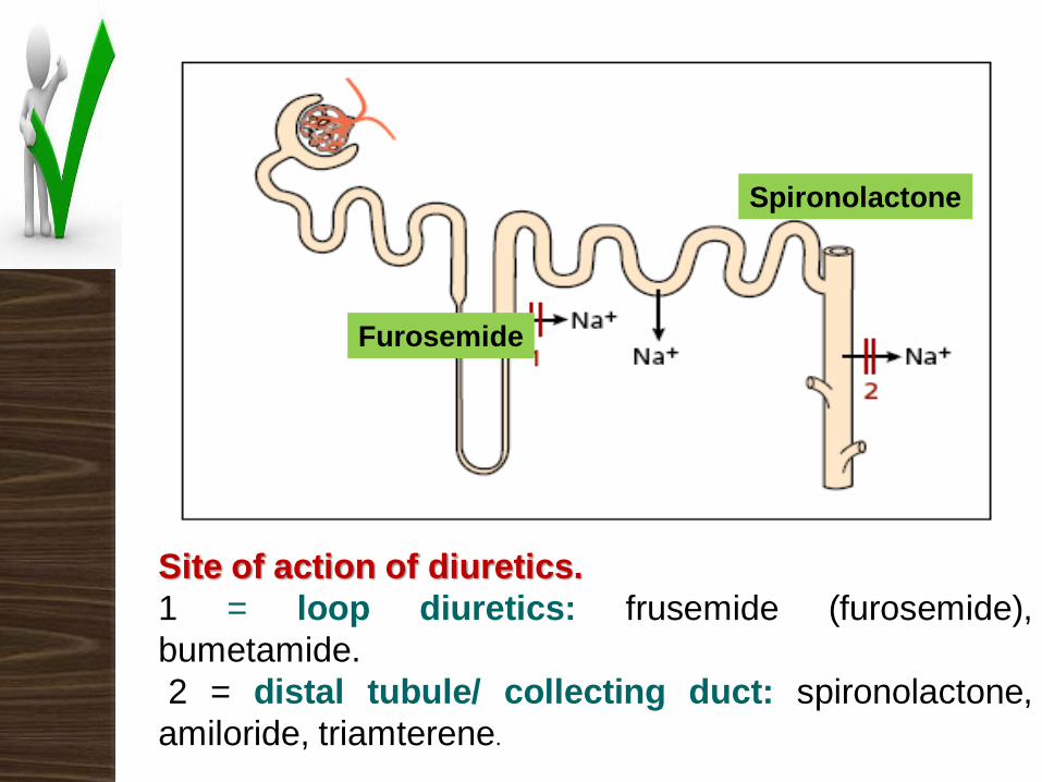
Site of action of diuretics.
1 = loop diuretics: frusemide (furosemide),
bumetamide.
2 = distal tubule/ collecting duct: spironolactone,
amiloride, triamterene.
Spironolactone
Furosemide
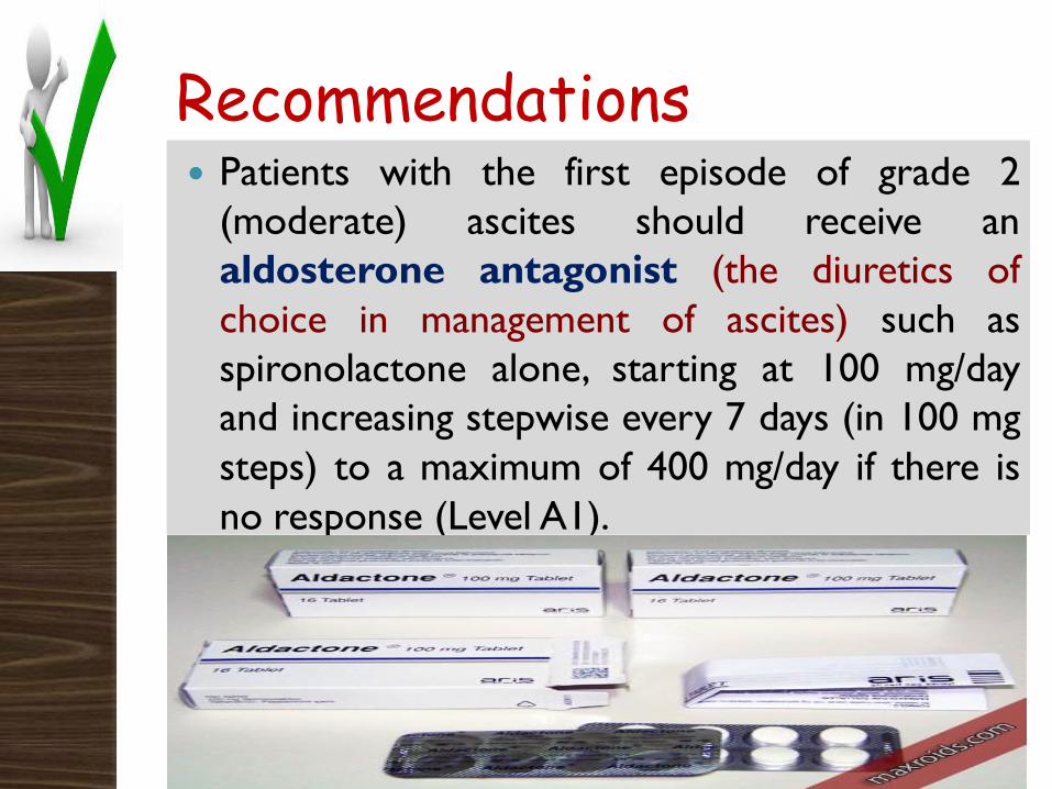
Recommendations Patients with the first episode of grade 2
(moderate) ascites should receive an
aldosterone antagonist (the diuretics of
choice in management of ascites) such as
spironolactone alone, starting at 100 mg/day
and increasing stepwise every 7 days (in 100 mg
steps) to a maximum of 400 mg/day if there is
no response (Level A1).
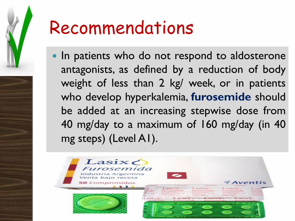
Recommendations
In patients who do not respond to aldosterone
antagonists, as defined by a reduction of body
weight of less than 2 kg/ week, or in patients
who develop hyperkalemia, furosemide should
be added at an increasing stepwise dose from
40 mg/day to a maximum of 160 mg/day (in 40
mg steps) (Level A1).
Recommendations
Patients with recurrent ascites should be
treated with a combination of an
aldosterone antagonist plus furosemide,
the dose of which should be increased
sequentially according to response, as
explained before (Level A1).
Recommendations
The maximum recommended weight loss
during diuretic therapy should be 0.5
kg/day in patients without edema and
1 kg/day in patients with edema
(Level A1).
Recommendations
The goal of long-term treatment is to maintain
patients free of ascites with the minimum dose
of diuretics. Thus, once the ascites has largely
resolved, the dose of diuretics should be
reduced and discontinued later, whenever
possible (Level B1).
Recommendations
All diuretics should be discontinued if
there is
1. Severe hyponatremia (serum sodium
concentration <120 mmol/L),
2. Progressive renal failure,
3. Worsening hepatic encephalopathy,
4. Incapacitating muscle cramps
(Level B1).
Recommendations
Furosemide should be stopped if there is severe
hypokalemia (<3 mmol/L).
Aldosterone antagonists should be stopped if
patients develop severe hyperkalemia (serum
potassium >6 mmol/L) (Level B1).
Recommendations
Diuretics are generally contraindicated in
patients with overt hepatic encephalopathy
(Level B1).
Gynaecomastia is common with the use of
aldosterone antagonists, but it does not usually
require discontinuation of treatment.
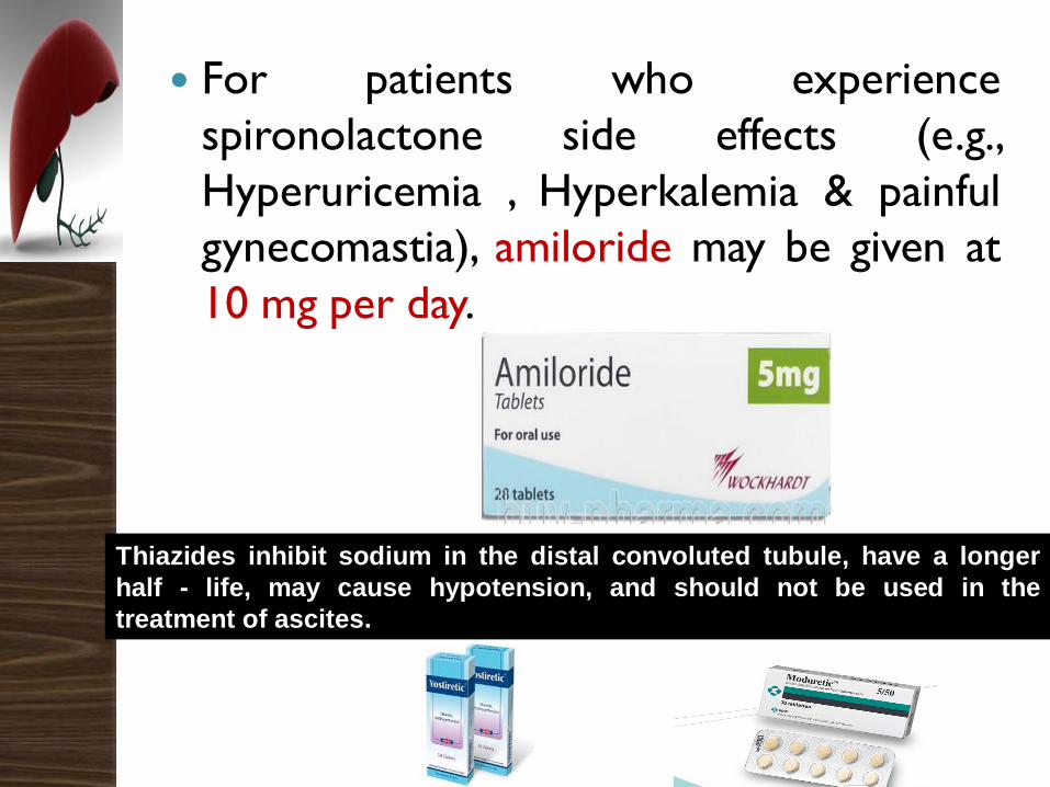
For patients who experience
spironolactone side effects (e.g.,
Hyperuricemia , Hyperkalemia & painful
gynecomastia), amiloride may be given at
10 mg per day.
Thiazides inhibit sodium in the distal convoluted tubule, have a longer
half - life, may cause hypotension, and should not be used in the
treatment of ascites.
Grade 3 or large ascites
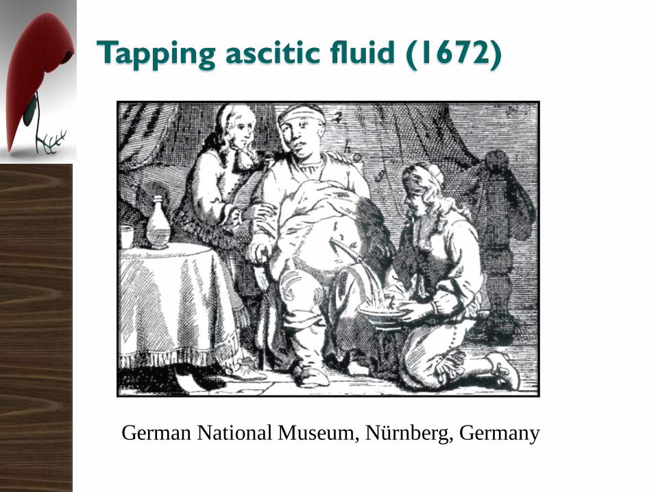
Tapping ascitic fluid (1672)
German National Museum, Nürnberg, Germany
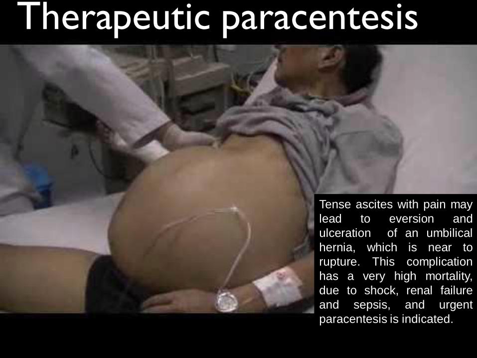
Therapeutic paracentesis
Tense ascites with pain may
lead to eversion and
ulceration of an umbilical
hernia, which is near to
rupture. This complication
has a very high mortality,
due to shock, renal failure
and sepsis, and urgent
paracentesis is indicated.
LVP should be performed under strict sterile
conditions using disposable sterile materials.
It is generally agreed that there are no
contraindications to LVP other than loculated
ascites, Hemorrhagic complications after LVP
are infrequent.
In one study, which also included patients with
INR >1.5 and platelet count <50,000/µl, only
two patients experienced minor cutaneous
bleedings out of 142 paracenteses.
Recommendations
Large-volume paracentesis (LVP) is the
first-line therapy in patients with large
ascites (grade 3 ascites) (Level A1).
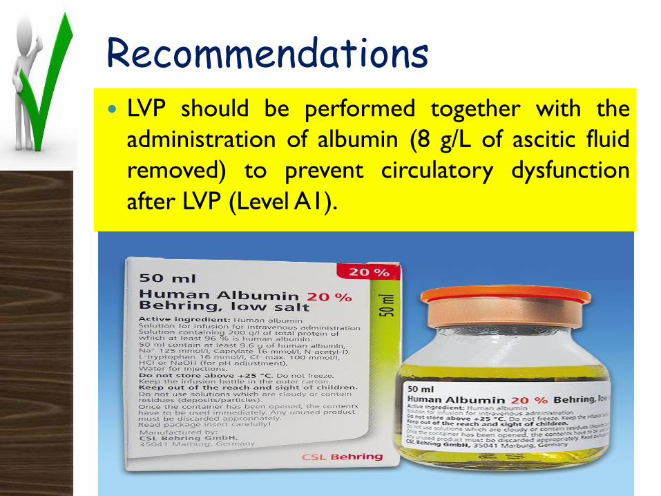
Recommendations LVP should be performed together with the
administration of albumin (8 g/L of ascitic fluid
removed) to prevent circulatory dysfunction
after LVP (Level A1).
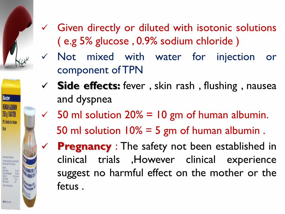
Given directly or diluted with isotonic solutions
( e.g 5% glucose , 0.9% sodium chloride )
Not mixed with water for injection or
component of TPN
Side effects: fever , skin rash , flushing , nausea
and dyspnea
50 ml solution 20% = 10 gm of human albumin.
50 ml solution 10% = 5 gm of human albumin .
Pregnancy : The safety not been established in
clinical trials ,However clinical experience
suggest no harmful effect on the mother or the
fetus .
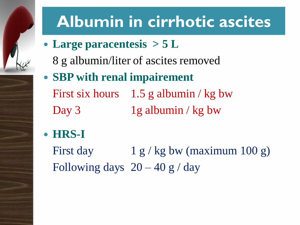
Albumin in cirrhotic ascites
Large paracentesis > 5 L
8 g albumin/liter of ascites removed
SBP with renal impairement
First six hours 1.5 g albumin / kg bw
Day 3 1g albumin / kg bw
HRS-I
First day 1 g / kg bw (maximum 100 g)
Following days 20 – 40 g / day
Recommendations
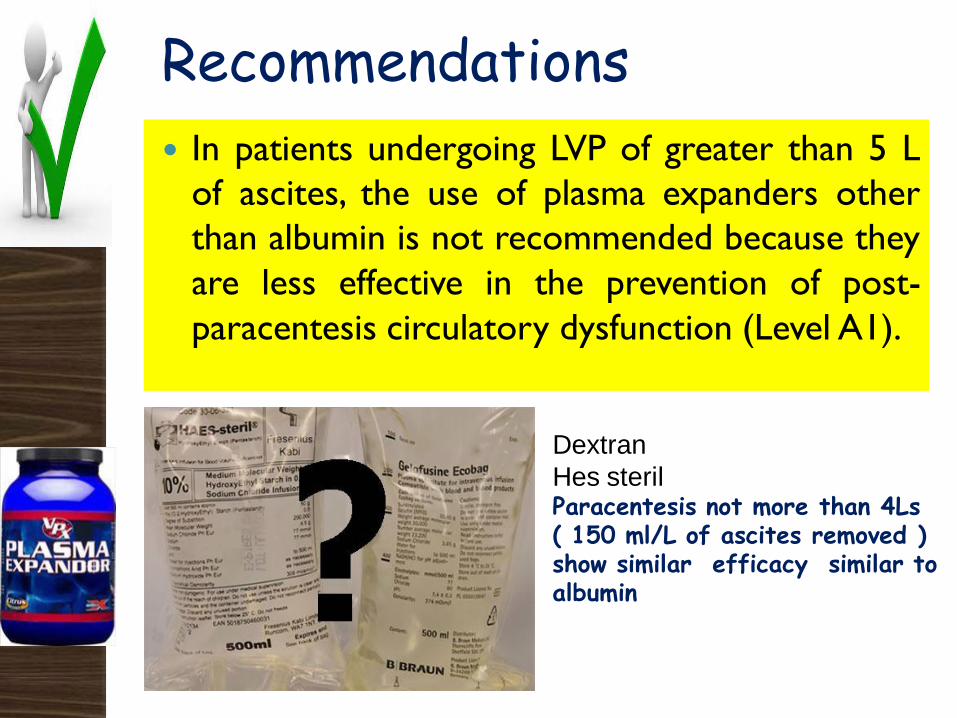
In patients undergoing LVP of greater than 5 L
of ascites, the use of plasma expanders other
than albumin is not recommended because they
are less effective in the prevention of post-
paracentesis circulatory dysfunction (Level A1).
Dextran
Hes sterilParacentesis not more than 4Ls( 150 ml/L of ascites removed ) show similar efficacy similar to albumin
Recommendations
After LVP, patients should receive the minimum
dose of diuretics necessary to prevent the
re-accumulation of ascites (Level A1).
In a controlled trial, serial large - volume
paracenteses (LVP) reduced hospital stay
compared with standard diuretic
treatment.
However, readmissions to hospital,
survival and causes of death did not differ
signifi - cantly between the LVP and
diuretic groups.
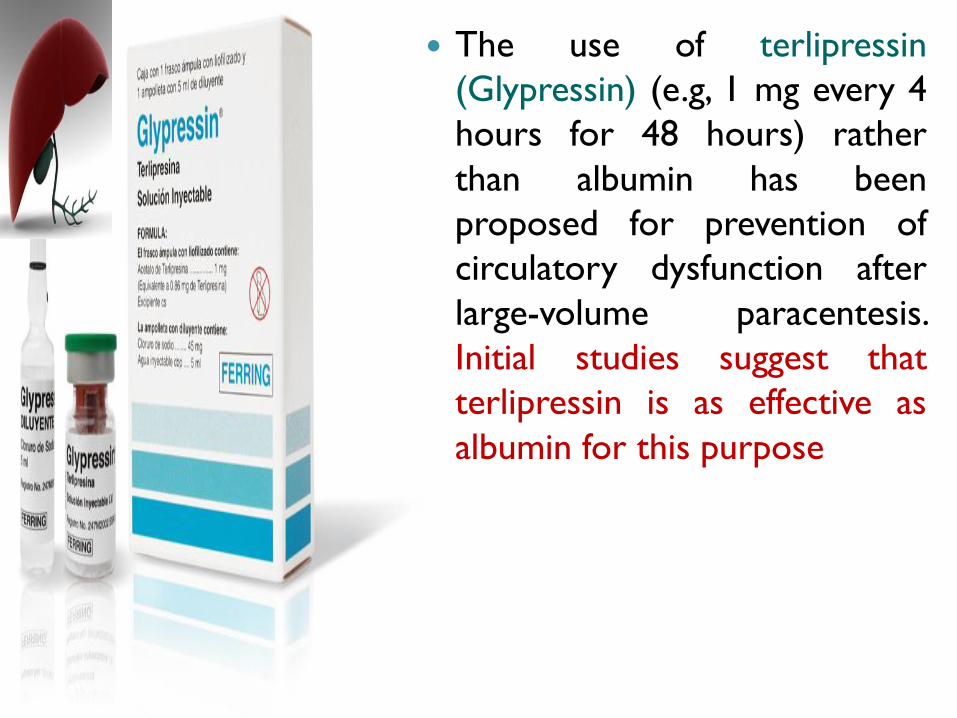
The use of terlipressin
(Glypressin) (e.g, 1 mg every 4
hours for 48 hours) rather
than albumin has been
proposed for prevention of
circulatory dysfunction after
large-volume paracentesis.
Initial studies suggest that
terlipressin is as effective as
albumin for this purpose
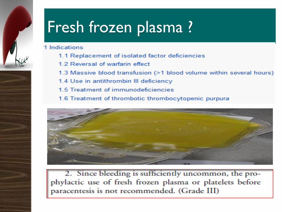
Fresh frozen plasma ?
There are no data to support the use of fresh
frozen plasma or pooled platelets before LVP, yet
in many centers these products are given if there
is severe coagulopathy (prothrombin activity less
than 40%) and/or thrombocytopenia (less than
40,000).
Evaluation of patients with refractory ascites
According to the criteria of the International
Ascites Club, refractory ascites is defined as
‘‘ascites that cannot be mobilized or the early
recurrence of which (i.e., after LVP) cannot be
prevented by medical therapy” .
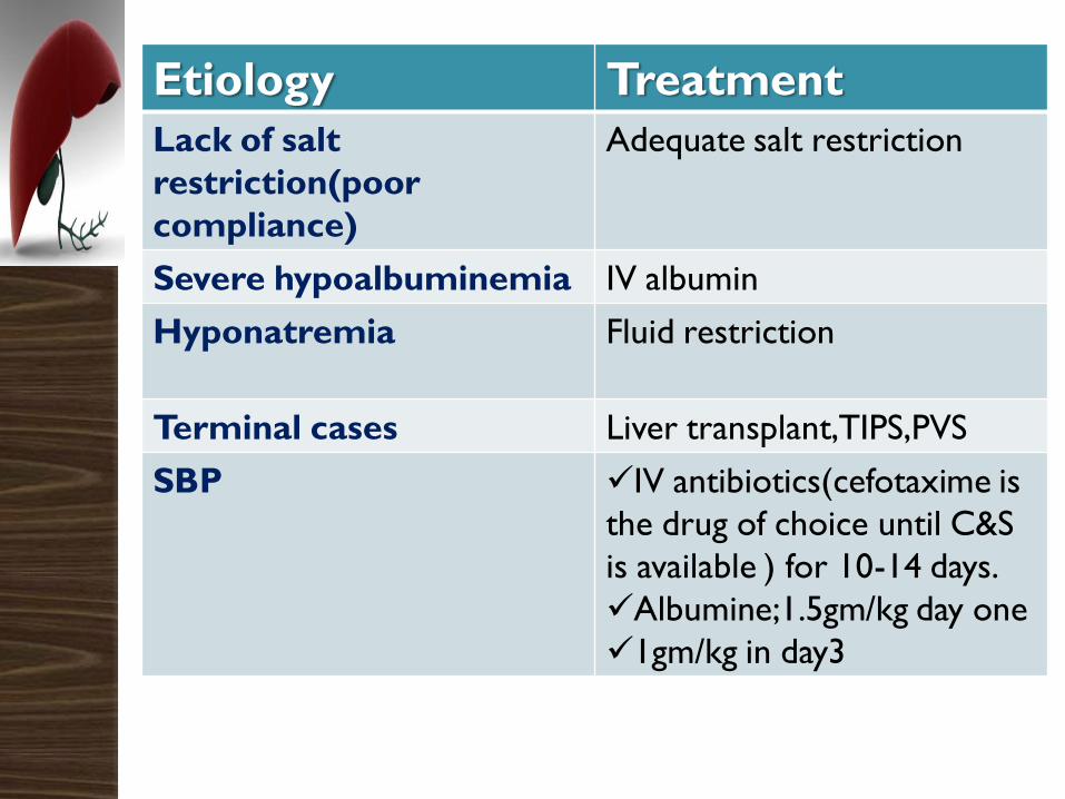
Etiology Treatment
Lack of salt
restriction(poor
compliance)
Adequate salt restriction
Severe hypoalbuminemia IV albumin
Hyponatremia Fluid restriction
Terminal cases Liver transplant,TIPS,PVS
SBP IV antibiotics(cefotaxime is
the drug of choice until C&S
is available ) for 10-14 days.
Albumine;1.5gm/kg day one
1gm/kg in day3
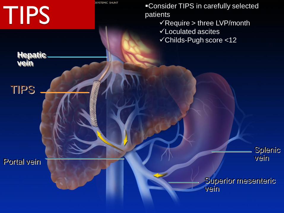
Hepatic vein
Portal vein
Splenicvein
Superior mesenteric vein
TIPS
THE TRANSJUGULAR INTRAHEPATIC PORTOSYSTEMIC SHUNT
TIPSConsider TIPS in carefully selected
patients
Require > three LVP/month
Loculated ascites
Childs-Pugh score <12
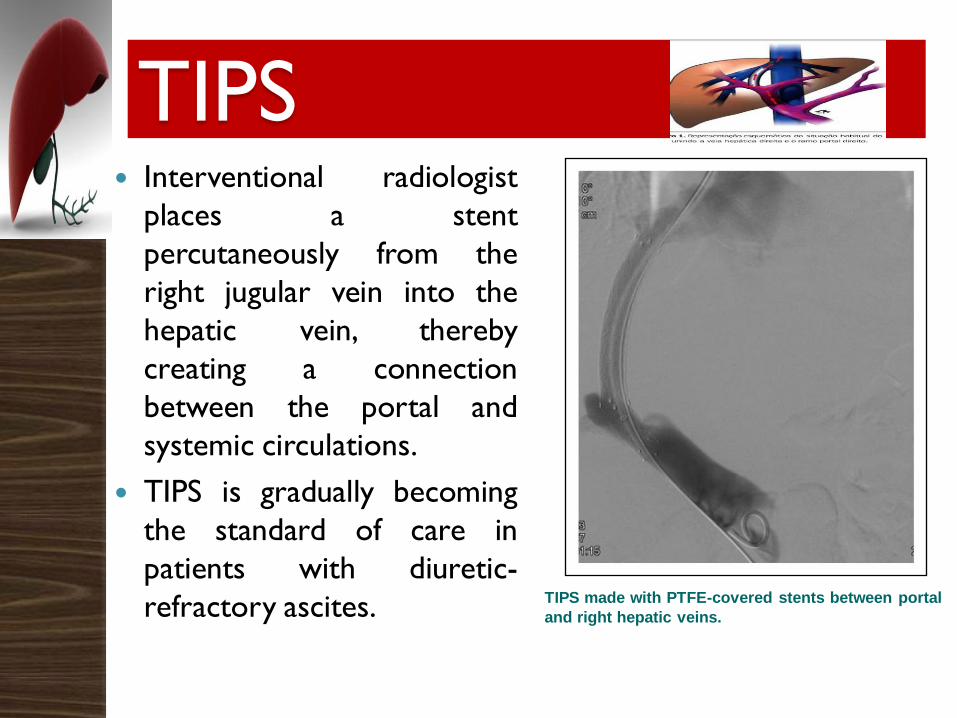
TIPS Interventional radiologist
places a stent
percutaneously from the
right jugular vein into the
hepatic vein, thereby
creating a connection
between the portal and
systemic circulations.
TIPS is gradually becoming
the standard of care in
patients with diuretic-
refractory ascites. TIPS made with PTFE-covered stents between portal
and right hepatic veins.
Benefits:
◦ Reduce portal pressure
◦ Rapid reduction or elimination of ascites
◦ Reduction or cessation of diuretic therapy
◦ Improve renal function
◦ Improve nutritional status
TIPS
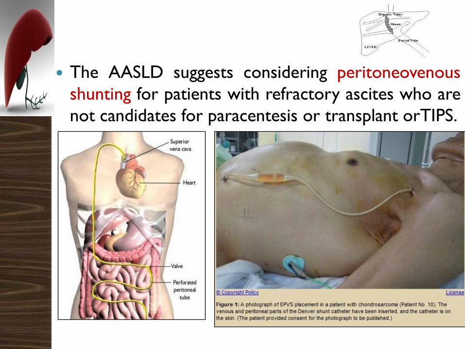
The AASLD suggests considering peritoneovenous
shunting for patients with refractory ascites who are
not candidates for paracentesis or transplant orTIPS.
Recommendations
TIPS should be considered in patients with very
frequent requirement of large-volume
paracentesis, or in those in whom paracentesis
is ineffective (e.g. due to the presence of
loculated ascites) (Level B1).
Recommendations
Resolution of ascites after TIPS is slow and
most patients require continued administration
of diuretics and salt restriction (Level B1).
Recommendations
TIPS cannot be recommended in patients with
1. Severe liver failure (serum bilirubin >5 mg/dl,
INR >2 or Child-Pugh score >11,
2. Current hepatic encephalopathy or chronic
hepatic encephalopathy), concomitant active
infection,
3. Progressive renal failure,
4. Severe cardiopulmonary diseases
(Level B1).
Recommendations
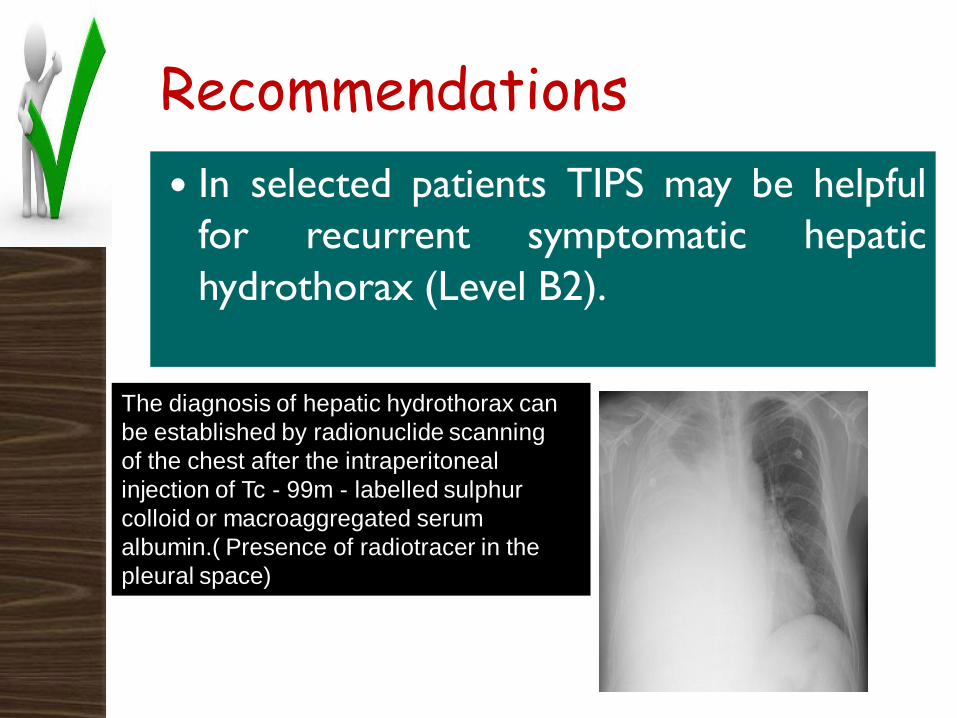
In selected patients TIPS may be helpful
for recurrent symptomatic hepatic
hydrothorax (Level B2).
The diagnosis of hepatic hydrothorax can
be established by radionuclide scanning
of the chest after the intraperitoneal
injection of Tc - 99m - labelled sulphur
colloid or macroaggregated serum
albumin.( Presence of radiotracer in the
pleural space)
Drugs
contraindicated in
patients with
ascites
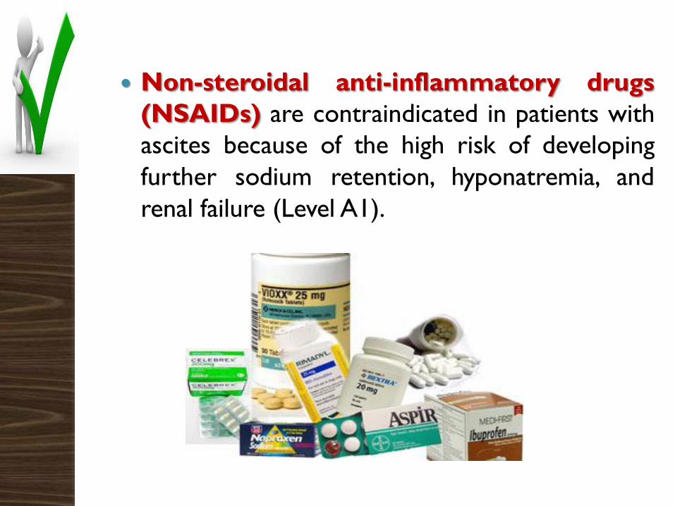
Non-steroidal anti-inflammatory drugs
(NSAIDs) are contraindicated in patients with
ascites because of the high risk of developing
further sodium retention, hyponatremia, and
renal failure (Level A1).
Preliminary data show that short-term
administration of selective inhibitors of
cyclooxygenase-2 does not impair renal
function and the response to diuretics.
However, further studies are needed to confirm
the safety of these drugs
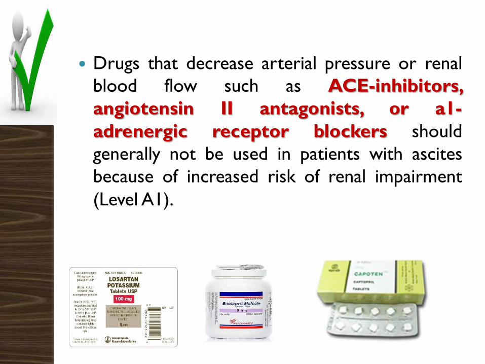
Drugs that decrease arterial pressure or renal
blood flow such as ACE-inhibitors,
angiotensin II antagonists, or a1-
adrenergic receptor blockers should
generally not be used in patients with ascites
because of increased risk of renal impairment
(Level A1).
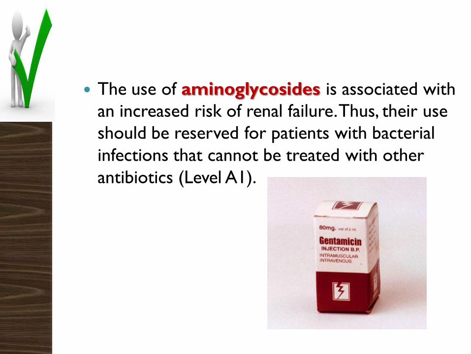
The use of aminoglycosides is associated with
an increased risk of renal failure. Thus, their use
should be reserved for patients with bacterial
infections that cannot be treated with other
antibiotics (Level A1).
In patients with ascites without renal failure, the
use of contrast media does not appear to be
associated with an increased risk of renal
impairment (Level B1).
Contrast media should be used with caution
and the use of general preventive measures of
renal impairment is recommended (Level C1).
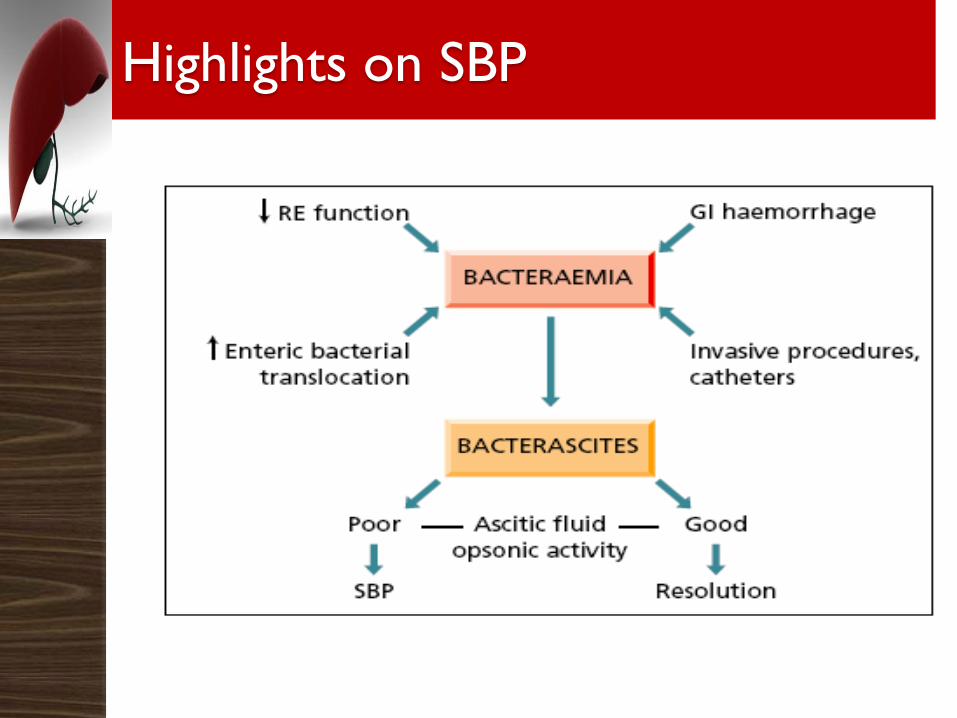
SBP is blood - borne and in 90% monomicrobial.
Bacteria of gut origin are the most commonly
isolated causative organisms.
Therefore, migration of enteric bacteria across
the intestinal mucosa to extraintestinal sites and
the systemic circulation (bacterial
translocation)
Highlights on SBP
Highlights on SBP
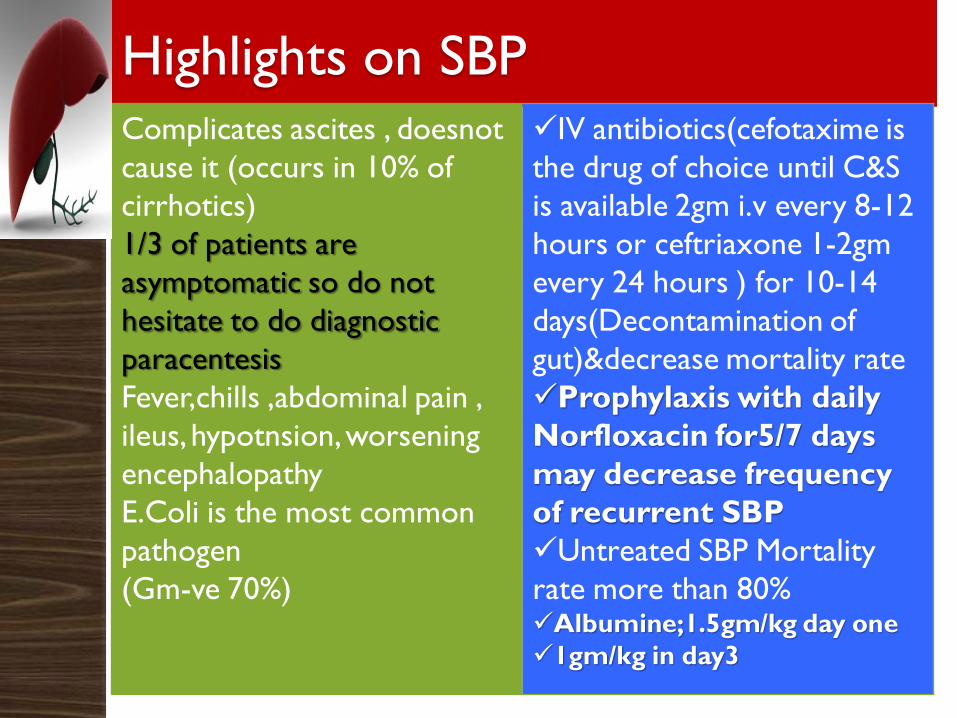
Highlights on SBPComplicates ascites , doesnot
cause it (occurs in 10% of
cirrhotics)
1/3 of patients are
asymptomatic so do not
hesitate to do diagnostic
paracentesis
Fever,chills ,abdominal pain ,
ileus, hypotnsion, worsening
encephalopathy
E.Coli is the most common
pathogen
(Gm-ve 70%)
IV antibiotics(cefotaxime is
the drug of choice until C&S
is available 2gm i.v every 8-12
hours or ceftriaxone 1-2gm
every 24 hours ) for 10-14
days(Decontamination of
gut)&decrease mortality rate
Prophylaxis with daily
Norfloxacin for5/7 days
may decrease frequency
of recurrent SBP
Untreated SBP Mortality
rate more than 80%Albumine;1.5gm/kg day one
1gm/kg in day3
Recommendations
Spontaneous bacterial pleural empyema
may complicate hepatic hydrothorax. The
diagnosis is based on positive pleural fluid
culture and increased neutrophil count of
>250/mm3 or negative pleural fluid culture and
>500 neutrophils/mm3 in the absence of
pneumonia (Level B1).
Recommendations
Patients with suspected secondary bacterial
peritonitis should undergo appropriate
radiological investigation such as CT scanning
(Level A1).
RecommendationsRecommendations. Empirical antibiotics should
be started immediately following the diagnosis
of SBP (Level A1).
Since the most common causative organisms of
SBP are Gram-negative aerobic bacteria, such as
E. coli, the first line antibiotic treatment are
third-generation cephalosporins (Level A1).
Recommendations
Alternative options include
amoxycillin/clavulanic acid and quinolones such
as ciprofloxacin or ofloxacin. However, the use
of quinolones should not be considered in
patients who are taking these drugs for
prophylaxis against SBP. (Level B1).

Recommendations
Resolution of SBP should be proven by
demonstrating a decrease of ascitic neutrophil
count to <250/mm3 and sterile cultures of
ascitic fluid, if positive at diagnosis (Level A1).
A second paracentesis after 48 h of start of
treatment may help guide the effect of antibiotic
therapy.
Recommendations
All patients who develop SBP should be
treated with broad spectrum antibiotics
and intravenous albumin (Level A2).
Recommendations Patients who recover from an episode of SBP
have a high risk of developing recurrent SBP.
In these patients, the administration of
prophylactic antibiotics reduces the risk of
recurrent SBP. Norfloxacin (400 mg/day, orally)
is the treatment of choice (Level A1).
Alternative antibiotics include ciprofloxacin
(750 mg once weekly, orally) or co-trimoxazole
(800 mg sulfamethoxazole and 160 mg
trimethoprim daily, orally), but evidence is not
as strong as that with norfloxacin (Level A2).
Recommendations
In patients with gastrointestinal bleeding and
severe liver disease ceftriaxone is the
prophylactic antibiotic of choice. (Level
A1).
Treatment initially is withdrawal of
diuretics and nephrotoxins, followed by
saline and/or albumin infusion.
Vasoactive agents, octreotide, midodrine,
and vasopressin, as well as TIPS have been
used with some encouraging results in
largely uncontrolled studies and liver
transplantation is the only definitive cure
Take home message
Ascites is the most common decompensating
event in cirrhosis
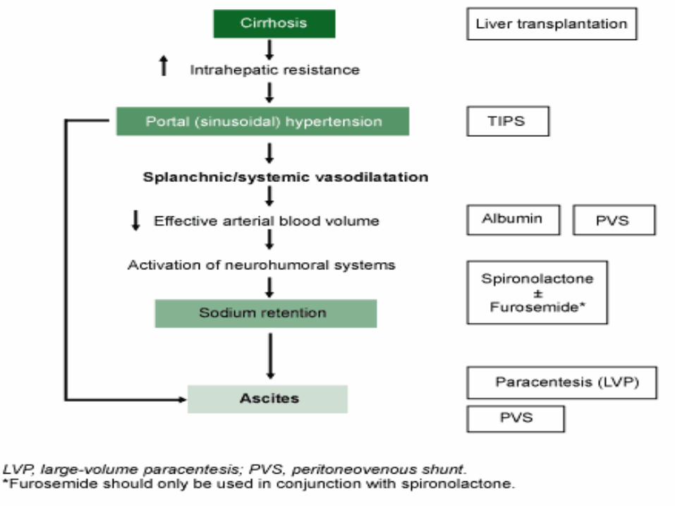
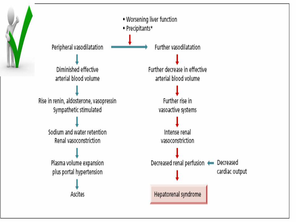
Its pathophysiology is mostly explained by
splanchnic and peripheral vasodilatation that
lead to a decrease in effective blood volume.
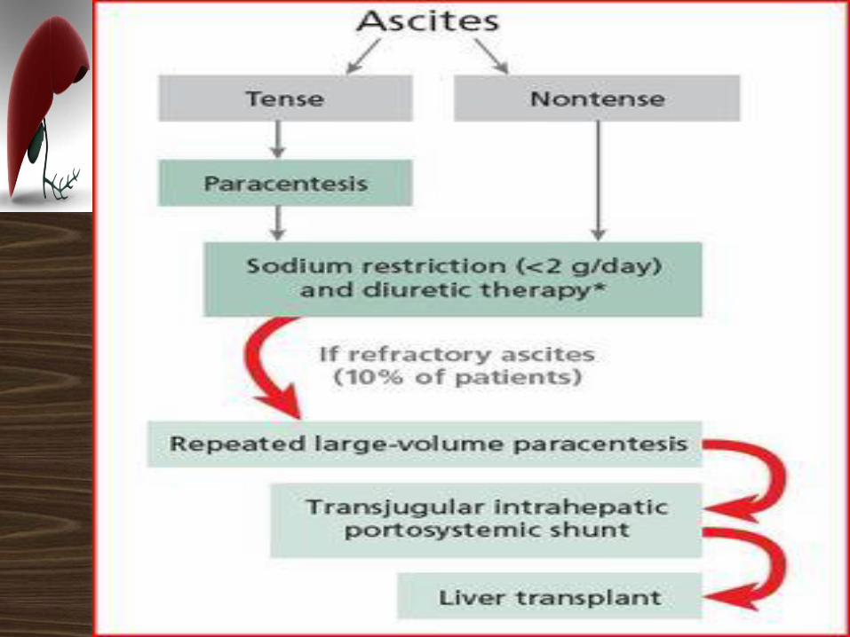
Most patients respond to diuretics. Patients
who no longer respond should be treated with
repeated large - volume paracenteses.
Transjugular intrahepatic portosystemic shunt
(TIPS) should be considered in those requiring
frequent paracenteses.
Take home message
Fluid restriction is recommended in patients
with hyponatraemia. Vasoconstrictors may
reverse hepatorenal syndrome and are useful as
a bridge to liver transplantation.
Take home message
Ascites per se is not lethal unless it becomes
infected (spontaneous bacterial peritonitis).
Infection often precipitates the hepatorenal
syndrome leading to death.
Antibiotic prophylaxis is indicated for secondary
prevention of spontaneous bacterial peritonitis
and in high - risk patients.
Take home message

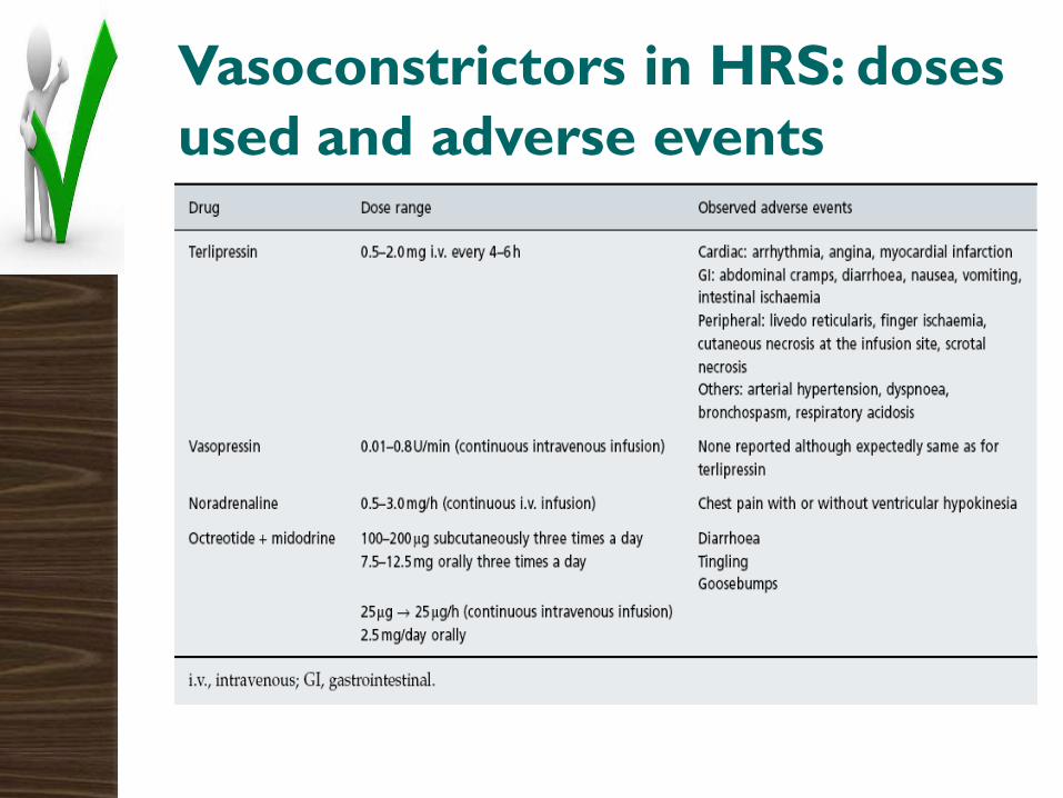
Vasoconstrictors in HRS: doses
used and adverse events