Embed Size (px)
Citation preview

CORONARY ANOMALYDr Siva Subramaniyan
PGIMER &Dr.RML Hospital
New Delhi

DEFINITION
• Defining what anatomy of the coronary arteries is normal can be challenging.
• Some normal features can be defined in numerical terms (for example, the number of coronary ostia), while in some other cases a more qualitative description is required.
• Angelini and coworkers proposed to classify “normal” as every feature with > 1% of frequency in an unselected general population
Angelini. Coronary Artery Anomalies. Circulation March 13, 2007

• According to this approach, a CAA can be defined as a coronary pattern or feature that is encountered in less than 1% of the general population.
coronary feature in two groups:
(1) normal coronary anatomy, defined as any morphological characteristics seen in > 1% of unselected sample. This group also includes normal anatomical variants, defined as alternative and relatively unusual morphological feature observed in > 1% of the population; and
(2) anomalous coronary anatomy, defined as morphological features found in < 1% of the population

IMPORTANCE
• Difficulty during cannulation
• Needs to know before going to any thoracic surgery
• Some anomaly can cause ischemia and prone for atherosclerosis
• More importantly some can cause SCD


NORMAL CORONARY
Villa ADM et al . Coronary artery anomalies. June 28, 2016|Volume 8|Issue 6|

LCA
• In the normal coronary anatomy, the right coronary artery originates from theright coronary sinus, and the left coronary artery trunk originates from the leftcoronary sinus.
• It posteriorly crosses the pulmonary trunk, bifurcating into anterior descendingand circumflex arteries.
• In approximately 37% of the individuals, there is a trifurcation of the leftcoronary trunk into anterior descending, circumflex and diagonal coronaryarteries or intermediate branch, which irrigates the free lateral wall of the leftventricle

• The anterior descending artery travels in the interventricular groove and gives off diagonal branches towards the anterolateral wall of the left ventricle.
• The circumflex artery travels in the left atrioventricular groove and varies both in size and extent, depending upon the coronary dominance. It gives off one to three marginal branches supplying the free wall of the left ventricle

RCA
• The right coronary artery travels down the right atrioventricular groove. In 50% of individuals, its first branch is the conus branch that supplies the right ventricle outflow tract.
• the second branch is the sinoatrial node branch that supplies the sinoatrial node and the right atrium.
• In 38% of cases, such a branch originates from the left coronary artery, and in 3%, from both arteries

• Also, there are branches towards the free wall of the right ventricle, and the branch located in the junction between medial and distal thirds of the right coronary artery is named obtuse marginal artery.
• In approximately 85% of the individuals, the right coronary artery crosses the crux cordis and gives the
- posterior descending branch (right dominant coronary supply),
- in 7% to 8% the circumflex artery gives PDA (left dominant coronary
supply), and
- in 7% to 8% of the cases the posterior interventricular septum is irrigated by branches of the right coronary and circumflex arteries (codominance)

INCIDENCE

CLASSIFICATION
Coronary anomalies may be classified according Angelini et al. as follows:
1) anomalies of origination and course;
2) intrinsic anomalies;
3) termination anomalies.


Angelini. Coronary Artery Anomalies. Circulation March 13, 2007


J.M. Pe´rez-Pomares et al. Congenital coronary artery anomalies (2016) 109, 204–216

functional classification


• Another classification divides coronary anomalies into hemodynamicallysignificant and non-hemodynamically significant.
• Anomalies classified as hemodynamically significant include:
1) anomalies of origination with interarterial course
2) anomalous origin in the pulmonary artery;
3) atresias;
4) congenital fistulas
Villa ADM et al . Coronary artery anomalies. June 28, 2016|Volume 8|Issue 6|

classification based on ischemia

Anomalies of origination and course
Absent left main trunk (split origination of LCA)
• LAD and Cx originates from LCS without common trunk
• Occurs in about 1%
• More frequent with BAV, left dominance
• No clinical consequences
• Coronary ostia are smaller
J.M. Pe´rez-Pomares et al. Congenital coronary artery anomalies (2016) 109, 204–216

CANNULATION
• Inject more dye so that Lcx also can be visualized
• If LAD is cannulated it is bulled back into aorta and clockwise torque is given to cannulate Lcx
• Use JL4 for LAD and longer curve for Lcx
• JL catheter for selective cannulation of LAD and amplatz left catheter for LCX


Anomalous location of coronary ostium within aortic root or near proper aortic sinus of Valsalva
a. High
b. Low
c. Commissural
• It is defined as origin of a coronary artery or left coronary trunk more than 1 cm above the sinotubular junction.
• It usually does not present with clinical problem, however the preoperativeidentification of this anomaly is important in case of ascending aorta surgery andmay cause difficulties in catheterization during angiography.
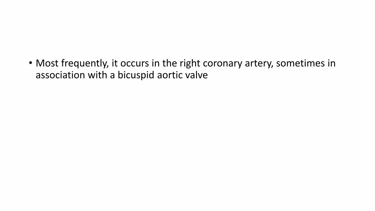
• Most frequently, it occurs in the right coronary artery, sometimes in association with a bicuspid aortic valve

CANNULATION
• High origin of LCA engaged with Amplatz left, MP catheter
• High origin of RCA engaged with Amplatz catheter

ALCAPA
ALCAPA
• LCA arises from PA usually from left posterior facing sinus
• Fetus-both coronary arteries receive forward flow
• Early after birth - Anterolateral infarct and slight retrograde flow from LCA to PA
• 15% of patients-myocardial blood flow can sustain myocardial function at rest or even during exercise
• Adult-Enlarged RCA and collaterals and significant retrograde flow into PA
J.M. Pe´rez-Pomares et al. Congenital coronary artery anomalies (2016) 109, 204–216

Clinical features
• Paroxysmal attacks of acute discomfort precipitated by feeding
• CHF at 2 to 3 month
• Physical examination-CHF,MR
• Abnormal Q waves in leads I, aVL, and precordial leads V4 to V6
• Older children and adults -may be asymptomatic or have dyspnea, syncope, or exertional angina
• Sudden death after exertion

Echo
• Abnormal origin of LCA
• Flow passes from RCA into PA
• Enlarged RCA
• RWMA and mitral regurgitation

Aortic root angiography
• Dilated RCA
• Absence of left coronary osteum
• large collaterals-filling of LAD and Lcx
• Later MPA and LMCA filled by LAD and LCX branch


Treatment
• Ligation of LCA at its origin
• Direct reimplantation of origin of LCA into aorta (with a button of PA around the origin)
• Ligation of origin of LCA and reconstitution of flow through it with subclavianarterial or SVG
• Takeuchi procedure- aortopulmonary window is created and a tunnel fashioned that directs blood from aorta to LCA

ACC/AHA 2008 Guidelines for Adults With CHD. December 2, 2008:e143–263

other artery
Anomalous Origination of RCA, LAD or Cx Artery From PA
• Benign condition
• Typically recognized by atypical angina, systolic heart murmur , abnormal stress test or angiography
• In absence of major clinical manifestations not an indication for surgery

ACAOS

Anomalous CA ostium location at improper aortic sinus–wrong sinus• RCA to left sinus
• LCA to right sinus
• LCX to RCA/ or sinus
• LAD to RCA/or sinus
• RCA or LCA to posterior sinus
with anomalous course: interaterial, prepulmonic, intraseptal, retroaortic, posterior atrioventricular groove or retrocardiac, postero-anterior interventricular groove
J.M. Pe´rez-Pomares et al. Congenital coronary artery anomalies (2016) 109, 204–216

Abnormal crossing pathways
Angelini et al
• Retrocardiac
• Path is behind mitral and tricuspid valves in posterior AV groove
Retrocardiac

• Retroaortic• Most common• Specifically involve
origination of Cx artery from right sinus of Valsalva
• Incidence in general population range from 0.1 to 0.9%
• No clinical consequences

• Intraseptal(supracristal)
• Located in upper anterior IVS(Derived from conal septum)
• Recogonised by systolic phasic narrowing during CAG

• Prepulmonary(precardiac)
• Common in TOF

INTRAMUARAL

ACAOS
• Anomalous origination of coronary artery from the opposite sinus (ACAOS) is anuncommon coronary anomaly. Its incidence is reported to be around 1.07% .
• It comprises anomaly of right coronary artery originated from left sinus (rightACAOS) and its opposite or left ACAOS.

Right coronary originating from the left coronary sinus
• Right coronary artery originating from the left coronary sinus or as a branch of a single coronary artery is found in 0.03% to 0.17% of the individuals submitted to angiography.
• The most common proximal pathway of the right coronary in such cases is interarterial, and can be associated with sudden cardiac death in up to 30% of patients

• Both right and left ACAOS have significant clinical consequence if the ectopic artery has an interarterial course or intramural intussusception
• A constant relationship is observed between left ACAOS and sudden death or ischemia during extreme exercise .
• Right ACAOS with an interarterial course is a type of ACAOS which poses high risk for myocardial ischemia or sudden death as well

The clinical picture of ACAOS can be divided into two spectra:
• the first is sudden death in the young and after strenuous physical activity orsport and
• the second is atypical clinical picture .
• Most of ACAOS patients are asymptomatic. Atypical chest discomfort is the mostprevalent symptom urging patients to refer to the health facility and to performthe coronary angiography to detect ACAOS

SUDDEN DEATH
• Hard activity causes dilatation of aortic root and pulmonary trunk whichcompresses slit-like ostium or particular segment of ectopic coronary artery. Thisoccurs especially in individuals with sufficient aortic distensibility, such as inyoung people or sportsmen.

Anomalies of origination and course
Sudden cardiac death associated with four risk factors
• Slit-like coronary orifice• Acute angle of take-off from
aorta• Presence of aortic
intramural coronary arteries • Inter-arterial course
between aorta and PA

Left coronary trunk originating from the right coronary sinus• Left coronary trunk originating from the right coronary sinus or as a branch of a
single coronary artery occurs in 0.09% to 0.11% of the individuals submitted toangiography .
• Proximal interarterial course occurs in 75% of such patients

Anterior descending or circumflex arteries originating from the right coronary sinus
• circumflex artery is the one that most commonly presents anomalous origin,occurring in 0.32% to 0.67% of the population. Retroaortic pathway is its mostcommon course, and there is no association with sudden death.
• The anterior descending artery with anomalous origin rarely occurs in individualswith a normal cardiac anatomy. It is generally associated with Fallot’s tetralogy,complex transposition and double right ventricular output tract

INTRAMURAL VS INTRA SEPTAL


ACC/AHA 2008 Guidelines for Adults With CHD. December 2, 2008:e143–263

Surgical and Catheterization-Based Intervention• Both surgical revascularization (eg, marsupialization, coronary bypass, or
coronary reimplantation) and limited cases of transcatheter stenting have beenreported to have short-term stability, without long-term follow-up
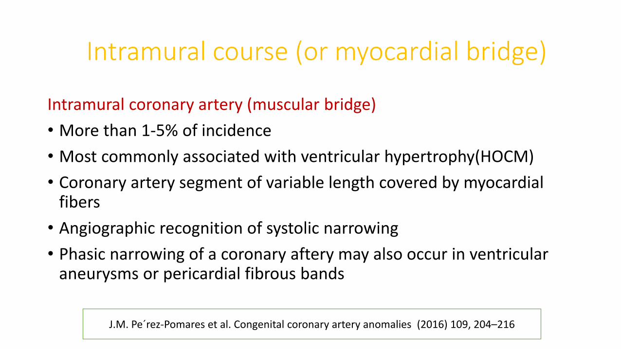
Intramural course (or myocardial bridge)
Intramural coronary artery (muscular bridge)
• More than 1-5% of incidence
• Most commonly associated with ventricular hypertrophy(HOCM)
• Coronary artery segment of variable length covered by myocardial fibers
• Angiographic recognition of systolic narrowing
• Phasic narrowing of a coronary aftery may also occur in ventricular aneurysms or pericardial fibrous bands
J.M. Pe´rez-Pomares et al. Congenital coronary artery anomalies (2016) 109, 204–216

• U sign- Artery's accentuated descent from its subepicaardial location
• Most commonly involve proximal LAD
• Systolic stenosis is unlikely to cause coronary flow reduction
• Rare reports of spasm, thrombus and atherosclerotic change


• By intravascular ultrasound clinically significant myocardial bridges are characterized by
- phasic systolic vessel compression,
- persistent reduction in diastolic lumen,
- increased blood flow velocities,
- retrograde systolic flow, and
- decreased coronary flow reserve.
management- surgical de-bridging and even stent implantation have been successfully carried out in symptomatic cases

CORONARY FISTULAS
• A sizable communication between a coronary artery and a cardiac cavity or any segment of systemic or pulmonary circulation
J.M. Pe´rez-Pomares et al. Congenital coronary artery anomalies (2016) 109, 204–216

• Fistulas from RCA, LCA, or infundibular artery to RV,RA,CS , SVC, PA, PV,LA, LV, Multiple(right+left ventricles)
• Originate from left coronary artery system (50-60%), right coronary artery system (30-40%), or both (2-5%)
• Most fistulas (90%) drain into right heart
• The haemodynamic consequences of CA fistulae depend mainly on the resistance (which is determined by fistula size, tortuosity, and length) and on the site of drainage.


Complications• Aneurysm formation• Intimal ulceration• Medial degeneration• Intimal rupture• Atherosclerotic deposition• Calcification• Side branch (nutrient) obstruction• Mural thrombosis• Coronary rupture

SURGERY
• Surgical fistula closure can be successful if CAVF is well defined and clear surgicalaccess is believed to be technically achievable.
• Recurrence may be a problem if anatomic definition is suboptimal, and surgerymay be difficult to perform owing to poorly visualized, typically distal fistulousconnections.
• Surgical closure of audible CAVF with appropriate anatomy is recommended in alllarge CAVFs and in small to moderate CAVFs in the presence of symptoms ofmyocardial ischemia, threatening arrhythmia, unexplained ventriculardysfunction, or left atrial hypertension

Catheterization-Based Intervention
• Numerous reports of transcatheter closure with coils or detachable devices describe near or complete CAVF occlusion in attempted closure procedures .
• Criteria for transcatheter closure of CAVF are similar to those used for surgical closure of CAVF.
• Transcatheter closure of CAVF should be performed only in centers with particular expertise in such intervention.

ACC/AHA 2008 Guidelines for Adults With CHD. December 2, 2008:e143–263

anomaly in congenital heart disease

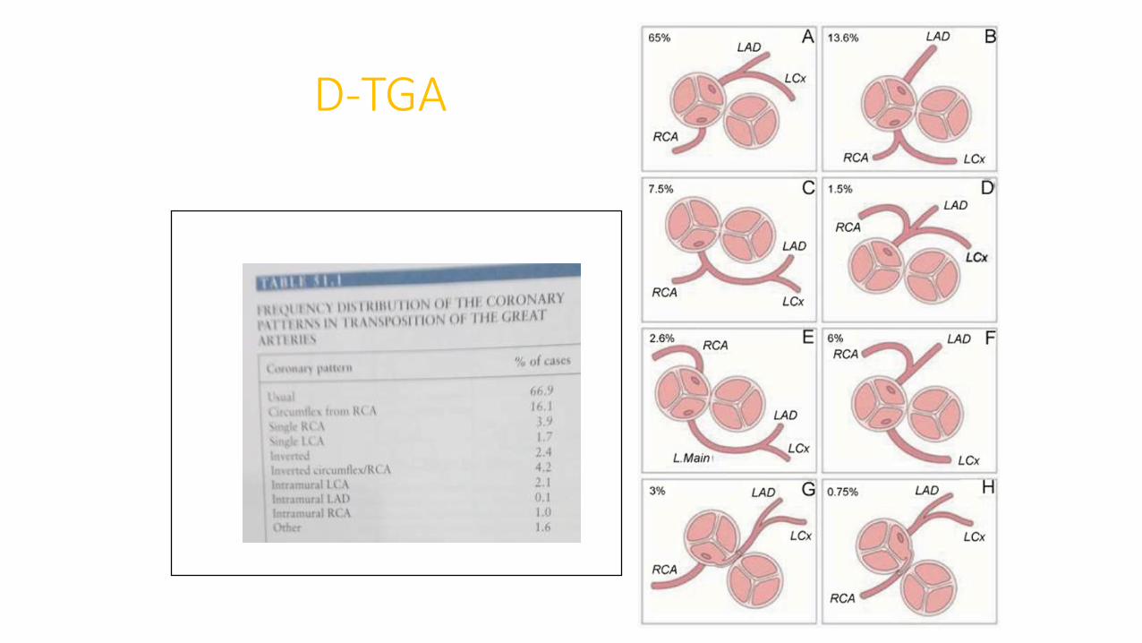
D-TGA
• Important to identify before switch Sx
• Facing sinuses -Sinuses adjacent to PA
• Nomenclature of facing sinuses depends on relationship of great vessels
• VSD or side-by-side GVs more associated with coronary anomalies
• Almost all coronaries arise from facing sinuses
• In 60% coronary arteries come from appropriate sinuses and branch normally
• Seen often with aorta anterior and to right of PA
D-TGA
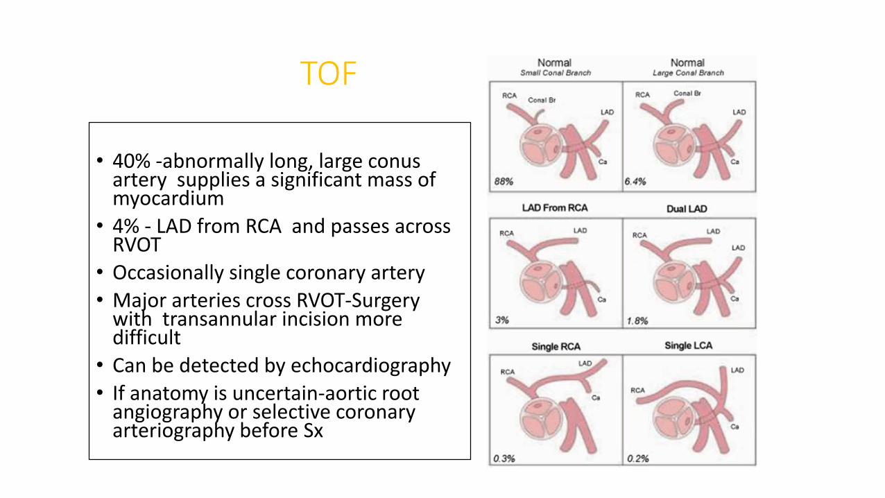
TOF
• 40% -abnormally long, large conus artery supplies a significant mass of myocardium
• 4% - LAD from RCA and passes across RVOT
• Occasionally single coronary artery
• Major arteries cross RVOT-Surgery with transannular incision more difficult
• Can be detected by echocardiography
• If anatomy is uncertain-aortic root angiography or selective coronary arteriography before Sx

CONCLUSION
• Coronary anomaly common, but rarely causes symptoms
• Most of them detected during angiography and will have difficulty in cannulation
• Atypical chest pain or TMT positive in athletes needs attention and evaluation
• It can cause SCD