Embed Size (px)
Citation preview
Title
Copyright © The McGraw-Hill Companies, Inc. Permission required for reproduction or display.
Chapter 3: AntigenCapture and
Presentation toLymphocytes
Dr. Hafez Sumairi
Learning outcomes
1.How do the rare lymphocytes specific for anymicrobial antigen find that microbe, especiallyconsidering that microbes may enter anywhere inthe body?
2.How does the immune system produce the effectorcells and molecules best able to eradicate aparticular type of infection, such as antibodies thatbind to extracellular microbes and CTLs that killinfected cells harboring microbes in theircytoplasm?
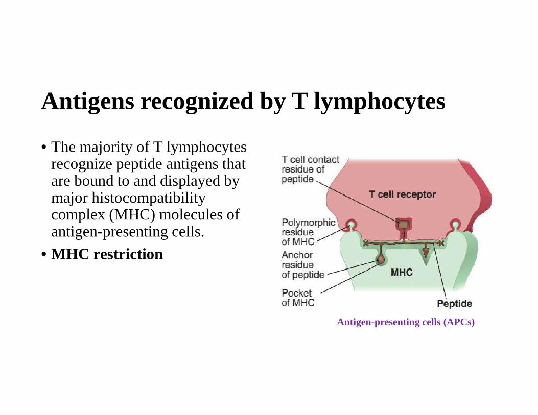
Antigens recognized by T lymphocytes
• The majority of T lymphocytesrecognize peptide antigens thatare bound to and displayed bymajor histocompatibilitycomplex (MHC) molecules ofantigen-presenting cells.
• MHC restriction
Antigen-presenting cells (APCs)
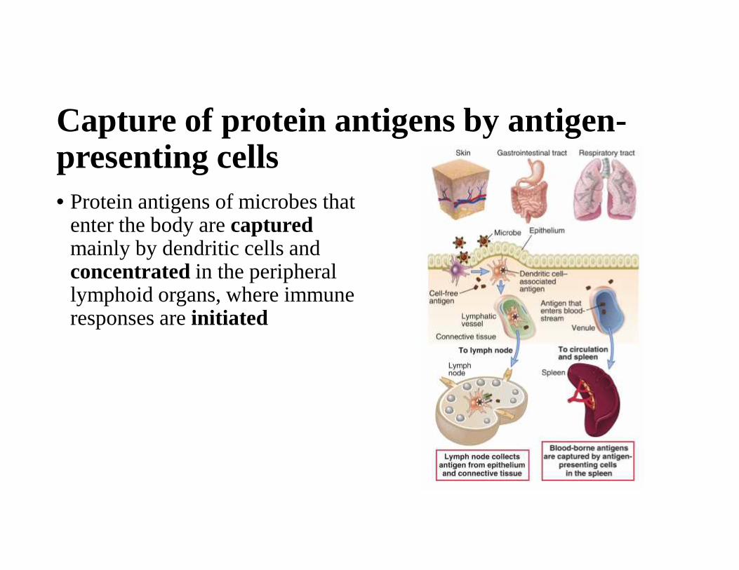
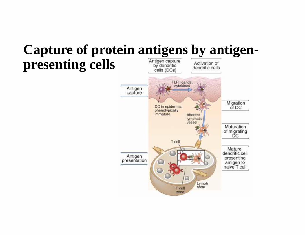
Capture of protein antigens by antigen-presenting cells• Protein antigens of microbes that
enter the body are capturedmainly by dendritic cells andconcentrated in the peripherallymphoid organs, where immuneresponses are initiated
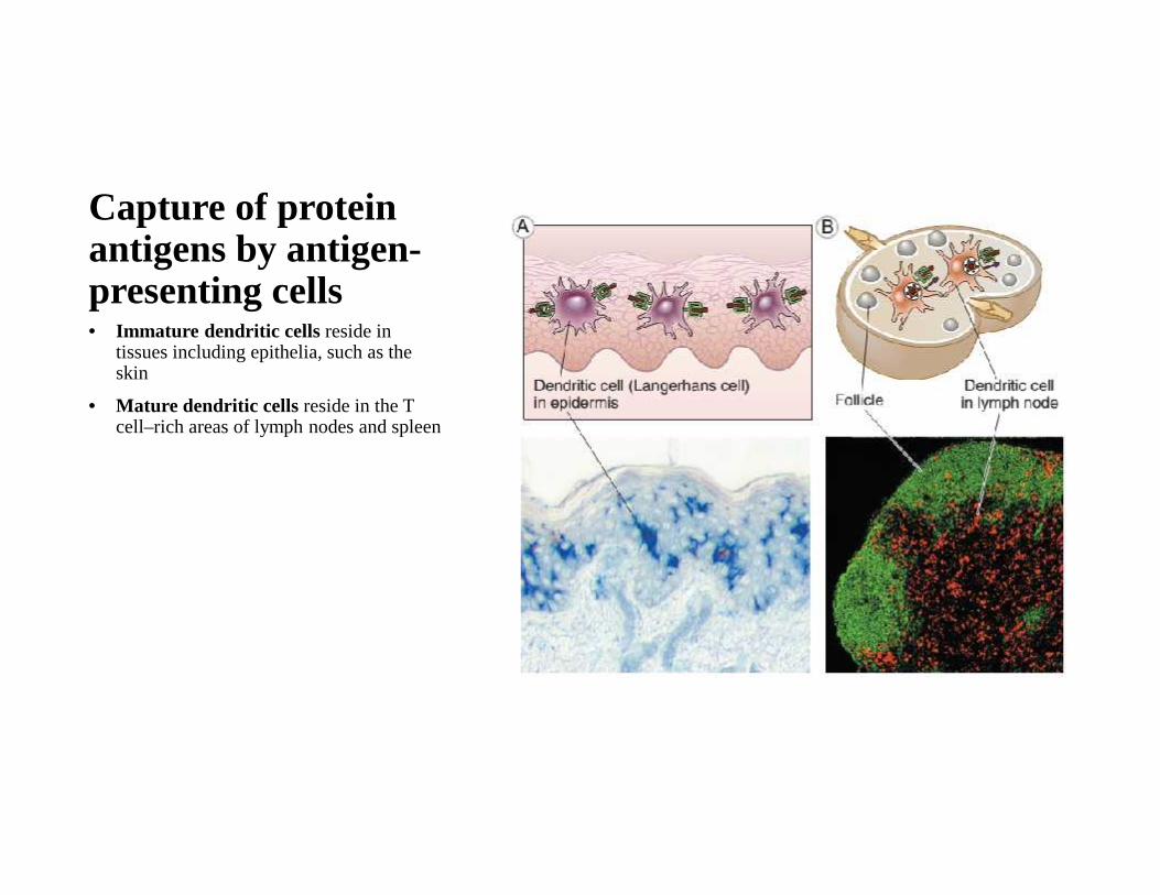
Capture of proteinantigens by antigen-presenting cells• Immature dendritic cells reside in
tissues including epithelia, such as theskin
• Mature dendritic cells reside in the Tcell–rich areas of lymph nodes and spleen

Capture of protein antigens by antigen-presenting cells
Capture of protein antigens by antigen-presenting cells
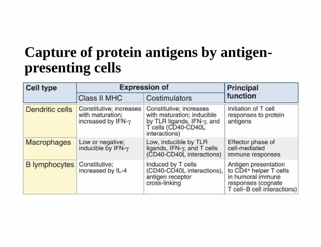
Capture of protein antigens by antigen-presenting cells
Now that we know how proteinantigens are captured, transportedto, and concentrated in peripherallymphoid organs, we next ask, howare these antigens displayed to Tlymphocytes?
Structure and function of majorhistocompatibility complex (MHC) molecules• MHC molecules are membrane proteins on APCs that display peptide
antigens for recognition by T lymphocytes
• Human MHC proteins are called human leukocyte antigens (HLAs)

Genes of the major histocompatibilitycomplex (MHC) locus
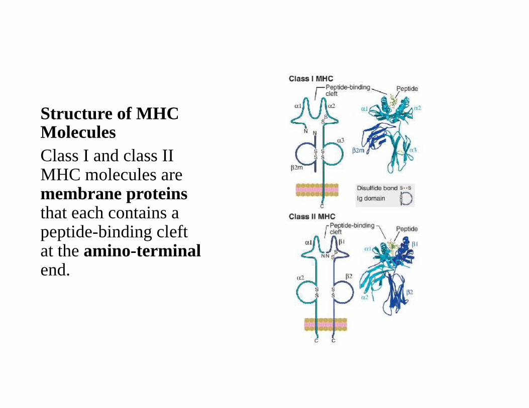
Structure of MHCMoleculesClass I and class IIMHC molecules aremembrane proteinsthat each contains apeptide-binding cleftat the amino-terminalend.
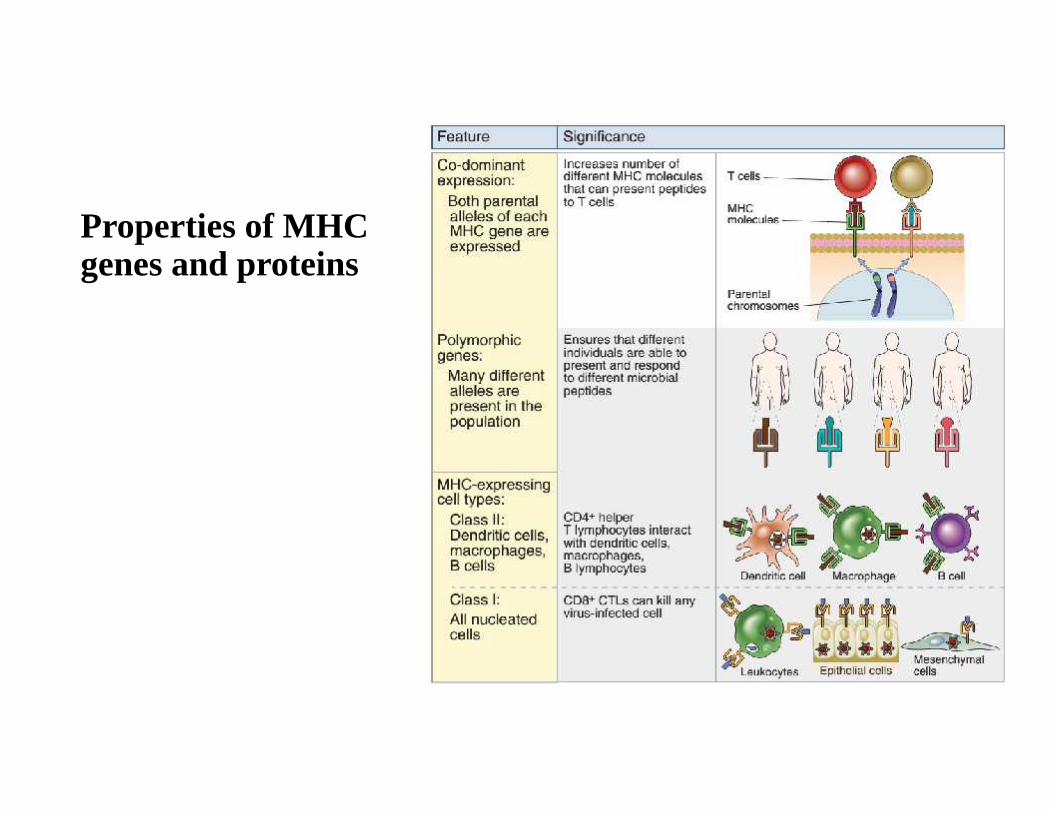
Properties of MHCgenes and proteins
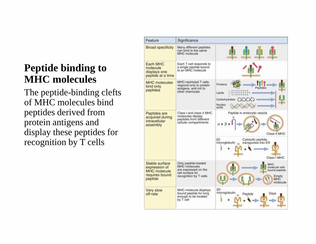
Peptide binding toMHC moleculesThe peptide-binding cleftsof MHC molecules bindpeptides derived fromprotein antigens anddisplay these peptides forrecognition by T cells
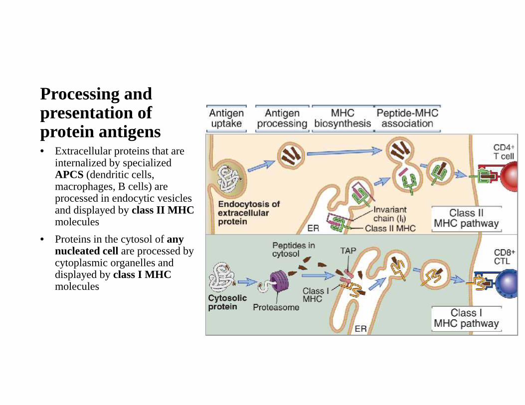
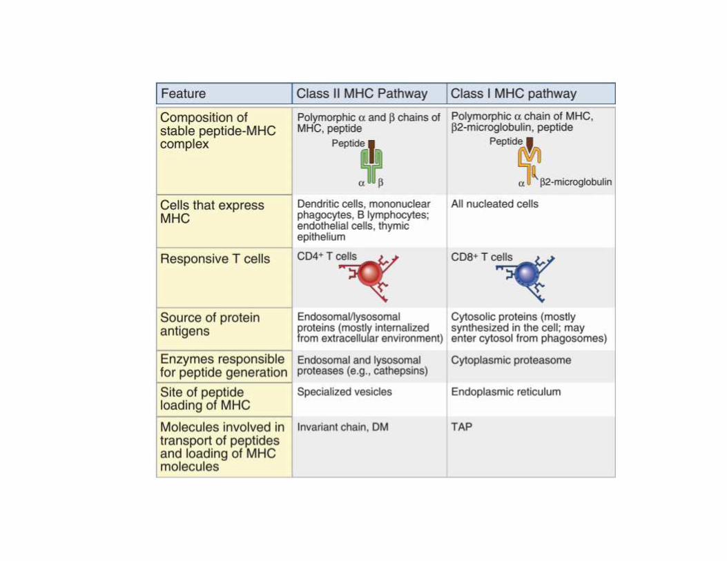
Processing andpresentation ofprotein antigens• Extracellular proteins that are
internalized by specializedAPCS (dendritic cells,macrophages, B cells) areprocessed in endocytic vesiclesand displayed by class II MHCmolecules
• Proteins in the cytosol of anynucleated cell are processed bycytoplasmic organelles anddisplayed by class I MHCmolecules

Processing ofinternalized antigensfor display by class IIMHC molecules
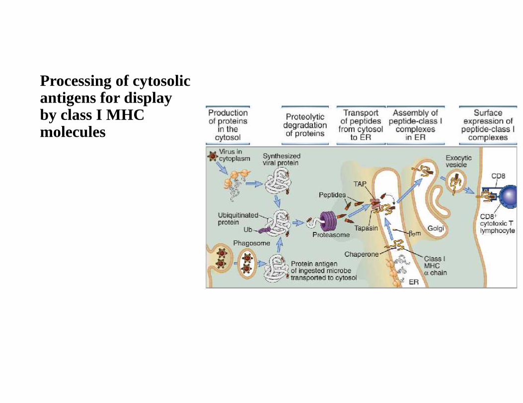
Processing of cytosolicantigens for displayby class I MHCmolecules
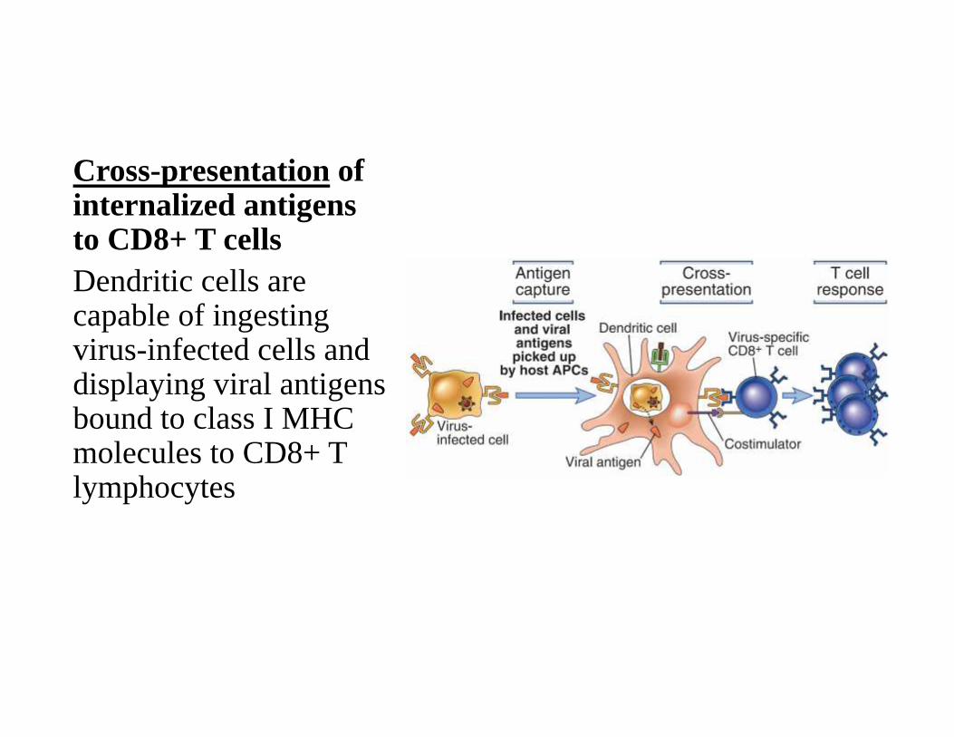
Cross-presentation ofinternalized antigensto CD8+ T cellsDendritic cells arecapable of ingestingvirus-infected cells anddisplaying viral antigensbound to class I MHCmolecules to CD8+ Tlymphocytes
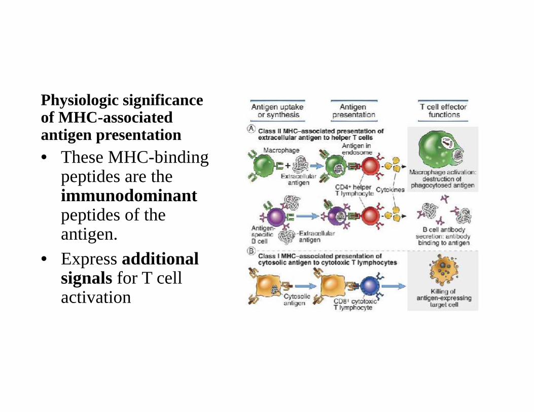
Physiologic significanceof MHC-associatedantigen presentation• These MHC-binding
peptides are theimmunodominantpeptides of theantigen.
• Express additionalsignals for T cellactivation
Antigens recognized by B cells and otherlymphocytes• B lymphocytes use membrane-bound antibodies
• Follicular dendritic cells (FDCs), whose function is to displayantigens to activated B cells
• Natural killer (NK) cells are specific for lipids displayed by class I–like CD1 molecules
• γδ T cells recognize a wide variety of molecules, some displayed byclass I–like molecules

Thank you