Embed Size (px)
Citation preview

BONEY TISSUES BONEY TISSUES HUMAN BODY 1 HB1 L3 HUMAN BODY 1 HB1 L3
by by Dr.Joseph Aziz Dr.Joseph Aziz
Asst. prof. of Anatomy& EmbryologyAsst. prof. of Anatomy& EmbryologyConsultant of Anatomy Consultant of Anatomy
1
BonesBonesBones of the skeleton are organs that
contain several different tissues.Bones are dominated by bone tissue but
also contain: ◦ Nervous tissue and nerves◦ Blood tissue and vessels◦ Cartilage in articular cartilages◦ Epithelial tissue lining the blood vessels
2
Function of Bones:Function of Bones:
Bones perform several important functions:◦Support◦Protection ◦Movement◦Mineral storage ◦Blood cell formation
3
Function of BonesFunction of Bones
SupportBones provide a hard
framework that supports the body
Bones provide support for internal organs
4
Function of BoneFunction of Bone
Protection
Fused bones provide a brain case that protects this vital tissue
Spinal cord is surrounded by vertebrae
Rib cage protects vital organs
5
Function of BoneFunction of Bone
Movement Skeletal muscle attached to bones use the bones as levers to move the body.
Arrangement of bones and joints determine the movements possible.
6
Function of BonesFunction of Bones
Mineral StorageBone serves as a
mineral reservoir.Phosphate and calcium
ions can be released into the blood steam.
7
Function of BonesFunction of Bones
Blood cell formation
In normal conditions: hematopoiesis occurs
within the marrow cavities of bones.
8
CLASSIFICATION OF CLASSIFICATION OF BONESBONES
9
Classification of Bones:Classification of Bones:I- according to shape:Bones vary in shape and size.The unique shape of each bone fulfills a particular need.Bones are classified by their shape as long, short, flat, or irregular bones.Bones differ in the distribution of compact and spongy osseous tissues.
10
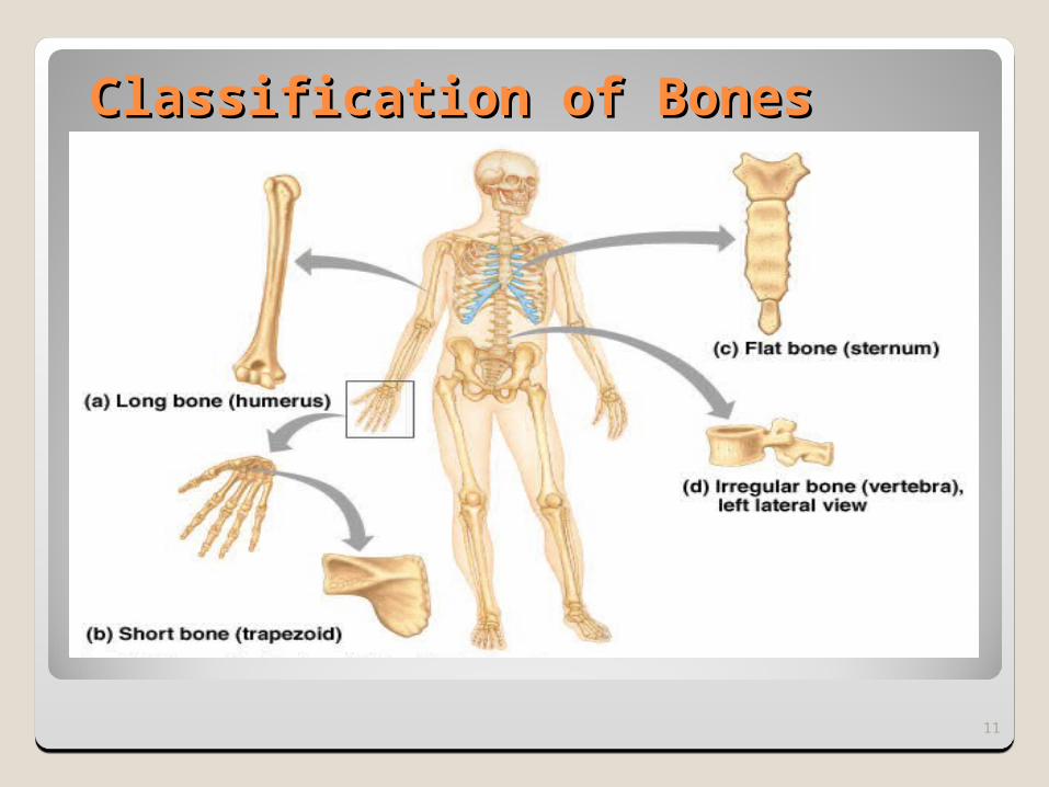
Classification of BonesClassification of Bones
11
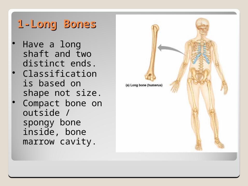
1-Long Bones1-Long Bones Have a long shaft
and two distinct ends.
Classification is based on shape not size.
Compact bone on outside / spongy bone inside, bone marrow cavity.
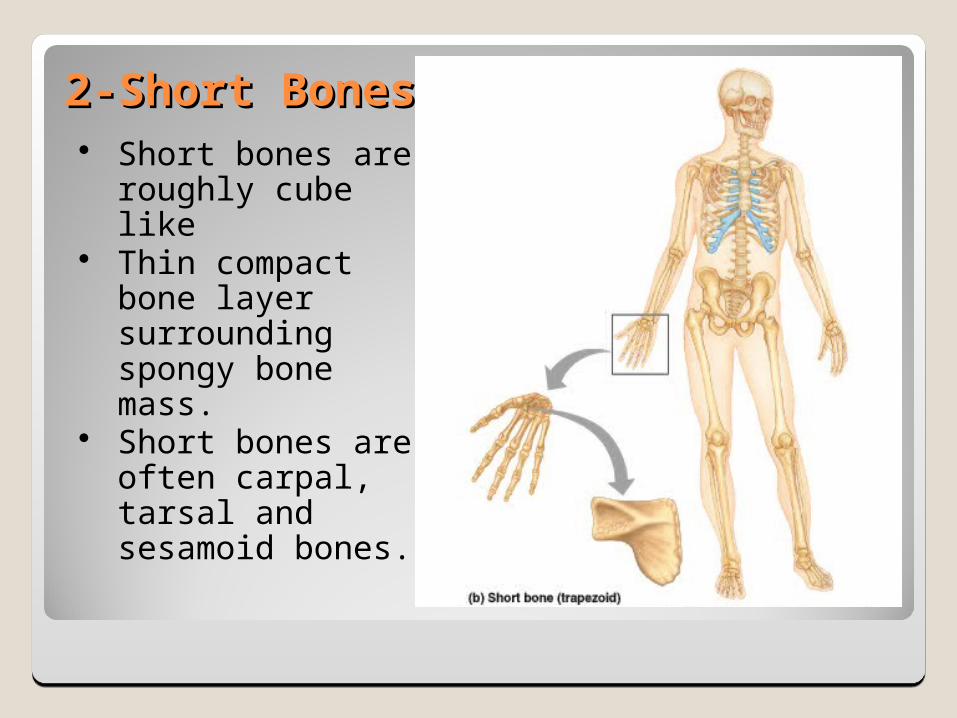
2-Short Bones2-Short Bones Short bones are
roughly cube like Thin compact
bone layer surrounding spongy bone mass.
Short bones are often carpal, tarsal and sesamoid bones.
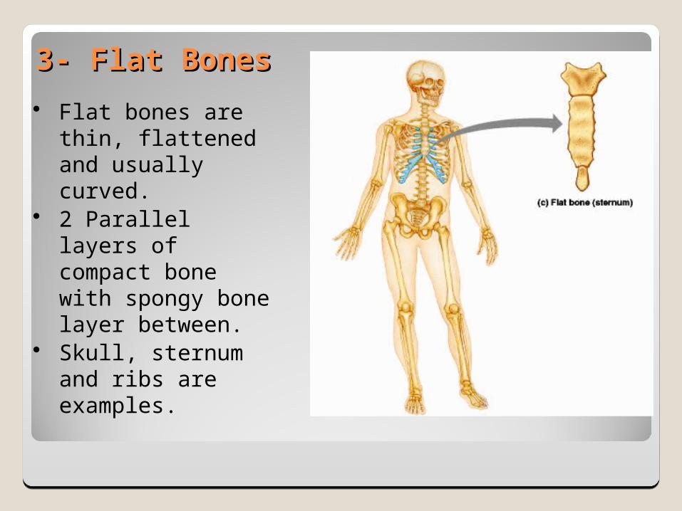
3- Flat Bones3- Flat Bones Flat bones are thin,
flattened and usually curved.
2 Parallel layers of compact bone with spongy bone layer between.
Skull, sternum and ribs are examples.
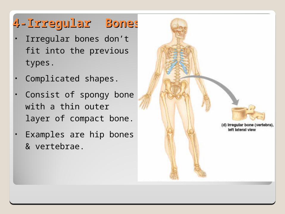
4-Irregular Bones4-Irregular Bones• Irregular bones don’t fit
into the previous types.• Complicated shapes.• Consist of spongy bone
with a thin outer layer of compact bone.
• Examples are hip bones & vertebrae.
II-Classification according to gross observation:1. Compact bone: Shows dense areas composed of solid lamellae without cavities. In case of compact long bones, bone marrow is contained in the medullary canal.2. Cancellous (spongy) bone: Shows numerous interconnecting cavities separated by bone trabeculae. Cavities of cancellous bone contain bone marrow.
16

Gross appearance of bone. Under the microscope, both compact bone and the trabeculae separating the cavities of cancellous bone have the same basic histological structure.
17
BONE STRUCTUREBONE STRUCTURE
18

Gross Gross AnatomyAnatomyLandmarks on
a typical long bone◦ Diaphysis ◦ Epiphysis◦ Membranes
Membranes◦ Periosteum◦ Endosteum
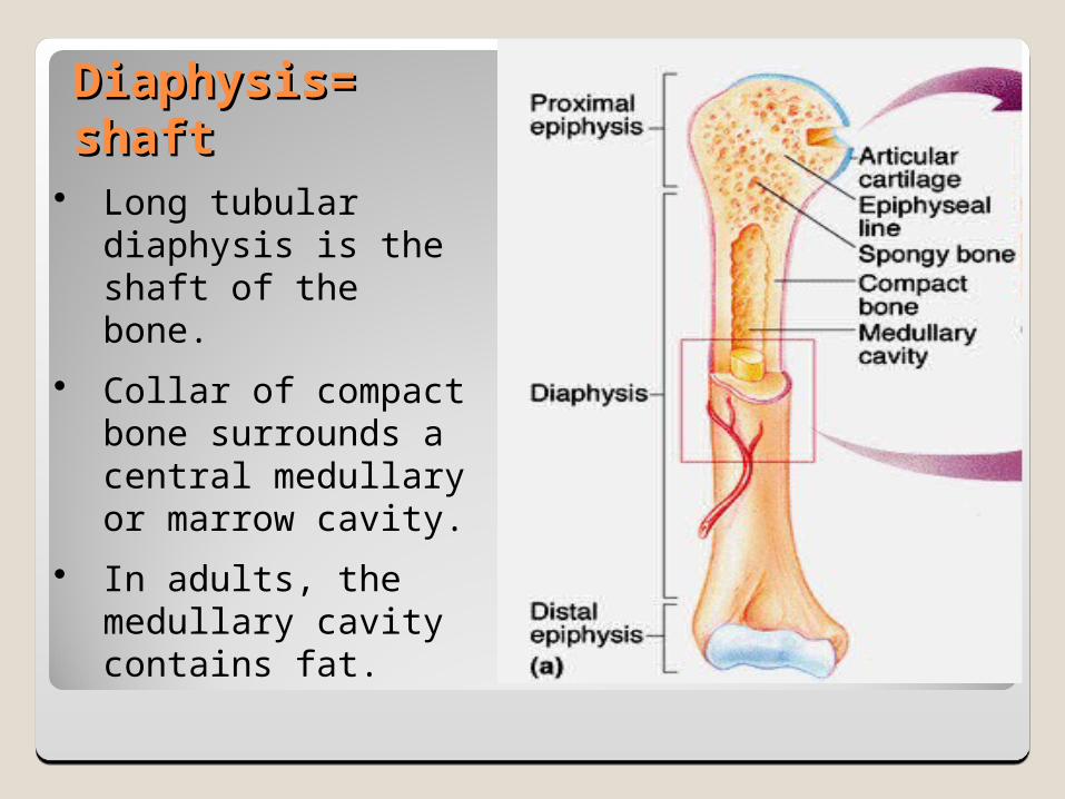
Diaphysis=Diaphysis=shaftshaft
Long tubular diaphysis is the shaft of the bone.
Collar of compact bone surrounds a central medullary or marrow cavity.
In adults, the medullary cavity contains fat.
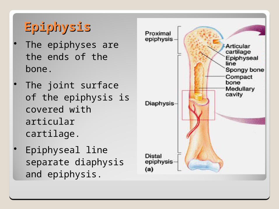
EpiphysisEpiphysis The epiphyses are
the ends of the bone.
The joint surface of the epiphysis is covered with articular cartilage.
Epiphyseal line separate diaphysis and epiphysis.
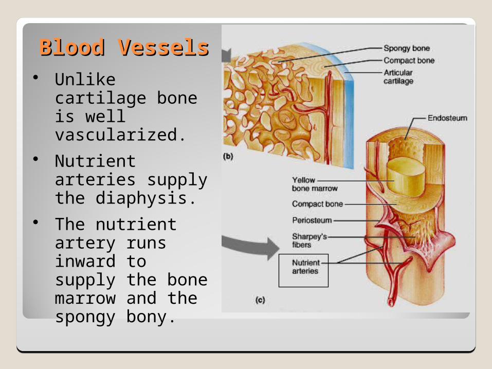
Blood VesselsBlood Vessels Unlike cartilage
bone is well vascularized.
Nutrient arteries supply the diaphysis.
The nutrient artery runs inward to supply the bone marrow and the spongy bony.
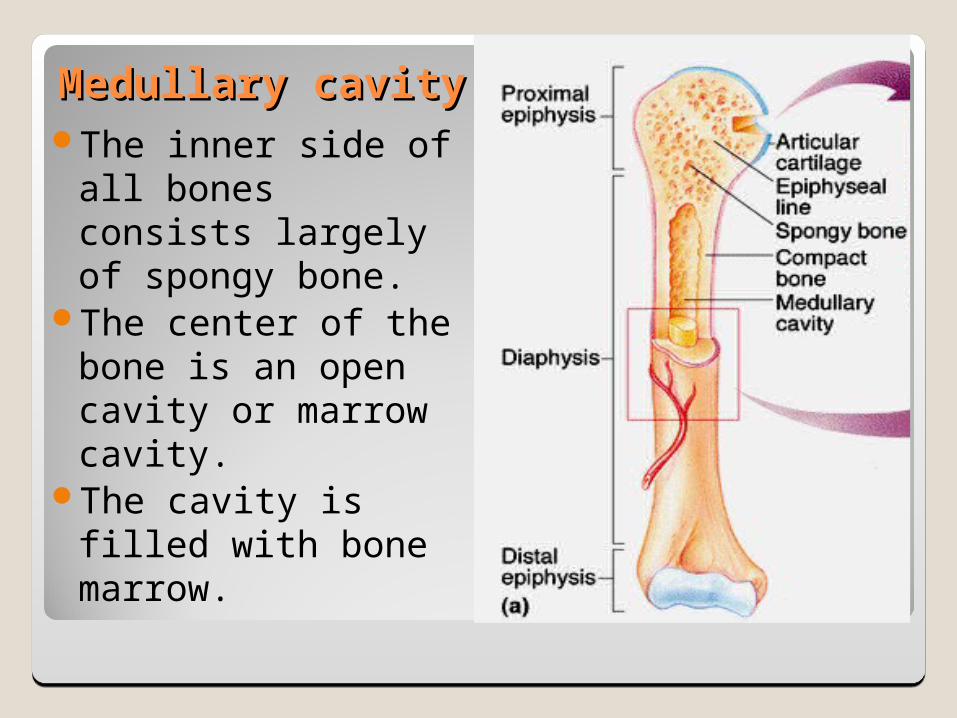
Medullary cavityMedullary cavityThe inner side of all
bones consists largely of spongy bone.
The center of the bone is an open cavity or marrow cavity.
The cavity is filled with bone marrow.
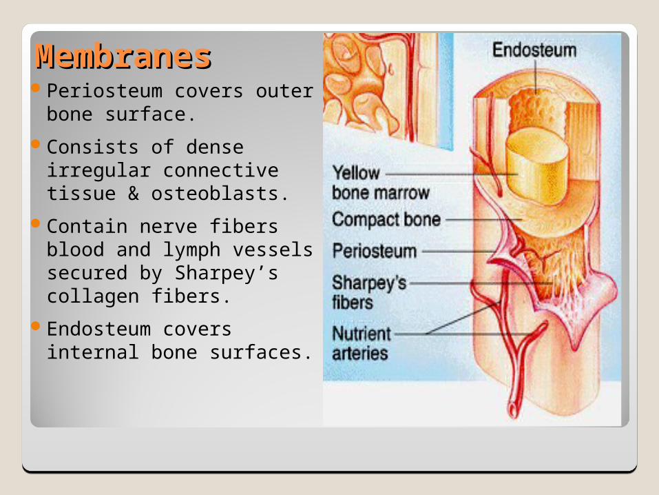
MembranesMembranesPeriosteum covers outer
bone surface.Consists of dense
irregular connective tissue & osteoblasts.
Contain nerve fibers blood and lymph vessels secured by Sharpey’s collagen fibers.
Endosteum covers internal bone surfaces.

Short, Irregular and Flat BonesShort, Irregular and Flat Bones Bones consist of
thin layers of compact bones over spongy bone.
No shaft, epiphysis or marrow cavity.
Spongy area between is a diploe.
Flat sandwich of bone.
Hematopoietic TissueHematopoietic TissueThe hematopoietic tissue, red marrow,
is typically found within the medullary cavities of long bones and in the diploe of flat bones.
These cavities are referred to as red marrow cavities.
In infants the medullary cavity and all areas of spongy bone contain red bone marrow.
26
In the adult the medullary cavity contains yellow bone marrow [fat-rich] that extends into the epiphysis and there is little red marrow present in spongy bone cavities.
Blood cell production occurs only in the head of the femur and humerus.
Most blood cell production occurs in the diploe areas of the sternum and hip bone.
Yellow marrow can revert to red marrow if the person becomes very anemic
27
Chemical Composition of BoneChemical Composition of Bone
The organic components:◦Osteoblasts (bone forming cells)◦Osteocytes (mature resting bone cells) ◦Osteoclasts (large cells which resorb matrix)◦Osteoid (the non calcified boney matrix)
28
Composition of BoneComposition of BoneThe inorganic components of bone (65% by
mass) consist mainly of calcium phosphate hydroxyapatites.
Tiny crystals of calcium salts are deposited in and around the collagen fibers of the extracellular matrix.
The crystals are hard and resist compression.Organic and inorganic components of matrix
allows a bone to be strong and not brittle
29
Histological Structure of Compact BoneHistological Structure of Compact Bone
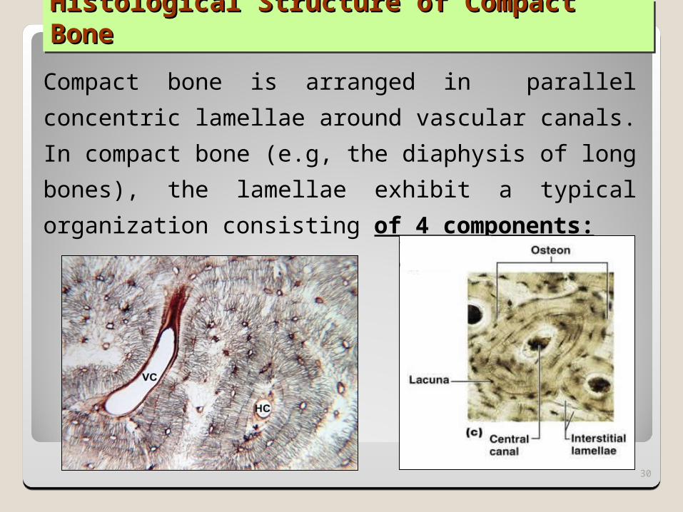
Compact bone is arranged in parallel concentric lamellae around vascular canals. In compact bone (e.g, the diaphysis of long bones), the lamellae exhibit a typical organization consisting of 4 components:
30

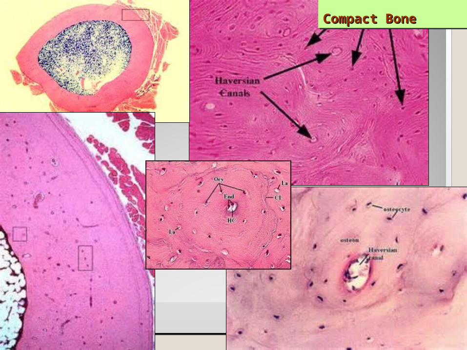
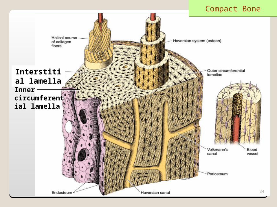
Compact BoneCompact Bone1. Haversian system, or osteon: The structural unit of compact bone composed of concentric lamellae of bone surrounding an endosteum-lined central canal [haversian canal] containing blood vessels, nerves, and loose connective tissue.
The haversian canals communicate with the marrow cavity, the periosteum, and with one another through transverse or oblique Volkmann's canals.
31
Compact BoneCompact Bone
32
Compact BoneCompact Bone
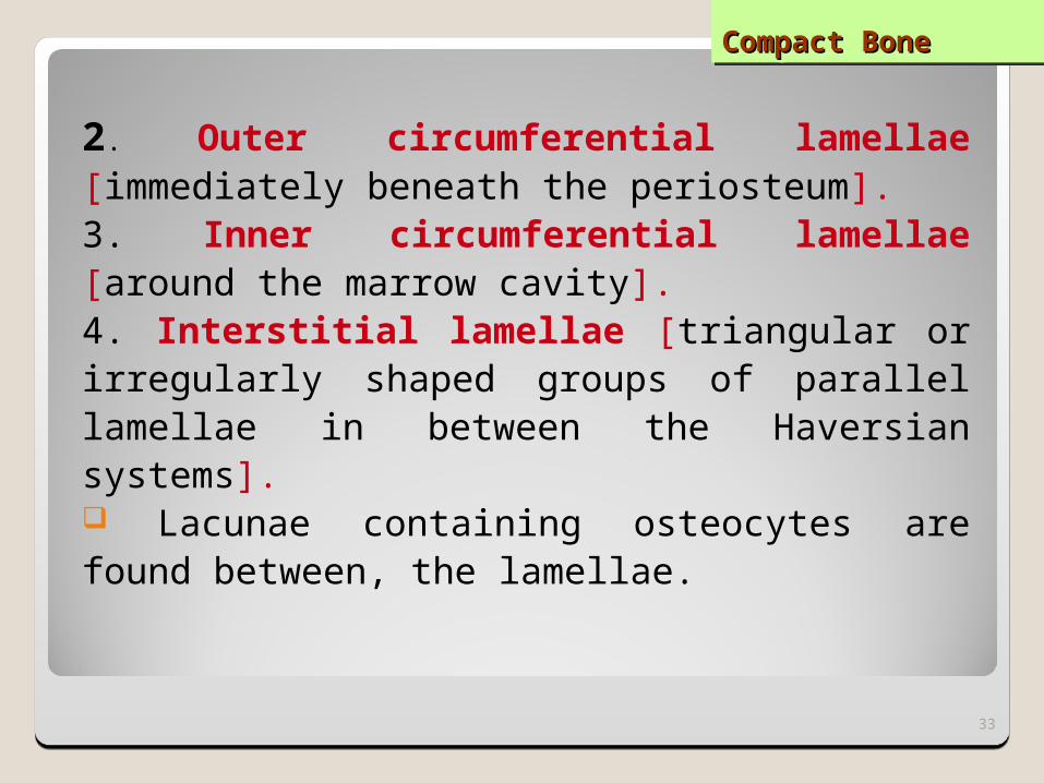
2. Outer circumferential lamellae [immediately beneath the periosteum]. 3. Inner circumferential lamellae [around the marrow cavity]. 4. Interstitial lamellae [triangular or irregularly shaped groups of parallel lamellae in between the Haversian systems]. Lacunae containing osteocytes are found between, the lamellae.
33
Interstitial lamella
Inner circumferential lamella
Compact Bone
34

References:1.Gray's Anatomy, 40th Edition By Susan Standring, PhD, DSc, FKC2.Junqueira's Basic Histology: Text and Atlas, Thirteenth Edition 2013 3.ISBN-13: 978-0071780339 ISBN-10: 0071780335 Edition: 13th 4.The histology tutor: http://www2.yvcc.edu/histologyzoomer/HistologyTutorials/histology_tutorials.htm5.2.http://www.highlands.edu/academics/divisions/scipe/biology/labs/rome/histology
6.3.http://krupp.wcc.hawaii.edu/BIOL100L/powerpoint/tissues
7.4. http://www.iteachbio.com/Anatomy-Physiology/BodyTissues
8.5. http://www.lavc.edu/instructor/watson_k/docs/Tissues
9.6. http://www.nakedscience.org/mrg/Anatomy%20Unit%204%20-%20Tissue%20Types
Bone DevelopmentBone DevelopmentHUMAN BODY 1 HB1 L4 HUMAN BODY 1 HB1 L4
by by Dr.Joseph Aziz Dr.Joseph Aziz
Asst. prof. of Anatomy & EmbryologyAsst. prof. of Anatomy & EmbryologyConsultant of Anatomy Consultant of Anatomy
36
Bone Histogenesis [Bone formation]Bone Histogenesis [Bone formation]Bone can be formed in two ways: 1. Intramembranous Ossification:Occur in a membrane of mesenchyme. Direct mineralization of matrix secreted by osteoblasts.
2. Endochondral [cartilaginous] Ossification: Deposition of bone matrix on a preexisting
cartilage template.
37
Intramembranous OssificationIntramembranous Ossification
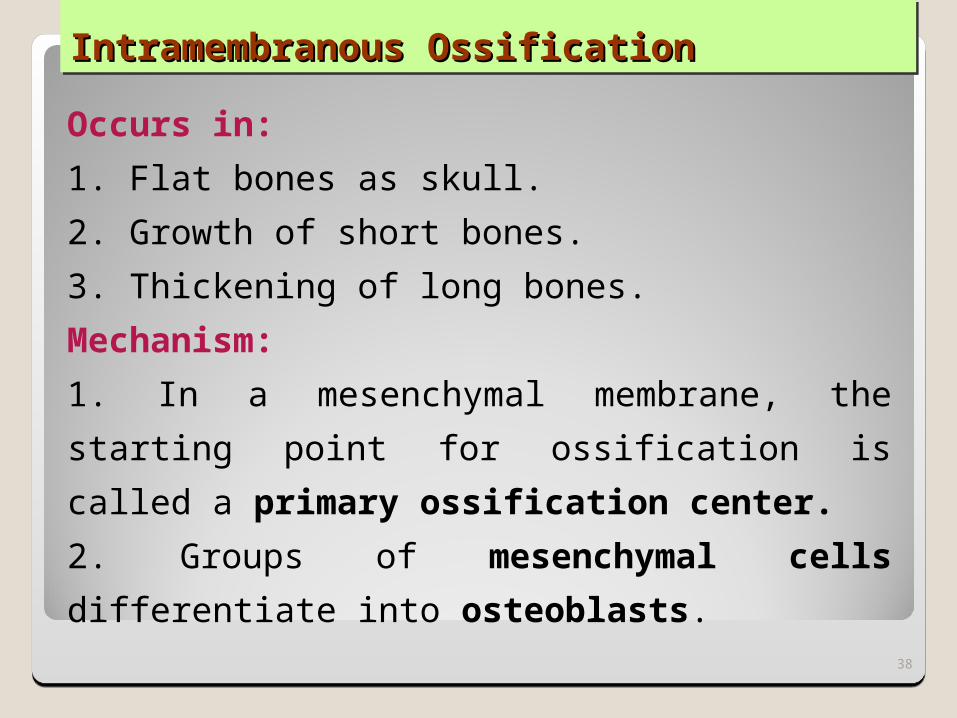
Occurs in: 1. Flat bones as skull.2. Growth of short bones.3. Thickening of long bones.Mechanism:1. In a mesenchymal membrane, the starting point for ossification is called a primary ossification center. 2. Groups of mesenchymal cells differentiate into osteoblasts.
38
Intramembranous Intramembranous OssificationOssification
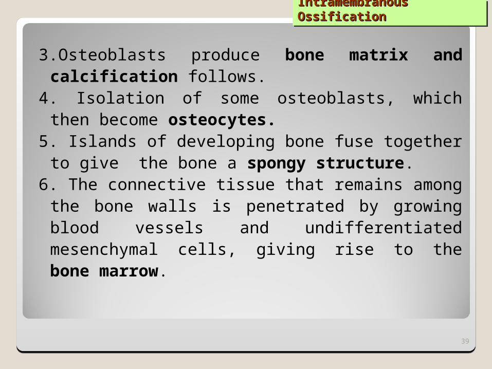
3.Osteoblasts produce bone matrix and calcification follows.
4. Isolation of some osteoblasts, which then become osteocytes.
5. Islands of developing bone fuse together to give the bone a spongy structure.
6. The connective tissue that remains among the bone walls is penetrated by growing blood vessels and undifferentiated mesenchymal cells, giving rise to the bone marrow.
39
Intramembranous Intramembranous OssificationOssification
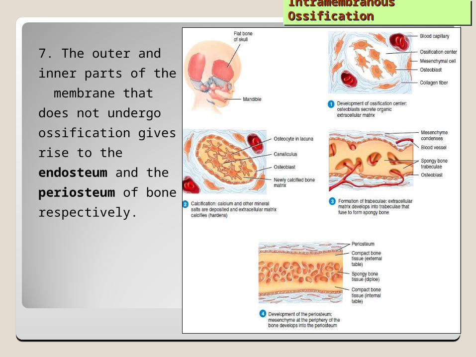
7. The outer and inner parts of the membrane that does not undergo ossification gives rise to the endosteum and the periosteum of bone respectively.
40
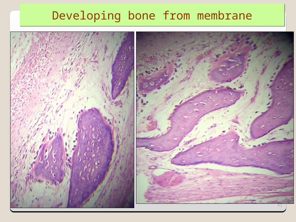
Developing bone from membrane
41

Endochondral OssificationEndochondral Ossification
Occurs in: short and long bones.
42
Endochondral Ossification
Enchondral OssificationEnchondral Ossification
Mechanism: 1.Bone collar, is produced by intramembranous
ossification within the local perichondrium. 2. The local cartilage undergoes a degenerative
process [cell death and matrix calcification] at the central portion of the cartilage model (diaphysis).
3. Blood vessels penetrate through the bone collar, bringing osteoprogenitor cells [source of osteoblasts] to this region.
4. Osteoblasts adhere to the calcified cartilage matrix and produce primary ossification center.
43
Cartilaginous Cartilaginous OssificationOssification
5. Secondary ossification centers appear at the epiphyses.6. Osteoblasts at primary and secondary ossification centers
produce continuous layers of bone and bone cavities that are gradually filled with bone marrow.
7. In the secondary ossification centers, cartilage remains in two regions:
a. Articular cartilage, which persists throughout adult life and does not contribute to bone growth in length.
b. Epiphyseal plate of cartilage which is responsible for the growth in length of the bone, and it disappears in adults at about 20 years .
44
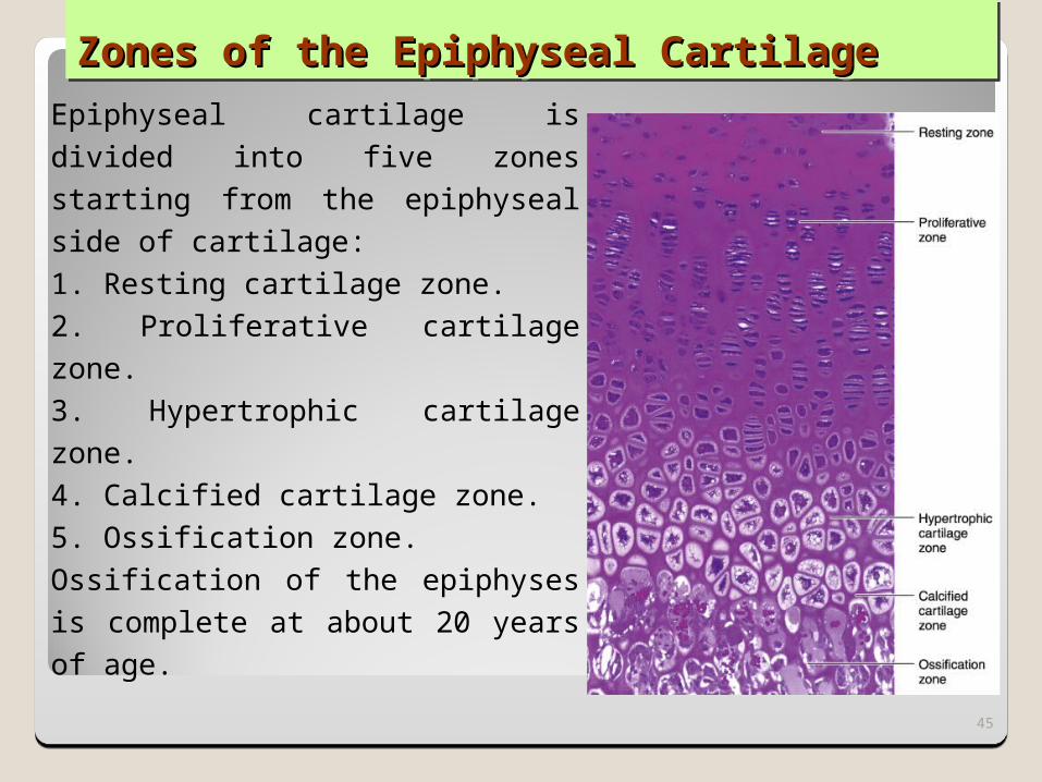
Zones of the Epiphyseal CartilageZones of the Epiphyseal CartilageEpiphyseal cartilage is divided into five zones starting from the epiphyseal side of cartilage: 1. Resting cartilage zone. 2. Proliferative cartilage zone.3. Hypertrophic cartilage zone. 4. Calcified cartilage zone. 5. Ossification zone. Ossification of the epiphyses is complete at about 20 years of age.
45
Bone RemodelingBone Remodeling
46
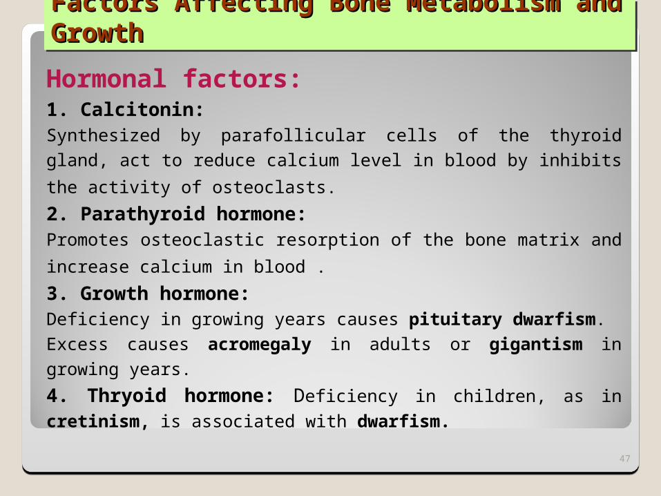
Factors Affecting Bone Metabolism and Factors Affecting Bone Metabolism and GrowthGrowth
Hormonal factors:1. Calcitonin:Synthesized by parafollicular cells of the thyroid gland, act to reduce calcium level in blood by inhibits the activity of osteoclasts. 2. Parathyroid hormone:Promotes osteoclastic resorption of the bone matrix and increase calcium in blood . 3. Growth hormone:Deficiency in growing years causes pituitary dwarfism.Excess causes acromegaly in adults or gigantism in growing years.4. Thryoid hormone: Deficiency in children, as in cretinism, is associated with dwarfism.
47
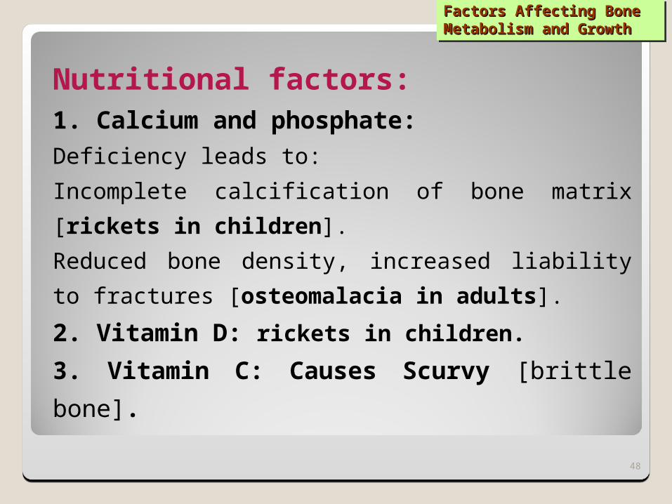
Factors Affecting Bone Factors Affecting Bone Metabolism and GrowthMetabolism and Growth
Nutritional factors:1. Calcium and phosphate:Deficiency leads to:Incomplete calcification of bone matrix [rickets in children]. Reduced bone density, increased liability to fractures [osteomalacia in adults].
2. Vitamin D: rickets in children. 3. Vitamin C: Causes Scurvy [brittle bone].
48
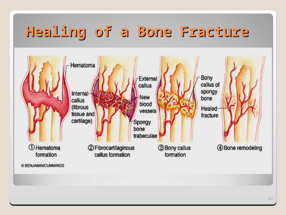
Healing of a Bone FractureHealing of a Bone Fracture
49

RECOMMENDED REFERENCES, RECOMMENDED REFERENCES, WEBSITESWEBSITESJunqueira's Basic
Histology: Text and Atlas, Thirteenth Edition 2013
ISBN-13: 978-0071780339 ISBN-10: 0071780335 Edition: 13th
The histology tutor:http://www2.yvcc.edu/
histologyzoomer/HistologyTutorials/histology_tutorials.htm
50