Embed Size (px)
Citation preview

BLEEDING DISORDERS
PRESENTED BY Dr Soundarya V
I YEAR

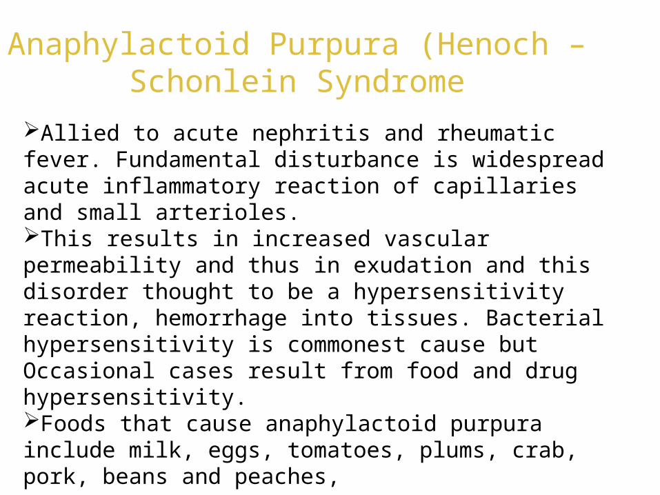
Classification of bleeding disorders
Definition
Bleeding disorders are hematological conditions
characterized by a functional impairment in the
hemostatic process.
The bleeding disorders are classified in two broad
categories: inherited or hereditary; and acquired.

Bleeding disorders
Vessel wall disorders
Platelet disorders
Coagulationdisorders
Bleeding disorders

VESSEL WALL DISORDERS• Scurvy (vit. C deficiency)• Cushing’s syndrome • Ehlers - Danlos syndrome• Rendu- Osler Weber Syndrome

• CONGENITAL Thromobocytopenic-quantitative platelet deficiency• May-Hegglin anomaly• Wiskott-Aldrich syndrome• Neonatal alloimmune thrombocytopenia
Nonthrombocytopenic-qualitative or functional platelet defect• Glanzmanns thrombasthenia• platelet type von willebrands disease• Bernard-Soulier syndrome
PLATELET DISORDERS

• ACQUIRED Thromobocytopenic-quantitative platelet deficiency
• Auto immune or idiopathic thrombocytopenic purpura• Thrombotic thrombocytopenic purpura• Cytotoxic chemotherapy• Drug induced (qunine,qunidine,gold salts,trimethoprim-
sulfamethaxazole,rifampicin)• Leukemia• Aplastic anaemia• SLE• DIC

• Nonthrombocytopenic-qualitative or functional platelet defect• Drug induced (aspirin,NSAIDs,pencillin,cephalosporin)• Uremia• Alcohol dependency• Liver disease• Myelopoliferative disorders• Acquired platelet type von willebrands disease
• CONGENITAL ACQUIRED• Hemophilia A Secondary to drugs• Hemophilia B or diseases process• F XI deficiency• F XII deficiency• F X deficiency• F V deficiency• F XIII & I deficiency• von Willibrands disease
COAGULATION DISORDERS

• Heparin• coumarin
ANTICOAGULANT RELATED COAGULOPATHIES

• Liver disease• Vitamin k deficiency• DIC• Fibrinolytic disorders
DISEASE RELATED COAGULOPATHIES


VESSEL WALL DISORDERS

Scurvy Scurvy results from Vitamin C deficiency. Hemorrhage is usual in scurvy and is the major feature of
scurvy. It is primarily due to increased capillary fragility which results from defective formation of intercellular substance of capillary wall.
Hemorrhages may occur any where in the skin, particularly common in the legs. Hemorrhages into muscles also occur, resulting in areas of brawny indurations and tenderness.

Oral effects chiefly involve the gingival and periodontal tissues.
The interdental and marginal gingival is bright red with a swollen,
smooth, shiny surface. Gingiva becomes boggy, ulcerates and
bleeds.
In severe chronic cases of scurvy, hemorrhage and swelling of
periodontal membrane occur followed by loss of bone, loosening of
teeth, which eventually exfoliate
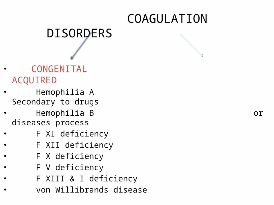
Anaphylactoid Purpura (Henoch – Schonlein Syndrome
Allied to acute nephritis and rheumatic fever. Fundamental disturbance is widespread acute inflammatory reaction of capillaries and small arterioles. This results in increased vascular permeability and thus in exudation and this disorder thought to be a hypersensitivity reaction, hemorrhage into tissues. Bacterial hypersensitivity is commonest cause but Occasional cases result from food and drug hypersensitivity.Foods that cause anaphylactoid purpura include milk, eggs, tomatoes, plums, crab, pork, beans and peaches,

Hereditary haemorrhagic telangiectasia (Osler-Rendu-Weber disease)
Present in the skin and mucous membranes
The telengiectases are lined by a thin layer of endothelial
cells, because of their thinness they bleed easily and
because they contract poorly bleeding is often prolonged
)

Ehlers-Danlos Disease It is a group of hereditary disorders of connective tissue. Atleast eight forms of disease are now recognized (EDS I to VIII).EDS IV is often called the ecchymotic type since rupture of even large arteries as well as intestine often occurs, producing a life threatening situation. Basic lesion is developmental abnormality of mesenchyme which results in increased fragility of blood vessels of skin, together with increased elasticity of skin and hyper extensibility of joints.The hemostatic defect results in occurrence of large haematomas following slight trauma.
Bleeding disorders
Vascular abnormalities
Platelet disorders
Coagulation disorders

Bleeding disorders
Platelet disorders
↓production
↑destruction
Primary/IdiopathicITP
Acute/Chronic
SecondaryDrugs, HIV
SequestrationHypersplenism
Platelet disorders

Platelet disorders • Thrombocytopenia =Reduced platelet number• Causes
– Decreased production of platelets• vitamin B12 or folic acid deficiency
– Decreased platelet survival• Immunologic or Nonimmunologic etiology
– Sequestration- Hypersplenism • ameliorated by splenectomy
– Dilutional• Massive transfusions
Thrombocytopenic purpura
Thrombocytopenia is a condition is which there is an abnormal
reduction in the number of circulating blood platelets.
Patients develops focal hemorrhages into various tissues and
organs, including skin and mucous membranes.
Two basic forms of thrombocytopenia recognized
– Primary
– Secondary

Idiopathic thrombocytopenic purpura
• Idiopathic thrombocytopenic purpura (ITP), also known as
primary immune thrombocytopenic purpura or autoimmune
thrombocytopenic purpura, is defined as isolated
thrombocytopenia with normal bone marrow and the absence of
other causes of thrombocytopenia.
• The clinical syndromes manifest as an acute condition in
children and a chronic condition in adults.

ITP is primarily a disease of increased peripheral platelet
destruction, with most patients having antibodies to specific
platelet membrane glycoproteins.
Relative marrow failure may contribute to this condition, since
studies show that most patients have either normal or diminished
platelet production.
Acute ITP often follows an acute infection and has a
spontaneous resolution within 2 months. Chronic ITP persists
longer than 6 months without a specific cause.
Females are affected 3-4 times more than males.

Type and site of bleeding
• Bleeding occurs spontaneously
• It occurs following trauma, surgery and dental procedures.
• Bleeding from wounds tends to occur at once ,ceases within 48 hrs
and does not recur.
• Skin is the most common site for hemorrhage and in mild cases it is
the only site.
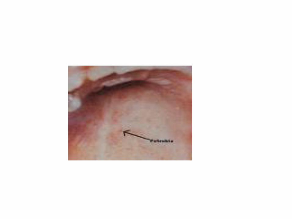
• Haemorrhage may take the form of multiple petichae or ecchymosis
or both.

Petichae spot size varies from pin point to pin head or somewhat larger.
They are not raised and do not blanch on pressure.
When fresh they are red in colour but with time they pass through the colour
changes of absorbing blood.
They occur in groups or crops especially on the arms and legs, the neck and
the upper part of the chest
Vary in number from a few scattered crops to innumerable crops covering
almost entirely whole of the body.

• Occasionally large haematomas are formed in the subcutaneous tissue
• Haemorrhages are not accompanied by urticaria or erythema
• Bleeding from mucous membrane is common
• Epistaxis and bleeding from gums are the most common forms
• Hematuria, menorrhagia, and malena are also seen.
• Petichae similar to the skin is seen in mouth and nose.
• Rarely is there hemorrhage into the pleura or peritoneal cavity.
• Bleeding into the internal organs are uncommon but may be serious
• Most important site is the nervous system especially the brain
• Cerebral hemorrhage is the most common cause of death in severe
thrombocytopenia.
• Haemorrhage into the spinal cord and meninges may occur.
• Bleeding into joints are rare.
• Spleen may be slightly enlarged
• Lymph nodes and liver are not palpable and there is no sternal tenderness.

• Jaundice is absent except when there is extensive tissue hemorrhage.
• Fever is usually absent but there may be moderate rise in temperature with
extensive hemorrhage into tissue or GIT.
• In the acute variety there is acute onset of hemorrhage into the skin and
• membrane
• Hemorrhage is severe,epistaxis is common

Bleeding ceases spontaneously after a period varying from a few days to a
few weeks, in about 10% the bleeding persists and the disorder runs the course
of chronic idiopathic thrombocytopenia.
Bleeding is most severe at onset and tends to lessen in severity as time
passes.
Death is uncommon and occurs within 1st 4 weeks of onset
Chronic type is seen in young to middle aged females.
Onset is less abrupt
Severity of symptoms varies from mild to (recurrent crops of petichae or easy
bruising) to severe bleeding from mucous membranes
• In chronic disease symptoms are often intermittent with remissions lasting
weeks, months or even years.
• In other cases symptoms persists but fluctuate in severity.
Blood Picture:
• There is reduction of the platelet count ranging from just below normal to 10
x 109/l .Lower count s tending to be associated with the acute disease.
• Platelet sometimes appears to be morphologically abnormal
• Prolonged bleeding time (upto 30 mins)
• Torniquet test may be positive
• Anaemia proprtional to the degree of blood loss may be present
• ESR is usually normal
• 30% of the cases have positive anticardiolipin antibodies in the serum

Bone marrow
• Megakaryocytes and their precursors are present in normal or increased numbers.
• Increase in the percentage of precursor cells.
• In some cases there is a moderate increase in the number of mature lymphocytes
or in eosinophils.
Treatment
• In the patient with known ITP, high-dose parenteral glucocorticoids and IV
immunoglobulin (IVIg), with or without platelet transfusions, are appropriate.
• Prednisone -- Useful in treating inflammatory and allergic reactions; may decrease
inflammation by reversing increased capillary permeability and suppressing PMN
activity.

• DOC for all adult patients with platelet counts <50,000/mm3.
Asymptomatic patients with platelet counts >20,000/mm3, or patients
with counts 30,000-50,000/mm3 with only minor purpura, may not
need therapy; withholding medical therapy may be appropriate for
asymptomatic patients, regardless of count.
• Platelet transfusion is indicated for controlling severe hemorrhage
• Splenectomy is reserved for patients in whom medical therapy fails
and when thrombocytopenia persist for more than 6 months.
• Emergent splenectomy is indicated in patients with life-threatening
bleeding in whom medical therapy fails.

Secondary Thrombocytopenia:
• The majority of the cases of thrombocytopenia seen in clinical
practice are secondary to some underlying disorder.
Drug induced thrombocytopenic purpura:
• Most cases occur as a complication of drug therapy but
occasionally are due to toxic action of chemicals used in the
industry or in the home.
• Mechanism:
i) They have a direct toxic effect on the bone marrow
ii) A hypersensitivity reaction in which the platelets are rapidly
destroyed in the peripheral blood ,possibly with associated
impairment of platelet formation by megakaryacytes.

• Drug combines with the plasma proteins to form antigens ,which results
in antibody formation.
• When the drug is readministered,the antibody combines with the antigen
to form an immune complex which is adsorbed onto the surface of the
platelet, resulting in its the removal by the reticuloendothelial system.
Clinical features:
• There is a H/O administration of the drug for days ,weeks or months
followed by bleeding in a matter of hours upto several days following the
last dose.
• In cases due to bone marrow depressing agents there is sometimes an
interval of weeks or more between the last dose and the onset of
bleeding

• The bleeding is sometimes mild and limited to the skin but frequently is
severe with extensive mucous membrane haemorrhage and blood blisters in
the mouth,as well as skin petichae and ecchymosis.
• Severe haemorrhage of sudden onset is especially characteristic of those
drugs in which hypersensitivity can be demonstrated, especially
quinidine ,quinine qnd digitoxin
• With acute severe bleeding,constitutional symptoms are common like chills,
headache, fever, abdominal pain, nausea ,vomiting, and itching of the skin.
• Withdrawal of the drug is followed by cessation of bleeding within few
hours or days.

Blood picture:
• There is thrombocytopenia without anaemia or neutropenia
• There may be moderate leucocytosis during the bleeding
• The bone marrow shows a normal or increased number of megakaryocytes, which
may show reduced or absent granularity,
Diagnosis:
• Based on the h/o of the drug ingested
• Presence of constitutional symptoms
• Spontaneous remission on cessation of the drug
Prognosis:
• The platelet returns to normal and the bleeding ceases within few hours to
few days after stopping the drug
• Recovery is nearly always complete within 7 -14 days
• Occasionally death occurs usually from cerebral haemorrhage
Treatment:
• Immediate cessation of the offending drug
• Administration of corticosteroids in those with severe bleeding
• Blood transfusion to replace blood loss
• In cases caused by antibodies, transfused platelets are rapidly destroyed

.
But if bleeding appears to be life threatening ,platelet transfusion should be given.
• Splenectomy is without beneficial effect and is contraindicated.

Leukemias:
• In acute leukemia thrombocytopenia is usually severe.
• Bleeding is a common manifestation and a common cause of death
• In chronic lymphocytic leukemia in the early stage thrombocytopenia is
usually mild and asymptomatic, but in the later stage it may be marked and
cause severe bleeding.
• In chronic granulocytic leukemia , the platelet count is initially normal or
raised , but it falls in the later stage of the disease.

Aplastic anaemia:
• Aplastic anaemia is characterized by anaemia, leucopenia, and
thrombocytopenia resulting from aplasia of the bone marrow.
• Thrombocytopenia is common in aplastic anaemia, especially in drug
induced cases.
• The drugs can cause hypoplasia of the bone marrow
• Onset is insidious with symptoms of progressively worsening anaemia and
an associated bleeding or
• Symptoms of anaemialike weakmess, easy fatiguability, lassitude,.dyspnoea
on exertion.

• The bleeding manifestations are petichae,ecchymosis,epistaxis,
mennorhagia, bleeding from gums and alimentary tract.
• Cerebral hemorrhage is not unusual and is a common fatal complication.
Bone marrow infiltration:
• Thrombocytopenia may occur as a consequence of secondary
carcinoma, multiple myeloma, myelofibrosis and malignant lymphoma.
• Occasionally thrombocytopenic bleeding may be the first manifestation of
secondary carcinoma of bone and multiple myeloma.

Disseminated lupus erythematosis:
• Thrombocytopenia is is quite common in this condition
• Although the history and clinical examination usually reveal one or more
of the other features of the disease, the disorder occasionally presents
with thrombocytopenia as the only manifestation.
Liver disease:
• Thrombocytopenia associated with liver disease is most often due to
hypersplenism caused by congestive splenomegaly associated with
cirrhosis of the liver
• Thrombocytopenia may also occur in severe acute infective hepatitis.

Alcoholism:
• Chronic thrombocytopenia is common in chronic alcoholics
• Usually considered to be due to hypersplenisim associated with cirrhosis
and congestive splenomegaly or to nutritional megaloblastic anaemia.
• However an acute transient thrombocytopenia may occur in alcoholics
without cirrhosis,related to drinking bouts
• The platelet count increases within a few days of cessation of alcohol.
• Thrombocytopenia is due to direct effect of alcohol intoxication on the
developing megakaryacytes

Massive blood transfusion:
• Thrombocytopenia causing severe bleeding is seen in patients
transfused with massive amounts of stored whole blood
• Thrombocytopenia is related to the amount of whole blood
transfused and the rate of infusion
• It occurs when there is replacement of 50% or more of the patients
blood volume.
• Dilution of the recipients platelets, the non viable state of the
platelets in stored blood , or their lack in packed red blood cells are
the cause of thrombocytopenia

• Platelet count usually returns to normal level within 3-5 days
• Transfusion of 6-8 platelet concentrates is indicated in the presence of
haemorrhage when the platelet count falls to 50,000/cmm in association
with massive transfusion.

Qualitative platelet defects:
Thrombasthenia ( Glanzmann’s disease)
It is familial disorder occurring in both sexes and transmitted as
an autosomal recessive gene
The bleeding time is long inspite of normal platelet count and
normal platelet morphology.

• Hereditary platelet disorders characterized by thrombocytopenia, giant
platelets, and a tendency toward bleeding
• The defect is on the membrane with a lack of glycoprotein IIb-IIIa
complex,which is the point of attachemnt with fibrinogen, which in turn is
required for platelet-platelet binding in the aggregation process.
• Clinically the disorder is characterized by tendency to bleed even after minor
trauma,excessive and prolonged bleeds after cuts and abrasion,
menorrhagia
• Deep haematoma and haemarthroses are rare.

Bernard-Soulier syndrome
• The underlying biochemical defect is the absent or decreased expression of the
glycoprotein Ib and associated IX complex on the surface of the platelets.
• This complex is the receptor for von Willebrand factor (vWF), and the result of
decreased expression is deficient binding of vWF to the platelet membrane at sites of
vascular injury, resulting in defective platelet adhesion.
• This is demonstrated by the lack of aggregation of platelets in response to ristocetin,
an antibiotic that normally causes platelets to aggregate
Treatment :
• The only available treatment is the use of platelet transfusion in case of surgery or
potentially life-threatening bleeding.

Thrombocytosis:
• Thrombocytosis is defined as an increase in the platelet count above
4lakhs/cmm
Idiopathic thrombocythaemia:
• It is classified as one of the myeloproliferative disorders.
• Extremely high platelet counts may be seen.

Clinical features:
• Disorder occurs in middle age and older age groups.
• Bleeding of varying severity and small blood vessel occlusion is seen.
• GI bleeding is most common but haematuria, haemoptysis, menorrhagia
and bleeding after minor trauma and surgery is also common.
• Spontaneous bruising occurs and large haematomas form only after mild
trauma
• Thrombosis is the most common complication and particularly affects
toes, feet, fingers and the cerebral circulationenlarged liver and spleen
may be present.

Thank you